•6/9/2011 •1 Nerve Entrapment Syndromes Dr. Tudor H. Hughes M.D., FRCR Department of Radiology University of California School of Medicine San Diego, California Nerve entrapment syndromes Introduction • Chronic entrapment • Commonly as the nerve passes through an osseoligamentous tunnel, or under an aponeurotic margin • One side fixed, one moves -> friction • More common in upper limb • More common in upper limb • Inflamed or thickened nerve • May see mass pressing on nerve – not true entrapment • Secondary changes in muscles of nerve distribution Nerve entrapment syndromes History • Initially described by: • Astley Cooper 1820 • James Paget 1850 • First surgical decompression • Learmonth 1930 • Common types • Carpal tunnel syndrome • Cubital tunnel - Ulnar nerve at the elbow • Guyon’s canal – Ulnar nerve at the wrist • Suprascapular syndrome • Chronic blunt injury -> ischemic changes • Edema • Dislocation of the nodes of Ranvier Nerve entrapment syndromes Pathology • Structural changes to myelin sheath and axon • Focal segmental demyelination is constant • Complete recovery is due to remyelination • Incomplete recovery due to Wallerian degeneration Myelinated axons osmic acid stain Nerve entrapment syndromes Clinical • Depends on nerve involved • Irritative sensory symptoms • Pain and paresthesias • Ablative sensory symptoms Ablative sensory symptoms • Numbness • Ablative motor signs • Weakness and atrophy • If mixed nerve – sympathetic dystrophy • Dry, thin, hairless skin Nerve entrapment syndromes Clinical • Most peripheral entrapped nerves Sens. and Motor • Notable exceptions: • Anterior interosseous nerve (motor) • Deep ulnar (motor) • Lateral femoral cutaneous (sensory) • Superficial branch of radial nerve (sensory) • Superficial branch of radial nerve (sensory) Nervi nervorum
Transcript
•6/9/2011
•1
Nerve Entrapment Syndromes
Dr. Tudor H. Hughes M.D., FRCRDepartment of RadiologyUniversity of California School of MedicineSan Diego, California
Nerve entrapment syndromes
Introduction
• Chronic entrapment
• Commonly as the nerve passes through an osseoligamentous tunnel, or under an aponeurotic margin
• One side fixed, one moves -> friction• More common in upper limb• More common in upper limb
• Inflamed or thickened nerve
• May see mass pressing on nerve – not true entrapment
• Secondary changes in muscles of nerve distribution
Nerve entrapment syndromes
History• Initially described by:
• Astley Cooper 1820• James Paget 1850
• First surgical decompression • Learmonth 1930
• Common types• Carpal tunnel syndrome• Cubital tunnel - Ulnar nerve at the elbow• Guyon’s canal – Ulnar nerve at the wrist• Suprascapular syndrome
• Chronic blunt injury -> ischemic changes• Edema• Dislocation of the nodes of Ranvier
Nerve entrapment syndromes
Pathology
• Structural changes to myelin sheath and axon• Focal segmental demyelination is constant• Complete recovery is due to remyelination
• Incomplete recovery due to Wallerian degenerationMyelinated axons osmic acid stain
Nerve entrapment syndromes
Clinical• Depends on nerve involved
• Irritative sensory symptoms• Pain and paresthesias
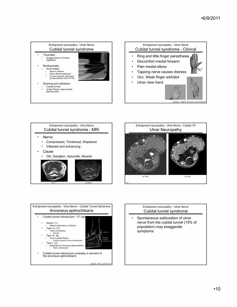
• Replaced by Anconeus epitrochlearis• Static compression
• Cubital tunnel retinaculum probably a remnant of the anconeus epitrochlearis
Entrapment neuropathy - Ulnar Nerve
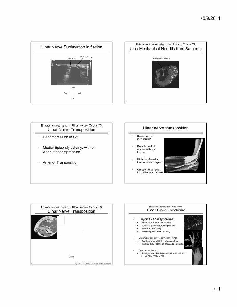
Cubital tunnel syndrome• Spontaneous subluxation of ulnar
nerve from the cubital tunnel (15% of population) may exaggerate symptoms
•6/9/2011
•11
Ulnar Nerve Subluxation in flexion
Ulnar NerveMedial epicondyle
Med
Lat
AntPost
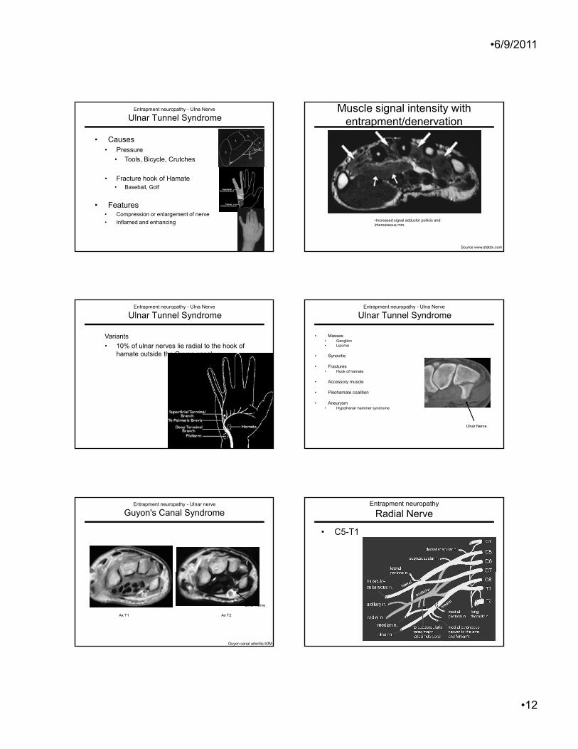
Entrapment neuropathy - Ulna Nerve - Cubital TS
Ulna Mechanical Neuritis from Sarcoma
Anconeus Epitrochlearis
7
• Decompression In Situ
• Medial Epicondylectomy, with or without decompression
Entrapment neuropathy - Ulnar Nerve - Cubital TS
Ulnar Nerve Transposition
p
• Anterior Transposition
Ulnar nerve transposition
• Resection of retinaculum
• Detachment of common flexor tendon
• Division of medial intermuscular septum
• Creation of anterior tunnel for ulnar nerve
Entrapment neuropathy - Ulnar Nerve - Cubital TS
Ulnar Nerve Transposition
s/p ulnar nerve transposition with medial-sided pain
Axial PD
3
Entrapment neuropathy - Ulna Nerve
Ulnar Tunnel Syndrome
• Guyon’s canal syndrome• Superficial to flexor retinaculum• Lateral to pisiform/flexor carpi ulnaris• Medial to ulnar artery• Roofed by transverse carpal lig
• Superficial sensory hypothenar branch• Proximal to canal 65% - silent paralysis• In canal 35% - additional pain and numbness
• Deep motor branch• Paralysis – AddPol, Interossei, ulnar lumbricals
• OpDM > FDM > AbDM
•6/9/2011
•12
Entrapment neuropathy - Ulna Nerve
Ulnar Tunnel Syndrome
• Causes• Pressure
• Tools, Bicycle, Crutches
F t h k f H t• Fracture hook of Hamate• Baseball, Golf
• Features• Compression or enlargement of nerve• Inflamed and enhancing
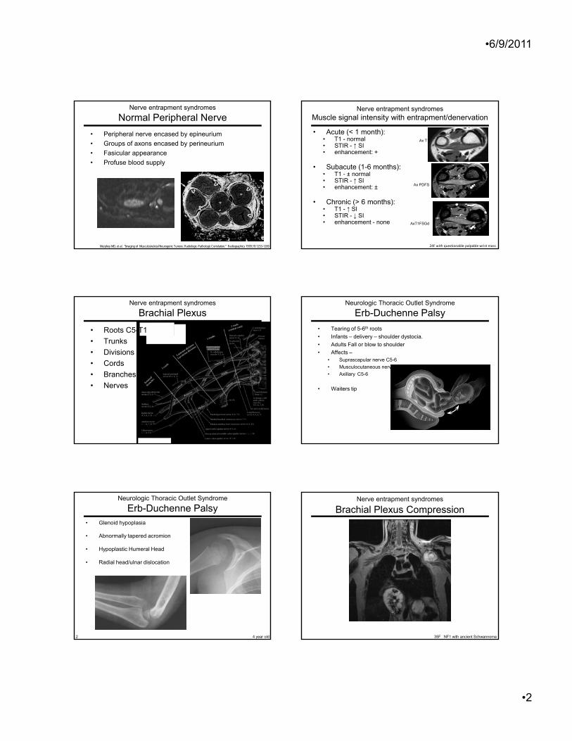
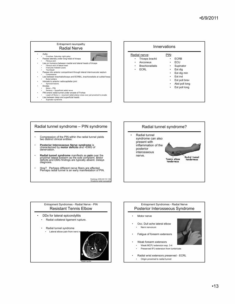
Muscle signal intensity with entrapment/denervation
Source www.statdx.com
•Increased signal adductor pollicis and interosseous mm.
Entrapment neuropathy - Ulna Nerve
Ulnar Tunnel Syndrome
Variants• 10% of ulnar nerves lie radial to the hook of
hamate outside the Guyon canal
Entrapment neuropathy - Ulna Nerve
Ulnar Tunnel Syndrome
• Masses• Ganglion• Lipoma
• Synovitis
• Fractures• Hook of hamate
• Accessory muscle
• Pisohamate coalition
• Aneurysm• Hypothenar hammer syndrome
Ulnar Nerve
Entrapment neuropathy - Ulnar nerve
Guyon's Canal Syndrome
Guyon canal arteritis 63M
Ax T1 Ax T2
Ulnar Nerve
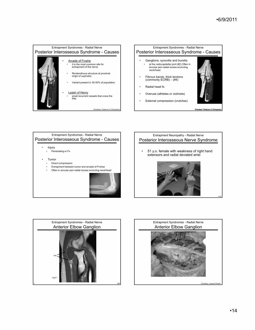
Entrapment neuropathy
Radial Nerve
• C5-T1
•6/9/2011
•13
Entrapment neuropathy
Radial Nerve• Axilla
• Crutches, Saturday night palsy• Passes laterally under long head of triceps
• Fibrous arch• Lies on humerus between medial and lateral heads of triceps
• Fibrous arch of lateral head• Fractures Holstein-Lewis• Tourniquet
P i t t i t t th h l t l i t l t• Passes into anterior compartment through lateral intermuscular septum• Compression
• Lies between brachialis/biceps and ECRB/L, brachioradialis at cubital fossa• Body builders
• Intimate to anterior radiocapitellar joint• Synovial lesions
• Divides• Motor – PIN• Sensory – Superficial radial nerve
• PIN enters radial tunnel under arcade of Frohse• Leash of Henry (← recurrent radial artery) cross over just proximal to arcade
• Lies between deep and superficial heads• Supinator syndrome
• Compression of the PIN within the radial tunnel yields two distinct clinical entities:
• Posterior Interosseous Nerve syndrome is characterized by motor deficits and +EMG of denervation.
• Radial tunnel syndrome manifests as pain over the proximal lateral forearm as the sole complaint. Motor deficits and EMG findings are typically absent. Delays diagnosis.
• How? Perhaps different nerve fibers are affected. Perhaps radial tunnel is an early manifestation of PIN.