PHYSIOLOGICAL RESEARCH ISSN 0862-8408 2003 Institute of Physiology, Academy of Sciences of the Czech Republic, Prague, Czech Republic Fax +420 241 062 164 E-mail: [email protected]http://www.biomed.cas.cz/physiolres Physiol. Res. 52: 1-30, 2003 MINIREVIEW Neurohumoral Control of Gastrointestinal Motility M. B. HANSEN Department of Surgical Gastroenterology D, Glostrup University Hospital of Copenhagen, Denmark Received December 6, 2001 Accepted April 25, 2002 Summary Neurohumoral substances and their receptors play a major part in the complex regulation of gastrointestinal motility and have therefore been the predominant targets for drug development. The numerous receptors involved in motility are located mainly on smooth muscle cells and neuronal structures in the extrinsic and intrinsic parts of the enteric nervous system. Within this system, receptor agonists and antagonists interacts directly to modify excitatory or inhibitory signals. In view of this complexity it is not surprising that our knowledge about the mechanisms of actions of the various neurohormones and drugs affecting gut motility has been rather fragmented and incomplete. However, recently substantial progress has been achieved, and drug therapy for gut dysmotility is emerging, based primarily on neurohumoral receptors. This paper presents a selective review of the neurohumoral regulatory mechanisms of gastrointestinal motility. In this context, the physiology and pharmacology of the smooth muscle cells, gastrointestinal motility and dysmotility, the enteric nervous system, gastrointestinal reflexes, and serotonin is presented. Further investigation and understanding of the transmitters and receptors involved in especially the reflex activation of peristalsis is crucial for the development of novel therapies for motility disorders. Key words 5-Hydroxytryptamine • Enteric Nervous System • Gastrointestinal • Hormones • Interstitial Cells of Cajal • Motility • Review • Serotonin 1. Introduction Gastrointestinal (GI) motility is an integrated process including myoelectrical activity, contractile activity, tone, compliance and transit. These different entities of motility can be generated and modulated by local and circulating neurohumoral substances. The importance of neurohumoral control of motility goes back to 1859, where C. Bernhard observed profuse diarrhea (”paralytic secretion”) and vigorous intestinal motility (”hunger contractions”) after external sympathetic denervation of the dog gut. Brunton and Pye-Smith attributed correctly this phenomenon to a disruption of neural pathways within the intestinal wall. Later, the term peristalsis was introduced (Modlin et al. 2000). During the last decade substantial new knowledge has been accomplished on especially the involvement of the enteric nervous system (ENS) in these processes. The purpose of this review is to present an overview and some new interesting findings on the mechanism by which GI motility is controlled and
Transcript
PHYSIOLOGICAL RESEARCH
ISSN 0862-8408
2003 Institute of Physiology, Academy of Sciences of the Czech Republic, Prague, Czech Republic Fax +420 241 062 164 E-mail: [email protected] http://www.biomed.cas.cz/physiolres
Physiol. Res. 52: 1-30, 2003
MINIREVIEW
Neurohumoral Control of Gastrointestinal Motility M. B. HANSEN Department of Surgical Gastroenterology D, Glostrup University Hospital of Copenhagen, Denmark Received December 6, 2001 Accepted April 25, 2002 Summary Neurohumoral substances and their receptors play a major part in the complex regulation of gastrointestinal motility and have therefore been the predominant targets for drug development. The numerous receptors involved in motility are located mainly on smooth muscle cells and neuronal structures in the extrinsic and intrinsic parts of the enteric nervous system. Within this system, receptor agonists and antagonists interacts directly to modify excitatory or inhibitory signals. In view of this complexity it is not surprising that our knowledge about the mechanisms of actions of the various neurohormones and drugs affecting gut motility has been rather fragmented and incomplete. However, recently substantial progress has been achieved, and drug therapy for gut dysmotility is emerging, based primarily on neurohumoral receptors. This paper presents a selective review of the neurohumoral regulatory mechanisms of gastrointestinal motility. In this context, the physiology and pharmacology of the smooth muscle cells, gastrointestinal motility and dysmotility, the enteric nervous system, gastrointestinal reflexes, and serotonin is presented. Further investigation and understanding of the transmitters and receptors involved in especially the reflex activation of peristalsis is crucial for the development of novel therapies for motility disorders. Key words 5-Hydroxytryptamine • Enteric Nervous System • Gastrointestinal • Hormones • Interstitial Cells of Cajal • Motility • Review • Serotonin 1. Introduction
Gastrointestinal (GI) motility is an integrated process including myoelectrical activity, contractile activity, tone, compliance and transit. These different entities of motility can be generated and modulated by local and circulating neurohumoral substances. The importance of neurohumoral control of motility goes back to 1859, where C. Bernhard observed profuse diarrhea (”paralytic secretion”) and vigorous intestinal motility (”hunger contractions”) after external
sympathetic denervation of the dog gut. Brunton and Pye-Smith attributed correctly this phenomenon to a disruption of neural pathways within the intestinal wall. Later, the term peristalsis was introduced (Modlin et al. 2000). During the last decade substantial new knowledge has been accomplished on especially the involvement of the enteric nervous system (ENS) in these processes.
The purpose of this review is to present an overview and some new interesting findings on the mechanism by which GI motility is controlled and
Hansen Vol. 52
2
modulated by neurohumoral substances with focus on the small intestine and serotonin (5-hydrotryptamine, 5-HT). 2. The smooth muscle of the gut Structure
Smooth muscle cells of the gut behave as unitary types and consists of a thin outer longitudinal layer and a thick, densely innervated circular layer. The thickness of the two layers varies both between species and regions (Olsson and Holmgren 2001). The layers are separated by laminar septae into bundles about 1 mm long, which act as contractile units. The muscle cells are embedded in a connective tissue matrix, a product of their synthetic and secretory activity consisting mainly of elastic and collagen fibrils. These layers include glial cells, fibroblasts, and a distinctive population of cells, the interstitial cells of Cajal. The muscle cell plasma membranes consist of two specialized structures known as caveolae and dense bands. The caveolae are basket-shaped and represents calcium stores. Clusters of caveolae are separated form each other by dense bands that occupy about one-half of the surface of the cell. The dense bands are points of attachment of thin actin filaments. Intermediate filaments link dense bands in the membrane to dense bodies in the cytoplasm and transmit the force generated by the contractile apparatus within the cell to the entire surface of the cell. Gap junctions are abundant in circular and rare in longitudinal muscle layer, and they permit movement of intracellular regulatory molecules, all suggesting a significant functional role (Makhlouf 1995, Horowitz et al. 1996, Murphy 1998, Daniel et al. 2001). Contractile filaments
Three types of filaments exist: thin actin, thick myosin and intermediate desmin filaments. These filaments interdigite with each other. Following electric or mechanical coupling, the essential first step in smooth muscle contraction is generated: phosphorylation by myosin light chain kinase. Several steps lead to the activation of the enzyme. Cytoplasmic calcium sequentially binds to the regulatory protein, calmodulin, which eventually greatly enhances the ability of actin to activate myosin Mg2+-ATPase and bring about the hydrolysis of adenosine triphosphate (ATP) bound to the myosin head. The interaction of myosin and actin with hydrolysis of ATP occurs in a cycle, the essential feature of which is a shift in the
affinity of myosin for actin (Makhlouf 1995, Horowitz et al. 1996, Murphy 1998). The signal transduction pathway
In smooth muscle undergoing contraction or relaxation, agonists act mainly by means of intracellular messengers to induce the release or stimulate the sequestration of calcium (Ca2+). The signal transduction pathway of an external neurohumoral signal into an internal signal is a process that involves sequential activation of at least three membrane proteins: a receptor, a guanosine triphosphate (GTP)-binding protein, and phospholipase C (PLC), which is capable of mobilizing intracellular Ca2+. Production of cyclic adenosine monophosphate (cAMP, e.g. by β-adrenergic agonists), cyclic guanylate monophosphate (cGMP, e.g. by nitric oxide, NO) or both (e.g. by vasoactive intestinal polypeptide, VIP), leads to activation of protein kinase A and C, respectively. These kinases cause a decrease in cytosolic Ca2+ and in the sensitivity of contractile proteins to Ca2+. PLC hydrolyses inositol phospholipids located in the plasma membrane, generating several metabolites, one being 1,4,5-trisphosphate (IP3) and another being diacylglycerol (DG). One metabolic product of IP3, IP4, regulates Ca2+
influx into the cell and its reuptake into the intracellular store. Accordingly, the exposure of smooth muscle cells derived from the circular muscle layer to a contractile agonist induces rapid contraction accompanied by an increase in IP3, cytosolic Ca2+, and net Ca2+ influx. The peak responses of IP3, cytosolic Ca2+, Ca2+ efflux and contraction are concentration dependent and closely correlated with each other. The mechanism in isolated cells of the longitudinal muscle layer is markedly different from those of the circular layer and less sensitive. IP3 participate little or not at all in longitudinal muscle cells, while DG seems involved instead (Murphy 1998). Electrical properties
The resting membrane potential of muscle cells of the gut is in the range of –40 to –80 mV and is largely determined by activity of the Na+-K+ pump and K+ channels. In addition to passive ion-selective channels, the plasma membrane contains ion-selective channels that can be regulated by membrane potential and by various neurohumoral agents. Especially, voltage-gated Ca2+ and several types of K+ channels have been identified. High conductance channels with mixed selectivity for K+ and Na+ carry an inward depolarizing current and are
2003 Regulation of Gut Motility
3
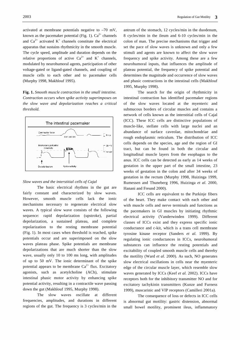
activated at membrane potentials negative to –70 mV, known as the pacemaker potential (Fig. 1). Ca2+ channels and Ca2+ activated K+ channels constitute the electrical apparatus that sustains rhythmicity in the smooth muscle. The cycle speed, amplitude and duration depends on the relative proportions of active Ca2+ and K+ channels, modulated by neurohumoral agents, participation of other voltage-gated or ligand-gated channels, and coupling of muscle cells to each other and to pacemaker cells (Murphy 1998, Makhlouf 1995). Fig. 1. Smooth muscle contraction in the small intestine. Contraction occurs when spike activity superimposes on the slow wave and depolarization reaches a critical threshold.
Slow waves and the interstitial cells of Cajal
The basic electrical rhythms in the gut are fairly constant and characterized by slow waves. However, smooth muscle cells lack the ionic mechanisms necessary to regenerate electrical slow waves. A typical slow wave consists of the following sequence: rapid depolarization (upstroke), partial depolarization, a sustained plateau, and complete repolarization to the resting membrane potential (Fig. 1). In most cases when threshold is reached, spike potentials occur and are superimposed on the slow waves plateau phase. Spike potentials are membrane depolarizations that are much shorter than the slow wave, usually only 10 to 100 ms long, with amplitudes of up to 50 mV. The ionic determinant of the spike potential appears to be membrane Ca2+ flux. Excitatory agonists, such as acetylcholine (ACh), stimulate intestinal phasic motor activity by enhancing spike potential activity, resulting in a contractile wave passing down the gut (Makhlouf 1995, Murphy 1998).
The slow waves oscillate at different frequencies, amplitudes, and durations in different regions of the gut. The frequency is 3 cycles/min in the
antrum of the stomach, 12 cycles/min in the duodenum, 8 cycles/min in the ileum and 6-10 cycles/min in the colon of man. The precise mechanisms that trigger and set the pace of slow waves is unknown and only a few stimuli and agents are known to affect the slow wave frequency and spike activity. Among these are a few neurohumoral inputs, that influences the amplitude of plateau potential, the frequency of spike potential and determines the magnitude and occurrence of slow waves and phasic contractions in the intestinal cells (Makhlouf 1995, Murphy 1998).
The search for the origin of rhythmicity in intestinal contraction has identified pacemaker regions of the slow waves located at the myenteric and submucous borders of circular muscles and contains a network of cells known as the interstitial cells of Cajal (ICC). These ICC cells are distinctive populations of muscle-like, stellate cells with large nuclei and an abundance of surface caveolae, mitochondriae and rough endoplasmic reticulum. The distribution of ICC cells depends on the species, age and the region of GI tract, but can be found in both the circular and longitudinal muscle layers from the esophagus to the anus. ICC cells can be detected as early as 14 weeks of gestation in the upper part of the small intestine, 23 weeks of gestation in the colon and after 34 weeks of gestation in the rectum (Murphy 1998, Huizinga 1999, Rumessen and Thuneberg 1996, Huizinga et al. 2000, Hanani and Freund 2000).
ICC cells are equivalent to the Purkinje fibers of the heart. They make contact with each other and with muscle cells and nerve terminals and functions as the pacemakers in GI muscles by initiating rhythmic electrical activity (Vanderwinden 1999). Different classes of ICCs exist and they express specific ionic conductance and c-kit, which is a trans cell membrane tyrosine kinase receptor (Sanders et al. 1999). By regulating ionic conductances in ICCs, neurohumoral substances can influence the resting potentials and excitability of coupled smooth muscle cells and thereby the motility (Ward et al. 2000). As such, NO generates slow electrical oscillations in cells near the myenteric edge of the circular muscle layer, which resemble slow waves generated by ICCs (Keef et al. 2002). ICCs have receptors both for the inhibitory transmitter NO and for excitatory tachykinin transmitters (Kunze and Furness 1999), muscarinic and VIP receptors (Camilleri 2001a).
The consequence of loss or defects in ICC cells is abnormal gut motility: gastric distension, abnormal small bowel motility, prominent ileus, inflammatory
Hansen Vol. 52
4
bowel disease, chronic idiopathic intestinal pseudo-obstruction (CIP), GI stromal tumors and multiple GI autonomic tumors, achalasia, intestinal obstruction with hypertrophy in the small intestine, Hirschsprung’s disease, juvenile and adult intestinal pseudoobstruction, slow transit constipation, anorectal malformations, post-infectious and Chagas disease (He et al. 2000, Sanders et al. 1999, Hagger et al. 2000, Takayama et al. 2000, Camilleri 2001a, Porcher et al. 2002). Smooth muscle contraction
The smooth muscle of the gut exhibits two distinct types of contractions: tonic and rhythmic phasic contractions to cause mixing and propulsive movements (i.e. segmentation). The motility function of each type of contraction and the neurohumoral, electric, and cellular mechanisms that regulate them, are very different. The fact that the small intestine becomes dilated in CIP and during the administration of glucagon, indicates that some level of basal tone exists under normal fasting conditions and following biomechanical stimulation. Two kinds of tone exist in the gut: neurogenic and myogenic. The neurogenic tone results from a constant low discharge of excitatory innervation, while the myogenic tone results from a property of the muscle itself (Gregersen et al. 1992, Gregersen and Christensen 2000). The rhythmic phasic contractions produce mixing and slow distal propulsion of luminal content in the fasting and the postprandial states. The maximal frequency and direction of propagation of these contractions are regulated by slow waves. Release of neurotransmitters from the motorneurons determines whether the smooth muscle will contract, increase tone or not. In the presence of excitatory neurotransmitters, muscle cell membrane receptors are activated to produce excitatory junction potentials and subsequently increase smooth muscle contraction, and visa versa. The response to physiological excitatory stimuli such as bradykinin, ACh and prostaglandin F2α (PGF2α) are characterized by an increase in sensitivity to agonists with a more tonic nature of motility (Sanders et al. 1999). The distance of propagation of individual phasic contractions depends on the length of the segment over which the excitatory neurotransmitter is released concurrently, and the distance over which the slow waves are synchronized. Phasic contractions also occur as groups, generating the migrating motor complex (MMC) and migrating clustered contractions (Sarna et al. 2000, Hansen 2002).
Age and gender Women and elderly persons have impaired
esophageal motor function, gastric emptying and colonic transit as compared to males and younger healthy adults, respectively. Plasma levels of oestrogen and progesterone does not seem be of importance, as the stage of the menstrual cycle does not affect gastric or jejunal myoelectric activity (Madsen 1992, Gianaros et al. 2001). It is likely, but uncertain, that the gender and aging is also of importance for small intestinal motility. For example one manometric study showed that the postprandial motility did not display gender difference in any parameter examined and that the majority of patterns of motility are similar in menstruating women and men, whereas certain aspects of the MMC, most conspicuously propagation velocity and phase III contraction amplitude, differ. The circadian variation of phase III contraction frequency is present in both women and men (Aytug et al. 2001). Another study showed more frequent cluster contractions and slower migration of the MMC in older subjects during fasting and postprandially (Husebye and Engedal 1992, Hansen 2002). However, the rest of basic patterns of fasting and stimulated motility and transit time are maintained throughout the process of aging and without any gender differences (Husebye and Engedal 1992). In another study, using a different method, women had slower small intestinal transit than men (Sadik et al. 2001). These differences could be due to lack of full relationship between myoactivity and transit time studies. 3. Regulation of gut motility
The regulation of smooth muscle activity and gut motility takes place at several levels. Hormones and neurotransmitters are the dominating components, which act and interact directly and indirectly on muscle cells. The use of knockout animals, in which the development or synthesis of particular neurohumoral transmitter substances or receptors has been prevented, has turned out to be a significant novel tool for studying neurohumoral control of gut motility (Spencer 2001).
The hormonal influence and the interplay with the ENS, takes place after and in between meals. The postprandial endocrine response includes release of insulin, neurotensin, cholecystokinin (CCK), gastrin, glucagon-like-peptides (GLP-1 and GLP-2), glucose-dependent insulinotropic polypeptide (GIP, previously known as gastric inhibitory peptide), but not motilin nor somatostatin (Medhus et al. 1999). These released hormones have all been demonstrated to have functional
2003 Regulation of Gut Motility
5
importance for digestion. For example, CCK is released into the circulation from the upper small intestine, causing direct contraction of muscle cells in the gallbladder and neurally mediated relaxation of muscle cells in the sphincter Oddi, which is mediated by VIP at the neuromuscular junction.
There is also hormonal release and subsequent neural activation arising within the lumen from the mechanical and chemical properties of food and digestive
secretions. Hormones are released locally from endocrine cells in the mucosal lining (e.g. 5-HT from enterochromaffin cells, EC cells) and modulate motility by activating receptors on sensory fibers, the extrinsic (e.g. vagal) and intrinsic primary afferent neurons (IPANs), and again back on the endocrine cells in an autoregulatory fashion. This system conveys sensory information, so the CNS can evaluate GI activity and modulate motility accordingly (Fig. 2 and 3).
Fig. 2. 5-HT and sensorimotor function. 5-HT is released from mucosal sources. 5-HT induce peristaltic motor activity. Intrinsic and extrinsic neuronal excitatory and inhibitory pathways and several 5-HT receptors are involved in this process. Acetylcholine, substance P, nitric oxide, vasoactive intestinal peptide and calcitonin gene-regulated peptide is involved in the processes. See Fig. 3 for further details.
Fig. 3. The peristaltic reflex in the small intestine. Following mucosal stimulation, 5-HT is released from enterochromaffin cells to intrinsic primary afferent neurons (IPANs with 5-HT1P, 5-HT3 and 5-HT4 receptors) and extrinsic vagal and spinal afferents (with 5-HT3 receptors). IPANs release substance (SP) acetylcholine (ACh), glutamate and calcitonin gene-regulated peptide (CGRP) to interneurons. Excitatory interneurons release SP and ACh orally to excitatory motorneurons, while 5-HT and ACh is released aborally to inhibitory motorneurons. Excitatory motorneurons release SP and ACh to muscles, while inhibitory motorneurons release nitric oxide (NO), vasoactive intestinal peptide (VIP) and adenosine triphosphate (ATP) to muscles. Efferent sympathetics release norepinephrine (NE), somatostatin (SOM) and neuropeptide I, while efferent parasympathetics release ACh (not shown).
Hansen Vol. 52
6
The neuronal regulation of GI motility involves
intrinsic as well as extrinsic nerves. The intrinsic innervation involves the enteric nervous system (ENS), which consists of ganglionated and non-ganglionated plexi. The extrinsic innervation involves the vagus nerve and splanchnic nerves to the stomach and upper intestine, while the pelvic nerves supply the distal intestines. Extrinsic neurons of the sympathetic and parasympathetic systems influence smooth muscle indirectly by acting on neurons of the myenteric plexus. However also neurons from the submucous plexus innervate the innermost layers of circular muscle, at least in large species. The smooth muscle cells form an electrical syncytium that is innervated by hundreds of excitatory and inhibitory neurons. In general, the control systems for motility are amazingly similar between species. Inhibitory stimuli (relaxing motorneurons) are exerted by VIP, pituitary adenylate cyclase activating polypeptide (PACAP) and NO, while excitatory stimuli (contracting motorneurons) are exerted by tachykinins, ACh and 5-HT. Again other neurotransmitters modulate the release of these transmitters from motorneurons. For example, reflex activation of myenteric neurons by stimuli, such as stretch or mucosal stimulation, causes the release of VIP, NO production, and muscle relaxation (Kunze and Furness 1999, Olsson and Holmgren 2001).
The GI tract is connected to the CNS through the autonomic nervous system (the brain-gut axis). The CNS is able to modulate, but not entirely control, the motor activity by sending instructions via the two components of the extrinsic autonomic nervous system: the sympathetic and parasympathetic nervous system. As such several peptidergic (e.g. opioids, thyrotropin-releasing hormone - TRH, corticotropin-releasing hormone - CRF, bombesin, calcitonin gene-regulated peptide - CGRP, neurotensin and CCK) and non-peptidergic (e.g. 5-HT, prostaglandins, dopamine and opioids) have been demonstrated to affect motility following stimulation directly of the CNS. For example, pancreatic polypeptide Y (PPY) is released into the circulation from the endocrine cell in the gut and directly influences central neuronal function and thereby gut motility (Rogers et al. 1995). The parasympathetic fibers transmit their instructions via the release of ACh to speed up motility, while the sympathetic fiber release norepinephrine (NE), somatostatin and neuropeptide Y (NPY) to slow motility. The activity of the autonomic nervous system is influenced by several factors,
including stress, emotion and eating. For example, nociception, following distension and inflammation of the gut, is mediated following the release of 5-HT and tachykinins and subsequent activating of neurogenic 5-HT1A, 5-HT3, 5-HT4 and neurokinin NK1, NK2 and NK3
receptors. They are activated in a very complex manner, some centrally, some peripheral, some stimulatory, some inhibitory, some in the stomach, some in the intestines, etc. (Gue and Bueno 1996).
In the small intestine, two populations of sensory neurons have been identified. The first, activated by mucosal stimuli, is wholly intrinsic, and the second, activated by muscle stretch (i.e. mechanosensory), has neuronal cell bodies in the dorsal root ganglia. Two types of mechanosensory neurons exist. Vagal afferents mediate physiological messages in low threshold Aδ and C fibers. Spinal afferents on the other hand mediates nociceptive messages in wide range of high threshold Aδ and C fibers. The IPANs in the myenteric ganglia have been identified to be of major importance for the physiological motor response to digestion. They are numerous and make direct contact with motorneurons and interneurons. The mucosa contains cells that facilitate GI function, such as the EC cells. EC cells release 5-HT and are endowed with at least stimulatory 5-HT3 and inhibitory 5-HT4 receptors, and maybe also stimulatory 5-HT1 and 5-HT2 receptors (Gebauer et al. 1993, Schworer and Ramadori 1998). The interneurons receive instructions from the sensory neurons, which forward information from the EC cells, and from postganglionic termini of extrinsic autonomic nerve fibers. The sensory neurons include connections to the mucosa and fibers that run through the circular muscles. Concentrated in the same areas are the ICC cells. The muscle cells in the longitudinal and circular muscle layer are innervated and therefore linked to the myenteric plexus. The longitudinal layer is innervated by excitatory motorneurons, whereas the circular layer is innervated by both inhibitory and excitatory motorneurons. Afferent extrinsic nerve fibers, which travel along the side of the autonomic nerves in the area, carry sensory information from the ENS, via the vagus and spinal cord, back to CNS (Kunze and Furness 1999).
Neurons in the myenteric plexus project their fibers to neurons in the same plexus, the submucous plexus and paravertebral ganglia, and to cells in the circular and longitudinal muscle layers. Neurons of the myenteric plexus of the small intestine fall into two broad categories: those that contain VIP with NO synthase, and
2003 Regulation of Gut Motility
7
those that contain SP with virtually no overlap of the two. VIP neurons also contain homologous peptides, e.g. peptide histidine-methionine. SP neurons also contain homologous peptides, e.g. substance K (SK). It is likely that ACh coexist with SP and SK in most neurons. Subpopulations of neurons within these categories contain one or more of the following: bombesin (i.e. gastrin-releasing peptide), NPY, the opioid peptides, dynorphin and met-enkephalin, and galanin. A subpopulation of neurons contain GABA, 5-HT or somatostatin. Neurons that contain 5-HT or somatostatin project their fibers exclusively within the myenteric plexus and influence smooth muscle cells only directly by means of other neurons. Receptors for most of these agents and others have been identified on smooth muscle cells of the gut. Some neurotransmitters have been identified as of specific importance for contraction and relaxation in a specific region of the gut. For example, NO and CCK seems important for relaxation of the lower esophageal sphincter (LES), the antrum and fundus of the stomach, the pylorus and the duodenum (Kuiken et al. 2002), while 5-HT and motilin are important for antroduodenojejunal contractions. Table 1. Dominating effect of some neurohumoral substances on intestinal contraction in vivo.
Stimulatory Inhibitory
ACh Adenosine Bombesin CCK GRP Histamin Motilin Neurokinin A Opioids PGE2 Serotonin SP TRH
Changes in the neurohumoral response to stimuli have been demonstrated in certain conditions involving dysmotility. In the carcinoid syndrome, small intestinal and colonic transit is accelerated (der Ohe et al. 1993), while in slow transit constipation, there is an increased secretion of proximal gut hormones and reduced secretion of distal gut hormones. Abnormal postprandial levels of motilin, CCK, neurotensin and somatostatin (Peracchi et al. 1999), and fewer colonic enteroglucagon- and 5-HT-immunoreactive cells are present (El Salhy et al. 1999). As such, treatment with neurotrophic factors improves gut motility for patients with constipation, by mainly increasing the sensitivity to excitatory transmitters and reducing the inhibitory innervation (Camilleri 2001a).
In general neurohormones can be divided into those primarily contracting or relaxing (Table 1), and predominantly modulating upper GI motility (CCK, PPY, gastrin, galanin, GLP-1, motilin, neurotensin and secretin) or lower intestinal motility (e.g. peptide YY) or both (e.g. 5-HT). 4. Serotonin
Serotonin (5-HT) is an important neurohumoral transmitter, that is synthesized and stored in several cell types, mainly in EC cells (90 %) and neurons (10 %) of the gut. 5-HT is released into the blood postprandially and in response to changes in pressure across the gut wall, as well as to noxious stimuli (Bearcroft et al. 1998). 5-HT is released into the gut wall from the basolateral stores of the EC cells and probably spills over into the lumen (Hansen and Skadhauge 1997). One of the reasons for the strategic location of the EC cell is the close proximity to the mucosal sensory nerve endings, and interganglionic neurons, which synapse on motor excitatory and inhibitory neurons.
Extensive investigations have been performed to determine the role of 5-HT in the physiological and pathological regulation of gut motility. However, the precise roles of 5-HT and 5-HT receptors are not fully understood yet. This is partly because of the large number of 5-HT receptor subtypes and their diverse locations and effects. The affinity of the neuronal receptors is much greater than that of smooth muscle. As a result the effects of administered 5-HT receptor agonists and antagonists are primarily as those resulting from the activation of neuronal receptors (Sarna et al. 2000). In the following, an attempt to summarize the current knowledge is presented.
Hansen Vol. 52
8
Exogenous intravenous 5-HT increases contraction amplitudes in the gastric antrum, duodenum, jejunum and ileum (Hopkinson et al. 1989, Nakajima et al. 1997). In the small intestine, 5-HT stimulates circular contractions during phase I of the manometric MMC, induce propagated contractions and more frequent and faster propagating MMC complexes (Ormsbee et al. 1984, Siegle et al. 1990, Valdovinos et al. 1993, Lordal et al. 1998, Hansen et al. 2000). Not only the circular muscles are stimulated by 5-HT, but also the longitudinal muscles in the human stomach, duodenum and jejunum (Fishlock et al. 1965). In the colon, exogenous intravenous 5-HT stimulates motility along the entire length, by inducing phasic contractions, but not giant motor complexes (GMCs) (Boeckxstaens et al. 1990, Nagakura et al. 1996a, Nagakura et al. 1996b). Also exogenous intraluminal 5-HT evokes hypermotility in animals and probably man (Gronstad et al. 1987, Ahlman 1992, own unpublished observations). Endogenous 5-HT
has been demonstrated to have similar effects as exogenous 5-HT. As such, selective 5-hydroxytryptamine reuptake inhibitors interferes with the stimulated esophageal motor responses (Boeckaert et al. 2001), reduce the interdigestive gastric phase III activity (Haga et al. 1996), reduce the mean MMC periodicity and increase the propagation velocity of phase III in the small intestine and reduce the orocecal transit time (Gorard et al. 1994) in healthy humans. The importance of serotonergic neurotransmission to the motility has also been demonstrated by experiments in which 5,7-dihydroxytryptamine was employed to selectively destroy serotonergic neurons. These experiments established that normal intestinal motility is diminished and transit down the bowel is slowed when serotonergic neurons are lost. The congenital loss of enteric serotonergic neurons, which occurs in mice with mutations in mash-1 gene, is associated with the lethal absence of intestinal motility (Gershon M, personal communication).
Table 2. Effect of 5-HT receptors on gastrointestinal motility. The overall dominating effect of 5-HT receptors is presented.
↓ - inhibition of motility or tone, ↑ - stimulation of motility or tone, ? - unknown effect. 5-HT receptors
A variety of 5-HT receptors have been identified and the locations and subtypes of these receptors vary among species. Fourteen different 5-HT receptors are classified into seven receptor subtypes. The roles of 5-HT1, 5-HT2, 5-HT3, 5-HT4 and 5-HT7 receptors have been studied in the gut (Fig. 2, Table 2). A great deal of work has also been done on 5-HT1P receptors, which might be similar or closely related to the 5-HT4 receptors. For 5-HT receptors located on smooth muscle cells, four types have been demonstrated: 5-HT2A, 5-HT2B, 5-HT4 and 5-HT7. Smooth muscle 5-HT receptors contract or relax: 5-HT2A and likely 5-HT2B receptors contract, while 5-HT4 and 5HT7 receptors relax. Neuronal 5-HT receptors enhance or inhibit transmitter release and thereby modulate contraction: 5-HT1A inhibits, while 5-HT3 and
5-HT4 (5-HT1P) receptors excite. Obviously, 5-HT receptors can therefore act in concert or have opposing effects. For example, 5-HT receptors coexist on smooth muscle cells in the human small intestine, where 5-HT2A receptors mediate contraction, while 5-HT4 receptors mediate relaxation (Borman and Burleigh 1997). Furthermore, the potencies of 5-HT receptor active agents are species and region-dependent.
An important aspect is the fact that most 5-HT receptors do not seem to affect normal function, but only in disease states. An example for this is 5-HT3 receptor antagonist, alosetron, which delays colonic transit in diarrhea-predominant (D)-IBS patients, but not in normals (Camilleri et al. 1999, De Ponti and Tonini 2001).
2003 Regulation of Gut Motility
9
5-HT1 receptor subtype There is growing evidence for the involvement
of 5-HT1 receptors in the control of gut motility. 5-HT1 agonism (e.g. by the 5-HT1A agonist, buspirone or the 5-HT1B/D agonist, sumatriptan) alters esophageal motility (Houghton et al. 1994) by preventing the natural decay in rate of transient LES relaxations postprandially (Sifrim et al. 1999, Foster et al. 1999). They also suppress gastric phase III activity (Tack et al. 1998), delay gastric emptying by increasing the lag period (Houghton et al. 1992, Coulie et al. 1997), increase fundic and antral relaxation (Vingerhagen et al. 2000), decrease postprandial antral motility (Coulie et al. 1997) in healthy volunteers, but induce duodenal phase III activity in patients suffering from abnormal upper GI motility (Mathis et al. 2001a). Most of these effects probably increase the occurrence of gastroesophageal reflux. In the small intestine, 5-HT1 agonism stimulate peristaltic activity (Buchheit and Buhl 1994), induce premature jejunal phase III activity and shortens the cycle length of the MMC on the expense of phase II activity (Coulie et al. 1997, Tack et al. 1998, Coulie et al. 1999, Tack 2000, Tack and Vanden Berghe 2000). Confusing the picture, sumatriptan in a recent study, increased the length of the MMC in both the stomach and duodenum by prolonging phase II, but not phase III (Calvert et al. 2001). In summary, the net effect of 5-HT1 agonism on antroduodenal MMC in man seems opposing. 5-HT1D receptors mediates contraction in the circular layer, while 5-HT1B receptors mediates contractile response to 5-HT in the longitudinal layer of the small intestine (Borman and Burleigh 1997). However, the non-selective 5-HT1 receptor antagonist, methiothepin, does not affect the response to 5-HT on circular contractions during phase I in the small intestine (Boeckxstaens et al. 1990). Maybe selective novel 5-HT1B/D receptor antagonists (e.g. GR-127935) will be of clinical importance (De Ponti and Tonini 2001). 5-HT1P receptors are present on submucous primary afferent neurons in the guinea-pig small intestine (Pan and Gershon 2000) and 5-HT1P antagonists inhibits atropine, hexamethonium and xylocaine-induced phase III contractions in dog jejunum (Tohara et al. 2000). Further data supporting the existence and a role for the 5-HT1P receptor in gut motility has been provided in rat colon, where 5-HT released by mucosal stimuli initiates peristalsis by activating sensory CGRP neurons (Grider et al. 1996). Whether these effects are mediated by the 5-HT4, and not by the 5-HT1P receptor, is still unclear. Finally, 5-HT1A receptor agonists relax and inhibit
colonic and rectal motility (Nagakura et al. 1996a and 1996b, De Ponti and Tonini 2001). 5-HT2 receptor subtype
The net effect of exogenous 5-HT is contraction of the stomach and intestines, reflecting the contracting 5-HT2A receptors present on smooth muscle cells (Boeckxstaens et al. 1990, Kuemmerle et al. 1995, Janssen et al. 2001). 5-HT activates 5-HT2A and 5-HT2C receptors located on postsynaptic cholinergic neurons in dog jejunum to stimulate phasic contractions and phase III activity (Graf and Sarna 1996), while the 5-HT2B receptor mediates contraction of longitudinal muscle in human ileum in vitro (Borman and Burleigh 1995). 5-HT also induces colonic and rectal contractions primarily by stimulation of smooth muscle 5-HT2A
receptors (Prins et al. 1997), and by stimulation of 5-HT2B receptors, which are expressed on both myenteric neurons and on smooth muscles (Borman et al. 2001). Despite these findings, 5-HT2 receptor antagonists, such as ketanserin, have been without convincing effects on the motility response to 5-HT (Boeckxstaens et al. 1990). 5-HT3 receptor subtype
The action of endogenous 5-HT on gastric activity is mediated partly by 5-HT3 receptors, as ondansetron inhibits gastric phase III activity and zacopride reduces the pyloric motor response to intraduodenal hydrochloric acid (Wilmer et al. 1993, Haga et al. 1996, Nakajima et al. 1997). 5-HT3 antagonists have failed to normalize gastric emptying for patients with gastric stasis or anorexia nervosa. However in reflux disease patients, 5-HT3 receptor antagonism, using granisetron, reduce gastroesophageal reflux by improving the function of the LES (Gonlachanvit et al. 2001). Thus 5-HT3 receptors seem to participate to some extend in the regulation of gastric emptying in man. Apparently stimulation of 5-HT3
receptors or their antagonism, affects small bowel transit and motility differently according to the species and segment. In guinea pig circular muscle of small intestine, 5-HT3 receptors play a role in the ascending excitatory reflex and these receptors may be on interneurons in the reflex pathway (Furness et al. 1993). In addition, ondansetron inhibits 5-HT3-mediated ACh release in guinea-pig ileum longitudinal muscle-myenteric plexus strips (Fox and Morton 1990). In the isolated mouse ileum, 5-HT3 receptors mediate 5-HT-induced contraction, however with a different
Hansen Vol. 52
10
pharmacological profile to that reported in guinea pig ileum (Tuladhar et al. 2000). In the rat YM-31636, a novel selective 5-HT3 agonist, increase GI motility, which is inhibited by ramosetron, which is a novel 5-HT3 receptor antagonist (Kiso et al. 2001). In dog small intestine, activation of the 5-HT3 receptor induce ascending contractions through an enteric excitatory pathway (Mizutani et al. 1992), and 5-HT3 receptor antagonists inhibits the digestive jejunal phase III contractions (Tohara et al. 2000). The excitatory pathway is formed by a series of cholinergic interneurons and motorneurons, apart from the anal contraction (Mizutani et al. 1992). The 5-HT3 receptor also regulates the ileocolonic junction, as 5-HT3 receptor agonists activate an ACh-mediated contraction and a relaxation mediated by an as yet unknown non-adrenergic-non-cholinergic (NANC) neurotransmitter (Boeckxstaens et al. 1990). In healthy volunteers, 5-HT3 receptor antagonists have no major impact on small intestinal transit time or mouth-to-cecum transit time (De Ponti and Tonini 2001), but selectively increase the interval between MMCs (Bush et al. 2000). PPY and neurotensin could be involved in this response, since the 5-HT3 receptor antagonist, GR 38032F, reduce postprandial PPY and neurotensin plasma levels (Talley et al. 1989). For colonic effects, a ondansetron-sensitive mechanism participates in guinea pig (Jin et al. 1999) and in the physiological contractile response in the young healthy human transverse and descending colon after ingestion of a meal (der Ohe et al. 1994). Ondansetron by itself does not affect fasting colonic tone or phasic contractions, but antagonism of 5-HT3 receptors blunts the gastrocolonic response, by actions on vagal afferent neurons, as well as in area postrema. Ondansetron retards colonic transit and inhibits the colonic motor response to meal in health without age or gender differences, but has no effect in diarrhea-dominated irritable bowel syndrome (D-IBS) patients (Talley et al. 1990). This could be because ondansetron has little effects on the increased frequency of GMCs in D-IBS (De Ponti and Tonini 2001). In carcinoid patients, ondansetron reduce postprandial colonic hypertonic response to normal level (der Ohe et al. 1994), suggesting ondansetron for treatment of carcinoid diarrhea. In summary, 5-HT3 antagonists may retard colonic transit in health by altering phasic contractions. 5-HT3 receptors, however, do not seem to be involved in the stimulation of GMCs in the colon or the small intestine. 5-HT3 receptor antagonists may, therefore, be
ineffective in retarding transit, if diarrhea is caused by an increase in the frequency of GMCs. 5-HT4 receptor subtype
5-HT4 receptors have been identified on myenteric neurons and muscles in the fundus, corpus and antrum of the stomach (Taniyama et al. 2000). 5-HT4 receptors do not seem to directly mediate nausea and vomiting, however if these symptoms are a result of delayed gastric emptying, they may be involved. In general, 5-HT4 receptor agonists stimulate gut motility. However, depending on the agonist, 5-HT4 receptor agonism has varying effects on gastric motility. 5-HT4 receptor agonists, SDZ-HTF-919 and procalopride, does not alter gastric emptying in dog or man (Nguyen et al. 1997, Bouras et al. 1999, Bouras et al. 2001). Tegaserod, another 5-HT4 receptor agonist, however accelerates gastric emptying, small intestinal and maybe colonic transit in man in health (Camilleri 2001b, Degen et al. 2001), accelerates gastric motility in diabetic mice (Mathis et al. 2001b) and postoperatively in rats (Zittel et al. 2000). However, in constipation-predominant (C)-IBS patients, tegaserod does not change gastric emptying (Prather et al. 2000, De Ponti and Tonini 2001). In the intestines, relaxing 5-HT4 receptors are present on intestinal circular and longitudinal smooth muscle cells, sensory CGRP-containing neurons (Foxx-Orenstein et al. 1996) and on NANC neurons, where they mediate relaxation by suppressing the amplitude and duration of phase III activity (Graf and Sarna 1996). 5-HT4 agonism with ML-10302 evokes myoelectric activity in dog small intestine (De Ponti et al. 2001). However small intestinal transit is not altered by SDZ-HTF-919 in dog (Nguyen et al. 1997), nor by prucalopride in healthy humans (Bouras et al. 1999). However, tegaserod accelerates small bowel transit in healthy volunteers (Camilleri 2001b) and stimulates the peristaltic reflex and increase postoperative motility in the small intestine of rodents (Buchheit and Buhl 1991, Zittel et al. 2000). Tegaserod reduce orocecal transit time in C-IBS patients (Scott and Perry 1999, Camilleri 2001b), while the 5-HT4 antagonist, SB-207266A (piboserod, now available for human studies), increase orocecal transit time towards normal in D-IBS patients (Houghton et al. 1999), but does not seem to affect the normal motility (De Ponti and Tonini 2001). Other 5-HT4 agonists, such as RS-67333, dose-dependently shortens the interval of phase III on the MMC in rat (Lordal M, personal communication) and increase phase II activity in dog small intestine (Grider et al. 1998). SB
2003 Regulation of Gut Motility
11
203-186, a selective 5-HT4 receptor antagonist, inhibits 5-HT-induced contractions in circular muscle strips of the equine ileum (Weiss et al. 2002), while GR 113808, another selective 5-HT4 receptor antagonist, itself disrupts the MMC and cause irregular spiking, but does not block the 5-HT-induced effects (Lordal M, personal communication). In the colon, 5-HT4 agonists accelerates motility and transit in guinea pig (Jin et al. 1999), dogs (Nguyen et al. 1997), healthy volunteers and C-IBS patients (Appel et al. 1997, Prather et al. 2000, Camilleri 2001b, Lefkowitz et al. 2001) without cardiac side effects (Rueegg et al. 2001). 5-HT4 receptors are likely to be involved in stress-induced colonic dysmotility. In dogs, prucalopride induces GMCs and causes proximal colon stimulation and distal colon inhibition of contractile motility (Briejer et al. 2001a). Prucalopride also accelerates colonic transit primarily by accelerating proximal colonic emptying, without affecting visceral sensitivity (Poen et al. 1999) in healthy human subjects (Bouras et al. 1999, Bouras et al. 2001). Prucalopride facilitates colonic motility through circular muscle relaxation and longitudinal muscle contraction (Prins et al. 1999, Borman et al. 2001). Various reports indicate that the 5-HT4 agonists increase stool frequency in a subset of patients with idiopathic constipation, primarily in the proximal and transversing colon, by stimulating GMCs and therefore segmental contractions, but not peristalsis (Yamato et al. 2001). The locus for this activation is not completely known. Segmental differences could exist, as prucalopride induce GMCs and stimulates motility in the proximal colon, while inhibiting in the distal colon (Briejer et al. 2001b). Finally, 5-HT4 receptors also seems involved in rectal motility, since agonism of 5-HT4 receptors induce relaxation of canine isolated rectum smooth muscle (Prins et al. 1999).
In summary, 5-HT4 receptor agonists are prokinetics and exhibit promise in accelerating gastric emptying and colonic transit, although the precise mechanism is not known, but an increase in GMC's activity is likely. 5-HT4 agonists may act on sensory neurons, motorneurons, or interganglionic neurons to achieve these effects. The differences in effect among the 5-HT4 agonists is probably a result of tachyphylaxis, as procalopride, which is a full agonist, seems to have less potential for tachyphylaxis than partial 5-HT4 agonists, such as tegaserod. Similar findings are present for cisapride versus metoclopramide on gastric emptying (Camilleri 2000).
5-HT7 receptor subtype 5-HT7 receptors have been identified in most
parts of the intestine (Hemedah et al. 2000, Krobert et al. 2001). 5-HT7 receptor agonists relax smooth muscles in the distal gut and as such inhibits colonic and rectal motility and mediates relaxation in man (Vanhoenacker et al. 2000, Borman et al. 2001, De Ponti and Tonini 2001). It might have opposing effects in the proximal gut, since 5-HT7 agonism induces jejunal contractions in the rat following activation of prejunctional receptors (McLean and Coupar 1996). Mediators of 5-HT-induced motility
5-HT activates microcircuits in the ENS, which in turn initiates peristaltic reflexes (Figs 2 and 3). 5-HT-induced contraction of circular muscles is blocked completely by atropine and hexamethonium in animals, indicating that 5-HT stimulates intestinal contractions almost entirely through the release of ACh at the neuromuscular junction. However, in humans, atropine only reduces phase III activity of the MMC in the small intestine (own unpublished observations). Activation of cholinergic neurons acting at a presynaptic site and at least one nicotinic synapse seems involved (Fox and Morton 1990, Mizutani et al. 1992, Taniyama et al. 2000), while in the colon, muscarinic M3 receptors are involved (Prins et al. 2001). In addition, NO (Coulie et al. 1999), tachykinins (Sarna et al. 2000) and CGRP (De Ponti and Tonini 2001) and maybe a unknown NANC neurotransmitter (Boeckxstaens et al. 1990) are involved as intermediate neurotransmitters for 5-HT signaling. Differences may exist between the small intestine and the colon, since tachykininergic pathways are involved in 5-HT4 receptor agonism (ML-10302) mediating colonic motor response in the dog, but not in the small bowel (De Ponti et al. 2001). 5. Other neurohumoral substances (Table 1)
ACh is well described as the major regulator of gut motility mainly through muscarinic M1 (Nelson et al. 1996) and M3 receptor contractile mechanisms (Olsson and Holmgren 2001). Two types of receptors for adenosine coexist on smooth muscle cells of the intestines. A2 receptors mediate relaxation through the increase of cAMP, while A1 receptors contract due to decrease in cAMP and mobilization of Ca2+. The net effect of adenosine is contraction, which can be greatly augmented if A2 receptors are blocked (Makhlouf 1995). Bombesin and CCK are stimulatory transmitters of
Hansen Vol. 52
12
contraction of primarily the circular layer of the intestines. However exogenous CCK relaxes the LES and the stomach, while endogenous CCK relax colonic motility (Scarpignato 1996). Part of the CCK effect is due to a concomitant release of ACh and SP. In surprise, most studies have however turned out negative for effects of CCK antagonists on improving gastric emptying (Liddle et al. 1986). In addition to a direct effect on the smooth muscle, also a central mechanism is likely for bombesin and CCK. As such, endogenous CCK in the paraventricular nucleus of the hypothalamus modulates colonic motility via CCK-B receptors (Monnikes et al. 2000a). This seems be a physiological effect, since CCK is mainly released by food intake. The action of CCK on intestinal motility follows a biological rhythm related to the light-dark cycle. CGRP is found in both the myenteric and submucous plexi and supply all layers of the intestinal wall along the entire gut (Rasmussen et al. 2001a). Only a small proportion can be observed in the mucosal endocrine cells, lamina propria, the muscularis mucosae, or in the circular and longitudinal outer smooth muscle layer (Timmermans et al. 1992). The effect of CGRP on gut motility is predominantly relaxation although the different studies indicate differences between species. For example, CGRP stimulates antral and ileal motility in the isolated perfused pig model in an atropine-sensitive, but NK1 and NK2 antagonist receptor antagonist-insensitive manner (Rasmussen et al. 2001b). Therefore, the site of excitation of CGRP on the contractile activity is probably on intramural cholinergic neurons rather than direct on the smooth muscle cells. Furthermore, CGRP seems to mediate 5-HT4 receptor induced afferent signals from the IPANs to induce the peristaltic reflex and motility (Grider et al. 1998). CRF is released from many sources in the body, including the CNS, the adrenal glands, immune cells, ENS and the EC cells. CRF slows gastric emptying and small intestinal transit, but increases colonic transit and defecation in healthy volunteers and causes an exaggerated colonic motility response in IBS patients (Monnikes et al. 2001). These actions are believed to be due to modulation of the vagal and sacral parasympathetic outflow. The mechanisms through which CRF activates colonic motor function seems to involve CRF1 receptor activation of myenteric ganglia as well as circuitry recruiting 5-HT and mast cells (Karalis et al. 1991, Schafer and Mestres 1997, Fukudo et al. 1998, Anton 1999). The role of cytokines has not yet been finally established (Vrees et al. 2002). The involvement of eicosanoids in gut motility now seems clear, since cyclooxygenase (COX) inhibitors
induce duodenal motility in rats, suggesting eicosanoids to exert a tonic inhibitory action on duodenal motility (Nylander et al. 2001). Furthermore, prostaglandins (PGE2, PGI2 and PGF2α) excite secretomotor and interneurons in the submucous plexus of the small intestine (Frieling et al. 1994). Two types of receptors for leukotrienes have been identified on gastric muscle cells, a specific receptor for leukotriene C4 and a separate common receptor for leukotrienes D4 and E4. All three leukotrienes cause contraction through an increase in IP3 and cytosolic Ca2+. The influence of these agents, as for other substances (e.g. histamine and 5-HT), is likely to be most pronounced when the smooth muscle is inflamed or hypersensitized. The cells from which they are released are in proximity to muscle cells and to myenteric neurons and their terminals. These agents can act directly on muscle cells and indirectly by stimulating or inhibiting the release of neurotransmitters and in this fashion influence motility (Makhlouf 1995). Galanin cause relaxation of the ileum via action on myenteric neurons (Ren et al. 2001). Gastrin relaxes the fundus and increase gastric wall compliance (Mearadji et al. 1999). GLP-1 and GLP-2 are secreted from L-cells in the distal small intestine and colon in response to ingestion. They inhibit fasting and postprandial gastric and antroduodenal motility and stimulate tonic and phasic contractile activity of the pylorus thereby probably mediating the ileal brake. They seem to mediate their effects by activating specific GLP receptors, which may also be located on neuronal structures. They inhibit cholinergic pathways probably by receptors at vagal and circumventricular organs in the CNS (Blazquez et al. 1998, Wettergren 2001). Glutamate influence gut motility but its overall effect and the receptors involved is unknown (Kirchgessner 2001). Ghrelin, a recently discovered peptide, stimulates gastric contractility via a vagal pathway (Chen et al. 2001). Interleukin-1 beta decreases ACh-induced intestinal contraction in a VIP-dependent manner (Aube et al. 2001). H1 and H2 histamine receptors coexist on smooth muscle cells of the gut. H1 receptors mediate contraction and H2 receptors mediate relaxation. The net effect of histamine is contraction, reflecting the dominant influence of H1 receptors. This has importance for the function of the gallbladder and the sphincter Oddi (S0), as they contain neuronal plexi, that are distinct from those of the intestines (Keinke et al. 1986). Species differences in the neurogenic control of SO are also present. For example, in man, histamine does not change SO tone, while in pig, histamine stimulates contraction by activating H1 receptors (Sand et al. 2000). Neurohumoral
2003 Regulation of Gut Motility
13
agents that act at the brain level to affect gut motility include 5-HT (5-HT1A and 5-HT2 and 5-HT3 receptor agonists), the adrenergic system (α2 receptor antagonists), eicosanoids (prostaglandin receptor agonists) and dopamine (D1 and D2 receptor agonists and D2 receptor antagonist) (Gue and Bueno 1996). Motilin is secreted from the endocrine (EC and non-EC) cells of the mucosa of the upper jejunum. Motilin, and its agonist erythromycin, stimulates gut contractility and regulates the interdigestive motility by triggering phase III activity and increase gastric motor activity in the stomach (Bruley et al. 1995) and directly excites circular smooth muscle from the human colon (Van Assche et al. 2001). The motilin receptor has been identified as a GTP protein binding receptor, which is located throughout the ENS and on gut smooth muscles (Depoortere and Peeters 1997), with decreasing density from the stomach to the lower intestinal tract. However, a centrally mediated effect of motilin is also likely, since motilin and motilin binding sites are also present in the cortex of the brain (Depoortere and Peeters 1997, Pandolfino et al. 2000). Despite these findings, the precise stimulus for motilin release remains unknown (Pandolfino et al. 2000) and treatment of dysmotility with motilin agonists has in large been unsuccessful in studies in man. Neurotensin slows gastric emptying and duodenal motility (Keinke et al. 1986), but contracts the colon (Azriel and Burcher 2001a and 2001b). NPY, somatostatin and opioids are inhibitory transmitters as they inhibit the release of excitatory transmitters in the ENS. However, NPY applied to the hypothalamus, stimulates colonic transit by peripheral cholinergic and central CRF pathways (Monnikes et al. 2000b). Opioids are considered to exert their motor stimulatory actions through mechanisms involving µ-, δ-, κ-receptors targeting the middle part of the small intestine. β-Endorphin and morphine acts mainly trough muscular µ-receptors, while methionine-enkephalin acts via neuronal δ-receptors and dynorphins through neuronal κ-receptors (Makhlouf 1995). VIP, PACAP and NO all causes relaxation and a unique interplay exist between these major relaxants (Yamamoto et al. 1999, Ekblad and Sundler 1997a and 1997b). Neural stimulation elicits simultaneous VIP release and NO production, which results in a separate and additive effect on muscle relaxation. NO is one of the inhibitory transmitters in the NANC neurons, which regulate gut motility. NO seems to regulate the MMC pattern, since reduction of NO production in healthy volunteers triggers the onset of phase III in the small intestine (De Man et al. 2000, Kuiken et al. 2002) and in the colon (Powell and
Bywater 2001). The interplay of somatostatin, opioids, and GABA neurons is expressed in the regulation of VIP release and NO production during the descending relaxation phase of peristalsis. GABA also relaxes the LES (Pandolfino et al. 2000). A recently discovered gut peptide located in human colonic myenteric neurons (Ehrhardt et al. 2001), orphanin FQ/nociception, has variable effects on stomach, small intestinal and colonic motility (Osinski and Brown 2000). Oxytocin is released as a neurotransmitter and excites dorsal vagal neurons in the inhibitory pathway to the stomach, while TRH activates the excitatory pathway to the stomach (Rogers and Hermann 1987, Wood 1995). PACAP is, together with NO and VIP, mediators of the inhibitory excitatory neurons and interneurons in the inhibitory arm of the peristaltic reflex in the small intestine (Camilleri 2001c). However, in the isolated model, it induces motility in the antrum of the pig stomach with a concomitant release of SP, VIP and somatostatin (Tornoe et al. 2001). Secretin is released from the mucosal S cells of the duodenum and inhibits pyloric, gastric antrum, small and large intestinal motility in addition to inducing biliary and pancreatic secretion (Leither et al. 1994). Tachykinins and their receptors are located on enteric neurons and smooth muscle cells to regulate the GMCs and retrograde contractions. SP slows small intestinal motility in the rat (Valdovinos et al. 1993). NK1 receptors are located on small and large intestinal motorneurons (Bian et al. 2000), and colonic circular smooth muscle cells and mediate colonic GMCs, whereas NK3 receptors are located on presynaptic neurons to mediate small intestinal GMCs (Sarna 1999) and colonic propulsion (Onori et al. 2001). In addition to NK1 receptors, also NK2 receptors have been identified directly on muscle cells, mediating contraction (Goldhill et al. 1999). Xenin, another recently discovered peptide produced by specific endocrine cells of the duodenal mucosa, induce duodenojejunal phase III activity in man in both the interdigestive and postprandial state (Feurle et al. 2001). 6. Whole gut motility – the importance of reflexes
Reflexes are present all the way down from the
pharynx to the anus, and encounter the ENS with signaling locally and over long distances involving the prevertebral ganglia (Xie et al. 1997). As such prevertebral sympathetic ganglia, vagal nerves and intermesenteric nerves transmit signals for entero-
Hansen Vol. 52
14
enteric inhibition and stimulation of intestinal motility (Szurszewski and Miler 1994).
The peristaltic reflex can be evoked by stroking or by circumferential stretch. The reflex consists of two components: descending relaxation caudal and ascending contraction oral to the site of stimulus. The contraction of the longitudinal muscle during the initial stages of filling is the “preparatory phase” and the propagating wave of the circular muscle contraction is called the “emptying phase” of peristalsis. This circular muscle activity involves myogenic, neuro-mechanical and a yet unknown mechanism (Brookes et al. 2001). Following stroking of the mucosa, the EC cells in the mucosa releases 5-HT, which binds to receptors located on the sensory neurons. The 5-HT3 and 5-HT4 receptors seems to mediate at least part of this response, as for example selective 5-HT4 agonists applied to the mucosa trigger the peristaltic reflex (Fig. 3) in human, rat and guinea-pig intestine (Grider et al. 1998). When activated, these sensory neurons release neurotransmitters (CGRP, ACh and SP) at the excitatory interneurons. This action along with the release of neurotransmitters from interneurons that arise distal and proximal to the area of stimulation activates the interneurons to release neurotransmitters to the excitatory and inhibitory motorneurons. The motorneurons, by the release of either ACh/tachykinins or NO/VIP, then cause the associated muscle cells to modify their spike potentials, allowing a propulsive wave to pass down the gut (Camilleri 2001a). NO provides a tonic inhibition, while the final mediator at the neuromuscular junction for the descending relaxation is probably VIP. In this manner, transmitters in the two major populations of motorneurons in the myenteric plexus regulate descending relaxation and ascending contraction. In summary, the oral excitatory component of the peristaltic reflex is probably mediated by ACh (mainly nicotinic receptors) and tachykinins (mainly SP via NK receptors), whereas the aboral inhibitory components is to be mediated by the release of VIP and NO. Other neurons and transmitters (for example ATP and GABA) seem to be involved in the peristaltic reflex released by stretch. This has been demonstrated in guinea-pig small intestine, where the motor response to NK1 receptor agonists involves release of ATP in addition to NO from the inhibitory motor neurons (Lecci et al. 1999, Shahbazian and Holzer 2000). Stretch activates aborally projecting somatostatin neurons, which then stimulate GABA neurons. GABA is an excitatory modulator of ascending
contractions in the myenteric plexus and participator of the peristaltic reflex by modulating the release of transmitters from motorneurons. Thus, release of somatostatin and GABA increase during descending relaxation, while the release of opioid decrease. This inhibited opioids activity results in VIP release and relaxation.
There is an extensive reflex modulation of gastric emptying and motor activity from distant regions of the gut and visa versa. For example, gastric distension abolishes fasting duodenal and jejunal motor activity (the gastroenteric reflex). The enterogastric reflexes involve an interaction of humoral responses with intrinsic and extrinsic (vagal) afferent and efferent neural pathways. For example, activation of 5-HT1A and 5-HT2 receptors block the duodenogastric inhibitory reflex elicited by duodenal distension (Bueno et al. 1997). These extended reflexes play an important role in regulating gastric emptying through excitatory or inhibitory effects on the fundus (the enterofundus reflex), antrum (the enteroantral reflex) and pylorus (the duodenopyloric reflex). The degree of inhibition of gastric emptying by intestinal feedback is dependent on the length and region exposed to the stimulus. During acute ileitis, as an example, contractility of the fundus is inhibited (Moreels et al. 2001). Maximal inhibition of liquid emptying from the stomach is seen with exposure of the most proximal 150 cm of small intestine to acid, glucose or oleic acid. This classical reflex is the intestinal brake, which is mediated by release of duodenal CCK, gastric gastrin, CGRP and maybe 5-HT, GLP-1, tachykinins and VIP (Olsson and Holmgren 2001). Ileal perfusion of especially lipids reduces gastric emptying, duodenal and jejunal motility and transit. These inhibitory effects define a phenomenon known as the ileal brake. The ileal brake probably serves a protective physiologic function to prevent the distal intestine from being overwhelmed by massive nutrient loads. In agreement with this theory, a high MMC frequency and increased cluster activity is present for patients with short bowel syndrome (Husebye 1999). Mediators of the ileal brake are incompletely characterized. The presence of ileal lipids in association with a meal is known to release enteroglucagon, neurotensin, and PYY, although the precise role of these mediators in the ileal brake is unknown (Husebye 1999). α1-Adrenergic and β1-adrenergic, CCK, opioids (naloxone) and 5-HT3 receptor antagonists reduce ileal lipid-induced motor inhibition and an intact extrinsic innervation seems necessary to inhibit the small intestinal motility (Balsiger et al. 2001). A reflex circuit known as
2003 Regulation of Gut Motility
15
the vago-vagal reflex involving the nucleus tractus solitarius, underlies moment to moment adjustments required for optimal digestive function in the upper gut. Under normal conditions, retroperistaltic contractions in the duodenum propel intestinal contents orally, known as the duodenogastric reflex, inducing duodenogastric reflux of bile, digestive enzymes and bicarbonate. Just as small intestinal stimulation can modulate gastric motility, perturbations of the stomach can alter small intestinal motor patterns. In contrast to the peristaltic reflex, the intestinointestinal reflexes are not mediated by mucosal receptors, but depends on the extrinsic innervation. Clinically, if there is distension due to mechanical obstruction or another cause, the bowel responds with a decrease in motility and tone. These intestinointestinal reflexes serve a protective function. Other inhibitory reflexes exist, such as the peritoneogastric reflex, which slows gastric emptying following stimulation of the peritoneum for example during a laparotomy. The rectocolonic/rectoanal and colointestinal reflexes are activated by rectal and colonic distension, respectively. They retard colonic and small bowel transit and emptying of digestants into the cecum, respectively (Makhlouf 1995, Sanders et al. 2000, Kunze and Furness 1999, Husebye 1999).
Only few positive forward reflexes have been identified, such as the gastroduodeno/jejunal/ ileal/colonic reflexes, which are characterized by an increase in myoelectric, motor and propulsive activity in the intestines postprandially. The gastroduodenoileal reflex is abolished by intestinal transection, indicating that the reflex is mediated by intrinsic neural pathway. Another positive forward reflex is the jejunal-ileal reflex, which accelerates ileal motility in response to entry of chyme in the jejunum (Husebye 1999). Similar positive forward integrated motor responses includes the colocolonic reflex (Makhlouf 1995, Hasler 1995, Kunze and Furness 1999, Husebye 1999, Sanders et al. 2000).
5-HT fits perfectly the role as key mediator of most of these reflexes, as it is located in the ”tasting” EC cells of the mucosa in the whole gut and it is an important neurotransmitter for the interneurons. Tachykinins also seems important, as demonstrated in rat, where NK1 receptors mediate the rectocolonic inhibitory reflex with the involvement of central structures (Julia et al. 1999). 7. Motility of the stomach
During swallowing, a vagal-mediated transient receptive relaxation occurs, followed by a more
prolonged relaxation known as accommodation. However fullness and hunger is related solely to antral accommodation. The stomach can accommodate up to 2 l of fluids with no increase in pressure.
The motility patterns of the stomach is region-specific. The fundus and cardia (pacemaker region) generates tonic contractions, while the distal body and antrum exhibits phasic motor activity. Factors, in addition to age and gender, which come into play for gastric emptying includes the volume, caloric density and osmolarity of the injected. A 300 ml bolus of saline will empty twice as fast as a 150 ml load. In general, inert liquids empty rapidly with a time 50 % of emptying from 8 to 18 min. Liquid emptying of nutrients is tightly controlled to a rate that delivers approximately 200 kcal/h into the duodenum. Liquids with high caloric density empty more slowly than foods that have fewer calories per unit volume. An increase in osmolarity also decreases the contractility response in the small intestine. In addition to caloric density, the characteristics of the nutrient itself are important regulators of liquid emptying. Carbohydrates and most amino acids modulate intestinal nutrient delivery in part by an action on small intestinal mucosal osmoreceptors, which activate neural feedback inhibitory pathways. L-tryptophan, the precursor of 5-HT, is distinguished from the other amino acids in that it is effective at delaying liquid emptying, suggesting specific L-tryptophan receptors in the mucosa. The effect of triglycerides on gastric motility is dependent on the fatty acid chain-length with differences in their CCK-releasing ability (Hasler 1995). In spite of these experimental findings, drastic and rapid dietary changes do not seem to change gastro-duodenal motility, at least not in pigs (Boudry et al. 2001). The acidity of the gastric content also seems important. Omeprazol eliminates the temporal relationship between intragastric pH and characteristic of the MMC and induces delay in gastric emptying of liquid and solid meal (Rasmussen et al. 1999). Furthermore, the temperature of the injected bolus has an impact, as cold inhibits emptying.
Neurohumoral control
The neurohumoral modulation of gastric emptying for liquids and solids involves the presence of intact vagal innervation. However, vagal nerves are not necessary for the initiation or temporal co-ordination of global fasting or postprandial gastroduodenal motility patterns, but are involved in modulating the pattern of contractions during phase III. Numerous neurohumoral
Hansen Vol. 52
16
substances modify the rate of gastric emptying. At least 10 different types of neurons with different transmitter combinations are involved in the excitatory control of stomach motility. Myenteric neurons of the gastric wall contain numerous neurotransmitters, including ACh, norepinephrine, 5-HT, SP, VIP, peptide histidine isoleucin (PHI) and enkephalins. Excitatory neurons contain ACh, SP or both and project directly to the circular muscle layer to control contraction. Inhibitory motorneurons contain VIP and NO and project in aboral directions to control relaxation (Olsson and Holmgren 2001).
During fasting, proximal gastric tone is maintained by vagally mediated cholinergic input. After a meal the proximal stomach relaxes probably through the activation of nitrergic neurons in the gastric wall. NO-induced relaxation involves the production of cGMP. As such, sildenafil (a selective phosphodiesterase-5 inhibitor), prolongs the activity of cGMP and thereby modifies esophageal contractions, reduce LES pressure (Zhang et al. 2001), inhibits interdigestive motor activity of the antrum and duodenum (Bortolotti et al. 2001), increases intragastric volumes after a meal and slows liquid emptying rate (Bortolotti et al. 2001). Conversely, a nitrergic pathway does not seem to be involved in fasting gastric tone and sensitivity to gastric distension in man (Sarnelli et al. 2001). Opiates of different receptor subclasses have both inhibitory and excitatory effects. 5-HT accelerates via 5-HT3 receptors gastric emptying, while somatostatin, neurotensin, oxytocin, PYY, GRP, enteroglucagon, oxyntomodulin, and PGE1 all slow gastric emptying. The role of CCK is still unclear (Hasler 1995).
Neural input from the CNS is a potent regulator of gastric motor activity and emptying. Mental stress prolongs the periodicity of the MMC. Anger increases phasic motor activity in the stomach, whereas fear and depression reduce gastric contractions. Cold pain and ischemic pain all delay gastric emptying. Multiple neural pathways are involved in these stress responses. These mediators of brain-gut interaction have been evaluated intensively. Intraventricular infusion of TRH accelerates, while CRF, CCK, opiates, bombesin, tachykinins, somatostatin, atrial natriuretic factor, GABA, calcitonin, and CGRP delays gastric emptying, partly via a vagal mechanism (Hasler 1995). In addition to reduce LESP and the number of transient lower esophageal sphincter relaxations (Lidums et al. 2000, Sifrim et al. 1999), GABA and 5-HT1 receptor agonism
induce gastric accommodation following activation of probably myenteric neurons (Tack et al. 2001, Tack and Peeters 2001). The proximal stomach
The proximal stomach exhibits slow relaxations and contractions. The fundus relaxes for about 15 min after ingestion of a meal before returning to resting tone. This accommodation is neurally mediated and is mediated by stimulation of the tension-sensitive mechanoreceptors in the gastric wall. With surgical resection of the fundus or following fundoplication for gastroesophageal reflux disease, intragastric pressure increases and liquid emptying is enhanced. If the antrum is surgically resected, the initial phase of liquid emptying is accelerated, suggesting that the distal stomach, like the proximal part, play a role in liquid emptying. The motor activity of the duodenum also appears to regulate the rate of liquid emptying from the stomach, as enhanced liquid emptying is noted after performance of a circular myotomy of the duodenal wall.
Several neurohumoral agents have been demonstrated to relax the proximal stomach. CCK seems to be one of special importance, as CCK-8 blocks ascending contraction elicited by electrical field stimulation of duodenal mucosa by means of simultaneous activation of CCK-A and CCK-B receptors (Giralt and Vergara 2000). Other relaxants of the fundus include secretin, VIP, gastrin, somatostatin, dopamine, gastrointestinal insulin-dependent peptide, glucagon, and bombesin, whereas motilin and TRH increase pressure in the fundus (Hasler 1995, Mearadji et al. 1999). However, except for gastrin, their physiologic roles have not been clarified.
The distal stomach
The rhythmic synchronized contractions of the distal stomach are controlled by electrical signals generated by a pacemaker region located on the greater curvature at rate of about 3/min. While contractions of the distal stomach are always associated with gastric slow wave, the slow wave persists in the absence of gastric contractile activity. Normal fasting antral motility is cyclical and this is termed the MMC, which in average takes about 100 min (phase I – 40 min, phase II – 50 min and phase III – 10 min) (Hansen 2002). Neurohumoral pathways also affects motor activity of the distal stomach. For example stimulation of efferent vagal low-threshold fibers evokes increased antral
2003 Regulation of Gut Motility
17
contractile activity that is atropine-sensitive, indicating that cholinergic inputs predominates in the antrum (Katschinski et al. 1996). In contrast, stimulation of high-threshold fibers decreases antral motor activity, most likely through the release of VIP and NO, with NO being the primary mediator for the muscle-relaxing effect of VIP (Murthy et al. 1996). VIP and PACAP are present in interneurons that project caudal within the myenteric plexus in motorneurons that project into the circular muscle layer. In the pig, PACAP 1-38 induce antral motility in an atropine- and SP antagonist-sensitive manner (Tornoe et al. 2001). ACh, CCK, bombesin and some of the endogenous opiates also increase antral contractions. In contrast secretin, somatostatin, glucagon, GIP, GRP, TRH, neurotensin, and PGE2 inhibit antral motility (Hasler 1995). The pyloric sphincter
The pyloric sphincter (PS) is a bundle of thickened circular muscles, which exhibit characteristic patterns of motility under fasting and fed conditions. During phase III of the MMC, the pylorus remains open, and fasting gastric contents are permitted to exit into the duodenum. Under fed conditions, the pylorus exhibits a complex motor response of prolonged periods of closure, with duodenal mixing and retropulsion. Relaxation of the PS is produced by strong inhibitory inputs received during gastric emptying. ICC cells interact with pyloric smooth muscle cells, and seems to act as pacemaker cells also for the pylorus. The pylorus is supplied with relatively high density of nerve fibers and neural arcs, which is quite distinct from the adjacent duodenum and distal stomach. It receives excitatory and inhibitory inputs from enteric motorneurons, which are influenced by both the extrinsic nerves, such as the vagus and sympathetics and hormonal factors. As such, electrical stimulation of the vagus results in pyloric contraction of low frequencies of stimulation and relaxation at high frequencies of stimulation, suggesting that both excitatory and inhibitory vagal pathways are present, using NO as the final mediator. In addition to NO, also VIP, PHI, galanin, PGE1, 5-HT (all inhibitory mediators), and ACh, SP, CCK, secretin and histamine (all excitatory mediators) regulates the PS (Katschinski et al. 1996). Descending inhibitory pathways from the gastric antrum to the pylorus controls closure. Distension, chemical and osmolar stimulation of the duodenum activates ascending excitatory motor pathways to the antro-pyloric region and thereby slow gastric emptying by a feedback mechanism (Yuan et al. 2001).
8. Dysmotility of the stomach
The rapid gastric emptying in the Zollinger-Ellison syndrome exemplifies the importance of neurohumoral substances (e.g. gastrin and 5-HT) in pathology. There seems to be at least three different states of functional dyspepsia of the stomach: delayed gastric emptying (gastroparesis), impaired gastric accommodation and gastric hypersensitivity (functional dyspepsia). All these states seem to have an abnormal neurohumoral component. For example, the normally rhythmic contractions of the distal stomach are disorganized in diabetic neuropathy. Sumatriptan seems to improve several of these conditions following activation of either the 5HT1A or 5-HT1P receptor (Tack et al. 2001, Tack and Peeters 2001). 9. Motility of the small intestine
The small intestinal wall has two organized
regions of muscles: the muscularis externa and the muscularis mucosa. The muscularis externa is the major effector of contractile activity. It consists of an outer longitudinal and an inner circular layer, oriented at 90 degree angle to each other. The circular layer is subdivided into inner and outer layers. The circular layer mediates the basic contractile pattern, segmentation (mixing and propulsion). The longitudinal muscle probably does not have potent propulsive capabilities, but shortens the gut length and accelerates transit. The role of the muscularis mucosa is poorly understood, but seems important for secretory processes. The interrelation between the layers remains unresolved.
The segmental and propulsive movements depend on co-ordinated contractions and relaxations of the muscle layers. Contractions may be phasic or tonic. Most contractions are phasic and are controlled by spike potentials. Relaxation results from removal of a contractile stimulus or application of an active relaxant agent. In general, relaxant agonists act through cAMP-dependent reduction in intracellular calcium levels, which results mainly in inhibition of spike potential activity (Hasler 1995).
The organized motor activity is achieved by means of segmentation (Hansen 2002). During fasting, segmentation still occurs, resulting in cleaning for undigested solids and sloughed enterocytes. Orocecal transit time for chymes is 2-4 h (Kutchai 1998). With meals low in nutrients, transit is accelerated via the action
Hansen Vol. 52
18
of mechanoreceptors in the small intestine, while infusion of lipids causes a delay due to the action of chemoreceptors. Both these actions are inhibited by 5-HT3 antagonists, which underline the importance of neurohumoral regulation (Read and Gwee 1994).
Because of the fibrous septum separating the duodenum from the pylorus, most of the gastric electrical activity is not propagated into the small intestine. The duodenum exhibits an electrical pacemaker distinct from that of the distal stomach and pylorus, with a dominant frequency of about 12 cycles/min. The duodenum exhibits other patterns of contractile activity in addition to propagated antroduodenal contractions (Castedal et al. 1997, Castedal and Abrahamsson 2001). If a surgical myotomy of the duodenum is performed, the resulting reduction in duodenal contractility is associated with enhanced distal movement of the luminal nutrients. In addition to the lower slow wave frequency in the ileum, there is a decrease in slow wave propagation velocity from about 15 cm/min in the duodenum to about 10 cm/min in the distal ileum. This phenomenon provides physiologic advantages for efficient digestion. In the proximal intestine, it is desirable to propel nutrients over a large surface area of mucosa for rapid digestion and absorption, but in the ileum, delay in propulsion permits absorption of more slowly digested and absorbed substances such as fats, bile, and fat soluble vitamins (Hasler 1995). Neurohumoral control
The small intestine is richly supplied with sensory fibers: mechano-, chemo-, thermo- and pain receptors, which relay information through afferent fibers. Intrinsic afferent neurons that mediate local neuronal reflex activities project within the myenteric and submucous plexuses. Information from activated sensory receptors is carried in vagal and spinal afferent nerves to the CNS (Fig. 3). Most of the fibers in the vagus are afferent and synapse with neurons in the nodose ganglia. Spinal afferent fibers, carried in the splanchnic nerves, have cell bodies in the dorsal root ganglia and synapse in the dorsal horn of the spinal tract, where they activate second order neurons, which relay information back to the gut or centrally trough ascending tracts. Furthermore, most of the fibers in the splanchnic nerves are efferent. The predominant neural influence under basal conditions is inhibitory. This has clinical implications, as demonstrated in humans with spinal cord injury. These patients frequently suffer from
gut dysmotility, including delayed gastric emptying and GI transit, depending on the level of the injury (Gore et al. 1981, Gondim et al. 1999).
The extrinsic supply is divided into efferent and afferent categories with information carried in parasympathetic and sympathetic nerve tracts, provided by the vagus and the splanchnic nerves. Most efferent parasympathetic and sympathetic fibers terminate in the myenteric plexus and form connections in enteric ganglia, although some sympathetic axons terminate directly on sphincteric smooth muscle.
Efferent vagal supply is maximal to the upper gut, including the proximal colon. The cell bodies of these efferent nerves reside predominantly in the vagal dorsal motor nucleus in the brain stem. The vagus nerves contain three groups of efferent fibers: preganglionic parasympathetic cholinergic nerves, which supply excitatory neurons in the enteric plexi, preganglionic cholinergic nerves, which supply inhibitory neurons in the myenteric plexus, and sympathetics from the cervical ganglia. Stimulation of efferent vagal cholinergic neurons principally activates nicotinic receptors within enteric ganglia, exiting motor activity.
Sympathetic innervation from the splanchnic nerves is different from the vagal parasympathetic innervation in that neuronal cell bodies reside outside the wall within the prevertebral ganglia (i.e. celiac, superior and inferior mesenteric ganglia). Preganglionic cholinergic neurons project from the spinal cord to the prevertebral ganglia, where they synapse through nicotinic receptors. The postganglionic neurons, which are noradrenergic, project to the enteric ganglia through the splanchnic nerves. Noradrenergic innervation from the splanchnic nerves generally inhibits excitatory cholinergic transmission within the myenteric plexus. The physiologic significance of these pathways is exemplified by the long inhibitory intestinal reflexes, which decrease motility through neural arcs involving the prevertebral ganglia.
The major intrinsic innervation to the intestinal smooth muscle project from the myenteric plexus at the interface between the longitudinal and circular layers, although the submucous plexus along the luminal aspect of the circular layer may play a minor role in the some reflex activities. The number of intrinsic neurons in the gut greatly exceeds the number of fibers in the vagus or splanchnic nerves. In humans, the ENS contains up to 100 million neurons, compared with only 2000 efferent
2003 Regulation of Gut Motility
19
fibers in the vagus, suggesting that intrinsic nerves may direct most reflex and control activities and that the extrinsic innervation may serve only a modulatory function. Excitatory and inhibitory motorneurons project only a few mm along the intestine, although some fibers extend for 30 mm. Excitatory fibers in the longitudinal axis run in a cephalic direction, but inhibitory reflexes project in a caudal direction. That some reflex response of the intestine project 100 cm. or more implies that extensive interneuronal connections are involved. Most myenteric neurons project axons to other myenteric neurons and to the circular muscle, with a lesser number projecting to the submucous ganglia. Submucous ganglia are smaller than myenteric ganglia and predominantly project to the inner circular muscle layer or to the myenteric plexus (Kunze and Furness 1999).
Depending on the species and segment, myenteric motorneurons have different chemical coding. In man, almost half contain tachykinins and ACh and almost the other half contains VIP and NO with any overlap. The tachykininergic pathway mediates the excitatory neuromuscular transmission, while the VIPenergic pathway provides the inhibitory neuromuscular transmission (Shuttleworth and Keef 1995). 5-HT, somatostatin, NPY, CGRP, GRP, and galanin are co-located and function as interneurons that modulate motor activity (Hasler 1995). Due to the complexity, species differences and release of many transmitters simultaneously, it not possible always to predict the response on motility. 10. Dysmotility of the small intestine
Small intestinal dysmotility is less frequent
than esophageal, gastric and colonic dysmotility. However, small intestinal motility has become the focus of investigation as a potential site for functional dyspepsia (Hansen 2002). As such, abnormal duodenal propagation patterns are present in the D-IBS (Simren et al. 2000). Patients with small intestinal dysmotility have a wide range of clinical manifestations, regardless of the underlying cause of the disorder. The spectrum range from asymptomatic to chronic intestinal pseudoobstruction. Common symptoms include functional dyspeptic symptoms, including intermittent postprandial epigastric or periumbilical abdominal pain, bloating, nausea, vomiting, constipation and diarrhea.
Patients with small intestinal dysmotility usually have also dysmotility of other parts of the gut (Quigley 1999, Kuemmerle 2000).
Dilatation of the small intestine is a typical general feature of pathology involving small intestinal dysmotility in systematic diseases, such as scleroderma, diabetes mellitus (DM) and spinal cord injury, although the small bowel is not as frequently or severely affected as the stomach or colon. Also genetic conditions can cause dysmotility, such as familial visceral myopathies, which is characterized by fibrosis of the muscles, while familial visceral neuropathies is characterized by degeneration of the myenteric plexus (Camilleri 2001a). The etiology (Table 3) is based mainly on histology findings, using trichrome stain for the smooth muscle, and Manson’s silver stain for the neurons. For example, in diabetic patients, demyelination of the proximal vagus nerve and sympathetic nerves supplying the bowel occurs, while the ENS appears to be unaffected morphologically. The functional impact on small intestinal motility has shown mixed results and actually normal MMCs are usually found in DM patients, although they suffer from gastroparesis. However, absence of intestinal phase III of the MMC has been demonstrated in some DM patients. These changes in MMC are not related to the hyperglycemia, which is known to delay gastric emptying (Quigley 1999, Hansen 2002). 11. Ileocecal junction
The ileocecal junction (ICJ) is a region of specialized smooth muscle and neural tissue. Its physiologic role seems to be to control flow of chyme from the terminal ileum to the cecum and safeguard against reflux of fecal back into the ileum. The ICJ share several properties with other sphincteric regions, like the pylorus. The vagus, the superior and the inferior mesenteric ganglia innervate the ICJ intrinsically by the myenteric plexus and extrinsically. Several neurohumoral substances exert significant effects on the ICJ. SP contracts ICJ, while 5-HT has a biphasic opposing effect. In pigs, using manometry, the ICJ displays myogenic tone, which is influenced by excitatory muscarinic and inhibitory nitrergic and β-adrenergic postganglionic pathways (Hasler 1995, Kajimoto et al. 2000).
Hansen Vol. 52
20
Table 3. Therapy of gastrointestinal dysmotility Contractility-reducing agents Alfa2- and β2-adrenoceptor agonists, botulinum toxin, calcium channel blockers, CCKA receptor antagonists, muscarinic receptor antagonists and nitrates. Contractility-augmenting agents Alfa2- and β2-adrenoceptor antagonists, D2 receptor antagonists, erythromycin, GABAB receptor agonists, muscarinic receptor agonists, neostigmin, nifedipine, nitric oxide synthase inhibitors and substituted benzamides. Developmental agents • 5-HT1A and 5-HT1B/D receptor agonists for functional dyspepsia and IBS. • 5-HT2A receptor agonists specific for the smooth muscle for C-IBS and 5-HT2A receptor antagonists for D-IBS. • 5-HT3 receptor antagonists for D-IBS, non-cardiac chest pain, functional dyspepsia, carcinoid diarrhea and maybe
the short-bowel syndrome. • 5-HT4 receptor agonists for C-IBS, GERD, gastroparesis, functional dyspepsia, ileus and pseudoobstruction. • 5-HT4 receptor antagonists for D-IBS and maybe the short-bowel syndrome. • 5-HT7 receptor agonists and antagonists for D-IBS. • Agents active at receptors and ion channels of the interstitial cell of Cajal membrane. • CCKA receptor agonists and antagonists for GERD and gastroparesis. • GABA receptor agonists for GERD. • Motilides for gastroparesis. • Motilin and PGE1 receptor agonists for constipation. • Muscarinic M3 receptor antagonists for D-IBS. • Neostigmin, neurotrophic factors and N-methyl-D-aspartate for slow transit constipation and C-IBS. • Nitric oxide donors for achalasia and nitric oxide synthase inhibitors for GERD. • Phosphodiesterase-5 inhibitor for esophageal and gastric spastic motor disorders. Adapted from (Huizinga et al. 1997, Gaster and King 1997, Sanger et al. 1998, Sifrim et al. 1999, Pandolfino et al. 2000, Vanhoenacker et al. 2000, Lidums et al. 2000, Trevisani et al. 2000, Camilleri 2001c, De Ponti and Tonini 2001, Sarnelli et al. 2001, Thomson et al. 2001).
12. Motility and dysmotility of the colon
Two types of myoelectrical activity are documented in the colon, slow waves and spike potentials. Colonic slow wave frequencies in normal volunteers are extremely variably. Phasic pressure waves are the most common manometric phenomenon. Other types of activity includes the short spike burst, long spike burst and GMCs. Cyclic contractile activity with a periodicity of 20 to 30 min, and perhaps analogous to the MMC of small intestine, is found. The motility index is increased 20 to 30 min after a meal and remains elevated for up to 3 h. This gastrocolonic reflex remains intact after gastrectomy and vagotomy (Philips 1995) and involves neuronal and possibly hormonal mechanisms, for example CCK, which is also a stimulator of contractile activity in the colon. A variety
of peptide receptors (GRP, Y2, PACAP type 1, CCK-A, neurotensin type 1, sst2, NK1 and VIP type 2) are expressed in the human myenteric plexus suggesting a role in motility for these peptides (Rettenbacher and Reubi 2001). However, the understanding of motility and control mechanisms for motility and migrating motor activity in the colon is largely unknown and warrants studies using genetic manipulation and the development of knockout animals in which the development or synthesis of particular neurohumoral transmitter substances or receptors has been prevented (Spencer 2001).
The two main dysmotility disorders of the colon is the CIP and the IBS. Qualitative analysis of manometric recordings provides complementary evidence for these conditions (Herbst et al. 1997). The pathophysiologic features of CIP can be broadly
2003 Regulation of Gut Motility
21
subdivided into a myopathic variety (e.g. progressive systemic sclerosis, amyloidosis, and hollow visceral myopathy) and a neuropathic variety (e.g. DM) (Philips 1995). Finally, total colonic manometry can be used as guide for surgical management of functional colonic obstruction (e.g. Hirschsprung's disease) (Martin et al. 2001). 13. Therapy for dysmotility
The targets for pharmacological control of gut motility are putatively numerous, such as the enteric neurons, EC cells, smooth muscle cells, ICC cells and mast cells. Finally, yet another therapeutic approach to motility disorders is to alter symptom perception without exerting a direct effect on smooth muscle contractility. It is beyond the scope of this paper to go into further
presentation of these therapeutic modalities except for the overview given in Table 3. Acknowledgements This work was kindly supported by Sofus Carl Emil Friis and his wife Olga Friis Foundation, The Carlsberg Foundation, The Enid Ingemann Foundation, Else and Mogens Wedell-Wedellsborg Foundation, Dagmar Marshall Foundation, The Lundbeck Foundation, The Novo Nordisk Foundation, C.C. Klestrup and his wife Henriette Klestrup Foundation, The Danish Medical Association Research Foundation, and the Danish Hospital Foundation for Medical Research in Region of Copenhagen, The Faroe Islands and Greenland. I thank Novartis Healthcare A/S, Denmark, for support and help on illustrations.
References ANTON PA: Stress and mind-body impact on the course of inflammatory bowel diseases. Semin Gastrointest Dis
10: 14-19, 1999. AHLMAN H. Serotonin and the carcinoid syndrome. In: Serotonin and the Cardiovascular System, PM VANHOUTTE
(ed), Raven Press, New York, pp 199-212, 1992. APPEL S, KUMLE A, HUBERT M, DUVAUCHELLE T: First pharmacokinetic-pharmacodynamic study in humans
with a selective 5-hydroxytryptamine4 receptor agonist. J Clin Pharmacol 37: 229-237, 1997. AUBE AC, CHERBUT C, ROZE C, GALMICHE JP: Vasoactive intestinal peptide is involved in the inhibitory effect
of interleukin-1 beta on the jejunal contractile response induced by acetylcholine. Gastroenterol Clin Biol 25:1090-1095, 2001.
AYTUG N, GIRAL A, IMERYUZ N, ENC FY, BEKIROGLU N, AKTAS G, ULUSOY NB: Gender influence on jejunal migrating motor complex. Am J Physiol 280: G255-G263, 2001.
AZRIEL Y, BURCHER E. Characterization and autoradiographic localization of neurotensin binding sites in human sigmoid colon. J Pharmacol Exp Ther 297: 1074-1081, 2001a.
AZRIEL Y, BURCHER E: Mechanisms of action of neurotensin differ in the human ascending and sigmoid colon. Neurogastroenterol Motil 13: 373, 2001b.
BALSIGER BM, OHTANI N, ANDING WJ, DUENES JA, SARR MG: Chronic extrinsic denervation after small bowel transplantation in rat jejunum: Effects and adaptation in nitrergic and non-nitrergic neuromuscular inhibitory mechanisms. Surgery 129: 478-489, 2001.
BEARCROFT CP, PERRETT D, FARTHING MJ: Postprandial plasma 5-hydroxytryptamine in diarrhoea predominant irritable bowel syndrome: a pilot study. Gut 42: 42-46, 1998.
BIAN XC, BERTRAND PP, FURNESS JB, BORNSTEIN JC: Evidence for functional NK1-tachykinin receptors on motor neurones supplying the circular muscle of guinea-pig small and large intestine. Neurogastroenterol Motil 12: 307-315, 2000.
BLAZQUEZ E, ALVAREZ E, NAVARRO M, RONCERO I, RODRIGUEZ-FONSECA F, CHOWEN JA, ZUECO JA: Glucagon-like peptide-1 (7-36) amide as a novel neuropeptide. Mol Neurobiol 18: 157-173, 1998.
BOECKXSTAENS GE, PELCKMANS PA, RAMPART M, BOGERS JJ, VERBEUREN TJ, HERMAN AG, VAN MAERCKE YM: Pharmacological characterization of 5-hydroxytryptamine receptors in the canine terminal ileum and ileocolonic junction. J Pharmacol ExpTher 254: 652-658, 1990.
BORMAN RA, BURLEIGH DE: 5-HT1D and 5-HT2B receptors mediate contraction of smooth muscle in human small intestine. Ann NY Acad Sci 812: 222-223, 1997.
Hansen Vol. 52
22
BORMAN RA, BURLEIGH DE: Functional evidence for a 5-HT2B receptor mediating contraction of longitudinal muscle in human small intestine. Br J Pharmacol 114: 1525-1527, 1995.
BORMAN RA, CAREY JA, DAY N, TILFORD NS, COLEMAN RA: Functional evidence that 5-HT2B receptors play a pivotal role in controlling motility in human colon. Gastroenterology 120: A138, 2001.
BORTOLOTTI M, MARI C, LOPILATO C, LA ROVERE L, MIGLIOLI M: Sildenafil inhibits gastroduodenal motility. Aliment Pharmacol Ther 15: 157-161, 2001.
BOUDRY G, LALLES JP, MALBERT CH, SEVE B: Effect of a switch from milk to cereal diets on gastroduodenal motility in young pigs. Neurogastroenterol Motil 13: 378, 2001.
BOURAS EP, CAMILLERI M, BURTON DD, MCKINZIE S: Selective stimulation of colonic transit by the benzofuran 5HT4 agonist, prucalopride, in healthy humans. Gut 44: 682-686, 1999.
BOURAS EP, CAMILLERI M, BURTON DD, THOMFORDE G, MCKINZIE S, ZINSMEISTER AR. Prucalopride accelerates gastrointestinal and colonic transit in patients with constipation without a rectal evacuation disorder. Gastroenterology 120: 354-360, 2001.
BRIEJER MR, PRINS NH, SCHUURKES JA: Effects of the enterokinetic prucalopride (R093877) on colonic motility in fasted dogs. Neurogastroenterol Motil 13: 465-472, 2001a.
BRIEJER MR, BOSMANS JP, VAN DAELE P, JURZAK M, HEYLEN L, LEYSEN L, PRINS NH, SCHUURKES JA: The in vitro pharmacological profile of prucalopride, a novel enterokinetic compound. Eur J Pharmacol 423: 71-83, 2001b.
BROECKAERT D, VOS R, SIFRIM D, JANSEN JB, FISCHLER B, TACK J: The influence of citalopram, a selective serotonin reuptake inhibitor, on oesophageal sensitivity and motility. Gastroenterology 120: A628, 2001.
BROOKES SJ, D'ANTONA G, ZAGORODNYUK VP, HUMPHREYS CM, COSTA M: Propagating contractions of the circular muscle evoked by slow stretch in flat sheets of guinea-pig ileum. Neurogastroenterol Motil 13: 519-531, 2001.
BRULEY d, V, PARYS V, ROPERT A, CHAYVIALLE JA, ROZE C, GALMICHE JP: Erythromycin enhances fasting and postprandial proximal gastric tone in humans. Gastroenterology 109: 32-39, 1995.
BUCHHEIT KH, BUHL T: Prokinetic benzamides stimulate peristaltic activity in the isolated guinea pig ileum by activation of 5-HT4 receptors. Eur J Pharmacol 205: 203-208, 1991.
BUCHHEIT KH, BUHL T: Stimulant effects of 5-hydroxytryptamine on guinea pig stomach preparations in vitro. Eur J Pharmacol 262: 91-97, 1994.
BUENO L, FIORAMONTI J, DELVAUX M, FREXINOS J: Mediators and pharmacology of visceral sensitivity: from basic to clinical investigations. Gastroenterology 112: 1714-1743, 1997.
BUSH TG, SPENSER NJ, WATTERS N, SANDERS KM:. Effects of alosetron on spontaneous migrating motor complexes in the isolated small and large intestine of the mouse. Gastroenterology 118: A401, 2000.
CALVERT EL, HOUGHTON LA, WHORWELL PJ: 5-HT1 receptor agonism prolongs the cycle length of the migrating motor complex (MMC) in the stomach and duodenum of man. Gastroenterology 120: A532, 2001.
CAMILLERI M, MAYER EA, DROSSMAN DA, HEATH A, DUKES GE, MCSORLEY D, KONG S, MANGEL AW, NORTHCUTT AR: Improvement in pain and bowel function in female irritable bowel patients with alosetron, a 5-HT3 receptor antagonist. Aliment Pharmacol Ther 13: 1149-1159, 1999.
CAMILLERI M. Abnormalities of enteric and extrinsic nervous system and motility disorders. In: Falk Symposium 112. Neurogastroenterology. From Basics to the Clinics, H KRAMMER, MV SINGER (eds), Kluwer Academic Publishers, Dordrecht, 2000, pp 424-440.
CAMILLERI M. Enteric nervous system disorders: genetic and molecular insights for the neurogastroenterologist. Neurogastroenterol Motil 13: 277-295, 2001a.
CAMILLERI M. Tegaserod. Aliment Pharmacol Ther 15: 277-289, 2001b. CAMILLERI M: Novel medications for the irritable bowel syndrome: motility and sensation. J Pediatr Gastroenterol
Nutr 32 (Suppl 1): S35-S37, 2001c. CASTEDAL M, ABRAHAMSSON H: High-resolution analysis of the duodenal interdigestive phase III in humans.
Neurogastroenterol Motil 13: 473-481, 2001. CASTEDAL M, BJORNSSON E, ABRAHAMSSON H: Duodenal juxtapyloric retroperistalsis in the interdigestive
state in humans. Scand J Gastroenterol 32: 797-804, 1997. CHEN C-Y, MILLION M, ADELSON D, ST-PIERRE S, TACHE Y: Intracisternal ghrelin stimulates gastric
contractility via vagal pathway. Neurogastroenterol Motil 13: 381, 2001.
2003 Regulation of Gut Motility
23
COULIE B, TACK J, MAES B, GEYPENS B, DE ROO M, JANSSENS J: Sumatriptan, a selective 5-HT1 receptor agonist, induces a lag phase for gastric emptying of liquids in humans. Am J Physiol 272: G902-G908, 1997.
COULIE B, TACK J, SIFRIM D, ANDRIOLI A, JANSSENS J. Role of nitric oxide in fasting gastric fundus tone and in 5-HT1 receptor-mediated relaxation of gastric fundus. Am J Physiol 276: G373-G377, 1999.
DANIEL EE, THOMAS J, RAMNARAIN M, BOWES TJ, JURY J: Do gap junctions couple interstitial cells of Cajal pacing and neurotransmission to gastrointestinal smooth muscle? Neurogastroenterol Motil 13: 297-307, 2001.
DE MAN JG, MOREELS TG, DE WINTER BY, BOGERS, JJ, HERMAN AG, VAN MARCK AG, PELCKMANS PA: Cholinergic and nitrergic neurotransmission in chronically inflamed ileum. Gastroenterology 118: A410, 2000.
DE PONTI F, TONINI M: Irritable bowel syndrome: new agents targeting serotonin receptor subtypes. Drugs 61: 317-332, 2001.
DE PONTI F, CREMA F, MORO E, NARDELLI G, CROCI T, FRIGO GM: Intestinal motor stimulation by the 5-HT4 receptor agonist ML10302: differential involvement of tachykininergic pathways in the canine small bowel and colon. Neurogastroenterol Motil 13: 543-553, 2001.
DEGEN L, MATZINGER D, MERZ M, APPEL-DINGEMANSE S, OSBORNE S, LUCHINGER S, BERTOLD R, MAECKE H, BEGLINGER C. Tegaserod, a 5-HT4 receptor partial agonist, accelerates gastric emptying and gastrointestinal transit in healthy male subjects. Aliment Pharmacol Ther 15: 1745-1751, 2001.
DEPOORTERE I, PEETERS TL: Demonstration and characterization of motilin-binding sites in the rabbit cerebellum. Am J Physiol 272: G994-G999, 1997.
DER OHE MR, CAMILLERI M, KVOLS LK: A 5HT3 antagonist corrects the postprandial colonic hypertonic response in carcinoid diarrhea. Gastroenterology 106: 1184-1189, 1994.
DER OHE MR, CAMILLERI M, KVOLS LK, THOMFORDE GM: Motor dysfunction of the small bowel and colon in patients with the carcinoid syndrome and diarrhea. N Engl J Med 329: 1073-1078, 1993.
EHRHARDT W, OSINSKI MA, GAUMNITZ EBP, FOCKE PJ, CONNER P, EPSTEIN ML: Nociceptin is found in the myenteric ganglia of human colon. Neurogastroenterol Motil 13: 389, 2001.
EKBLAD E, SUNDLER F: Distinct receptors mediate pituitary adenylate cyclase-activating peptide- and vasoactive intestinal peptide-induced relaxation of rat ileal longitudinal muscle. Eur J Pharmacol 334: 61-66, 1997a.
EKBLAD E, SUNDLER F: Motor responses in rat ileum evoked by nitric oxide donors vs. field stimulation: modulation by pituitary adenylate cyclase-activating peptide, forskolin and guanylate cyclase inhibitors. J Pharmacol Exp Ther 283: 23-28, 1997b.
EL SALHY M, NORRGARD O, SPINNELL S. Abnormal colonic endocrine cells in patients with chronic idiopathic slow-transit constipation. Scand J Gastroenterol 34:1007-1011, 1999.
FEURLE GE, PFEIFFER A, SCHMIDT T, DOMINGUEZ-MUNOZ E, MALFERTHEINER P, HAMSCHER G. Phase III of the migrating motor complex: associated with endogenous xenin plasma peaks and induced by exogenous xenin. Neurogastroenterol Motil 13: 237-246, 2001.
FISHLOCK DJ, PARKS AG, DEWELL JV: Action of 5-hydroxytryptamine on the human stomach, duodenum, and jejunum in vitro. Gut 6: 338-342, 1965.
FOSTER JM, HOUGHTON LA, WHORWELL PJ, MORRIS J: Altered oesophageal motility following the administration of the 5-HT1 agonist, sumatriptan. Aliment Pharmacol Ther 13: 927-936, 1999.
FOX A, MORTON IK: An examination of the 5-HT3 receptor mediating contraction and evoked [3H]-acetylcholine release in the guinea-pig ileum. Br J Pharmacol 101: 553-558, 1990.
FOXX-ORENSTEIN AE, KUEMMERLE JF, GRIDER JR: Distinct 5-HT receptors mediate the peristaltic reflex induced by mucosal stimuli in human and guinea pig intestine. Gastroenterology 111: 1281-1290, 1996.
FRIELING T, RUPPRECHT C, KROESE AB, SCHEMANN M. Effects of the inflammatory mediator prostaglandin D2 on submucosal neurons and secretion in guinea pig colon. Am J Physiol 266: G132-G139, 1994.
FUKUDO S, NOMURA T, HONGO M: Impact of corticotropin-releasing hormone on gastrointestinal motility and adrenocorticotropic hormone in normal controls and patients with irritable bowel syndrome. Gut 42: 845-849, 1998.
FURNESS JB, BORNSTEIN JC, POMPOLO S, YOUNG HM, KUNZE WA, YUAN SY, KELLY H: The nerve circuits for motility control in the gastrointestinal tract. J Smooth Muscle Res 29: 143-144, 1993.
GASTER LM, KING FD: Serotonin 5-HT3 and 5-HT4 receptor antagonists. Med Res Rev 17: 163-214, 1997.
Hansen Vol. 52
24
GEBAUER A, MERGER M, KILBINGER H: Modulation by 5-HT3 and 5-HT4 receptors of the release of 5-hydroxytryptamine from the guinea-pig small intestine. Naunyn-Schmiedebergs Arch Pharmacol 347: 137-140, 1993.
GIANAROS PJ, REH A, BURKE K, STERN RM: Stage of the menstrual cycle does not affect gastric myoelectric activity or symptoms of motion sickness. Gastroenterology 18: A129, 2001.
GIRALT M, VERGARA P: Inhibition by CCK of ascending contraction elicited by mucosal stimulation in the duodenum of the rat. Neurogastroenterol Motil 12: 173-180, 2000.
GOLDHILL J, PORQUET MF, SELVE N: Antisecretory and relaxatory effects of tachykinin antagonists in the guinea-pig intestinal tract. J Pharm Pharmacol 51: 1041-1048, 1999.
GONDIM FA, OLIVEIRA GR, GRACA JR, GONDIM RB, ALENCAR HM, DANTAS RP, ROLA FH: Neural mechanisms involved in the delay of gastric emptying of liquid elicited by acute blood volume expansion in awake rats. Neurogastroenterol Motil 11: 93-99, 1999.
GONLACHANVIT S, CHEN YH, LIEN H-C, SUN W-M, OWUANG C: Blockage of 5-HT3 pathways reduces gastroesophageal reflux (GER) induced by intraduodenal chocolate in gastroesophageal reflux disease patients. Gastroenterology 120: A629, 2001.
GORARD DA, LIBBY GW, FARTHING MJ. 5-Hydroxytryptamine and human small intestinal motility: effect of inhibiting 5-hydroxytryptamine reuptake. Gut 35: 496-500, 1994.
GORE RM, MINTZER RA, CALENOFF L: Gastrointestinal complications of spinal cord injury. Spine 6: 538-544, 1981.
GRAF S, SARNA SK: 5-HT-induced jejunal motor activity: enteric locus of action and receptor subtypes. Am J Physiol 270: G992-G1000, 1996.
GREGERSEN H, CHRISTENSEN J: Gastrointestinal tone. Neurogastroenterol Motil 12: 501-508, 2000. GREGERSEN H, ORVAR K, CHRISTENSEN J. Biomechanical properties of duodenal wall and duodenal tone during
phase I and phase II of the MMC. Am J Physiol 263: G795-G801, 1992. GRIDER JR, FOXX-ORENSTEIN AE, JIN JG: 5-Hydroxytryptamine4 receptor agonists initiate the peristaltic reflex in
human, rat, and guinea pig intestine. Gastroenterology 115: 370-380, 1998. GRIDER JR, KUEMMERLE JF, JIN JG: 5-HT released by mucosal stimuli initiates peristalsis by activating 5-HT4/
5-HT1P receptors on sensory CGRP neurons. Am J Physiol 270: G778-G782, 1996. GRONSTAD K, DAHLSTROM A, FLORENCE L, ZINNER MJ, AHLMAN J, JAFFE BM: Regulatory mechanisms in
endoluminal release of serotonin and substance P from feline jejunum. Dig Dis Sci 32: 393-400, 1987. GUE M, BUENO L: Brain-gut interaction. Semin Neurol 16: 235-243, 1996. HAGA N, MIZUMOTO A, SATOH M, MOCHIKI E, MIZUSAWA F, OHSHIMA K, ITOH Z: Role of endogenous
5-hydroxytryptamine in the regulation of gastric contractions by motilin in dogs. Am J Physiol 270: G20-G28, 1996.
HAGGER R, FINLAYSON C, KAHN F, DE OLIVEIRA R, CHIMELLI L, KUMAR D: A deficiency of interstitial cells of Cajal in Chagasic megacolon. J Auton Nerv Syst 80: 108-111, 2000.
HANANI M, FREUND HR: Interstitial cells of Cajal – their role in pacing and signal transmission in the digestive system. Acta Physiol Scand 170: 177-190, 2000.
HANSEN MB: Small intestinal manometry. Physiol Res 51: 541-556, 2002. HANSEN MB, SKADHAUGE E: Signal transduction pathways for serotonin as an intestinal secretagogue. Comp
Biochem Physiol A 118: 283-290, 1997. HANSEN MB, GREGERSEN H, HUSEBYE E, WALLIN L: Effect of serotonin and ondansetron on upper GI
manometry in healthy volunteers. Neurogastroenterol Motil 12: 281, 2000 HASLER LH. Motility of the small intestine. In: Textbook of Gastroenterology, T YAMADA (ed), JB Lippincott,
Philadelphia, 1995, pp 181-225 . HE CL, BURGART L, WANG L, PEMBERTON J, YOUNG-FADOK T, SZURSZEWSKI J, FARRUGIA G:
Decreased interstitial cell of cajal volume in patients with slow-transit constipation. Gastroenterology 118: 14-21, 2000.
HEMEDAH M, COUPAR IM, MITCHELSON FJ: Characterisation of a 5-HT7 binding site in mouse ileum. Eur J Pharmacol 387: 265-272, 2000.
HERBST F, KAMM MA, MORRIS GP, BRITTON K, WOLOSZKO J, NICHOLLS RJ: Gastrointestinal transit and prolonged ambulatory colonic motility in health and faecal incontinence. Gut 41: 381-389, 1997.
2003 Regulation of Gut Motility
25
HOPKINSON GB, HINSDALE J, JAFFE BM: Contraction of canine stomach and small bowel by intravenous administration of serotonin. A physiologic response? Scand J Gastroenterol 24: 923-932, 1989.
HOROWITZ A, MENICE CB, LAPORTE R, MORGAN KG: Mechanisms of smooth muscle contraction. Physiol Rev 76: 967-1003, 1996.
HOUGHTON LA, FOSTER JM, WHORWELL PJ, MORRIS J, FOWLER P: Is chest pain after sumatriptan oesophageal in origin? Lancet 344: 985-986, 1994.
HOUGHTON LA, FOWLER P, KEENE ON, READ NW: Effect of sumatriptan, a new selective 5HT1-like agonist, on liquid gastric emptying in man. Aliment Pharmacol Ther 6: 685-691, 1992.
HOUGHTON LA, JACKSON NA, WHORWELL PJ, COOPER SM. 5-HT4 receptor antagonism in irritable bowel syndrome: effect of SB-207266-A on rectal sensitivity and small bowel transit. Aliment Pharmacol Ther 13: 1437-1444, 1999.
HUIZINGA JD: Gastrointestinal peristalsis: joint action of enteric nerves, smooth muscle, and interstitial cells of Cajal. Microsc Res Tech 47: 239-247, 1999.
HUIZINGA JD, THUNEBERG L, VANDERWINDEN JM, RUMESSEN JJ: Interstitial cells of Cajal as targets for pharmacological intervention in gastrointestinal motor disorders. Trends Pharmacol Sci 18: 393-403, 1997.
HUIZINGA JD, ROBINSON TL, THOMSEN L: The search for the origin of rhythmicity in intestinal contraction; from tissue to single cells. Neurogastroenterol Motil 12: 3-9, 2000.
HUSEBYE E: The patterns of small bowel motility: physiology and implications in organic disease and functional disorders. Neurogastroenterol Motil 11: 141-161, 1999.
HUSEBYE E, ENGEDAL K: The patterns of motility are maintained in the human small intestine throughout the process of aging. Scand J Gastroenterol 27: 397-404, 1992.
JIN JG, FOXX-ORENSTEIN AE, GRIDER JR: Propulsion in guinea pig colon induced by 5-hydroxytryptamine (HT) via 5-HT4 and 5-HT3 receptors. J Pharmacol Exp Ther 288: 93-97, 1999.
JULIA V, SU X, BUENO L, GEBHART GF: Role of neurokinin 3 receptors on responses to colorectal distention in the rat: electrophysiological and behavioral studies. Gastroenterology 116: 1124-1131, 1999.
KAJIMOTO T, DINNING PG, GIBB DB, DE CARLE DJ, COOK IJ: Neurogenic pathways mediating ascending and descending reflexes at the porcine ileocolonic junction. Neurogastroenterol Motil 12: 125-134, 2000.
KARALIS K, SANO H, REDWINE J, LISTWAK S, WILDER RL, CHROUSOS GP. Autocrine or paracrine inflammatory actions of corticotropin-releasing hormone in vivo. Science 254: 421-423, 1991.
KATSCHINSKI M, SCHIRRA J, BEGLINER C, LANGBEIN S, WANK U, D'AMATO M, ARNOLD R: Intestinal phase of human antro-pyloro-duodenal motility: cholinergic and CCK-mediated regulation. Eur J Clin Invest 26: 574-583, 1996.
KEEF KD, ANDERSON U, O'DRISCOLL K, WARD SM, SANDERS KM. Electrical activity induced by nitric oxide in canine colonic circular muscle. Am J Physiol 282: G123-G129, 2002.
KEINKE O, WULSCHKE S, EHRLEIN HJ: Neurotensin slows gastric emptying by a transient inhibition of gastric and a prolonged inhibition of duodenal motility. Digestion 34: 281-288, 1986.
KIRCHGESSNER AL. Glutamate in the enteric nervous system. Curr Opin Pharmacol 1: 591-596, 2001. KISO T, ITO H, MIYATA K, KAMATO T, NAITOH, Y, IWAOKA K, YAMAGUSHI T. A novel 5-HT3 receptor
KROBERT KA, BACH T, SYVERSVEEN T, KVINGEDAL AM, LEVY FO. The cloned human 5-HT7 receptor splice variants: a comparative characterization of their pharmacology, function and distribution. Naunyn-Schmiedebergs Arch Pharmacol 363: 620-632, 2001.
KUEMMERLE JF, MURTHY KS, GRIDER JR, MARTIN DC, MAKHLOUF GM. Coexpression of 5-HT2A and 5-HT4 receptors coupled to distinct signaling pathways in human intestinal muscle cells. Gastroenterology 109: 1791-1800, 1995.
KUEMMERLE JF. Motility disorders of the small intestine: new insights into old problems. J Clin Gastroenterol 31: 276-281, 2000.
KUIKEN SD, TYTGAT GNJ, BOECKXSTAENS GE. Role of endogenous nitric oxide in regulating antropyloroduodenal motility in humans. Am J Gastroenterol 97: 1661-1667, 2002.
Hansen Vol. 52
26
KUNZE WA, FURNESS JB. The enteric nervous system and regulation of intestinal motility. Annu Rev Physiol 61: 117-142, 1999.
LECCI A, DE GIORGIO R, BARTHO L, STERNINI C, TRAMONTANA M, CORINALDESI R, GIULIANI S, MAGGI CA: Tachykinin NK1 receptor-mediated inhibitory responses in the guinea-pig small intestine. Neuropeptides 33: 91-97, 1999.
LEFKOWITZ M, LIGOZIO G, GLEBAS K, HEGGLAND JE, RUEEGG PC: Tegaserod provides relief of symptoms in female patients with irritable bowel syndrome (IBS) suffering from abdominal pain and discomfort, bloating and constipation. Gastroenterology 120: A22, 2001.
LEITHER AB, CHEY WY, KOPIN AS: Secretin. In: Gut Peptides: Biochemistry and Physiology, JH WALSH, GJ DOCKRAY (eds), Raven Press, New York, 1994, pp 147-173.
LIDDLE RA, MORITA ET, CONRAD CK, WILLIAMS JA. Regulation of gastric emptying in humans by cholecystokinin. J Clin Invest 77: 992-996, 1986.
LIDUMS I, LEHMANN A, CHECKLIN H, DENT J, HOLLOWAY RH: Control of transient lower esophageal sphincter relaxations and reflux by the GABAB agonist baclofen in normal subjects. Gastroenterology 118: 7-13, 2000.
LORDAL M, WALLEN H, HJEMDAHL P, BECK O, HELLSTROM PM: Concentration-dependent stimulation of intestinal phase III of migrating motor complex by circulating serotonin in humans. Clin Sci (Colch) 94: 663-670, 1998.
MADSEN JL. Effects of gender, age, and body mass index on gastrointestinal transit times. Dig Dis Sci 37: 1548-1553, 1992.
MAKHLOUF GM. Smooth muscle of the gut. In: Textbook of Gastroenterology, T YAMADA (ed), J B Lippincott, Philadelphia, 1995, pp 87-107.
MARTIN MJ, STEELE SR, NOEL JM, WEICHMANN D, AZAROW KS: Total colonic manometry as a guide for surgical management of functional colonic obstruction: preliminary results. J Pediatr Surg 36: 1757-1763, 2001.
MATHIS C, SCHETTELER-DUNCAN VA, CROWELL MD, LACY BE: Effects of sumatriptan on antroduodenal motility in patients with gastrointestinal dysmotility. Gastroenterology 120: A241, 2001a.
MATHIS C, LACY BE, YU S, NASS P, CROWELL MD: Tegaserod maleate, a highly selective 5-HT4 receptor partial agonist, accelerates gastric emptying in a murine of diabetic gastroparesis. Neurogastroenterol Motil 13: 412, 2001b.
MCLEAN PG, COUPAR IM: Characterisation of a postjunctional 5-HT7-like and a prejunctional 5-HT3 receptor mediating contraction of rat isolated jejunum. Eur J Pharmacol 312: 215-225, 1996.
MEARADJI B, STRAATHOF JW, LAMERS CB, MASCLEE AA. Effect of gastrin on proximal gastric motor function in humans. Neurogastroenterol Motil 11: 449-455, 1999.
MEDHUS AW, SANDSTAD O, NASLUND E, HELLSTROM PM, HUSEBYE E: The influence of the migrating motor complex on the postprandial endocrine response. Scand J Gastroenterol 34: 1012-1018, 1999.
MIZUTANI M, NEYA T, NAKAYAMA S: Ascending contraction mediated by 5-hydroxytryptamine3 receptors in canine small intestine. Am J Physiol 263: G306-G311, 1992.
MODLIN IM, KIDD M, FARHADI J: Bayliss and Starling and the nascence of endocrinology. Regul Pept 93: 109-123, 2000.
MONNIKES H, TEBBE J, GROTE C, SONNTAG A, PLUNTKE K, STURM K, ARNOLD R: Cholecystokinin in the paraventricular nucleus of the hypothalamus stimulates colonic motility via CCK-B receptors. Gastroenterology 118: A131, 2000a.
MONNIKES H, TEBBE J, BAUER C, GROTE C, ARNOLD R: Neuropeptide Y in the paraventricular nucleus of the hypothalamus stimulates colonic transit by peripheral cholinergic and central CRF pathways. Neurogastroenterol Motil 12: 343-352, 2000b.
MONNIKES H, TEBBE JJ, HILDEBRANDT M, ARCK P, OSMANGLOA E, ROSE M, KLAPP B, WIEDEMANN B, HEYMANN-MONNIKES I: Role of stress in functional gastrointestinal disorders. Evidence for stress-induced alterations in gastrointestinal motility and sensitivity. Dig Dis 19: 201-211, 2001.
2003 Regulation of Gut Motility
27
MOREELS TG, DE MAN JG, DE WINTER BY, TIMMERMANS JP, HERMAN AG, PELCKMANS PA: Effect of 2,4,6-trinitrobenzenesulphonic acid (TNBS)-induced ileitis on the motor function of non-inflamed rat gastric fundus. Neurogastroenterol Motil 13: 339-352, 2001.
MURTHY KS, GRIDER JR, JIN JG, MAKHLOUF GM: Interplay of VIP and nitric oxide in the regulation of neuromuscular function in the gut. Ann N Y Acad Sci 805: 355-362, 1996.
NAGAKURA Y, KAMATO T, NISHIDA A, ITO H, YAMANO M, MIYATA K: Characterization of 5-hydroxytryptamine (5-HT) receptor subtypes influencing colonic motility in conscious dogs. Naunyn-Schmiedebergs Arch Pharmacol 353: 489-498, 1996a.
NAGAKURA Y, NAITOH Y, KAMATO T, YAMANO M, MIYATA K: Compounds possessing 5-HT3 receptor antagonistic activity inhibit intestinal propulsion in mice. Eur J Pharmacol 311: 67-72, 1996b.
NAKAJIMA M, SHIIHARA Y, SHIBA Y, SANO I, SAKAI T, MIZUMOTO A, ITOH Z: Effect of 5-hydroxytryptamine on gastrointestinal motility in conscious guinea-pigs. Neurogastroenterol Motil 9: 205-214, 1997.
NELSON DK, PIERAMICO O, DAHMEN G, DOMINGUEZ-MUNOZ JE, MALFERTHEINER P, ALDER G: M1-muscarinic mechanisms regulate interdigestive cycling of motor and secretory activity in human upper gut. Dig Dis Sci 41: 2006-2015, 1996.
NGUYEN A, CAMILLERI M, KOST LJ, METZGER A, SARR MG, HANSON RB, FETT SL, ZINSMEISTER AR: SDZ HTF 919 stimulates canine colonic motility and transit in vivo. J Pharmacol Exp Ther 280: 1270-1276, 1997.
NYLANDER O. HALLGREN A, SABABI M: COX inhibition excites enteric nerves that affect motility, alkaline secretion, and permeability in rat duodenum. Am J Physiol 281: G1169-G1178, 2001.
OLSSON C, HOLMGREN S: The control of gut motility. Comp Biochem Physiol A 128: 481-503, 2001. ONORI L, AGGIO A, TADDEI G, CICCOCIOPPO R, SEVERI C, CARNICELLI V, TONINI M: Contribution of NK3
tachykinin receptors to propulsion in the rabbit isolated distal colon. Neurogastroenterol Motil 13: 211-219, 2001.
ORMSBEE HS, SILBER DA, HARDY FE: Serotonin regulation of the canine migrating motor complex. J Pharmacol Exp Ther 231: 436-440, 1984.
OSINSKI MA, BROWN DR: Orphanin FQ/nociceptin: a novel neuromodulator of gastrointestinal function? Peptides 21: 999-1005, 2000.
PAN H, GERSHON MD: Activation of intrinsic afferent pathways in submucosal ganglia of the guinea pig small intestine. J Neurosci 20: 3295-3309, 2000.
PANDOLFINO JE, HOWDEN CW, KAHRILAS PJ: Motility-modifying agents and management of disorders of gastrointestinal motility. Gastroenterology 118 (Suppl 1): S32-S47, 2000.
PERACCHI M, BASILISCO G, TAGLIABUE R, TERRANI C, LOCATI A, BIANCHI PA, VELIO P: Postprandial gut peptide plasma levels in women with idiopathic slow-transit constipation. Scand J Gastroenterol 34: 25-28, 1999.
PHILIPS SF. Motility disorders of the colon. In: Textbook of Gastroenterology, T YAMADA (ed), J.B. Lippincott, Philadelphia, 1995, pp 1856-1867.
POEN AC, FELT-BERSMA RJ, VAN DONGEN PA, MEUWISSEN SG: Effect of prucalopride, a new enterokinetic agent, on gastrointestinal transit and anorectal function in healthy volunteers. Aliment Pharmacol Ther 13: 1493-1497, 1999.
PORCHER C, BALDO M, HENRY M, ORSONI P, JULE Y, WARD SM: Deficiency of interstitial cells of Cajal in the small intestine of patients with Crohn's disease. Am J Gastroenterol 97: 118-125, 2002.
POWELL AK, BYWATER RA: Endogenous nitric oxide release modulates the direction and frequency of colonic migrating motor complexes in the isolated mouse colon. Neurogastroenterol Motil 13: 221-228, 2001.
PRATHER CM, CAMILLERI M, ZINSMEISTER AR, MCKINZIE S, THOMFORDE G: Tegaserod accelerates orocecal transit in patients with constipation-predominant irritable bowel syndrome. Gastroenterology 118: 463-468, 2000.
PRINS NH, BRIEJER MR, SCHUURKES JA. Characterization of the contraction to 5-HT in the canine colon longitudinal muscle. Br J Pharmacol 120: 714-720, 1997.
QUIGLEY EM. Disturbances in small bowel motility. Baillieres Best Pract Res Clin Gastroenterol 13: 385-395, 1999. RASMUSSEN L, OSTER-JORGENSEN E, QVIST N, PEDERSEN SA: The effects of omeprazole on intragastric pH,
intestinal motility, and gastric emptying rate. Scand J Gastroenterol 34: 671-675, 1999. RASMUSSEN TN, SCHMIDT P, POULSEN SS, HOLST JJ: Effect of calcitonin gene-related peptide (CGRP) on
motility and on the release of substance P, neurokinin A, somatostatin and gastrin in the isolated perfused porcine antrum. Neurogastroenterol Motil 13: 353-359, 2001b.
RASMUSSEN TN, SCHMIDT P, POULSEN SS, HOLST JJ: Localisation and neural control of the release of calcitonin gene-related peptide (CGRP) from the isolated perfused porcine ileum. Regul Pept 98: 137-143, 2001a.
READ NW, GWEE KA: The importance of 5-hydroxytryptamine receptors in the gut. Pharmacol Ther 62: 159-173, 1994.
REN J, HU HZ, STARODUB AM, WOOD JD: Galanin suppresses calcium conductance and activates inwardly rectifying potassium channels in myenteric neurones from guinea-pig small intestine. Neurogastroenterol Motil 13: 247-254, 2001.
RETTENBACHER M, REUBI JC: Localization and characterization of neuropeptide receptors in human colon. Naunyn-Schmiedebergs Arch Pharmacol 364: 291-304, 2001.
ROGERS RC, HERMANN GE: Oxytocin, oxytocin antagonist, TRH, and hypothalamic paraventricular nucleus stimulation effects on gastric motility. Peptides 8: 505-513, 1987.
ROGERS RC, MCTIGUE DM, HERMANN GE: Vagovagal reflex control of digestion: afferent modulation by neural and "endoneurocrine" factors. Am J Physiol 268: G1-G10, 1995.
RUEEGG PC, BALDAUF CD, DIGEMANSE SA: Tegaserod, a 5-HT4 receptor agonist, devoid of significant electrocardiographic effects. Gastroenterology 120: A643, 2001.
RUMESSEN JJ, THUNEBERG L: Pacemaker cells in the gastrointestinal tract: interstitial cells of Cajal. Scand J Gastroenterol 216 (Suppl): 82-94, 1996.
SADIK R, ABRAHAMSSON H, AGERFORS P, STOTZER P-O: Gender differences in small bowel transit shown by a novel radiological procedure. Neurogastroenterol Motil 13: 425, 2001.
SAND J, ARVOLA P, PORSTI I, JANTTI V, OJA OS, BAER G, NORDBACK I: Histamine in the control of porcine and human sphincter of Oddi activity. Neurogastroenterol Motil 12: 573-579, 2000.
SANDERS KM, KOH SD, KONG ID, BAYGUINOV O: Regulation of gastrointestinal motility muscles by enteric neurotransmitters. In: Falk Symposium 112. Neurogastroenterology. From the Basic to the Clinics, H KRAMMER, MV SINGER (eds). Kluwer Academic Publishers, Dordrecht, 2000, pp 151-157.
SANDERS KM, ORDOG T, KOH SD, TORIHASHI S, WARD SM: Development and plasticity of interstitial cells of Cajal. Neurogastroenterol Motil 11: 311-338, 1999.
SANGER GJ, BANNER SE, SMITH MI, WARDLE KA: SB-207266: 5-HT4 receptor antagonism in human isolated gut and prevention of 5-HT-evoked sensitization of peristalsis and increased defaecation in animal models. Neurogastroenterol Motil 10: 271-279, 1998.
SARNA SK, BRIEJER MR, SCHUURKES JA: 5-HT3/5-HT4 receptors in motility disorders. In: Drug Development: Molecular Targets for GI Diseases, TS GAGINELLA, A GUGLIETTA (eds), Humana Press, Totowa (NJ), 2000, pp 177-203.
SARNA SK: Tachykinins and in vivo gut motility. Dig Dis Sci 44 (Suppl): 114S-118S, 1999. SARNELLI G, JANSSENS J, TACK J. Effect of intranasal sumatriptan on gastric tone and sensitivity to distension.
Dig Dis Sci 46: 1591-1595, 2001. SCARPIGNATO C. Prokinetic drugs. In: Update Gastroenterology, GALMICHE JP (ed), J. Libbey Eurotext, 1996,
pp 115-141. SCHAFER KH, MESTRES P: Human newborn and adult myenteric plexus grows in different patterns. Cell Mol Biol
43: 1171-1180, 1997.
2003 Regulation of Gut Motility
29
SCHWORER H, RAMADORI G: Autoreceptors can modulate 5-hydroxytryptamine release from porcine and human small intestine in vitro. Naunyn-Schmiedebergs Arch Pharmacol 357: 548-552, 1998.
SCOTT LJ, PERRY CM: Tegaserod. Drugs 58: 491-496, 1999. SHAHBAZIAN A, HOLZER P: Differences in circular muscle contraction and peristaltic motor inhibition caused by
tachykinin NK1 receptor agonists in the guinea-pig small intestine. Neurogastroenterol Motil 12: 197-204, 2000.
SHUTTLEWORTH CW, KEEF KD: Roles of peptides in enteric neuromuscular transmission. Regul Pept 56: 101-120, 1995.
SIEGLE ML, BUHNER S, EHRLEIN HJ: 5-Hydroxytryptophan modulates postprandial motor patterns of canine proximal small intestine. Can J Physiol Pharmacol 68: 1495-1502, 1990.
SIFRIM D, HOLLOWAY RH, TACK J, ZELTER A, MISSOTTEN T, COULIE B, JANSSENS J: Effect of sumatriptan, a 5HT1 agonist, on the frequency of transient lower esophageal sphincter relaxations and gastroesophageal reflux in healthy subjects. Am J Gastroenterol 94: 3158-3164, 1999.
SIMREN M, CASTEDAL M, SVEDLUND J, ABRAHAMSSON H, BJORNSSON E: Abnormal propagation pattern of duodenal pressure waves in the irritable bowel syndrome (IBS). Dig Dis Sci 45: 2151-2161, 2000.
SPENCER NJ: Control of migrating motor activity in the colon. Curr Opin Pharmacol 1: 604-610, 2001. SZURSZEWSKI JH, MILER MM: Physiology of preverterbral ganglia. In: Physiology of the Gastrointestinal Tract,
LR JOHNSON (ed), Raven Press, New York, 1994, pp 795-877. TACK J, PEETERS T: What comes after macrolides and other motilin stimulants? Gut 49: 317-318, 2001. TACK J, VANDEN BERGHE P. Neuropeptides and colonic motility: it's all in the little brain. Gastroenterology 119:
to gastric distention in functional dyspepsia. Gastroenterology 121: 526-535, 2001. TACK J, COULIE B, WILMER A, PEETERS T, JANSSENS J: Actions of the 5-hydroxytryptamine 1 receptor agonist
sumatriptan on interdigestive gastrointestinal motility in man. Gut 42: 36-41, 1998. TACK J: Receptors of the enteric nervous system: Potential targets for drug therapy. Gut 47 (Suppl 4): 20-22, 2000. TAKAYAMA I, SETO E, ZAI H, OHNO D, TEZUKA H, DAIGO Y, DAIGO Y: Changes of in vivo gastrointestinal
motor pattern in pacemaker-deficient (WsRC-Ws/Ws) rats. Dig Dis Sci 45: 1901-1906, 2000. TALLEY NJ, PHILLIPS SF, HADDAD A, MILLER LJ, TWOMEY C, ZINXMEISTER AR, MACCARTY RL,
CIOCIOLA A. Effect of selective 5HT3 antagonist (GR 38032F) on small intestinal transit and release of gastrointestinal peptides. Dig Dis Sci 34: 1511-1515, 1989.
TALLEY NJ, PHILLIPS SF, HADDAD A, MILLER LJ, TWOMEY C, ZINXMEISTER AR, MACCARTY RL, CIOCIOLA A: GR 38032F (ondansetron), a selective 5HT3 receptor antagonist, slows colonic transit in healthy man. Dig Dis Sci 35: 477-480, 1990.
TANIYAMA K, MAKIMOTO N, FURUICHI A, SAKURAI-YAMASHITA Y, NAGASE Y, KAIBARA M, KANEMATSU T: Functions of peripheral 5-hydroxytryptamine receptors, especially 5-hydroxytryptamine4 receptor, in gastrointestinal motility. J Gastroenterol 35: 575-582, 2000.
THOMSON AB, KEELAN M, THIESEN A, CLANDININ MT, ROPELESKI M, WILD GE. Small bowel review: normal physiology part 2. Dig Dis Sci 46: 2588-2607, 2001.
TIMMERMANS JP, SCHEUERMANN DW, BARBIERS M, ADRIANSEN D, STACH W, WAN HEE R, GROODT-LASSEEL MH: Calcitonin gene-related peptide-like immunoreactivity in the human small intestine. Acta Anat (Basel) 143: 48-53, 1992.
TOHARA K, UCHIDA Y, SUZUKI H, ITOH Z: Initiation of phase III contractions in the jejunum by atropine, hexamethonium and xylocaine in conscious dogs. Neurogastroenterol Motil 12: 11-21, 2000.
TORNOE K, HANNIBAL J, GEORG B, SCHMIDT PT, HILSTED L, FAHRENKRUG J, HOLST JJ: PACAP 1-38 as neurotransmitter in the porcine antrum. Regul Pept 101: 109-121, 2001.
TREVISANI GT, HYMAN NH, CHURCH JM: Neostigmine: safe and effective treatment for acute colonic pseudo-obstruction. Dis Colon Rectum 43: 599-603, 2000.
TULADHAR BR, WOMACK MD, NAYLOR RJ: Pharmacological characterization of the 5-HT receptor-mediated contraction in the mouse isolated ileum. Br J Pharmacol 131: 1716-1722, 2000.
VALDOVINOS MA, THOMFORDE GM, CAMILLERI M: Effect of putative carcinoid mediators on gastric and small bowel transit in rats and the role of 5-HT receptors. Aliment Pharmacol Ther 7: 61-66, 1993.
Hansen Vol. 52
30
VAN ASSCHE G, DEPOORTERE I, THIJS T, MISSIAEN L, PENNINCKX F, TAKANASHI H, GEBOES K, JANSSENS J, PEETERS TL: Contractile effects and intracellular Ca2+ signalling induced by motilin and erythromycin in the circular smooth muscle of human colon. Neurogastroenterol Motil 13: 27-35, 2001.
VANDERWINDEN JM: Role of Interstitial Cells of Cajal and their relationship with the enteric nervous system. Eur J Morphol 37: 250-256, 1999.
VANHOENACKER P, HAEGEMAN G, LEYSEN JE: 5-HT7 receptors: current knowledge and future prospects. Trends Pharmacol Sci 21: 70-77, 2000.
VINGERHAGEN S, HAUSKEN T, GILJA OH, BERSTAD A: Influence of a 5HT1 receptor agonist on gastric accommodation and initial transpyloric flow in healthy subjects. Neurogastroenterol Motil 12: 95-101, 2000.
VREES MD, PRICOLO VE, POTENTI FM, CAO W: Abnormal motility in patients with ulcerative colitis: the role of inflammatory cytokines. Arch Surg 137: 439-445, 2002.
WARD SM, BECKETT EA, WANG X, BAKER F, KHOYI M, SANDERS KM: Interstitial cells of Cajal mediate cholinergic neurotransmission from enteric motor neurons. J Neurosci 20: 1393-1403, 2000
WEISS R, ABEL D, SCHOLTYSIK G, STRAUB R, MEVISSEN M: 5-Hydroxytryptamine mediated contractions in isolated preparations of equine ileum and pelvic flexure: pharmacological characterization of a new 5-HT4 agonist. J Vet Pharmacol Ther 25: 49-58, 2002.
WETTERGREN A: Glucagon-like peptide-1 gastrointestinal function and possible mechanism of action. Dan Med Bull 48: 19-28, 2001
WILMER A, TACK J, COREMANS G, JANSSENS J, PEETERS T, VANTRAPPEN G: 5-hydroxytryptamine-3 receptors are involved in the initiation of gastric phase-3 motor activity in humans. Gastroenterology 105: 773-780, 1993
WOOD JD: The brain-gut axis. In: Textbook of Gastroenterology, T YAMADA (ed) JB Lippincott, Philadelphia, 1995, pp 71-86.
XIE P, REN J, BARDAN E, MITTAL RK, SUI Z, SHAKER R: Frequency of gastroesophageal reflux events induced by pharyngeal water stimulation in young and elderly subjects. Am J Physiol 272: G233-G237, 1997.
YAMAMOTO H, KUWAHARA A, FUJIMURA M, MAEDA T, FUJIMIYA M: Motor activity of vascularly perfused rat duodenum. 2. Effects of VIP, PACAP27 and PACAP38. Neurogastroenterol Motil 11: 235-241, 1999.
YAMATO S, SHODA R, MURAOKA A, AKIYAMA J-I: Luminal distension induces segmental contractions in the proximal colon and peristalsis in the distal colon of guinea pig: selective 5-HT4 agonist enhances the proximal colonic motility but not distal one. Gastroenterology 120: A753, 2001.
YUAN SY, COSTA M, BROOKES SJ Neuronal control of the pyloric sphincter of the guinea-pig. Neurogastroenterol Motil 13: 187-198, 2001.
ZHANG X, TACK J, JANSSENS J, SIFRIM DA: Effect of sildenafil, a phosphodiesterase-5 inhibitor, on oesophageal peristalsis and lower oesophageal sphincter function in cats. Neurogastroenterol Motil 13: 325-331, 2001.
ZITTEL TT, HUGE A, KREIS ME, BECKER HD, JEHLE EC: Tegaserod (HTF 919), a new 5-HT4 receptor agonist, increases postoperative gastric and small intestinal motility in rats. In: Falk Symposium 112 Neurogastroenterology. From the Basic to the Clinics, H KRAMMER, MV SINGER (eds), Kluwer Academic Publishers, Dordrecht, 2000, pp 214-221.
Reprint requests Mark Berner Hansen, Chief Surgeon, DMSc, Department of Surgical Gastroenterology K, Bispebjerg University Hospital of Copenhagen, Bispebjerg Bakke 23, 2400 Copenhagen NV, Denmark , e-mail: [email protected]