+ All Categories
Home > Documents > Neurological Complications after allogeneic HSCT · disfagia ecc.. 100% risolve > 4 mesi Composti...

Neurological Complications after allogeneic HSCT · disfagia ecc.. 100% risolve > 4 mesi Composti...

Date post: 20-Mar-2019
Category:
Upload: dokiet
View: 217 times
Download: 0 times
Share this document with a friend
25
28/05/2018 1 Neurological Complications after allogeneic HSCT Raffaella Greco, MD Hematology and Bone Marrow Transplantation Unit, San Raffaele Scientific Institute, Milano Neurological Diagnostic Challenges after HSCT Malignancies Psychiatric manifestations IST neurotoxicity Ischemic stroke Intracerebral hemorrhage PRES Infections Seizures Neuromuscolar complications GvHD
Transcript

28/05/2018

1

Neurological Complications

after allogeneic HSCT

Raffaella Greco, MDHematology and Bone Marrow Transplantation Unit,

San Raffaele Scientific Institute, Milano

Neurological Diagnostic Challenges after HSCT

MalignanciesPsychiatric

manifestations

IST

neurotoxicity

Ischemic stroke

Intracerebral

hemorrhage

PRES

InfectionsSeizures

Neuromuscolar

complications

GvHD

28/05/2018

2

� The growth of allogeneic transplantation as a therapeutic modality in the past 5

decades, and the increased survival in many transplant recipients, has been associated

with the emergence of new patterns of disease including a range of acquired

neurological disorders some of which had never been characterized in the pre-

transplant era.

� Transplant-associated neurological complications

involve the central and/or peripheral nervous systems

may occur in the acute posttransplant setting or months to years later.

� It is estimated that one third of transplant recipients experiences clinically significant

neurological alterations:

incidences ranging from 11% to 70% depending on the populations studied and

the types of neurological diseases identified.

� Autopsy studies suggest that an even higher proportion of transplanted patients have

neurological injury which may not be clinically recognized

Neurological Diagnostic Challenges after HSCT

Integrated Diagnostic Approach

• blood count, schistocytes, LAD

• liver and kidney function tests, electrolytes

• immunosuppressive serum drug levels

• serological and microbiological tests for known pathogens

• PCR assays for viral pathogens

• exclusion of malignancies

• CT scan

• MRI of brain and spinal cord

• electroencephalogram (EEG)

• electromyogram (EMG)

• nerve conduction studies (NCS)

28/05/2018

3

Neurological Diagnostic Challenges after HSCT

MalignanciesInfections

Psychiatric

manifestations

Ischemic stroke

Intracerebral

hemorrhage

IST

neurotoxicity,

PRES

Seizures

Neuromuscolar

complications

GvHD

Neurotoxicity is common with calcineurin

inhibitors (CNIs):

occurs more commonly with tacrolimus (21% )

than CsA (12% ).

Reported clinical manifestations: fine tremor

responsive to b -blockade, neuralgia, and peripheral

neuropathy. More severe presentations affect 5% to

10% of patients: psychosis, cortical blindness,

seizures, cerebellar ataxia, motor weakness,

obtundation, and coma.

Neurotoxicity of Immunosuppressive Agents

Drug-Related Neurotoxicity

� Immunophilin Ligands (CNIs)

� Corticosteroids

� OKT3 and Other Monoclonal Antibody

(alemtuzumab, rituximab)

� Vincristine, cisplatin, ifosfamide, MTX

� RT

� Cytarabine

� Busulfan

� L-asparaginase

� Steroids

Clinical manifestations:

� Leukoencephalopathy

� Posterior Reversible Encephalopathy Syndrome (PRES)

� Peripheral Neuropathy

28/05/2018

4

Steroid-Related Neurotoxicity

Steroid-related cerebral pseudoatrophy

Atrofia cerebrale

non correla il grado di funzione

neuropsicologica di questi pazienti

Citarabine-Related Encephalopathy

� Sindrome cerebellare ± disfunzioni

cerebrali reversibili: compare 6-8 gg dalla

somm, è dose dipendente (> 54 g/m2) (

Lazarus HM, Cancer. 1981)

� Reversible posterior leukoencephalopathy

syndrome (PRES): dose indipendente

� Mielopatia: ARA-C it /ev (

Dunton SF et al Cancer 1986 )

Bungo Saito, American Journal of Hematology (2007)

28/05/2018

5

MTX-Related Leucoencephalopathy

“On MRI, methotrexate leukoencephalopathy is basically similar to RT leukoencephalopathy”

� Risparmio della S.G. e delle fibre “u” sub-corticali

� Demielinizzazione focale o diffusa

� Aree di necrosi della SB (soprattutto posteriormente ai ventricoli)

� Microangiopatia mineralizzante

� Calcificazioni

� Danno della glia

AJNR AM J

Neuroradiol,

2005

Busulfan-Related Neurotoxicity

� Frequenza: 10% (adulti), 7.5% (bambini)

� Dose dipendente (alte dosi 600mg/m2)

� Clinica: Convulsioni tonico-cloniche/miocloniche (spesso dopo le ultime dosi: prolungata

esposizione al Busulfano e/o ai suoi metaboliti)

� Profilassi anticonvulsiva (Eberly AL Pharmacotherapy, 2008 ):

1° scelta Clonazepam (anche i.c),lorazepam

2° scelta fenintoina (tox epatica) carbamazepina

Iniziare 2-3 gg prima del Bus e scalare dopo 72-96 ore dopo ultima dose

(Meloni G, Haematologica, 1995)

28/05/2018

6

L-Asparaginase Neurotoxicity

Diatesi emorragica + Diatesi trombotica:

� Infarti corticali (spesso multipli)

� Trombosi dei seni

� Emorragie cerebrali (rare)

� PRES (Rathi B Pediatr Neurosurg. 2002)

� Neurotossicità compare rapidamente (entro 1° gg)

Peripheral Neuropathy

Farmaco % Onset Sensorio % Motorio % Out come

Alcaloidi della Vinca (VCR, Vindesina, Vinblastina,

Vinorelbide)

75%*

2 sett

30-40%

Distale simm.

(parestesie,ileoparalitico ecc..)

Disautonomie

5-10%

Muscoli estensori mani/piedi e dorsi-flessori dei piedi Debolezza muscolare simmetrica,disartria,

disfagia ecc..

100% risolve

> 4 mesi

Composti contenenti Platino: Cisplatino

(dose cumulativa > 300 mg/m2 S.C.), Carbo,Oxa-platino

10-40% Distale simm.

(+/- dolorosa)

Normale 20-60% risolve

>molti mesi

Talidomide 20-40% 20-40%

Distale simm.

Rara debolezza muscolare

J. of Peripheral Nervous System, 2008

Late Effects of Childhood Cancer. chapter 2a

Neuropatie Sensoriali e/o Motorie:

Dose cumulativa e durata dipendenti

28/05/2018

7

Posterior Reversible Encephalopathy

Syndrome (PRES)

Clinical–radiographic syndrome of headache, visual disturbances (cortical blindness), confusion,

seizures, or coma.

� A pattern of vasogenic edema involving the parietal and occipital white matter—with abnormalities

resolving within weeks following treatment or correction of the precipitating circumstance.

� Common precipitating factors are acute hypertension, immunosuppressive and chemotherapeutic

agents, eclampsia, infection, and autoimmune diseases.

� The incidence of PRES after allogeneic HSCT using MAC preconditioning and cyclosporine or

tacrolimus is 3% to 16% , depending on the aggressiveness of the preconditioning and TBI.

� Management of PRES is centered on

(a) identifying and treating the underlying precipitating factor (discontinuing CNI therapy) and (b)

treating brain edema and seizures.

Reversibility of symptoms and brain lesions, however irreversible damage may occur in selected

patients in whom PRES is complicated by cerebral infarction, or hemorrhage.

Posterior Reversible Encephalopathy

Syndrome (PRES)

Farmaci responsabili di PRES

Età pediatrica

CsA

Tacrolimus

MTX

L-asparaginase

Cisplatino

ATRA

Età adulta

CsA,Tacrolimus

Bevacizumab, Thalidomide,Rituximab

Quasi tutti i CT: Gemcitabine,Doxorubicin,CY,VCR,

Cisplatinum,5-Flourouracil,MTX,ARA-C,

Carboplatin, Bleomycin,VP16,Irinotecan,

Oxaliplatin,Paclitaxel,Vinblastine(Marinella MA Int.Med.Jour 2008)

Second to renal toxicity, neurotoxicity is the most serious side effect with cyclosporine affecting

25% to 59% of transplant patients

Biochemical mechanisms of cyclosporine neurotoxicity

Serkova NJ, Christians U, Benet LZ Mol Interv 2004;4:97-107

Neurotossicita’ da CsA

28/05/2018

8

Posterior Reversible Encephalopathy

Syndrome (PRES)

Fattori scatenanti la PRES da CNIs:

Altro:

�Ipomagnesemia

�Reazioni anafilattiche,infezioni

�Alcune terapie concomitanti

Tossicità diretta (anche se i livelli sono spesso nei range):

�Alterazione della barriera emato-encefalica

�Rilascio di vasocostrittori (endotelina-tromboxano)

�Anomalo metabolismo della CSA (disfunzioni epatiche)

�Ipocolesterolemia

Danno endoteliale:

�Microangiopatia trombotica (TTP)

�GvHD acuta severa (>2 grado)

�Ipertensione arteriosa

Posterior Reversible Encephalopathy

Syndrome (PRES)

Patogenesi della PRES

� Danno endoteliale: rilascio di endotelina, prostacicline e tromboxane A

� “Cytokine storm”: ipertensione arteriosa

� Autoregolazione-Vasocostrizione

� L’innervazione simpatica è più rara nella circolazione posteriore

� “Breakdown” dei meccanismi autoregolatori

� Vasodilatazione

� Aumento della pressione capillare idrostatica

� Fuoriuscita di fluidi

� Edema vasogenico

Due opposte teorie:

1. la grave ipertensione supera i meccanismi autoregolatori cerebrali: edema cerebrale

2. l’ipertensione induce una autoregolazione del flusso cerebrale con vasocostrizione, ischemia e conseguente

edema cerebrale

28/05/2018

9

Posterior Reversible Encephalopathy

Syndrome (PRES)

MRI:

T2 and FLAIR hyperintensity, prevailingly subcortical, occipito-parietal.

Neurological Diagnostic Challenges after HSCT

MalignanciesInfections

IST

neurotoxicity

Ischemic stroke

Intracerebral

hemorrhage

PRES

Psychiatric

manifestations

Seizures

Neuromuscolar

complications

GvHD

Anxiety disorders, mood disorders, and suicide

Posttraumatic stress disorder, depression, mania, and

psychosis

Corticosteroids may have very prominent behavioral effects

Influence of the life-threatening nature of the patients’ medical conditions

28/05/2018

10

Neurological abnormalities in TA-TMA

Neurological symptoms in TA-TMA are typically transient and

fluctuating.

At diagnosis of TA-TMA up to 60% of patients can present

neurological abnormalities, 90% during the course of disease.

Neurological clinical manifestations:

� Altered mental status

(confusion, disorientation)

� Focal neurological signs

� Headache

� TIA, aphasia, dysarthria

� Paresthesia, Paresis, Paralysis

� Consciousness loss

� Ataxia, dizziness

� Visual alterations

Ischemic stroke

Intracerebral

hemorrhage

� TA-TMA, a significant complication of HSCT.

� Family of thrombotic microangiopathies (HUS, TTP).

� TA-TMA occurs when endothelial injury in the context of HSCT causes microangiopathic

hemolytic anemia and platelet consumption, resulting in thrombosis and fibrin deposition

in the microcirculation.

� The kidney is most commonly affected, but neurological injury has been reported.

� Mortality rates often high.

� Close association with other post-HSCT complications such as GVHD and infections.

Transplantation-associated thrombotic microangiopathy

(TA-TMA)

28/05/2018

11

Vascular injuries to the endothelium post SCT

Initiating events, such as CT and RT may lead directly to endothelial damage and/or

dysfunction. A procoagulant phenotype expressed by injured endothelial cells promotes

activation and adhesion of platelets.

Further events during the course of transplantation, such as the development of GVHD,

administration of CsA and possibly infection, may lead to further endothelial cell dysfunction

or apoptosis mediated directly or through the elaboration of proinflammatory cytokines.

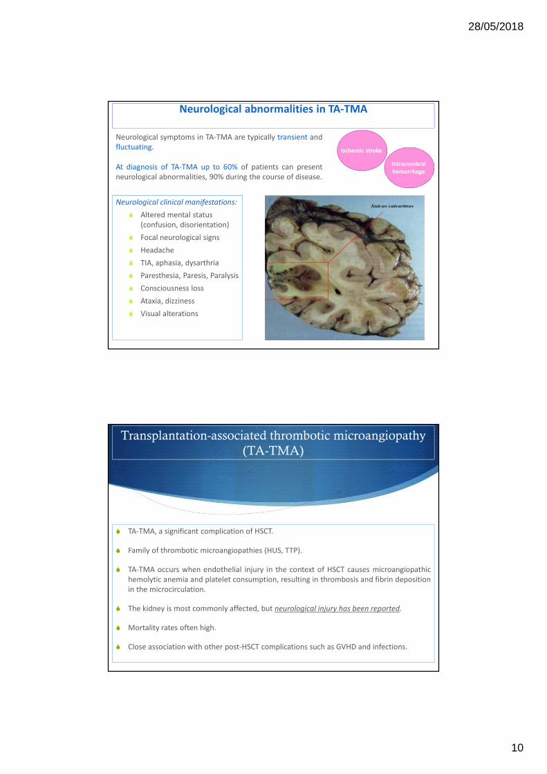
AS Daly, et al. BMT, 2002.Carreras E, et al. BMT, 2011

Classical diagnostic pentad

(40%) of TA-TMA:

� Microangiopathic

hemolytic anemia

� Thrombocytopenia

� Renal failure

� Fever

� Neurological

abnormalities

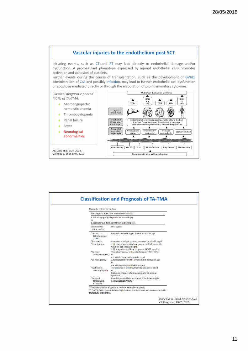
Classification and Prognosis of TA-TMA

Jodele S et al, Blood Reviews 2015.

AS Daly, et al. BMT, 2002.

28/05/2018

12

Brain MRI:Hyperintense FLAIR signal involving thebilateral (left N right) cortex and subcorticalwhite matter.Effacement of sulci suggests associatedswelling.Findings are suggestive of PRES.

Jodele S et al, Blood Reviews 2015

Although neurologic deficits have been

reported in up to half of all patients with TA-

TMA, a detailed understanding of central

nervous system (CNS) disease remains elusive.

Manifestations can include confusion,

headaches, hallucinations, or seizures.

Although the CNS vasculature can certainly be

affected by TA-TMA, the most common

TA-TMA-related CNS injury is likely due to

acute uncontrolled TMA-associated

hypertension, including PRES that may result in

CNS bleeding. PRES may present with

headaches, visual disturbances, mental status

changes, or seizures.

Neuroimaging reveals signal abnormalities in

posterior portions of the brain but can include

the brainstem, cerebellum and basal ganglia,

and symptoms are often preceded by

significant hypertension.

TA-TMA: CNS involvement

Neurological manifestations in hemolytic–uremic syndrome

Weissenborn, Neuroradiology (2013).

28/05/2018

13

A discrepancy often exists between bland or mild cerebral MRI findings and severe neurological

symptoms.

Hypothesis: the metabolic–toxic changes predominantly affect brain function on a microstructural level

instead of a macrostructural level, resulting in inconspicuous conventional MRI findings.

Quantitative analysis of routine clinical MRI sequences in the acute phase of STEC–HUS can be more

sensitive: prolonged T2 relaxation time indicates cerebral microstructural damages.

Cerebral damage in hemolytic–uremic syndrome

Neurological Diagnostic Challenges after HSCT

DIFFERENTIAL DIAGNOSIS OF ENCEPHALITIS

Malignancies Drug-

induced

Bacterial, Lyme disease,

Neurosyphilis

Parasitic

Sarcoidosis Adenovirus,

Enterovirus

Fungal HSV 1-2,

HHV6

Cerebral vasculitis TBC Toxoplasmosis VZV, EBV, CMV

MalignanciesPsychiatric

manifestations

IST

neurotoxicity

Ischemic stroke

Intracerebral

hemorrhage

PRES

Infections

Seizures

Neuromuscolar

complications

GvHD

28/05/2018

14

HHV-6 Acute Limbic Encephalitis: MRI

Viral infections: Encephalitis

Headache, confusion, memory problems and fatigue.

� Infections such as CMV and HHV-6 have been associated in case reports with TA-TMA.

EEG:

sharp waves in both of the temporal lobes,

epileptic seizures in right temporal lobe.

CSF analysis: PCR assays for viral pathogens.

Human-herpes-virus-6-related acute encephalitis

as a major diagnostic and therapeutic challenge

after haploidentical stem cell transplantation

28/05/2018

15

Post-HSCT HHV-6 disease

• Well known in Cord blood transplant1

• Demonstered impact2 on

– Engraftment (delayed platelet engraftment)

– Acute GVHD

– Overall mortality

• Multi-organ involvement

– Skin rash

– Cholitis/diarrhea

– Mielosuppression

– Encephalitis

1. Scheurer et al., Bone Marrow Transplantation (2013) 48, 574–580

2. Dulery R et al., Biol. Blood Marrow Transplant. 2012 Jul;18(7):1080-9

HHV-6 related encephalitis

• limbic encephalitis + HHV-6 in CS fluid(typical clinical and MRI signs)

• even without the evidence of HHV-6 in CSF– limbic encephalitis

– plasmatic high viral load

– exclusion of other causes of CNS disease

28/05/2018

16

Patients

• 9 years (2009-2017)

• 16 cases HHV-6 encephalitis after haplo-HSCT– M 10, F 6

– median age 52y

– PBSC graft

– 8 AML; 2 ALL; 4 NHL; 2 HD

– rapamycine-based GvHD prophylaxis • 5 anti-thymocyte globulin + rapa

• 8 PT cyclophosphamide + rapa

– 3 T-cell depleted graft

Patients

• Baseline prophylaxis:13/16 pts receiving acyclovir (10 mg/Kg bid)

• Time of onset: 25 days after HSCT (median); range 2-218

28/05/2018

17

Clinical presentation

• acute onset (all cases)

• disorientation (common)

• confusion (common)

• pruritus (common)

• memory loss (4)

• seizures (3)

• hyponatremia (common; severe in 2 pts)

• hypothermia (1)

• axial myoclonus (1)

Lab

• Quantitative PCR

• Plasma (reactivation)

– all pts

– median viral load of 15443 copies/µl

(range 550- 101332)

• Cerebrospinal fluid

– LP performed in 9/16 patients

– HHV-6 positivity in all cases

– Median viral load 206400 copies/µl

(range 3187-961350)

28/05/2018

18

MRI

• Limbic

encephalitis– Insula

– Hyppocampi

• Gd enhancement

• Negative (5/16)

EEG

• Performed in 14/16 pts

– Disrupted general organisation (common)– Focal slow abnormalities (common)

– epileptiform abnormalities (4/14)

28/05/2018

19

Treatment

• All patients received antiviral pharmacological

treatment– Gancyclovir: 5/16

– Foscarnet: 5/16

– Foscarnet + Gancyclovir: 6/16

• Gancyclovir 5 mg/Kg bid

• Foscarnet 90 mg/Kg bid (adj for GFR)

• Descalation strategy

Outcome

Poor (mortality rate: 50%)

- Resolution in 8 cases:- 6/8 treated with foscarnet-containing regimen

(FSC+GCV or FSC)

- with sequelae (common): memory loss, mild

cognitive impairment

- Immune reconstitution- 67% in pts with >100 CD3+/µl

- 28% in pts with <100 CD3+/µl

28/05/2018

20

Conclusion

• Poor prognosis after haplo-HSCT

• Common antiviral prophylaxis not effective

• Early (combined) antiviral therapy

• Regular plasma HHV6 monitoring?

� prospective study

28/05/2018

21

40-50% of patients undergoing HSCT experience HHV-6 reactivation.

Clinical manifestations:• Fever • Skin rash• Gut involvement• Pulmunary complications• Neurological disorders• Delayed engraftment• Poor Graft Function.

HHV6 reactivation after HSCT is a predictor for poor clinical outcome

Higher NRM rates

Higher incidence of aGvHD

De Pagter. et al, BMT 2013Durely R. et al, BBMT 2012

2009-2013: we evaluated haematological patients who developed positivity toHHV-6 after allogeneic HSCT (54 pts).

At the moment of HHV6 reactivation all patients were receiving acyclovir asantiviral prophylaxis except 5 (3 off antiviral therapy, 2 on ganciclovir) .

Viral DNA was isolated from different specimens (peripheral blood, bone marrow,BAL, gastrointestinal biopsy, cerebrospinal fluid) using a quantitative PCR(Nanogen).

Median time of onset: 34 days

Organ involvement was documented also in patients with negative plasmaDNAemia test

28/05/2018

22

79% fever

37% skin rash

31% hepatitis

46% diarrhoea

10 cases of HHV6

encephalitis

31% cytopenia

21% delayed engraftment

29/54 pts: acute GvHD

(III-IV grade predominance)

Clinical Manifestations

Greco R et al. BBMT 2016

• necessary in 63% cases

• 67% received foscarnet

• Mortality rate was relatively high in this population, mainly

related to severe infections or GvHD.

• OS±SE at 1 year after HHV-6 reactivation was 38%± 7%.

Antiviral therapy

Outcome

Immunoreconstitution

p= 0,00011

CD3+≥ 200/mcl

CD3+< 200/mcl

OS from HHV6 Reactivation OS (univariate) HR (CI 95%) p-value

CR vs disease 0,26 (0,07-0,89) 0,032

aGvHD 3/4 2,08 (1,08-4,03) 0,029

CD3+ ≥ 200/mcl 0,27 (0,13-0,54) 0,0002

OS was not significantly influenced by steroids administration, time after alloSCT, type of antiviral prophylaxis, plasma viral load and organ involvement

Greco R et al. BBMT 2016

28/05/2018

23

0 200 400 600 800 1000

0.0

0.2

0.4

0.6

0.8

1.0

Days

Pro

babi

lity

0 10 2

HHV6+: 62%

HHV6 Incidence after HSCT

TRM: 7.6%

OSR Prospective Study in Allogeneic HSCT

Greco R et al. In preparation

Donors were:

� family haploidentical (104),

� HLA identical sibling (39),

� unrelated (63),

� cord blood (7).

Graft source: PBSCs (87%).MAC 84%.GvHD prophylaxis: ATG 40%,PT-Cy 49%.

February 2013- October 2015: prospective observational study in 213consecutive adult patients who received allo-HSCT for high-riskhematological malignancies (AL 56%, CR 41%, DRI high/very high 56%).

aGvHD: 56% (16% grade III-IV). OS 54%.

HHV6 Positivity and Clinical Manifestations

HHV6+ 131 pts (62%). Median time to HHV6 positivity: 25 days after HSCT. 2nd reactivation and/or organ involvement: 40%.

Antiviral treatment (GCV, FSC) in 40% of reactivating patients.

Only 40% of reactivating patients with clinically relevant infection.

Clinical manifestations:� hepatitis (19),� diarrhoea (28),� encephalitis (5),� BM suppression (30),� delayed engraftment (13),� fever (42),� skin rash (57).

Viral load: weekly monitored by quantitative PCR in plasma.

aGvHD in HHV6+ pts:48% (54% after HHV6).

28/05/2018

24

Clinica

lly re

levan

t

Not Rele

vant

020406080

100250300350400450500 ***

HHV6 Reactivation

N. S

pot H

HV

6

P <0.0001a)MEAN +/- SEM 81,46± 15,78 N=39b)MEAN +/- SEM 4,500± 0,9927 N=38

HHV6 Specific T-Cells Are Predictive Biomarker of Active HHV6 Infection

HHV6-specific T-cells were

higher in HHV6 reactivating

patients (p= 0.0149; n= 43.48

per 10^5 PBMC) than in non-

reactivating patients (n= 12.57

per 10^5 PBMC), especially in

presence of active and

clinically relevant HHV6

infection (p<0,0001; n= 81.46

per 10^5 PBMC).

� Polyclonal immune reconstitution biomarkers were not sufficient to predict clinically relevant HHV6 infection.

� Reconstitution of functional HHV6 immunity is able to predict subsequent severe clinical manifestations:

� To validate the thresholds of HHV6-specific IFN-g spots as a surrogate marker in larger prospective trials.

� To standardize the IFN-g ELISpot protocol.

CONCLUSIONS

FUTURE PLANS

�Fine tune anti-viral treatment of patients

� Improved risk stratification

�Better patient management, fewer toxic side-effects

�Significant potential cost savings on anti-viral drugs

28/05/2018

25

� Transplant recipients are prone to a spectrum of neurological disorders including

encephalopathy, seizures, infections, stroke, CPM, neuromuscular disorders, and CNS

malignancies.

� These complications may result from GVHD, radiation, chemotherapy,

immunosuppressants, surgery as well as from a clinical and neuro-infectious etiology.

� Definitive etiological diagnosis frequently presents a challenge due to a relative low

specificity of neuroimaging studies, warranting a tissue biopsy.

� Management is focused on mitigating neurological injury by careful consideration of risk

versus benefit ratio of immune suppressive regimens, and therapies directed to specific

etiologies.

Overview of key points

50


Recommended