76
Neurological Nuclear Medicine Department of Nuclear Medicine Renji Hospital
| Date post: | 25-Dec-2015 |
| Category: |
Documents |
| Upload: | maximillian-todd |
| View: | 253 times |
| Download: | 1 times |
Neurological Nuclear Medicine
Department of Nuclear Medicine
Renji Hospital
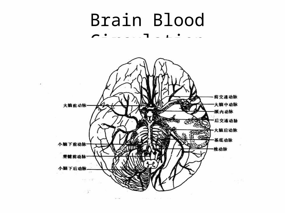
Brain Blood Circulation
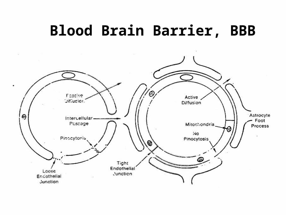
Blood Brain Barrier, BBB
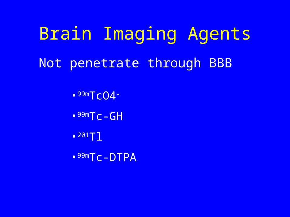
Brain Imaging Agents
Not penetrate through BBB
•99mTcO4-
•99mTc-GH
•201Tl
•99mTc-DTPA
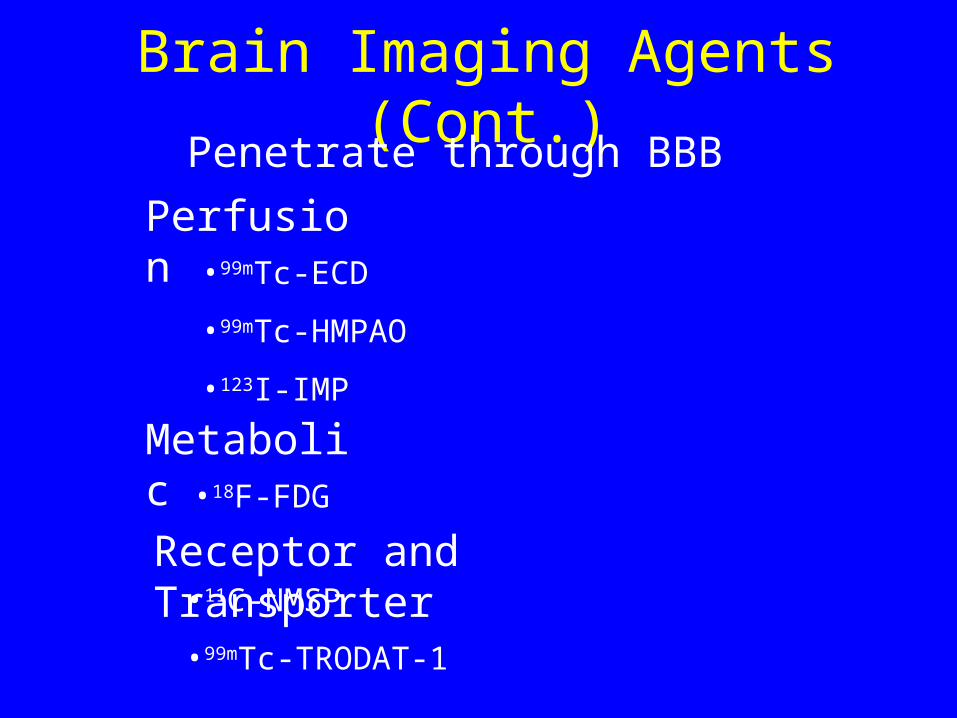
Brain Imaging Agents (Cont.)Penetrate through BBB
•99mTc-ECD
•99mTc-HMPAO
•123I-IMP
Perfusion
Metabolic•18F-FDG
Receptor and Transporter•11C-NMSP
•99mTc-TRODAT-1
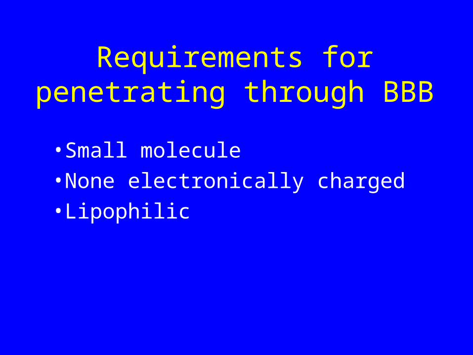
Requirements for penetrating through BBB
•Small molecule
•None electronically charged
•Lipophilic
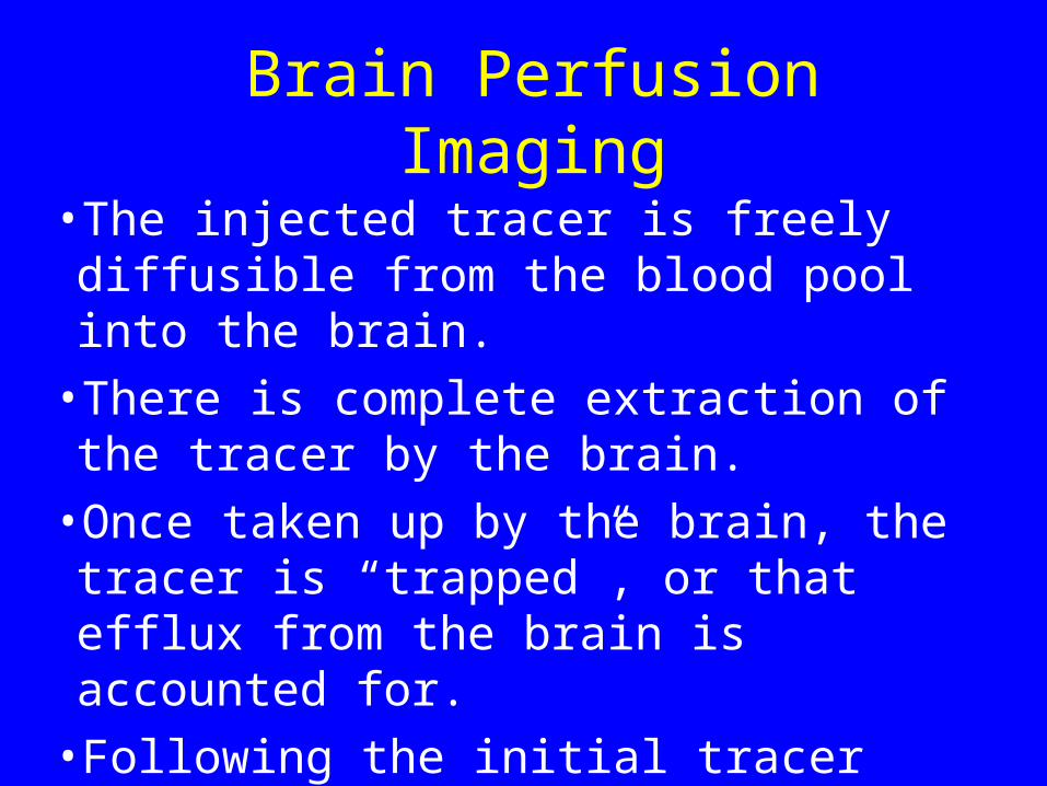
Brain Perfusion Imaging
• The injected tracer is freely diffusible from the blood pool into the brain.
• There is complete extraction of the tracer by the brain.
• Once taken up by the brain, the tracer is “trapped”, or that efflux from the brain is accounted for.
• Following the initial tracer uptake, there is no subsequent redistribution.
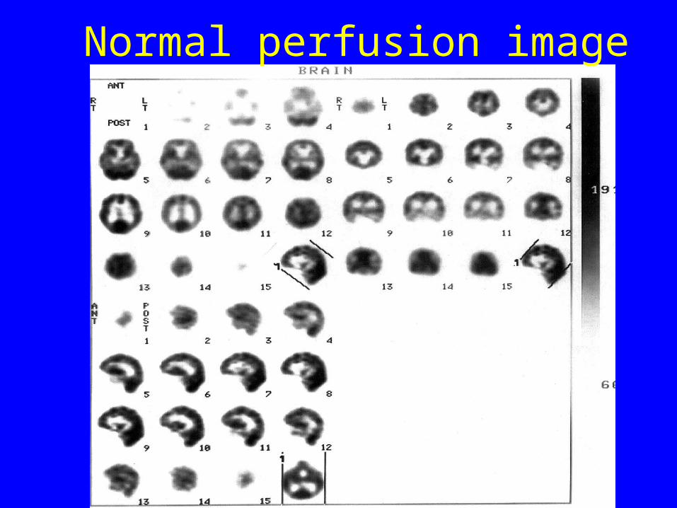
Normal perfusion image
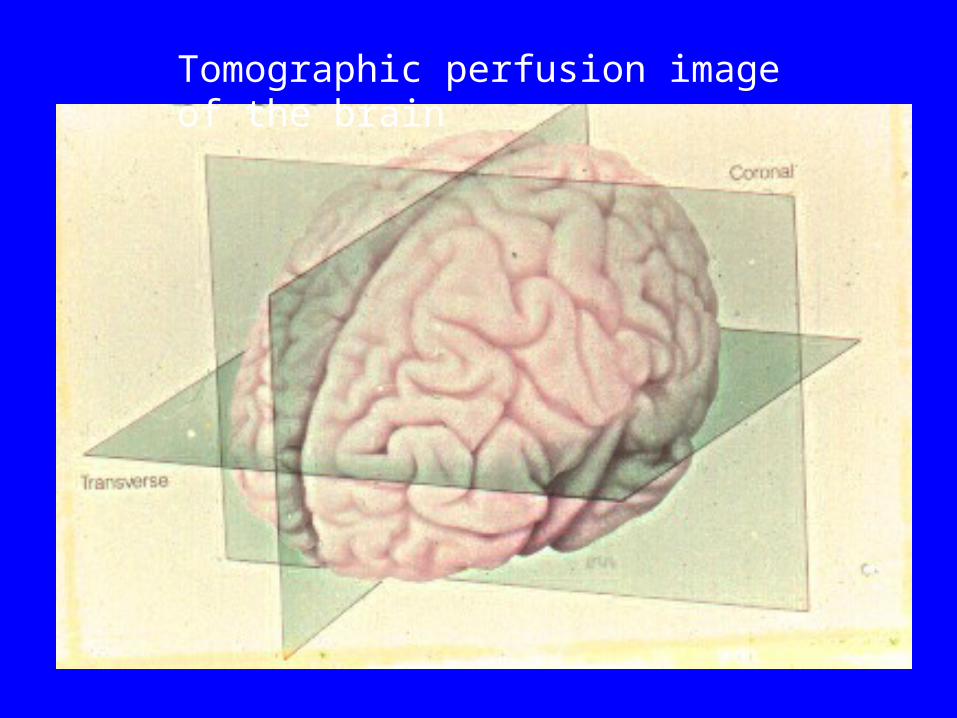
Tomographic perfusion image of the brain
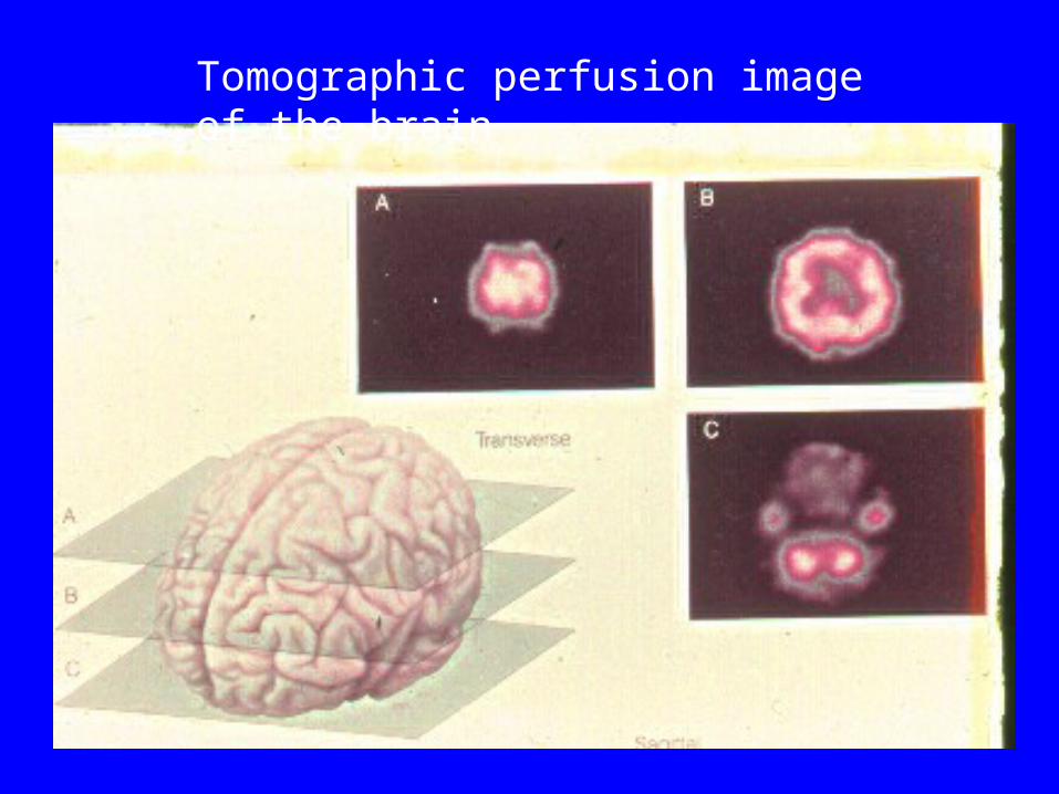
Tomographic perfusion image of the brain
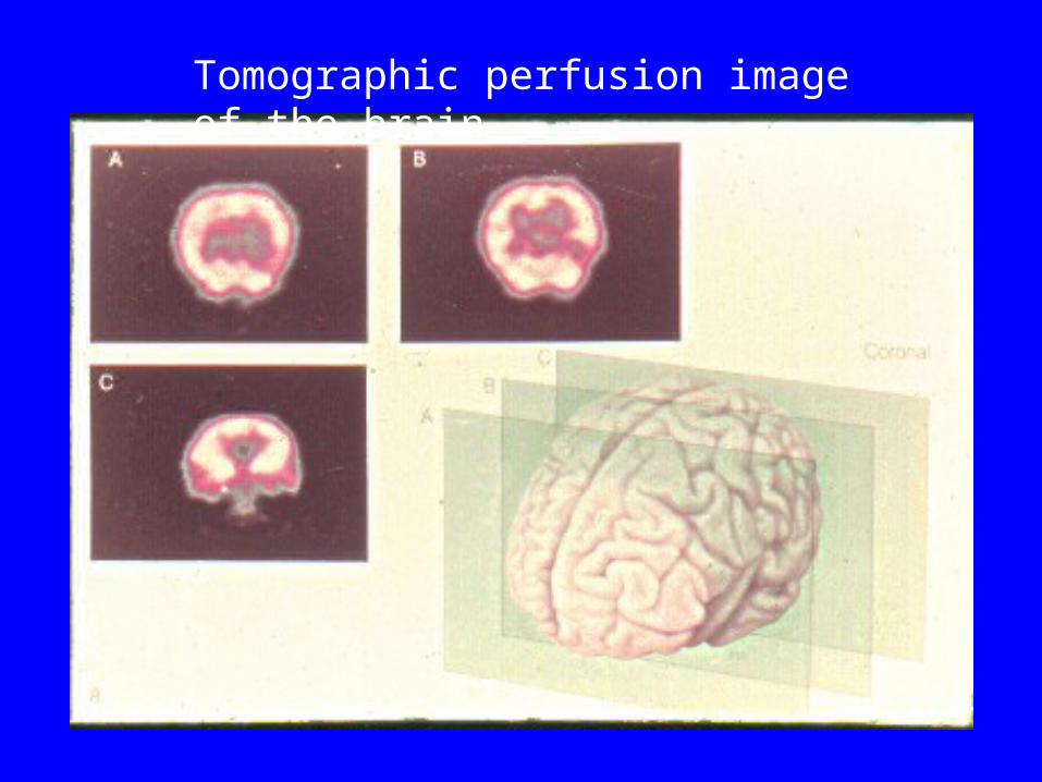
Tomographic perfusion image of the brain
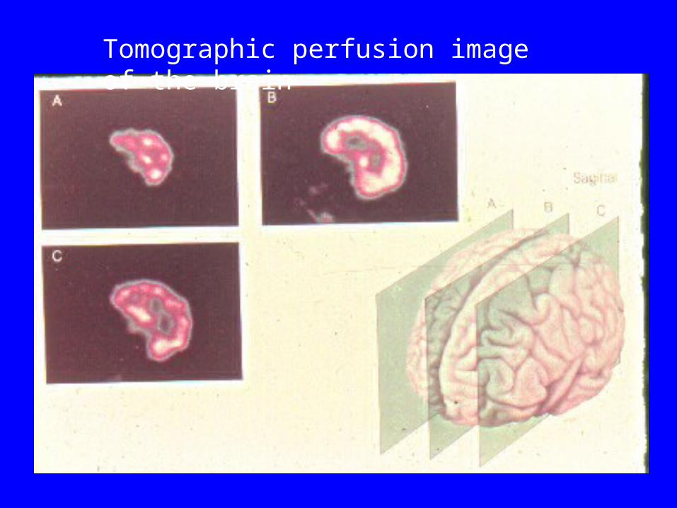
Tomographic perfusion image of the brain
Abnormal Patterns
Focal decreased uptakeFocal increased uptakeCrossed cerebellar diaschisisEnlargement of white matter or midline shift
Structure disorderAbnormal distribution of the tracercerebral atrophyDissymmetric distribution
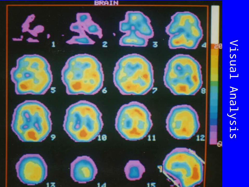
Visual A
nalysis
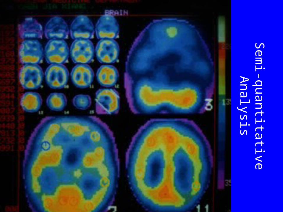
Sem
i-quantitative Analysis
Clinical Indications of Brain Perfusion Imaging
• Cerebral ischemia
• Dementia
• Seizures
• Alzheimer diseases
• psychiatric diseases
• Brain death
• Parkinson's Disease
Clinical Applications of Brain Perfusion Imaging
Cerebral Vascular Diseases
• Acute CNS Ischemia/Infarction
• Transient Ischemic Attacks(TIA)
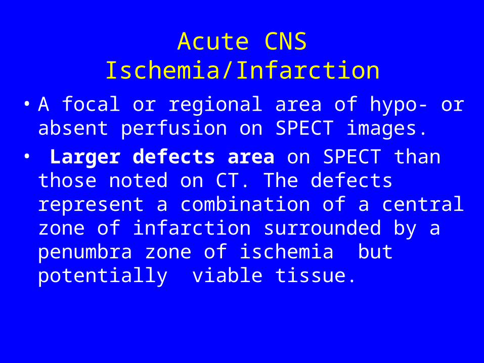
Acute CNS Ischemia/Infarction
• A focal or regional area of hypo- or absent perfusion on SPECT images.
• Larger defects area on SPECT than those noted on CT. The defects represent a combination of a central zone of infarction surrounded by a penumbra zone of ischemia but potentially viable tissue.
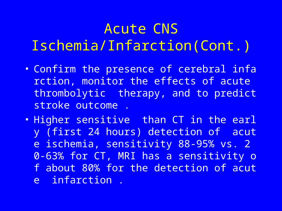
Acute CNS Ischemia/Infarction(Cont.)
• Confirm the presence of cerebral infarction, monitor the effects of acute thrombolytic therapy, and to predict stroke outcome .
• Higher sensitive than CT in the early (first 24 hours) detection of acute ischemia, sensitivity 88-95% vs. 20-63% for CT, MRI has a sensitivity of about 80% for the detection of acute infarction .
Subacute Phase Infarction
• Size of the infarct may be grossly underestimated due to luxury perfusion
• Luxury perfusion :uncoupling of flow and metabolism following an infarct. Apparently increased or normal tracer uptake despite the absence of metabolism in the involved area possibly related to either local breakdown in the blood-brain barrier or hyperemia from local tissue acidosis
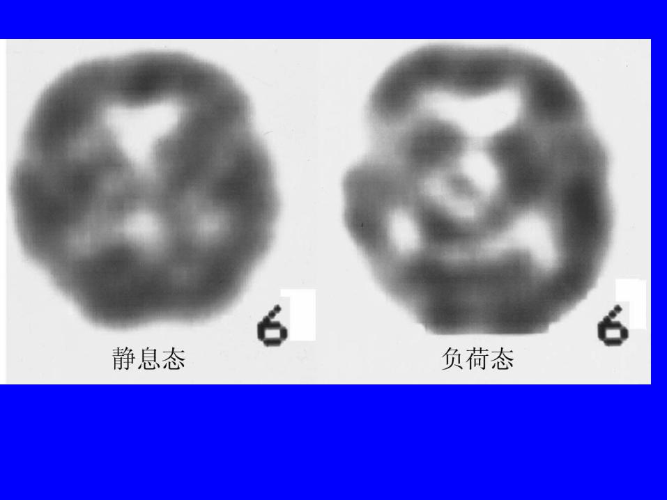
Crossed Cerebellar Diaschisis • Crossed cerebellar diaschisis (CCD): decreased
cerebellar perfusion contralateral to the cortical infarct during the acute and subacute phases of middle cerebral artery territory strokes.
• Mechanism: loss of axons interconnecting the infarcted cortical regions with other brain structures
• intact vascular reserve: activity increase in these areas following the administration of diamox due to the increased perfusion to these areas.
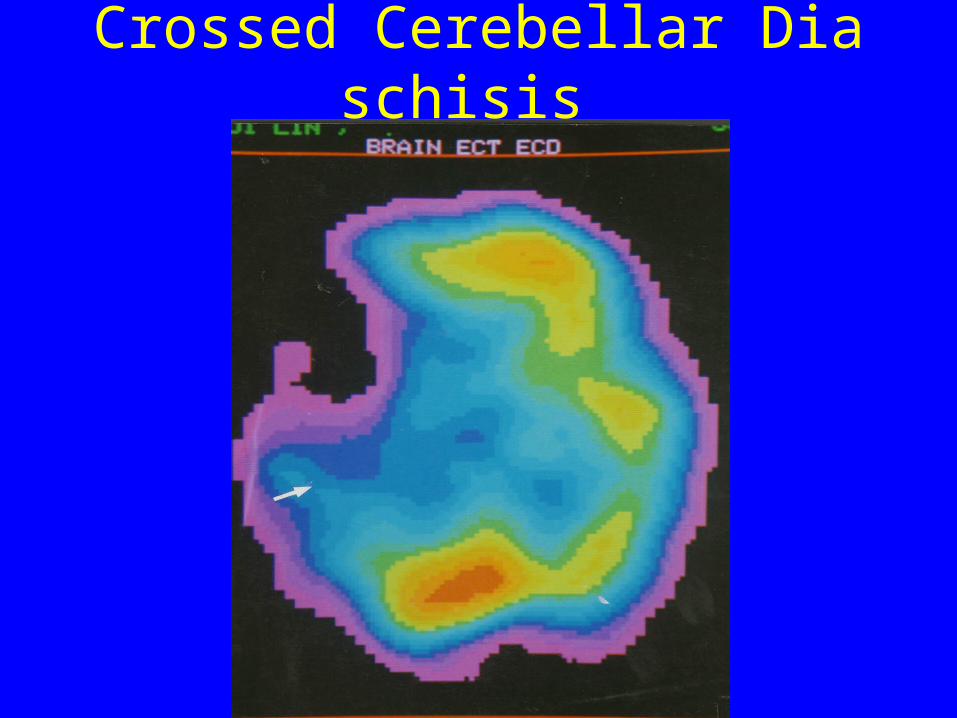
Crossed Cerebellar Diaschisis
Transient Ischemic Attacks
• It occurs in 10 to 20% of stroke patients. One third of these patients suffer a stroke within 5 years without treatment.
Transient Ischemic Attacks(Cont.)
• Single or multiple cerebral blood perfusion defect or abscent
• Early detecting ischemia region with SPECT compared to CT or MRI.
• Sensitivity is about 55-60% with SPECT, the sensitivity declines with time.
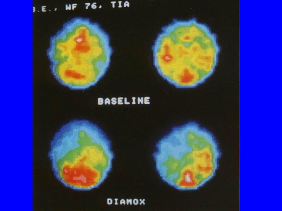
• SPECT CBF stress test with Diamox has been shown to increase the likelihood of detection of residual blood flow changes after TIA.
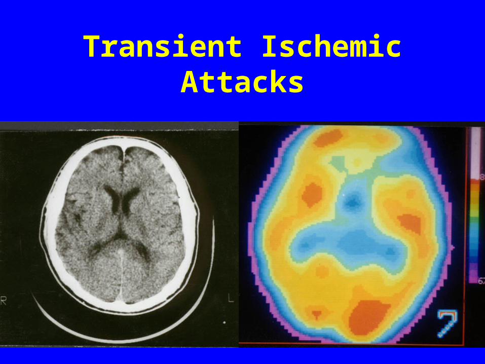
Transient Ischemic Attacks

DEMENTIA
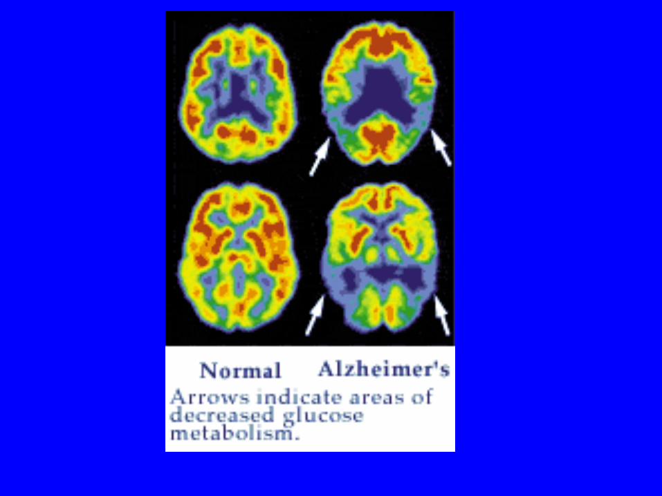
Alzheimer's Dementia• Dementia affects 10% of people over the
age of 60 years and Alzheimer's accounts for roughly 50% of these cases. Alzheimer's disease (AD) has a prevalence of 0.3% in patients aged 60-69 years, but increases to nearly 11% in 80-89 year olds . The mental degeneration associated with Alzheimer's is insidious and progressive memory loss is the most important symptom.
Alzheimer's Dementia(cont.)
• Very early stage: normal CBF perfusion
• Early stage: unilateral or bilateral temporoparietal perfusion defect
• Moderate to severe stage:bilateral temporoparietal perfusion defect
• Advanced stage: bilateral temporoparietal and frontal lobe perfusion defect
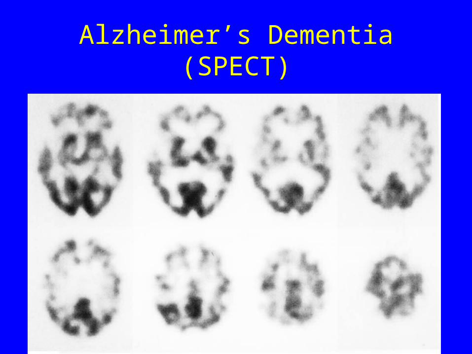
Alzheimer’s Dementia (SPECT)
Alzheimer's Dementia(Cont.)• A correlation has been described between the seve
rity of these defects, and the severity of the patient's dementia .
• The cerebellum, primary visual areas, and primary sensorimotor areas along the central sulcus remain relatively intact.
• SPECT perfusion imaging has a sensitivity of 63%, a specificity of 82%, a positive predictive value of 81%, a negative predictive value of 65%, and an accuracy of 71% for the diagnosis of Alzheimer's .
Multi-infarct Dementia
• Multi-infarct dementia (MID) is characterized clinically by multiple cerebral infarcts that occur sporadically and produce a step-wise deterioration in intellectual function. MID is the second most common cause of dementia in the elderly.
Multi-infarct Dementia (Cont.)
• multiple, bilateral, and randomly distributed cortical perfusion defects that follow vascular territories. The basal ganglia, motor, and sensory cortices may also be involved (spared in Alzheimer's).
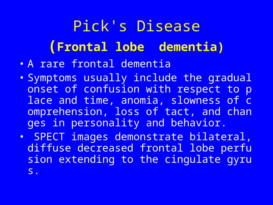
Pick's Disease (Frontal lobe dementia)
• A rare frontal dementia • Symptoms usually include the gradual onset
of confusion with respect to place and time, anomia, slowness of comprehension, loss of tact, and changes in personality and behavior.
• SPECT images demonstrate bilateral, diffuse decreased frontal lobe perfusion extending to the cingulate gyrus.
Epilepsy
• Epilepsy is one of the most prevalent neurological disorders.Seizures can be classified as either partial (focal) or generalized. Partial seizures originate in a given area of the brain and can be divided into simple (with no impairment of consciousness) and complex (with impairment of consciousness).
• About 10-20% of patients with partial complex seizures have inadequate control on medical treatment.Patients unresponsive to anti-convulsant therapy may be surgical candidates which can render the patient seizure free.
Epilepsy (Cont.)
• Scalp EEG often fails to accurately localize the seizure focus and although depth EEG is much more accurate, it is also extremely invasive and suffers from regional under sampling .
• CT and MRI have low sensitivity for seizure foci detection, 17% and 34% respectively. The role of brain SPECT is to localize the seizure focus.
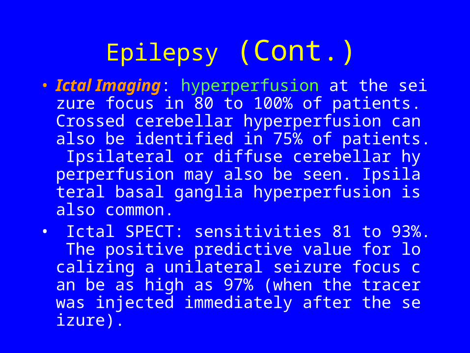
Epilepsy (Cont.)• Ictal Imaging: hyperperfusion at the seizure fo
cus in 80 to 100% of patients. Crossed cerebellar hyperperfusion can also be identified in 75% of patients. Ipsilateral or diffuse cerebellar hyperperfusion may also be seen. Ipsilateral basal ganglia hyperperfusion is also common.
• Ictal SPECT: sensitivities 81 to 93%. The positive predictive value for localizing a unilateral seizure focus can be as high as 97% (when the tracer was injected immediately after the seizure).
Epilepsy (Cont.)
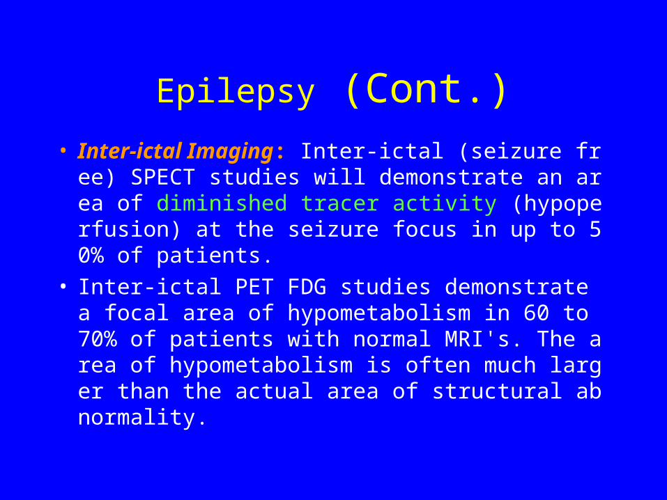
• Inter-ictal Imaging: Inter-ictal (seizure free) SPECT studies will demonstrate an area of diminished tracer activity (hypoperfusion) at the seizure focus in up to 50% of patients.
• Inter-ictal PET FDG studies demonstrate a focal area of hypometabolism in 60 to 70% of patients with normal MRI's. The area of hypometabolism is often much larger than the actual area of structural abnormality.
Epilepsy (Cont.)
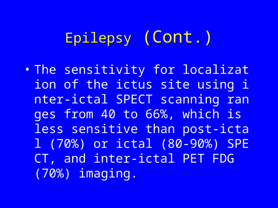
• The sensitivity for localization of the ictus site using inter-ictal SPECT scanning ranges from 40 to 66%, which is less sensitive than post-ictal (70%) or ictal (80-90%) SPECT, and inter-ictal PET FDG (70%) imaging.
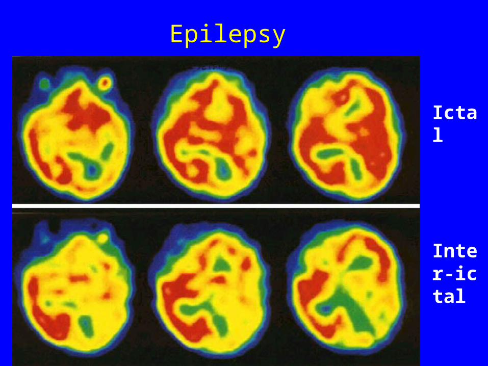
Ictal
Inter-ictal
Epilepsy
Brain Death• Lack of intracranial arterial flow: the carotid
arteries are visualized in the neck, but there is an abrupt cut-off of activity at the skull base
• Sagittal/Venous sinuses are not visualized on subsequent static images
• The "hot nose" sign: due to increased flow in the external carotid circulation.
• SPECT imaging in patients with brain death demonstrates no cerebral or cerebellar accumulation of the radiotracer
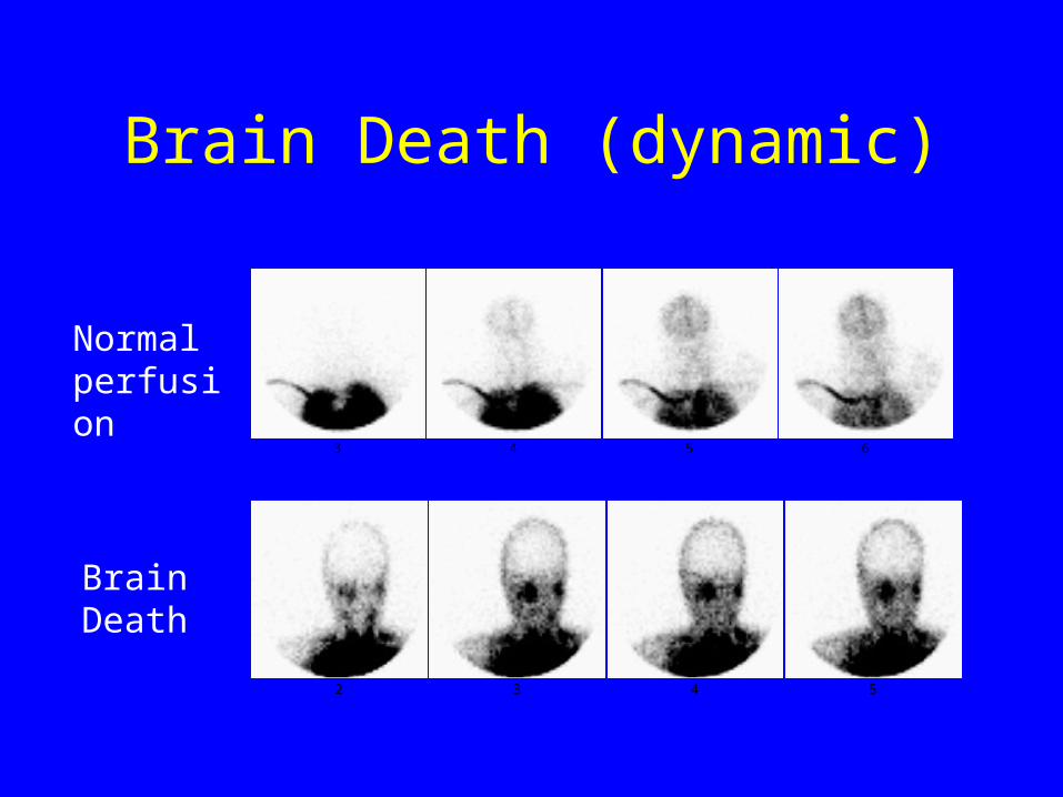
Brain Death (dynamic)
3 4 5 6
2 3 4 5
Normal perfusion
Brain Death
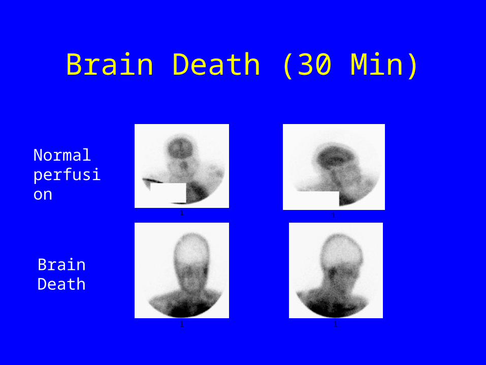
Brain Death (30 Min)
1 1
1 1
Normal perfusion
Brain Death
CNS Trauma
• SPECT is more sensitive than CT or MRI in detecting post traumatic CNS abnormalities both in the acute and remote stages. In the setting of acute trauma, small, non-focal frontal or occipital defects are associated with a favorable prognosis. Large or multifocal defects involving the parietal or temporal lobes, the cerebellum, or the brainstem are associated with an unfavorable prognosis.
Psychiatric Disorders
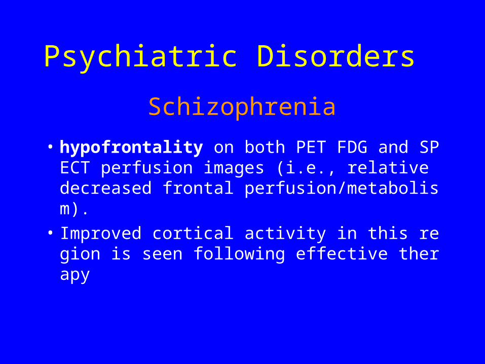
Schizophrenia
• hypofrontality on both PET FDG and SPECT perfusion images (i.e., relative decreased frontal perfusion/metabolism).
• Improved cortical activity in this region is seen following effective therapy
Psychiatric Disorders

Drug abuse
• Cocaine abuser: multiple small perfusion defect in brain cortex
• Chronic alcoholics:reversible decreasing frontal lobe flow that reverts to normal with abstinence
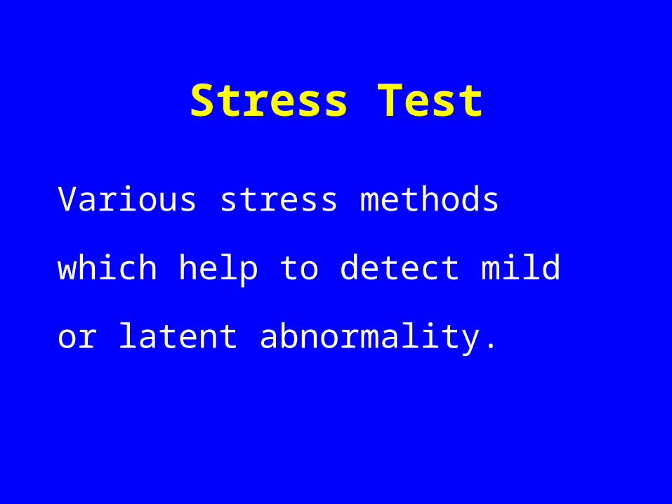
Stress Test
Various stress methods which help to
detect mild or latent abnormality.
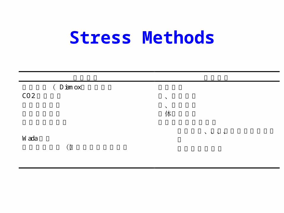
Stress Methods
负荷试验 激活试验
乙酰唑胺( Diamox)负荷试验 运动负荷 CO2负荷试验 上、下肢运动
过度换气试验 视、听觉刺激
立位负荷试验
颈动脉压迫试验 Wada试验
药物诱发试验(如美解眠诱发癫痫)
躯体感觉刺激
高级脑神经机能激活
包括记忆、言语、计算、认知、想象
威斯康星试验等
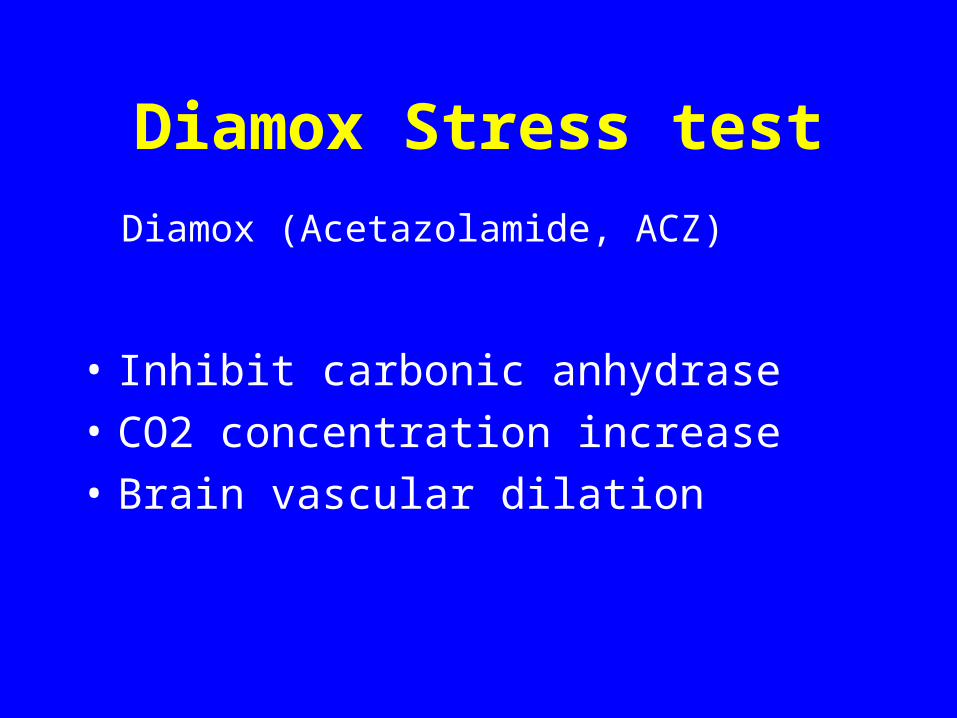
Diamox Stress test
• Inhibit carbonic anhydrase
• CO2 concentration increase
• Brain vascular dilation
Diamox (Acetazolamide, ACZ)
Diamox Stress test Procedures
• Get baseline image
• Diamox 15-20mg/kg iv before tra
cer injection.
• Stress image
Clinical Applications of Diamox Stress Test
• Silent brain ischemia detection
• Cerebrovascular reserve assessment
• Cerebrovascular reaction in CCD
• Prognosis prediction of cerebrovascular disease
18F-FDG Brain Imaging
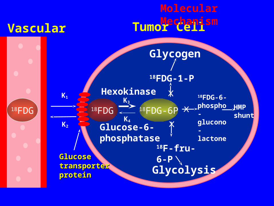
GlucoseGlucosetransportertransporterproteinprotein
K3
K4
Hexokinase
Tumor Cell
Glucose-6-phosphatase
18FDG-1-P
Glycogen
18F-fru-6-P
Glycolysis
18FDG-6-phospho-glucono-lactone
HMPshunt
18FDG
Vascular
K1
K2
18FDG 18FDG-6P
Molecular Mechanism
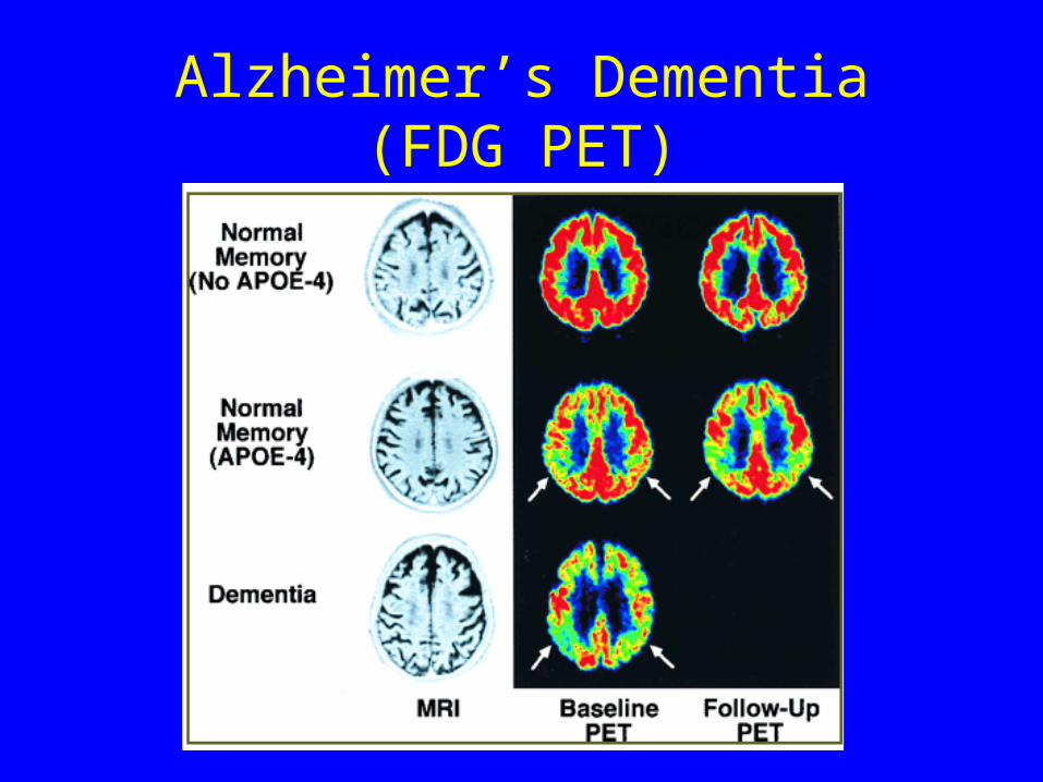
Alzheimer’s Dementia (FDG PET)
Parkinson's Disease
• Parkinson's is a progressive neurodegenerative disorder resulting from the progressive death of dopaminergic neurons in the nigrostriatal pathway.
• Symptoms consist of rigidity, bradykinesia, difficulty in initiating and stopping movement, and a resting tremor
Parkinson's Disease(Cont.)
• perfusion pattern in these patients is non-specific and demonstrates either normal or mild global cortical deficits. A pattern of bilateral posterior parietal/temporal defects indistinguishable from Alzheimer's may be observed in patients with Parkinson's disease with dementia.
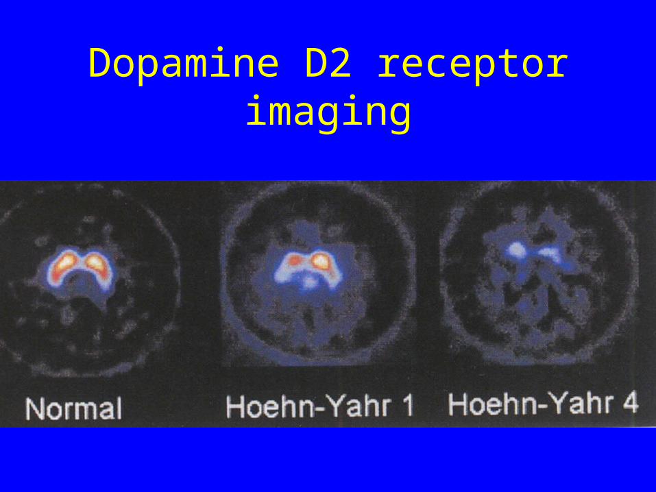
Dopamine D2 receptor imaging
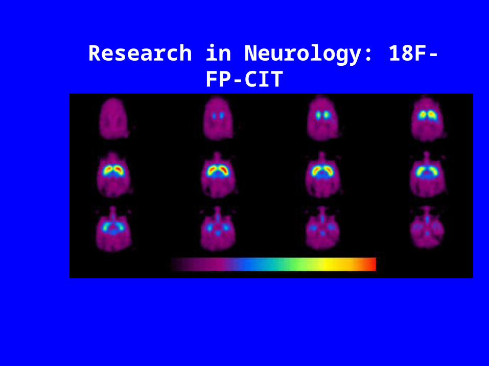
Research in Neurology: 18F-FP-CIT
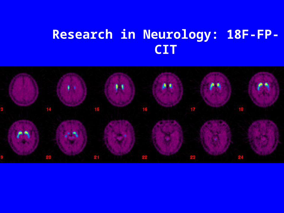
Research in Neurology: 18F-FP-CIT
Brain Tumors
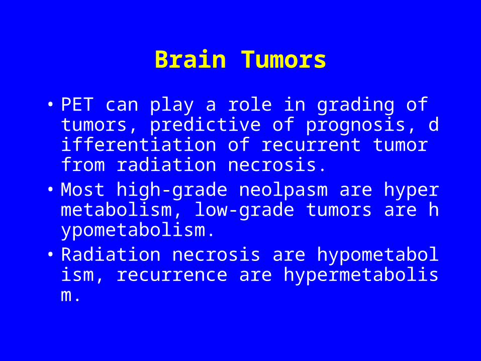
• PET can play a role in grading of tumors, predictive of prognosis, differentiation of recurrent tumor from radiation necrosis.
• Most high-grade neolpasm are hypermetabolism, low-grade tumors are hypometabolism.
• Radiation necrosis are hypometabolism, recurrence are hypermetabolism.
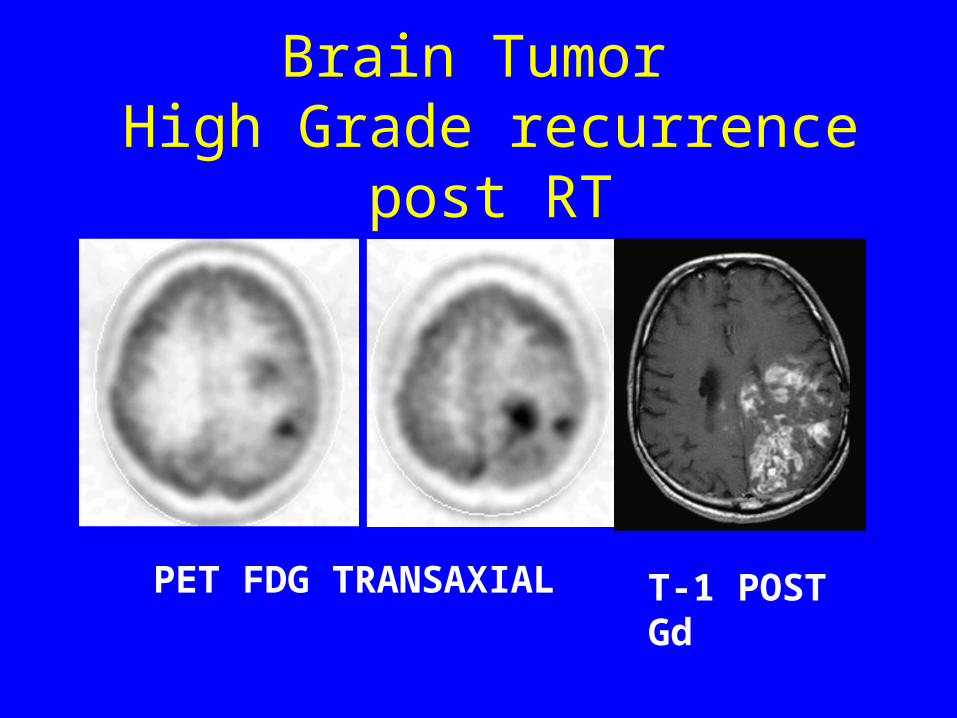
Brain Tumor High Grade recurrence post RT
PET FDG TRANSAXIAL T-1 POST Gd
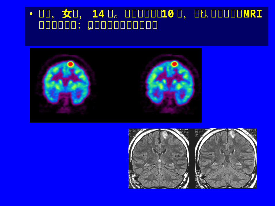
• 患者,女性, 14 岁。癫痫反复发作 10 年,近一年发作频繁。 MRI 平扫及增强见:胶质增生或良性胶质瘤。
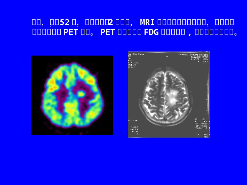
患者,男, 52 岁,顶叶胶质瘤 2 级术后, MRI 示顶叶手术部位高信号,为鉴别复发或瘢痕进行 PET 检查。 PET 示左侧顶叶 FDG 代谢增高灶 , 手术证实肿瘤复发。
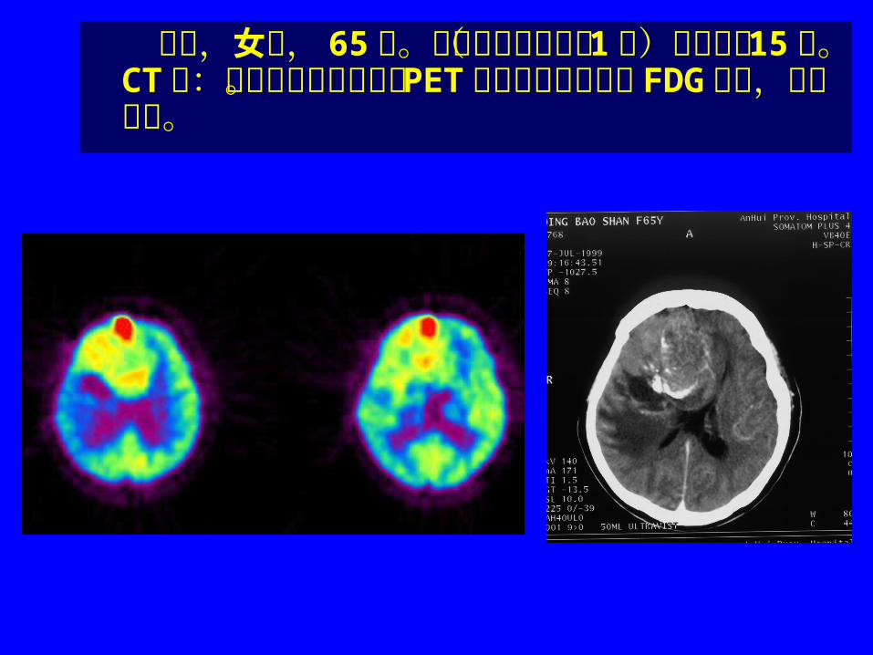
患者,女性, 65 岁。右额顶胶质瘤术( 1 级)后并放疗 15 年。 CT 示:原肿瘤部位有增强。 PET 显像见该部位明显 FDG 摄取,考虑复发。
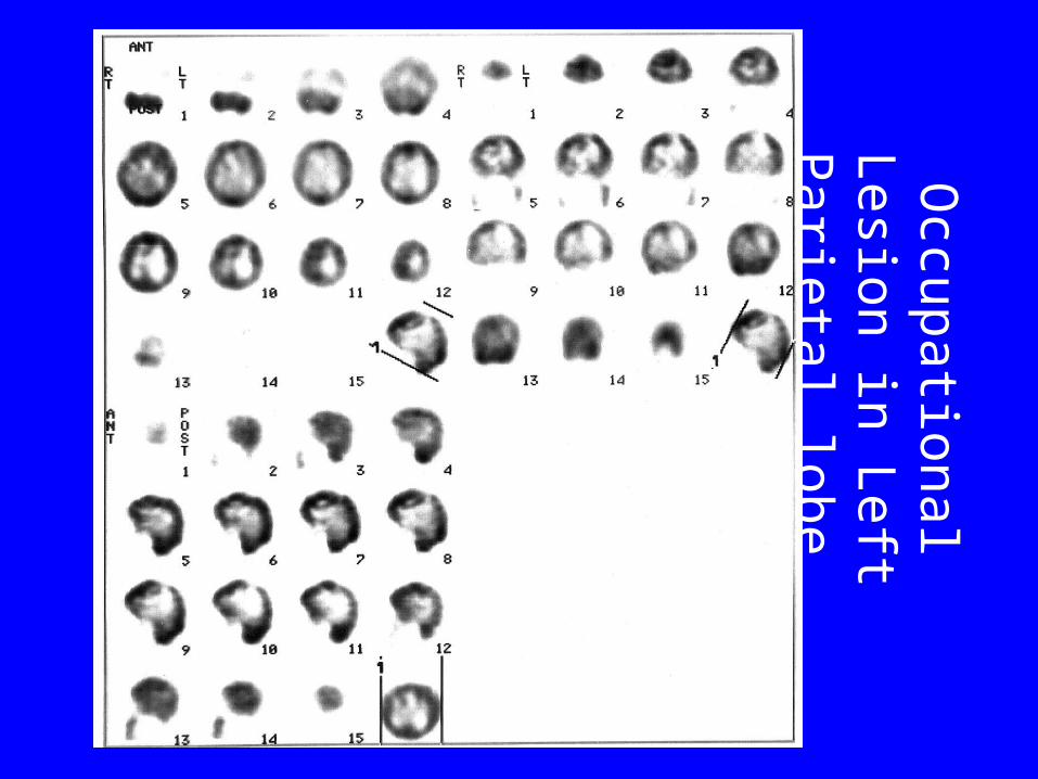
Occupational L
esion in L
eft Parietal lobe
Brain Function Study
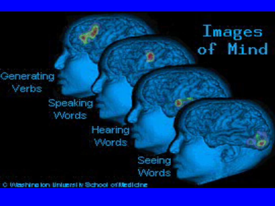

Hearing
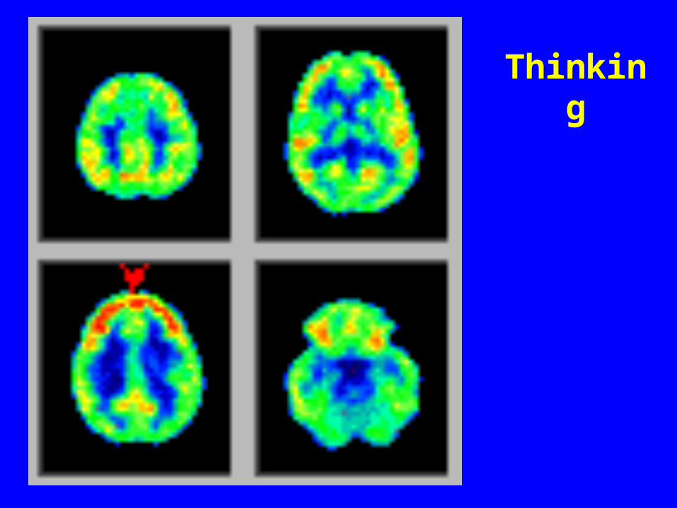
Thinking
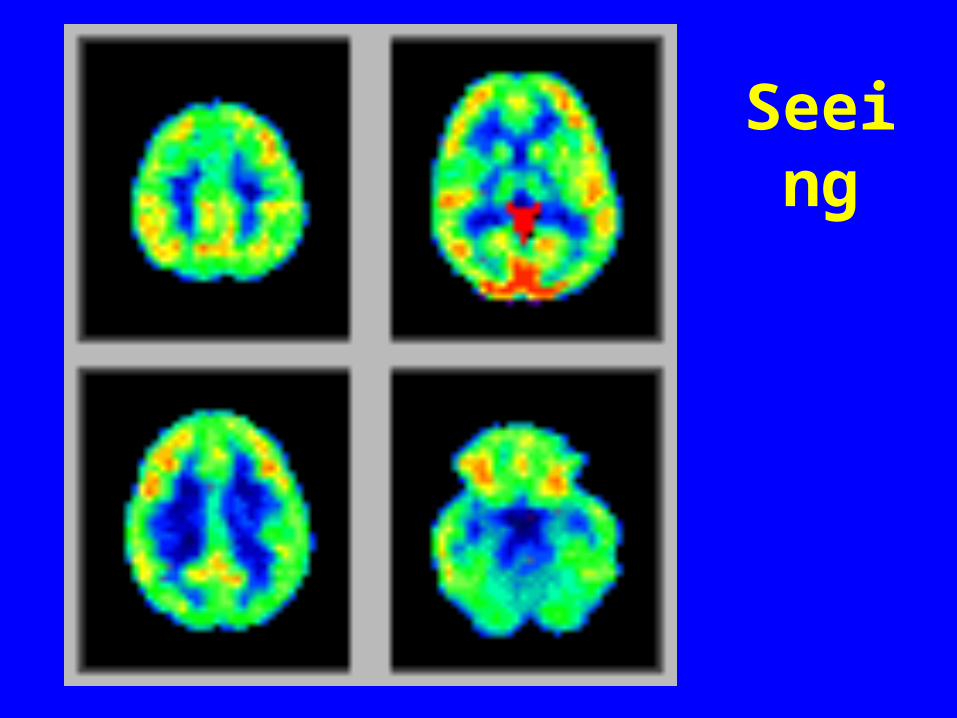
Seeing
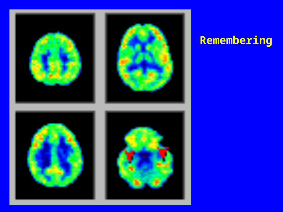
Remembering
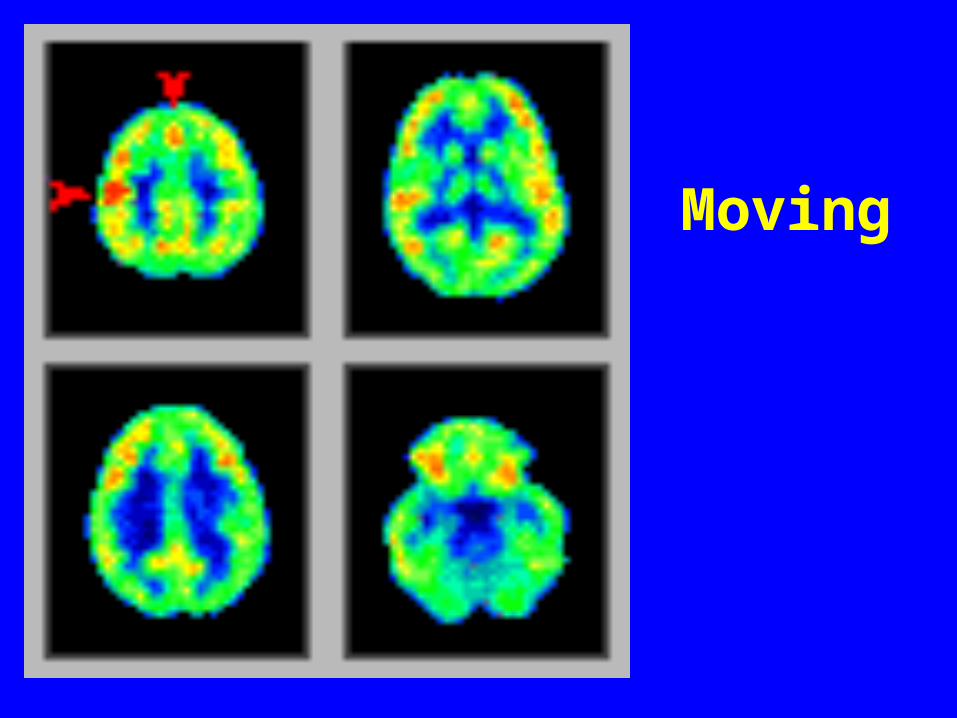
Moving


