Page 1
Neurology Boards Review
S. Andrew Josephson MD Carmen Castro Franceschi and Gladyne K. Mitchell Neurohospitalist Distinguished Professor
Senior Executive Vice Chairman, Department of Neurology
Director, Neurohospitalist Program
Medical Director, Inpatient Neurology
University of California, San Francisco
The speaker has no disclosures
Page 2
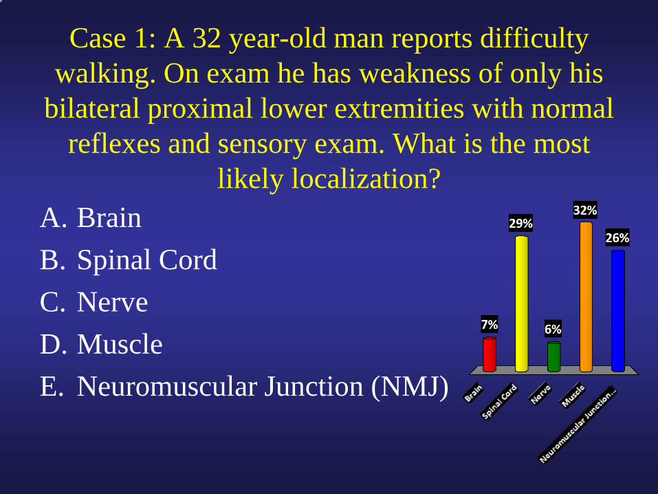
Case 1: A 32 year-old man reports difficulty
walking. On exam he has weakness of only his
bilateral proximal lower extremities with normal
reflexes and sensory exam. What is the most
likely localization?
A. Brain
B. Spinal Cord
C. Nerve
D. Muscle
E. Neuromuscular Junction (NMJ)
Page 3
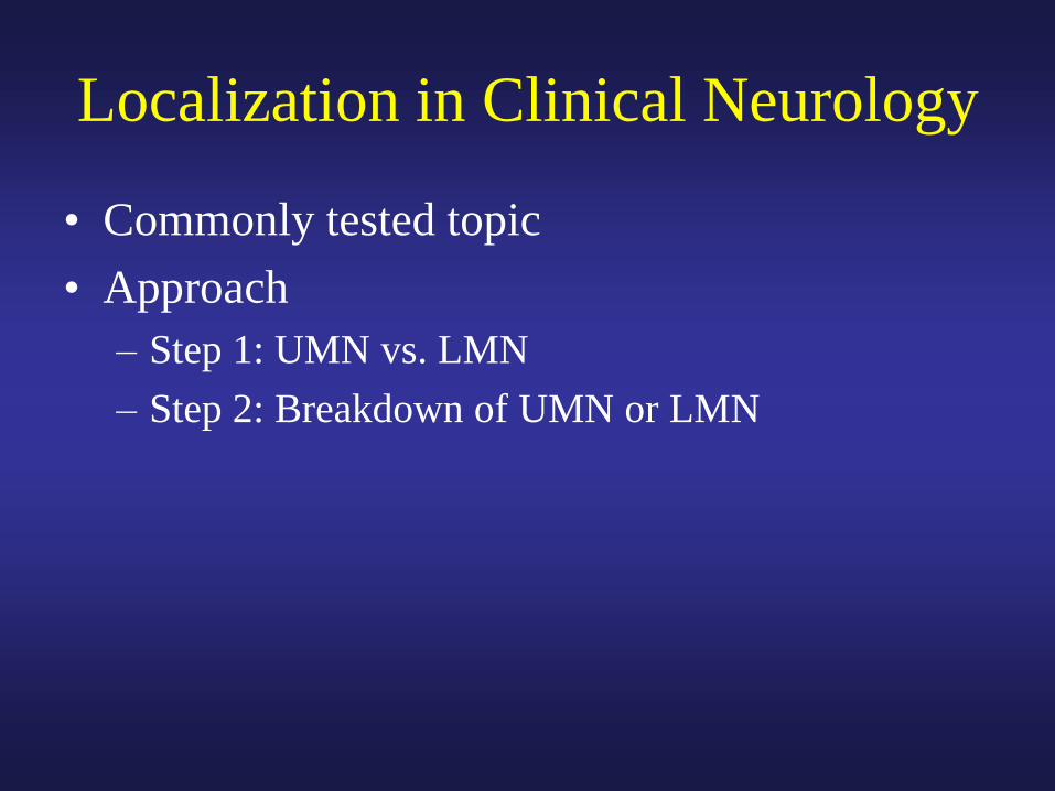
Localization in Clinical Neurology
• Commonly tested topic
• Approach
– Step 1: UMN vs. LMN
– Step 2: Breakdown of UMN or LMN
Page 4
Upper Motor Neurons
Predictable Pyramidal Pattern of Weakness:
UE Distal Extensors and LE Distal (Dorsi)Flexors
Page 5
Step 1: UMN vs. LMN
Pattern of weakness
Function/Dexterity
Tone
Tendon Reflexes
Other signs
Pyramidal Variable
Slow alternating
movements Variable
Increased Decreased
Increased Decreased, absent
or normal
Babinski sign,
other CNS signs
Atrophy (except for
NMJ disorders)
UMN LMN
Page 6
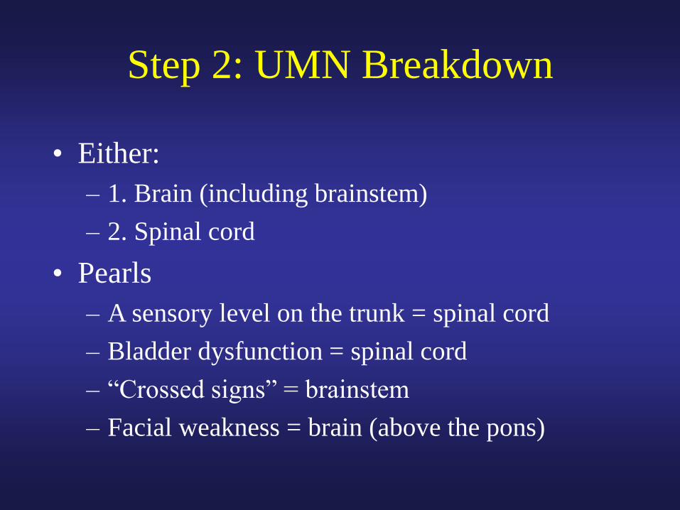
Step 2: UMN Breakdown
• Either:
– 1. Brain (including brainstem)
– 2. Spinal cord
• Pearls
– A sensory level on the trunk = spinal cord
– Bladder dysfunction = spinal cord
– “Crossed signs” = brainstem
– Facial weakness = brain (above the pons)
Page 7
Step 2: LMN Breakdown
• Either:
– 1. Anterior Horn Cell (AHC)
– 2. Nerve
– 3. Neuromuscular Junction (NMJ)
– 4. Muscle
Page 8
Step 2: LMN Breakdown Pearls
• Sensory Symptoms or Signs = Nerve
• Reflexes Decreased = Nerve (or AHC)
• Proximal Weakness = Muscle or NMJ
– Fatigue or fluctuating weakness = NMJ
• Fasciculations = AHC (or Nerve)
• Combination of UMN and LMN signs = ALS
Page 9
Case 2
• A 65 year-old man with a history of DM, HTN
presents with 9 hours of L sided binocular
visual loss
• Examination shows left-sided homonymous
hemianopia and is otherwise unremarkable.
• The patient is on ASA 81mg daily
Page 10
What treatment should you initiate?
A. IV t-PA
B. Heparin
C. Aspirin 325 mg
D. Plavix (clopidogrel)
E. Aggrenox (ER dipyridamole +
ASA)
Page 11
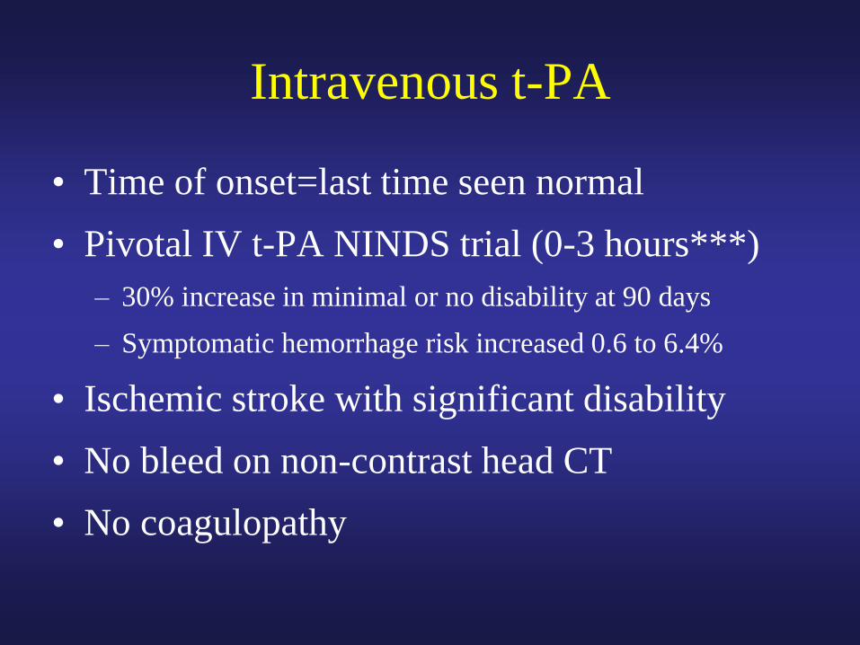
Intravenous t-PA
• Time of onset=last time seen normal
• Pivotal IV t-PA NINDS trial (0-3 hours***)
– 30% increase in minimal or no disability at 90 days
– Symptomatic hemorrhage risk increased 0.6 to 6.4%
• Ischemic stroke with significant disability
• No bleed on non-contrast head CT
• No coagulopathy
Page 12
Shrinking Indications for
Anticoagulation in Stroke
1. Atrial Fibrillation
2. Some other cardioembolic sources
– Thrombus seen in heart
– ?EF<35
– ?PFO with associated Atrial Septal Aneurysm
3. Vertebral and carotid artery dissection
4. Rare hypercoagulable states: APLA
Page 13
Antiplatelet Options
• 1. ASA
– 50mg to 1.5g equal efficacy long-term
• 2. Aggrenox
– 25mg ASA/200mg ER Dipyridamole
• ESPS-2, ESPRIT (Lancet 5/06)
• 3. Clopidogrel (Plavix) • MATCH (Lancet 7/04), FASTER (Lan Neurol 11/07)
• PRoFESS trial: Aggrenox and Plavix equal
Page 14
Antiplatelet Options
• If on no antiplatelet medication
– ASA or Plavix or Aggrenox
• If already on ASA
– Switch to Aggrenox or Plavix
Page 15
When to Fix the Carotid?
• NASCET in early 1990s
– Benefit of endarterectomy in patients with
symptoms ipsilateral to 70-99% stenosis
• Comparison: best medical management at the time
• In stroke management don’t miss carotid
disease or atrial fibrillation
Page 16
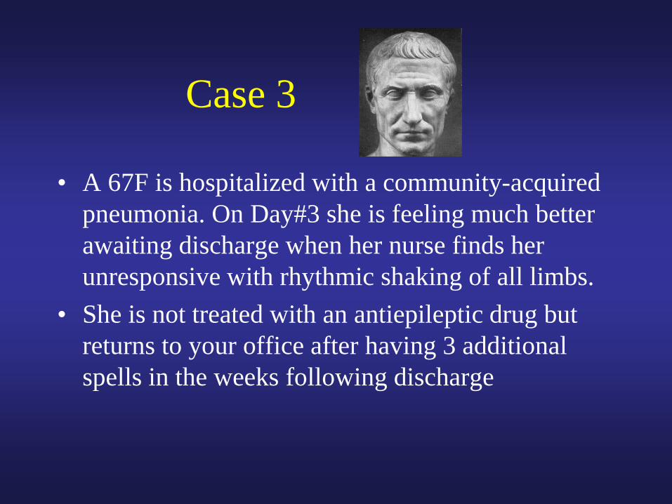
Case 3
• A 67F is hospitalized with a community-acquired
pneumonia. On Day#3 she is feeling much better
awaiting discharge when her nurse finds her
unresponsive with rhythmic shaking of all limbs.
• She is not treated with an antiepileptic drug but
returns to your office after having 3 additional
spells in the weeks following discharge
Page 17
Monotherapy for Seizures
• 70 percent of epilepsy can be managed with monotherapy, most on first drug tried
• Concept of Maximal Tolerated Dose (MTD)
• Rarely check levels
– Assess compliance
– Steady state level
– Not practically available with newer AEDs
N Engl J Med. 2000 Feb 3;342(5):314-9
Page 18
New Drugs: Clinical Pearls
• IV formulations: VPA, DPH, PHB, LVT
• Levels to Monitor: VPA, DPH, PHB, CBZ
• Lamotrigine (Lamictal)
– Rash (1/1000) progressing to Stevens-Johnson
• Levetiracetam (Keppra)
– No drug interactions (useful on HAART)
• Topiramate (Topamax)
– Well tolerated?: weight loss and cognitive side effects
Page 19
New Drugs: Clinical Pearls
• Oxcarbazepine (Trileptal)
– Tegretol pro-drug
– Hyponatremia
• Felbamate (Felbatol)
– Aplastic Anemia with required registry
• Gabapentin (Neurontin)
– Not a great AED
Page 20
Case 4
• A 45 yo man presents with 2 days of
progressive tingling and weakness of the
lower extremities. He now is having trouble
walking and rising from a chair.
Page 21
Case 4
• Exam
– MS, CN normal
– Motor: normal tone throughout, normal power in upper ext., 4/5 throughout in the lower extremities
– Sensory: decreased PP/Vib/temp patchy in lower extremities, Sensory level to PP at T4
– Reflexes: 1 and symmetric throughout, toes neutral
Page 22
What test should you next order?
A. MRI Brain
B. MRI Thoracic Spine
C. MRI Lumbar/Sacral Spine
D. Lumbar Puncture
E. Blood Cultures
Page 23
Workup of Myelopathy
• First step: MRI with contrast
– Pick appropriate level
– Excludes structural disease
• Second Step: If MRI negative, usually
proceed to lumbar puncture
– Pleocytosis = autoimmune or infectious
transverse myelitis
– No Pleocytosis = metabolic, vascular, genetic
Page 24
Case 5
A 44 man presents with bilateral numbness of the
legs, urinary incontinence and spinal fluid with an
elevated white blood cell count consisting of mainly
lymphocytes. Upon his death, a pathological
specimen of his spinal cord is shown. What is the
etiology of his symptoms?
Page 25
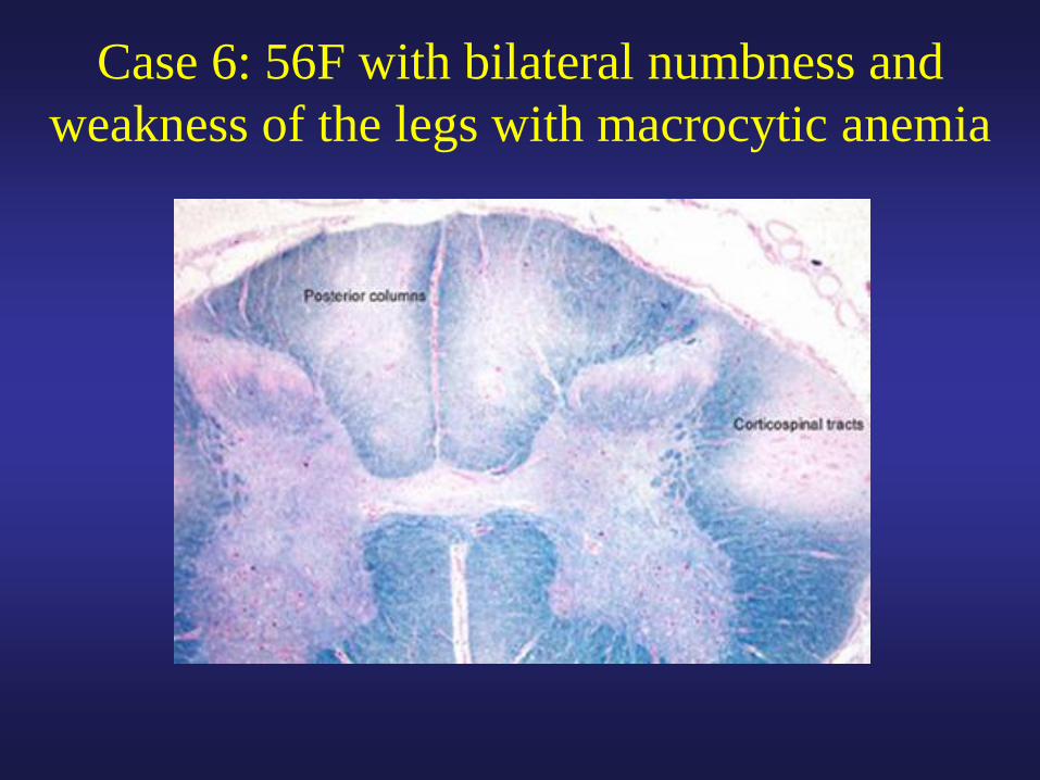
Case 6: 56F with bilateral numbness and
weakness of the legs with macrocytic anemia
Page 26
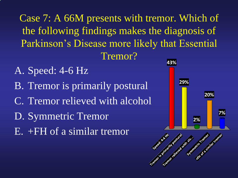
Case 7: A 66M presents with tremor. Which of
the following findings makes the diagnosis of
Parkinson’s Disease more likely that Essential
Tremor?
A. Speed: 4-6 Hz
B. Tremor is primarily postural
C. Tremor relieved with alcohol
D. Symmetric Tremor
E. +FH of a similar tremor
Page 27
Speed
Symmetry
Most Common
Component
Helped by EtOH
FH?
5-10Hz 4-6 Hz
Symmetric Asymmetric
Postural Rest
Yes No
Common
Autosomal Dominant Rare
ET PD
Any sign of Parkinsonism in an ET pt should lead to
questioning the diagnosis
Page 28
Parkinson’s Treatment
1. Give L-Dopa
-Levodopa/Carbidopa
2. Dopamine Receptor Agonists
-Pramipexole, Ropinirole
3. Alter Dopamine/L-Dopa Metabolism
-MAO-B Inhibitors: selegeline, rasagiline
-COMT Inhibitors: entacapone
4. Decrease Cholinergic tone (rarely used)
5. Surgery: Deep Brain Stimulation (DBS)
1
3
2
Page 29
Parkinson’s Treatment
Pearls
• Only treat when function is impaired
• Start with L-dopa or a dopamine agonist
– Agonists are best in those age <70 as they have
cognitive side effects (hallucinations) in the elderly
• If L-dopa is “wearing off”, increase the
frequency or add COMT inhibitor
• Reduce the dose if peak-dose dyskinesias are
impairing function
www.healthandage.com
Page 30
Case 8: A 54M comes to the ED with the worst
headache of his life. Which of the following
findings would be most worrisome for SAH?
A. Extremely severe head pain
B. Photophobia
C. Pain reaching maximum intensity in
seconds
D. Vomiting
E. No relief with high doses of opioids
Page 31
Migraine Therapy
• Abortive Therapy
– Begin with NSAIDs, ASA, acetaminophen
– Triptains
• Contraindicated with CAD
• Caution with OCPs, especially in smokers age >35
• Avoid if hx of complicated migraine
• Prophylactic therapy
– Start if >4-6 spells per month
– Multiple agents to choose from
Page 32
Cluster Headache
• Males ages 20-50
• Unilateral, periorbital pain, often with
autonomic symptoms
• Occur at the same time each day
• Occur in clusters over time
• EtOH can trigger
• Treatment: High flow O2, Lithium, Prednisone,
Triptans, Nasal Lidocaine
Page 33
Trigeminal Neuralgia
• Electric shocks in trigeminal distribution
• No objective sensory deficits
• Unilateral
• Trigger sites for many patients
• Consider MRI to exclude brainstem lesion, especially in young
• Treatment: carbamazepine
• Refractory cases: surgical decompression
Page 34
Temporal Arteritis
• Consider in any new HA over 50
• Consider in any new visual change over 50
– Missed diagnosis leads to permanent visual loss
• Most helpful for diagnosis
– Elevated ESR
– Jaw claudication
• Begin steroids immediately, prior to Bx
library.med.utah.edu
Page 35
A neurologist’s least favorite line on any brain
radiology report is….
“…can’t exclude demyelinating disease”
Page 36
Diagnosis of Multiple
Sclerosis
• Lesions separated by time and space
• MRI clues
– Involvement of corpus callosum
– Some lesions with enhancement in acute period
• History of optic neuritis helpful
– Visual Evoked Potentials (VEPs)
• Spinal Fluid
– Oligoclonal bands, elevated IgG index
• VERY non-specific for MS
Page 37
Case 9
• A 40 yo man comes to the ED with
increasing weakness and dyspnea. The
patient states that he has a history of
myasthenia gravis diagnosed at an OSH two
weeks ago but “things are going downhill.”
He is on Mestinon (pyridostigmine) 60mg
PO q4hrs and Prednisone 60mg PO qd. MIF
is –10, FVC 250cc
Page 38
Myasthenic Crisis
• True crisis vs. cholinergic crisis
• Triggers
– Infection, surgery, initial steroids
• Management
– Usually stop all anti-cholinesterase meds
– Pheresis or IVIg
– ICU, intubation, DVT/PE prophylaxis
• Follow MIF and FVC
– Drugs to Avoid: NMJ blockers, aminoglycosides
Page 39
Myasthenia Gravis: Key Points
• Two types of myasthenia
– Young F>M
– Old M=F
• Weakness fatigues with exercise and throughout the day
• Pure ocular form common (ptosis, diplopia)
• Diagnosis
– Antibodies (90% in generalized mysathenia)
– EMG with repetitive stimulation, single fiber EMG
Page 40
Myasthenia Gravis: Key Points
• Management
– Pyridostigmine (Mestinon)
– Immunosuppression
• Prednisone first then Imuran/CellCept/Cytoxan
– What about the Thymus?
Page 41
Case 10
• A 63yo man comes to the ED with 3 days of
inability to walk. The patient reports a 2
week history of tingling in his hands and
feet while also stating that he has been
stumbling while walking for five days.
Page 42
Case 10
• Exam
– General exam nl with stable vitals
– Mental status, cranial nerves normal
– Motor exam with mild-moderate symmetric weakness prox>distal in the upper ext., distal>prox in the LEs
– Sensory exam completely normal
– Reflexes 2+ throughout except 0 ankles, plantar response flexor bilaterally
Page 43
Case 10:
Additional Tests
FVC/MIF: 1.2L, -30
Lumbar Puncture: Opening pressure normal, 2
WBC, Zero RBC, Protein 87, Glucose normal
Page 44
Guillain Barre Syndrome:
Key Points • Clinically must think in the setting of paresthesias
and weakness
– Normal sensory exam, weakness not always ascending
– Areflexia the rule, but not early in the disease
– High protein with no cells on LP the rule, but not early in the disease
• EMG/NCS for diagnosis
– Axonal and Demyelinating forms
• Antecedent illness or infection only 30%
• Other Variants: Miller Fisher variant w/ GQ1bAbs
Page 45
Guillain Barre Syndrome:
Key Points • What will kill the patient
– Respiratory Failure: Intubate for less than 20cc/kg
• Frequent MIF/FVC
• ICU or stepdown care always
– DVT/PE: SQ heparin
– Autonomic instability: cardiac (telemetry), ileus
• Treatment
– IVIg or Pheresis, NOT steroids
– The earlier the better
Hughes RA, et al: Neurology 61:736, 2003
Page 46
Mononeuritis Multiplex Differential
• Inflammatory/Immune – Vasculitis
– Cryoglobulinemia
– Sarcoid
• Infectious – Leprosy
– Lyme
– Hep C
– CMV
• Neoplastic – Lymphoma
– Carcinomatous meningitis
• Others: DM (rare), Waldenström’s, Amyloid
Page 47
Answers to Questions
• Case 1 = D
• Case 2 = D or E
• Case 3 = C
• Case 4 = B
• Case 5 = Neurosyphillis
• Case 6 = B12 deficiency
• Case 7 = A
• Case 8 = C