20
Neurology Revision Dr Jordi M Morell Manchester Royal Infirmary 19 th December 2014
| Date post: | 25-Dec-2015 |
| Category: |
Documents |
| Upload: | hubert-parker |
| View: | 222 times |
| Download: | 0 times |
Neurology Revision
Dr Jordi M Morell
Manchester Royal Infirmary
19th December 2014
Neurology Revision
• The Basics• Examination tips• Common Conditions• Question Time

Neurology Basics
• Where is the lesion?– Central vs. Peripheral– Which lobe?– Spinal cord tracts– Determine the level– Upper or lower motor neurone (or both)
• The answer is usually: get a scan

Examination Tips
• Practice• Practice some more• Systematic approach• Look like this isn’t your first time• Say what you see• Don’t forget to assess/ask about function
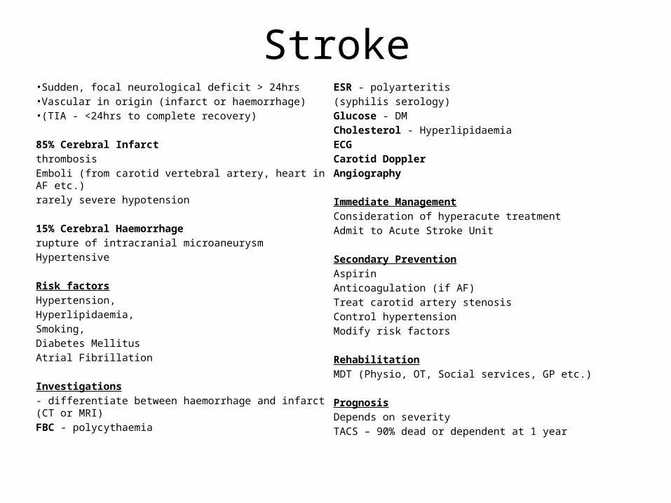
Stroke• Sudden, focal neurological deficit > 24hrs• Vascular in origin (infarct or haemorrhage)• (TIA - <24hrs to complete recovery)
85% Cerebral Infarct
thrombosis
Emboli (from carotid vertebral artery, heart in AF etc.)
rarely severe hypotension
15% Cerebral Haemorrhage
rupture of intracranial microaneurysm
Hypertensive
Risk factors
Hypertension,
Hyperlipidaemia,
Smoking,
Diabetes Mellitus
Atrial Fibrillation
Investigations
- differentiate between haemorrhage and infarct (CT or MRI)
FBC - polycythaemia
ESR - polyarteritis
(syphilis serology)
Glucose - DM
Cholesterol - Hyperlipidaemia
ECG
Carotid Doppler
Angiography
Immediate Management
Consideration of hyperacute treatment
Admit to Acute Stroke Unit
Secondary Prevention
Aspirin
Anticoagulation (if AF)
Treat carotid artery stenosis
Control hypertension
Modify risk factors
Rehabilitation
MDT (Physio, OT, Social services, GP etc.)
Prognosis
Depends on severity
TACS – 90% dead or dependent at 1 year
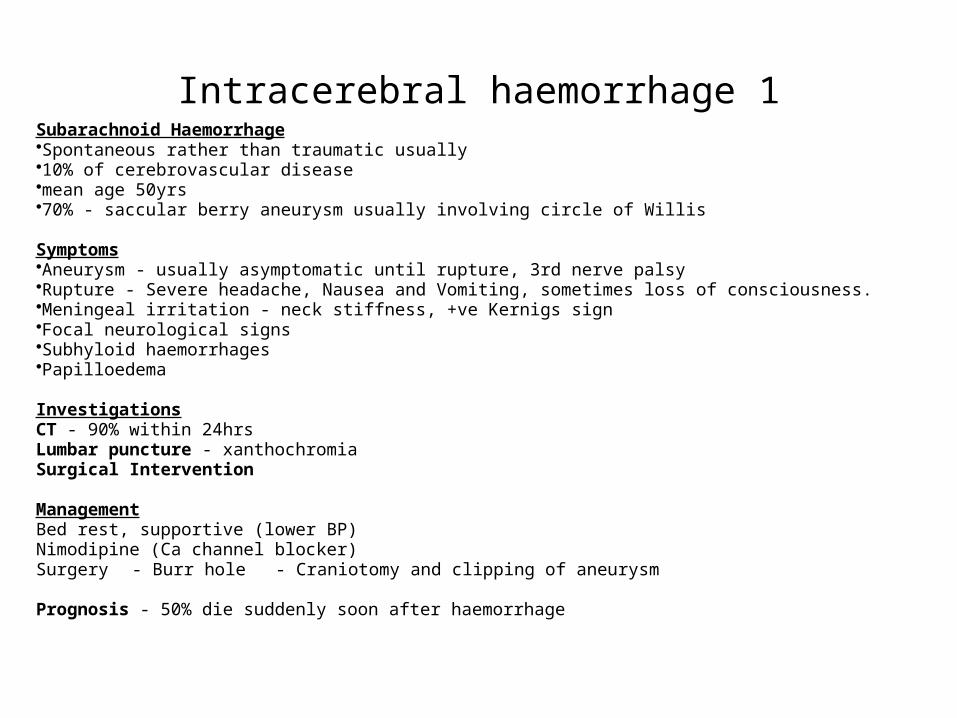
Intracerebral haemorrhage 1Subarachnoid Haemorrhage•Spontaneous rather than traumatic usually•10% of cerebrovascular disease•mean age 50yrs•70% - saccular berry aneurysm usually involving circle of Willis Symptoms•Aneurysm - usually asymptomatic until rupture, 3rd nerve palsy•Rupture - Severe headache, Nausea and Vomiting, sometimes loss of consciousness.•Meningeal irritation - neck stiffness, +ve Kernigs sign•Focal neurological signs•Subhyloid haemorrhages•Papilloedema InvestigationsCT - 90% within 24hrsLumbar puncture - xanthochromiaSurgical Intervention ManagementBed rest, supportive (lower BP)Nimodipine (Ca channel blocker)Surgery - Burr hole - Craniotomy and clipping of aneurysm Prognosis - 50% die suddenly soon after haemorrhage

Intracerebral haemorrhage 2
Subdural HaematomaBlood accumulating in subdural space after rupture of vein running from hemisphere to sagittal sinusDue to head injury (often minor)Latent interval (weeks, months)
Susceptible - alcoholics, elderly Symptoms - headache, drowsiness, confusion (fluctuate) CT diagnosis Treatment - surgical removal
Extradural HaemorrhageDamage to temporal bone - rupture of middle meningeal artery
Clinical picture- Head injury- loss of consciousness, followed by recovery, then sudden deterioration with focal neurological signs and reduced consciousness Treatment - surgical drainage

(a) Cerebral haemorrhage in thalamus(b) MR T2 weighted: Infarction of posterior branches of middle cerebral artery(c) MR T2 weighted: Cervical cord and C5/6 disc causing compression (d) CT: Communicating hydrocephalus (IIIrd, lateral and IVth ventricles)(e) CT: Subarachnoid haemorrhage (blood around brainstem)(f) DSA, lateral: Aneurysm of anterior cerebral artery(g) MR T1 weighted: Bilateral subdural haematomas(h) MR T1 weighted: Glioblastoma multiforme(i) MR T1 weighted: Cerebellar metastases(j) MR T1 weighted: Skull base meningioma

Epilepsy 1
CLASSIFICATION
A. Idiopathic Generalised Epilepsies
1. Tonic-clonic (Grand mal) fits
Tonic phase
Clonic phase
Post-ictal
2. Absence Attacks (Petit Mal)
3. Myoclonic Epilepsy
B. Localisation-Related Epilepsy
1. Focal Motor Attacks
2. Focal Sensory Attacks
3. Temporal Lobe Epilepsy
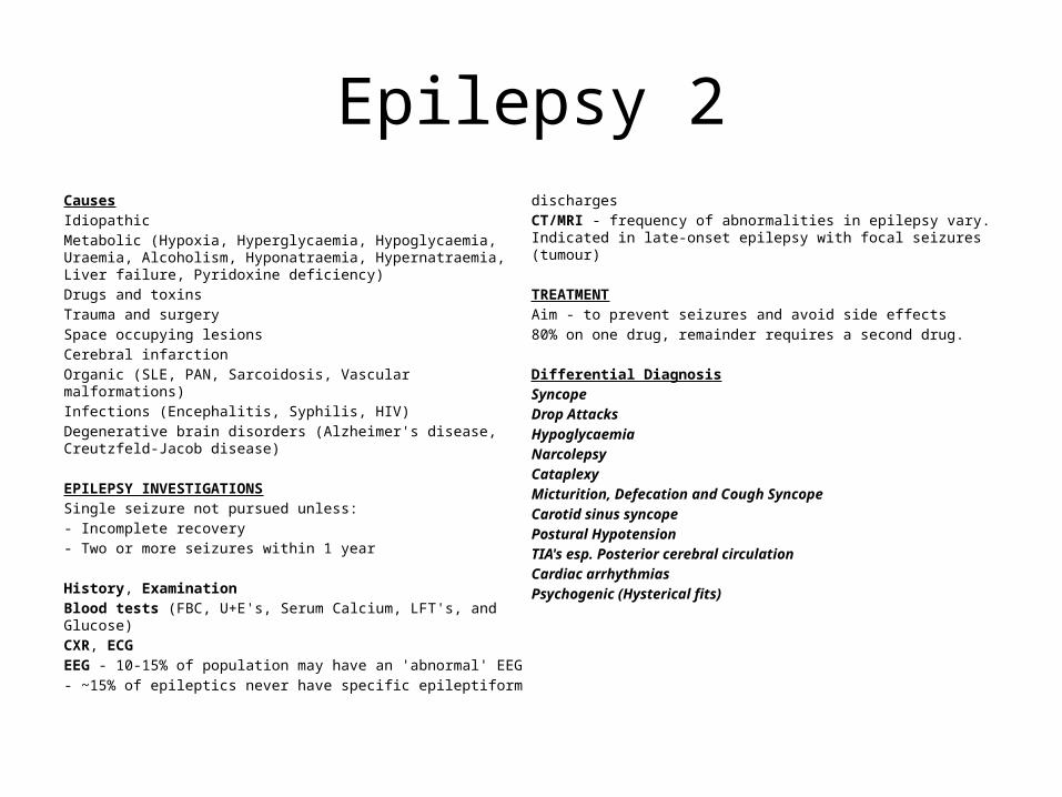
Epilepsy 2CausesIdiopathicMetabolic (Hypoxia, Hyperglycaemia, Hypoglycaemia, Uraemia, Alcoholism, Hyponatraemia, Hypernatraemia, Liver failure, Pyridoxine deficiency)Drugs and toxins Trauma and surgery Space occupying lesionsCerebral infarctionOrganic (SLE, PAN, Sarcoidosis, Vascular malformations)Infections (Encephalitis, Syphilis, HIV)Degenerative brain disorders (Alzheimer's disease, Creutzfeld-Jacob disease) EPILEPSY INVESTIGATIONSSingle seizure not pursued unless:- Incomplete recovery- Two or more seizures within 1 year History, ExaminationBlood tests (FBC, U+E's, Serum Calcium, LFT's, and Glucose)CXR, ECGEEG - 10-15% of population may have an 'abnormal' EEG- ~15% of epileptics never have specific epileptiform discharges
CT/MRI - frequency of abnormalities in epilepsy vary. Indicated in late-onset epilepsy with focal seizures (tumour) TREATMENTAim - to prevent seizures and avoid side effects80% on one drug, remainder requires a second drug. Differential DiagnosisSyncopeDrop AttacksHypoglycaemiaNarcolepsyCataplexyMicturition, Defecation and Cough SyncopeCarotid sinus syncope Postural HypotensionTIA's esp. Posterior cerebral circulationCardiac arrhythmiasPsychogenic (Hysterical fits)

Movement DisordersParkinson’s Disease• The Shaking Palsy• Tremor, rigidity and akinesia• Commonly asymmetrical• Response to Levodopa
• Side effects of treatment• ‘Wearing off’• Psychiatric aspects
Drug Induced/Vascular ParkinsonismEssential TremorChoreaHemiballismusMyoclonusTic disorderTorsion dystonias

Multiple Sclerosis• Demyelination with the brain and spinal cord• No single group of signs or symptoms is entirely diagnostic of MS• Two principal patterns:
– relapsing and remitting MS with lesions occurring in different parts of the CNS at different times
– chronic progressive MS (some 30% of cases)• Optic Neuropathy• Brainstem demyelination• Spinal cord lesion
Investigations• Imaging (MRI)• CSF examination• Electrophysiology (VER)
Management• Steroids• Immunosuppression• Beta-Interferon• Disease modifying drugs• Physiotherapy
Acquired condition Characterized by weakness and fatiguability of proximal limb, ocular and bulbar muscles. CLINICAL FEATURES Fatiguability The proximal limb muscles, the extraocular muscles, and the muscles of mastication, speech and facial expression commonly affected in the early stages. Respiratory difficulties may occur. Complex extraocular palsies, ptosis and a typical fluctuating proximal weakness The reflexes are initially preserved but may be fatiguable. Muscle wasting - late sign INVESTIGATIONS Tensilon (edrophonium) testSerum acetylcholine receptor Nerve stimulation COURSE AND MANAGEMENT Severity fluctuates but most cases have a protracted course. Important to recognize respiratory impairment,
dysphagia and nasal regurgitation;Emergency assisted ventilation may be required in myasthenic crises. Exacerbations are usually unpredictable and unprovoked but may be brought on by infections, by aminoglycosides or other drugs. Enemas (magnesium sulphate) may provoke severe weakness. TREATMENT Oral anticholinesterases Pyridostigmine (60 mg tablet) (most widely used drug). Thymectomy Immunosuppressant drugs Plasmapheresis and immunoglobulin
Myasthenia Gravis

Motor Neurone Disease
• Progressive degeneration of upper and lower motor neurones
• Three patterns…– Progressive muscular
atropthy– Amyotrophic lateral
sclerosis– Progressive bulbar palsy
• Same condition
Diagnosis• No diagnostic test• EMG/NCS
Management• Riluzole• Supportive treatment• Palliative care
Case 135yr old man presents with sudden onset of headachePain is on the left side just above his ear He woke up this morning with the pain this morning. Worse on coughing, sneezing and stooping down. He has experienced some blurred and double vision today.He is currently feeling quite nauseous but has not vomited.He has a temperature since yesterday evening when he began to feel unwell. His wife reports that he had a fit in bed this morning and again before coming into hospital. He has no memory of these events. He is worried it may be epilepsy. Past Medical HistoryEar infection on return from holiday in GreeceNil else Medication3 days worth of Amoxycillin for infection.Nothing currently No family history, non-smoker, unemployed On ExaminationPyrexialPain localising to Left side above earPapilloedema and diplopiaSigns of ear infection

Case 222yr old women presents with weakness in Left arm She noticed this when she dropped a cup of tea. This has never happened before. She has no related symptoms When questioned she says she experienced blurred vision in both eyes a few weeks ago. This lasted a few minutes and was related to exercise/ On flexing her neck she experiences parasthesiae in legs and an 'electric shock' sensation down her back PMHNone MedicationNone Family HistoryMother has Multiple Sclerosis InvestigationsCSF - Pleocytosis, raised protein, IgG

Case 380 yr old man presents with weakness in left arm and problems with speech.He woke up yesterday morning with weakness and has so far noticed no improvement.He is right handed PMHTIA'sHigh BPType II Diabetic for 20yrs, well controlledMI - 10yrs ago SHSmokerNon-drinker Family HistoryIschaemic heart disease On ExaminationAphasiaRight sided hemianopiaLeft arm - reduced power, tone and reflexesNo change in sensationLeft leg unaffectedOther limbs - Normal power, tone and reflexes GI, Lungs and Heart - all normal
Case 440yr old male. Attending GP at request of his wife - he doesn't think anything is wrong. Collateral History from wifeHe has 'funny turns' when he doesn't respond to her and appears to walk round in a dreamy state but continues with normal activities. On careful questioning the man admits to experiencing some strange things, there is often a smell, like sewerage, just before his wife notices these episodes. He also experiences Déjà vu frequently.His wife reports that he sometimes responds to voices which she cannot hear when he is having one of these 'funny turns'. No PMH No Family History


Merry Christmas!


