51
Neuropathy Review Yessar Hussain, MD Austin Neuromuscular Center Assistant Professor UT Austin Dell Medical School

Neuropathy Review
Yessar Hussain, MD Austin Neuromuscular Center
Assistant Professor UT Austin
Dell Medical School

Over review
• Nerve anatomy
• Nerve physiology
• Approach to Neuropathy
• Common Neuropathy
• Electrodiagnostic studies (EMG/NCS)

Nerve Anatomy

Motor units
• Motor unit: Composed of one motor neuron and all the muscle fibers that it innervates
• There are many motor units in a muscle
• The number of fibers innervated by a single motor neuron varies (from a few to thousand)
• The fewer the number of fibers per neuron the finer the movement (more brain power)
• Which body part will have the largest motor units? The smallest?


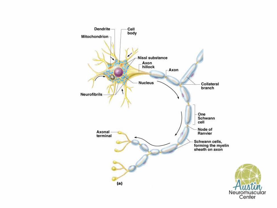
Structure of a nerve
(a)
(b)
Fascicle
Perineurium Blood vessels
Endoneurium Nerve fibers
Axon
Endoneurium
Perineurium
Epineurium
Myelin sheath
Blood vessels
Fascicle

Spinal Nerves and Nerve Plexuses
• Characteristics: – Somatic sensation (conscious) and somatic motor control (voluntary control)
of skeletal muscles. – Includes cranial nerves: I, II, IV-VI, VIII, XI and XII. – Spinal nerves: 31
• Cervical: 8 (above C1, and below C1-C7) • Thoracic: 12 (below T1-T12) • Lumbar: 5 (below T1-T5) • Sacral: 5 ( below S1-S5) • Coccygeal: 1 exit coccyx
– Mixed nerves • Sensory • Motor
– Dorsal and ventral rami (nerve branches) plexuses (network of nerves)
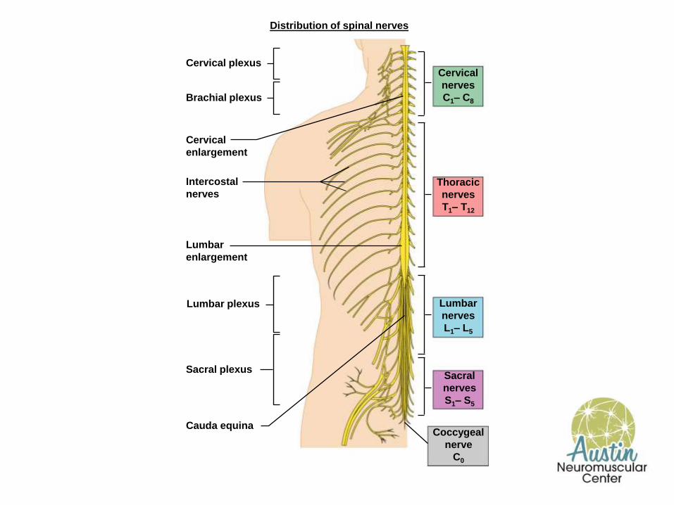
Distribution of spinal nerves
Cervical nerves C1– C8
Thoracic nerves T1– T12
Lumbar nerves L1– L5
Sacral nerves S1– S5
Coccygeal nerve
C0
Cervical plexus
Intercostal nerves
Cervical enlargement
Lumbar enlargement
Cauda equina
Brachial plexus
Lumbar plexus
Sacral plexus
Nerve Physiology
Action Potentials
• Action potentials are the mechanism by which the nervous system carries out its fundamental task: communication
• Action potentials are present in many cell types (including plants), but the neuron is specialized to generate and propagate these potentials
Building a Potential
• An action potential is a rapid change in polarity of the cell relative to the extracellular space
• In order to change polarity, there must be a “resting potential” – Related to the excess of intracellular negative ions
• A stimulus causes a depolarization, which propagates across the cell membrane
Starting a Nerve Impulse
Copyright © 2003 Pearson Education, Inc. publishing as Benjamin Cummings
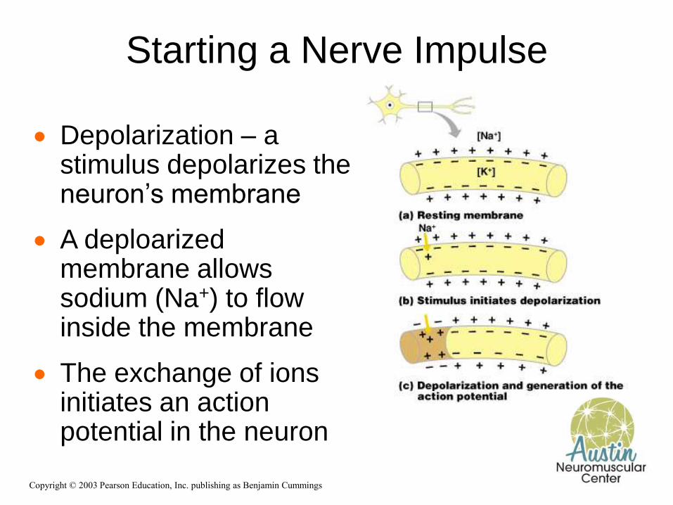
Depolarization – a stimulus depolarizes the neuron’s membrane
A deploarized membrane allows sodium (Na+) to flow inside the membrane
The exchange of ions initiates an action potential in the neuron

Neuropathy
• Peripheral neuropathy (PN) is damage to or disease affecting nerves, which may impair sensation, movement, gland or organ function, or other aspects of health, depending on the type of nerve affected.
• Common causes include systemic diseases (such as diabetes or leprosy), vitamin deficiency, medication (e.g., chemotherapy), traumatic injury, radiation therapy, excessive alcohol consumption, immune system disease or viral infection. It can also be genetic (present from birth) or idiopathic (no known cause).
• Neuropathy (neuro-, "nervous system" and -pathy, "disease of)
Pattern
• Neuropathy affecting just one nerve is called "mononeuropathy"
• Neuropathy involving multiple nerves in roughly the same areas on both sides of the body is called "symmetrical polyneuropathy" or simply "polyneuropathy."
• When two or more (typically just a few, but sometimes many) separate nerves in disparate areas of the body are affected it is called "mononeuritis multiplex," "multifocal mononeuropathy," or "multiple mononeuropathy
Pattern
• Peripheral neuropathy may be chronic (a long term condition where symptoms begin subtly and progress slowly) or
• Acute (sudden onset, rapid progress, and slow resolution). Acute neuropathies demand urgent diagnosis.
• Motor nerves (that control muscles),sensory nerves, or autonomic nerves (that control automatic functions such as heart rate, body temperature, and breathing), may be affected.
• More than one type of nerve may be affected at the same time. Peripheral neuropathies may be classified according to the type of nerve predominantly involved, or by the underlying cause.
• Where the cause is unknown it is described as idiopathic neuropathy.

Symptoms
• Neuropathy may cause painful cramps, fasciculations (fine muscle twitching), muscle loss, and changes in the skin, hair, and nails. M
• Motor neuropathy may cause impaired balance and coordination or, most commonly, muscle weakness.
• Sensory neuropathy may cause numbness to touch and vibration, reduced position sense causing poorer coordination and balance, reduced sensitivity to temperature change and pain, spontaneous tingling or burning pain, or skin allodynia (severe pain from normally nonpainful stimuli, such as light touch)
• Autonomic neuropathy may produce diverse symptoms, depending on the affected glands and organs, but common symptoms are poor bladder control, abnormal blood pressure or heart rate, and reduced ability to sweat normally.

Common Neuropathies
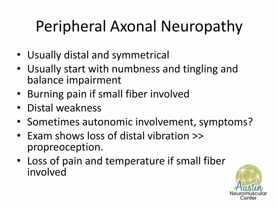
Peripheral Axonal Neuropathy
• Usually distal and symmetrical • Usually start with numbness and tingling and
balance impairment • Burning pain if small fiber involved • Distal weakness • Sometimes autonomic involvement, symptoms? • Exam shows loss of distal vibration >>
propreoception. • Loss of pain and temperature if small fiber
involved
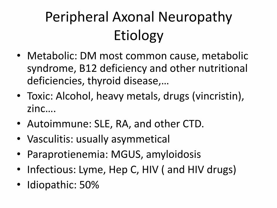
Peripheral Axonal Neuropathy Etiology
• Metabolic: DM most common cause, metabolic syndrome, B12 deficiency and other nutritional deficiencies, thyroid disease,…
• Toxic: Alcohol, heavy metals, drugs (vincristin), zinc….
• Autoimmune: SLE, RA, and other CTD.
• Vasculitis: usually asymmetical
• Paraprotienemia: MGUS, amyloidosis
• Infectious: Lyme, Hep C, HIV ( and HIV drugs)
• Idiopathic: 50%
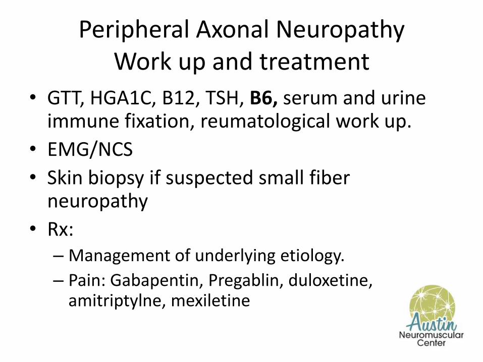
Peripheral Axonal Neuropathy Work up and treatment
• GTT, HGA1C, B12, TSH, B6, serum and urine immune fixation, reumatological work up.
• EMG/NCS
• Skin biopsy if suspected small fiber neuropathy
• Rx: – Management of underlying etiology.
– Pain: Gabapentin, Pregablin, duloxetine, amitriptylne, mexiletine

Small Fiber Neuropathy remember “ fibromyalgia”
• Definition:
– Peripheral neuropathy manifest by dysesthesias, with findings of small fiber dysfunction on neurologic exam, specialized electrodiagnostic testing or pathologic studies
• Loss of vibration at toes and absent ankle DTRs may be present
• Loss of vibration at ankles, decreased proprioception at toes, distal wasting or weakness, generalized areflexia, abnormal findings on routine NCSs/EMG may not be present

Signs and Symptoms
• Burning, cold, tingling, prickling shock- like, itching
• Stocking-glove distribution
• Can be patchy, proximal, diffuse
• Stimulus-evoked pain • Allodynia - Pain due to stimulus that
does not normally provoke pain
• Hyperalgesia – An increased response to a stimulus that is normally painful
• Cardiac • Syncope • Orthostatic hypotension • Change in heart rate or BP
• Gastrointestinal • Constipation/diarrhea • Emesis • Abdominal distention
• Urinary • Nocturia • Incontinence
• Genital • Impotence
• Sweating • Anhidrosis/hyperhidrosis
• Ocular • Dry eyes, dry mouth • Ptosis, miosis
Autonomic

Causes of painful/small fiber neuropathy
• Non-immune mediated Idiopathic Glucose dysmetabolism
• Diabetes • Glucose intolerance
Hypertriglyceridemia Alcoholism HIV Leprosy Restless leg syndrome Vitamin deficiency
• Niacin • Thiamine • Post gastroplasty
• Immune mediated Guillain-Barré syndrome Systemic amyloidosis Paraproteinemia HCV, cryoglobulinemia Celiac disease Paraneoplastic disease Vasculitis Sarcoidosis Anti-sulfatide antibodies Connective tissue disease
• Sjögren's syndrome • SLE

Causes of painful/small fiber neuropathy
• Hereditary diseases Fabry’s disease
• -galactosidase mutation
Tangier disease (An--lipoproteinemia) • ATP binding cassette transporter (ABC1)
mutation
Porphyria • Porphobilinogen deaminase mutation
Amyloidosis • Transthyretin mutation
Hereditary sensory neuropathies • HSN I/HSAN1
– Serine palmitoyltransferase mutation
• Congenital sensory neuropathy with anhidrosis (HSN4/HSAN 4)
– TRKA mutation
• Hereditary sensory neuropathy with loss of pain perception
– Nerve growth factor-β mutation
Familial Erythromelalgia • SCN 9A mutation
• Neurotoxins Metals
Thallium
Arsenic
Drugs • Metronidazole
• Nitrofurantoin
• Taxol
• Thalidomide
• Perhexiline
• Vinca alkaloids
– Vincristine, vinblastine, vindesine
• Nucleoside analogs
– Zalcitabine (ddC), Stavudine (d4T), Didanosine (ddI)
– Lamivudine (3TC)
Marine toxins • Ciguatera

Painful peripheral neuropathy Laboratory testing
• Routine studies Glucose tolerance test,
HgbA1c Metabolic profile Complete blood count Lipid profile Serum/urine immunofixation Quantitative
immunoglobulins Cryoglobulins Anti-sulfatide antibodies Thiamine (Other B vitamins) ANA, SS-A/SS-B, RF, ESR Cryoglobulins
• Additional studies HIV Anti-Hu antibodies Anti-endomysial/anti-gliadin
antibodies Urine for heavy metals -Aminolevulinic
acid/Porphobilinogen (24-hour urine) -galactosidase assay -lipoproteins Transthyretin gene test
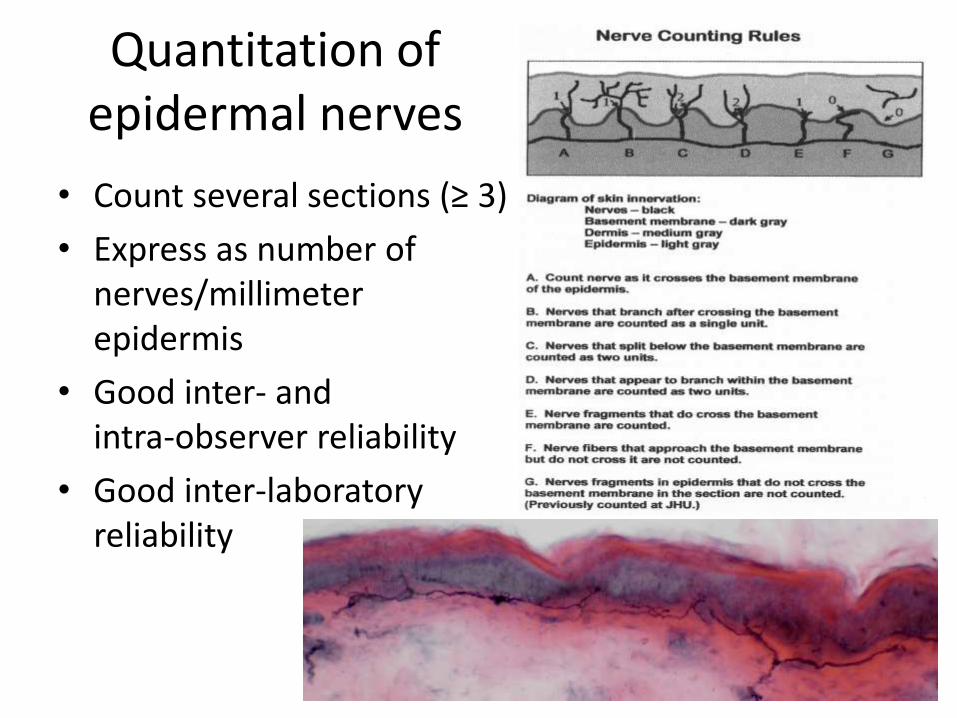
Quantitation of epidermal nerves
• Count several sections (≥ 3)
• Express as number of nerves/millimeter epidermis
• Good inter- and intra-observer reliability
• Good inter-laboratory reliability

Intraepidermal nerve fibers Normal Abnormal

Guillain-Barré syndrome
Guillain-Barré syndrome is a heterogeneous condition with several variant forms. Most often, GBS presents as an acute monophasic paralyzing illness provoked by a preceding infection.
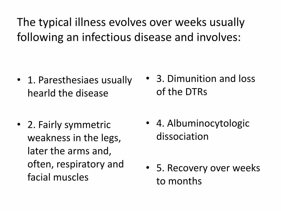
The typical illness evolves over weeks usually following an infectious disease and involves:
• 1. Paresthesiaes usually hearld the disease
• 2. Fairly symmetric weakness in the legs, later the arms and, often, respiratory and facial muscles
• 3. Dimunition and loss of the DTRs
• 4. Albuminocytologic dissociation
• 5. Recovery over weeks to months
History
Landry 1859 Miller Fisher 1956
Guillain, Barre & Strohl 1916

Predisposing factors- infections
• Well established- viral URI, GI illness (C. Jejuni)
• Well established- M. pneumoniae, H. influenzae, EBV, CMV
• Reported last year- VZV ( primary infection AND reactivation), Ricketsia typhi, Hepatitis A
• H1N1 associated GBS (at least 2 reports)
Predisposing factors- others reported over the last year
• Medications: Rituximab, Ecolizumab
• Paraneoplastic: lymphoma, SCLC, esophageal carcinoma
• Others: Graft-versus-host disease, ulcerative colitis
• Vaccinations
Treatment
Supportive:
• Respiratory failure: Bulbar and neck weakness are good predictors.
• Autonomic dysfunction: Avoid B blockers if possible
• DVT & PE
• Pain
Immunemodulating:
IVIG and PLEX

Chronic Inflammatory Demyelinating Polyneuropathy
Chronic Inflammatory Demyelinating Polyradiculoneuropathy was first described as an entity in 1975 (Dyck et al. 1975), although numerous case reports exists in the earlier literature describing patients with relapsing polyneuropathy responsive to steroids (Branagen et al., 2008)
CIDP
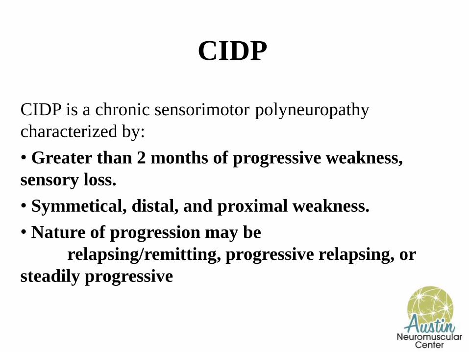
CIDP is a chronic sensorimotor polyneuropathy characterized by:
• Greater than 2 months of progressive weakness, sensory loss.
• Symmetical, distal, and proximal weakness. • Nature of progression may be relapsing/remitting, progressive relapsing, or steadily progressive

CIDP-Epidemiology
•Estimates of prevalence vary (1-7.7 per 100,000)
•Incidence increases with age
•Men more likely to be affected than women
•Prevalence in Men ages 70-79 is 1 in 10,000

CIDP: Diagnostic Studies
•No laboratory blood test helpful •CSF analysis elevated CSF protein in 80% of patients •Electrophysiological Nerve conduction studies extremely useful •Pathology Nerve biopsy sometimes helpful

CIDP: Treatment Options
Immunomodulatory Therapy
-Prednisone
-Intravenous Immunoglobulin (IVIg)
-Plasmapheresis
All have been shown to be effective in controlled clinical trials.
In refractory patients, other agents are used with variable results (cyclophosphamide, mycophenolate, azathioprine, interferons, cyclosporine)

Entrapment Neuropathies

• Nerve entrapment syndromes arise because of increased pressure applied to a nerve as it traverses a closed space.
• Common syndromes: Ulnar neuropathy, Median neuropathy (CTS), and peroneal neuropathy.
CTS
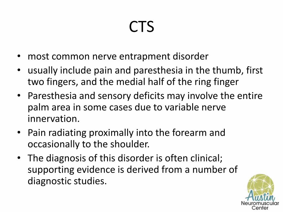
• most common nerve entrapment disorder
• usually include pain and paresthesia in the thumb, first two fingers, and the medial half of the ring finger
• Paresthesia and sensory deficits may involve the entire palm area in some cases due to variable nerve innervation.
• Pain radiating proximally into the forearm and occasionally to the shoulder.
• The diagnosis of this disorder is often clinical; supporting evidence is derived from a number of diagnostic studies.

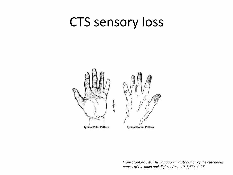
CTS sensory loss
From Stopford JSB. The variation in distribution of the cutaneous nerves of the hand and digits. J Anat 1918;53:14–25
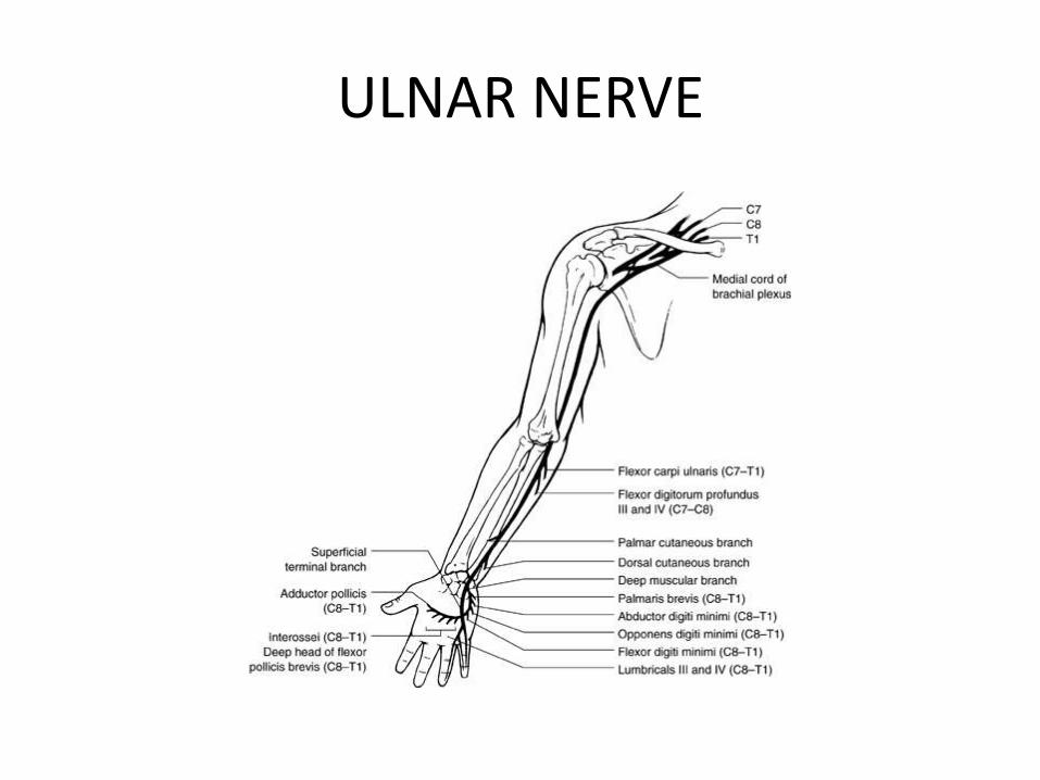
ULNAR NERVE

• Etiology: result of chronic mechanical compression or stretch, at – the groove
• external compression and repeated trauma • Rare, ganglia, tumors, fibrous bands, or accessory muscles. • tardy ulnar palsy-- a result of elbow fracture, often years before,
with subsequent arthritic changes of the elbow joint
– the cubital tunnel • distal to the groove • compression of the ulnar nerve under the humeral–ulnar
aponeurosis
• Weakness is the leading symptoms with minimal sensory symptoms

Electrodiagnostic Studies
Nerve conduction studies • Motor • Sensory • Late responses: F wave, H wave, Blink reflex • Autonomic • Repetitive Nerve Stimulation: Slow, Fast rates
EMG • Routine EMG studies • Single Fiber EMG

Electrodiagnosis: Practical Approach
• NCS/EMG is an extension of the neurological examination
• NCS/EMG used mainly for the evaluation & diagnoses of peripheral nerve, NMJ & muscle disorders
• Aims of the electrodiagnostic studies • Localization of the lesion (the major aim) • Pathophysiology of the lesion • Severity of the lesion • Temporal course of the disorder • Assess Recovery
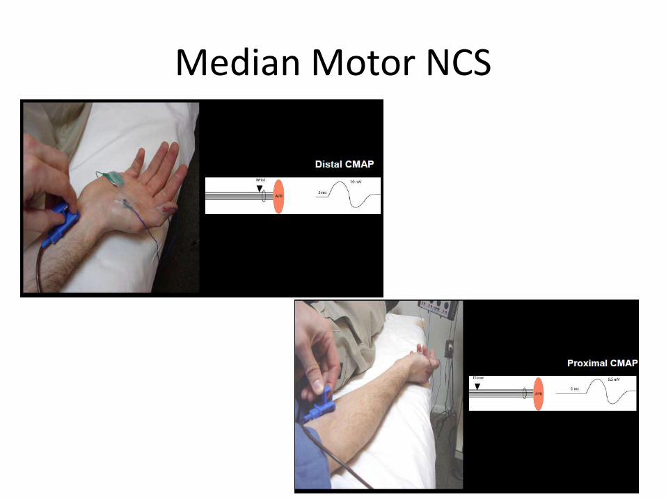
Median Motor NCS

EMG

Thank you


