Neuromuscular exercise as treatment for knee osteoarthritis in middle aged patients Research Unit for Musculoskeletal Function and Physiotherapy Faculty of Health Sciences Department of Sports Science and Clinical Biomechanics University of Southern Denmark, Denmark Brian Clausen PhD Thesis 2016
Transcript
Neuromuscular exercise as treatment for knee osteoarthritis in middle aged patients Research Unit for Musculoskeletal Function and Physiotherapy Faculty of Health Sciences Department of Sports Science and Clinical Biomechanics University of Southern Denmark, Denmark
Brian Clausen PhD Thesis 2016
2
3
Table of content Preface 4
List of papers 5
Thesis at a glance 6
Contributors 9
Abbreviations and definitions 11
Introduction 12
Theoretical framework 15
The role of external load on knee OA 17
Development of a novel knee joint load measure 19
Neuromuscular exercise (NEMEX) and knee biomechanics 20
Pharmacological pain relief and joint load in knee OA 21
Aims 23
General aims 23
Specific aims 23
Material and methods 24
Subjects 24
Sample size calculation 25
The EXERPHARMA trial (papers III and IV) 25
The neuromuscular exercise program (papers II, III and IV) 27
Pharmacological pain relief (papers III and IV) 29
Outcomes 30
Statistical analysis 35
Summary of results 37
Subjects and compliance (paper IV) 37
Development of a sensitive knee load index (paper I) 37
Feasibility of NEMEX-KOA (paper II) 39
The EXERPHARMA trial (paper IV) 41
Discussion 43
Main findings 43
Methodological considerations 43
Subjects 44
Outcomes 44
Interventions 47
Conclusions 49
Clinical implications and future perspectives 50
Summary 51
Resumé [Danish] 53
Acknowledgements 56
References 57
Papers I-IV Fejl! Bogmærke er ikke defineret.
4
Preface This PhD thesis was accomplished at the Department of Sports Science and Clinical Biomechanics,
Faculty of Health Sciences, University of Southern Denmark, Odense, Denmark. The main supervisor was
Professor Ewa M. Roos from the Research Unit for Musculoskeletal Function and Physiotherapy and co-
supervision was provided by Associate Professor Anders Holsgaard-Larsen, the Orthopaedic Research
Unit, Department of Orthopaedics and Traumatology, Odense University Hospital (OUH), Odense,
Denmark.
The study subjects were recruited via primary care general practitioners (GPs) in the communities of
Odense and Middelfart, Denmark, and from advertisements in local clubs, libraries, print media, and
Facebook. Outcome assessments at baseline and after treatment were carried out at the motion
analysis laboratory at OUH, Odense, Denmark.
The studies contained in this thesis were funded by the Region of Southern Denmark PhD fund, €67,000;
the Region of Southern Denmark research fund, €67,000; a University of Southern Denmark scholarship,
€55,000; Odense University Hospital free research funds, €44,000; The Danish Rheumatism Association,
€42,000; The Association of Danish Physiotherapists, €5,000; Family Hede Nielsens fund, €2,600; and a
Danish Rheumatism Association Ryholts grant, €2,000. None of the funders have any role in the study
other than to provide funding.
5
List of papers This thesis is based on the following four papers, which will be referred to by their Roman numerals in
the text:
I. Clausen B, Andriacchi T P, Nielsen D B, Roos E M, Holsgaard-Larsen A. The Knee Index and its
composition – a novel measure of knee joint load in subjects with mild to moderate knee
osteoarthritis. In manuscript
II. Clausen B, Holsgaard-Larsen A, Roos E M. An 8-week Neuromuscular Exercise Program in
Middle-aged Subjects with Mild to Moderate Knee Osteoarthritis – a Case-series. Submitted
III. Clausen B, Holsgaard-Larsen A, Søndergaard J, Christensen R. Andriacchi T P, Roos E M. The
effect on knee-joint load of instruction in analgesic use compared with neuromuscular exercise
in patients with knee osteoarthritis: study protocol for a randomized, single-blind, controlled
trial (the EXERPHARMA trial). Trials 2014; 15:444
IV. Holsgaard-Larsen A, Clausen B, Søndergaard J, Christensen R, Andriacchi T P, Roos E M. The
effect on knee-joint load of instruction in analgesic use compared with neuromuscular exercise
in patients with knee osteoarthritis: a randomized, single-blind, controlled trial (the
EXERPHARMA trial). In manuscript
6
Thesis at a glance This thesis comprises four studies describing the design and quality assurance of a randomized
controlled trial evaluating two different pain relief interventions for subjects with mild to moderate
knee osteoarthritis (OA). Paper I reported the properties of a novel biomechanical variable, the Knee
Index, for evaluating total knee joint load. Paper II reported the feasibility of the neuromuscular exercise
intervention for subjects with mild to moderate knee OA. Paper III described the protocol for the
randomized controlled trial evaluating the effect on knee joint load of two pain relief interventions:
neuromuscular exercise or drug use. Paper IV reported the effectiveness of the two pain relief
interventions: neuromuscular exercise or drug use.
7
Between-subject variation:
percentage distribution of the
contributors to the Knee Index
(study knee) in the 44 individuals.
Individuals are ordered from
lowest to highest for frontal plane
contribution – Mean of 5 trials.
Paper I – Is a novel measure of knee joint load, the Knee Index, suitable for use as a primary outcome in a randomized controlled trial?
Subjects: The first 44 subjects (23 females) with a mean age 57.5 years, included in the EXERPHARMA
trial.
Method: A cross-sectional study. Using three dimensional gait analysis, we describe a novel
biomechanical index of knee joint loading in patients with mild to moderate knee OA by reporting: i) the
relative contribution and inter-subject variation of each plane to the Knee Index, and ii) how other
biomechanical variables may be associated with the Knee Index.
Conclusion: Despite large variation in the composition of the Knee Index, it was primarily composed of
the frontal and sagittal planes, indicating that some subjects with mild to moderate knee OA have a
movement strategy dominated by frontal or sagittal moment. Depending on the knee-loading strategy,
the Knee Index was associated with both the frontal and sagittal planes. The current results hold
promise for the Knee Index as both a sensitive and responsive biomechanical outcome.
Paper II – Is a neuromuscular exercise program (NEMEX-KOA) a feasible intervention for subjects with
mild to moderate knee OA?
Subjects: The first 23 (12 females) subjects undergoing an 8-week neuromuscular exercise program.
Method: A case-series study describing the feasibility of a NEMEX program (NEMEX-KOA) in terms of
progression in exercise, exertion, pain, adverse events, and adherence to exercise for subjects with mild
to moderate KOA.
Conclusion: In subjects with mild to severe pain in activity at baseline, NEMEX-KOA was found feasible.
Progression was achieved with few incidents of clinically relevant increase in pain and no adverse
events. Jumping activities were, however, not feasible.
Paper III – A protocol for a randomized controlled trial on two pain-relieving treatments for mild to
moderate knee OA. The EXERPHARMA trial.
Subjects: 100 subjects, aged 40-70 years with mild to moderate knee OA, Kellgren and Lawrence grade
0-3, were to be recruited.
Method: A protocol describing a randomized controlled trial, evaluating the effectiveness of
neuromuscular exercise and optimized use of analgesics and anti-inflammatory drugs, on measures of
knee joint load during gait, patient-reported outcomes and functional performance in subjects with mild
to moderate knee OA.
Conclusion: The results of the EXERPHARMA trial will help to determine whether 8 weeks of
neuromuscular exercise is superior to optimized use of analgesics and anti-inflammatory drugs
regarding knee joint load, pain and functional performance in individuals with mild to moderate knee
OA.
Vs.
Paper IV – The effect on knee joint load of instruction in analgesic use compared with neuromuscular
exercise in patients with knee osteoarthritis: a randomized, single-blind, controlled trial (the
EXERPHARMA trial).
Subjects: 93 subjects (85% women, 58 ± 8 years with a BMI of 26.9 ± 3), were randomized to either the
NEMEX-group (n = 47) or the Pharma-group (n = 46). Data from 44 and 41 patients, respectively, were
available at follow-up. Subjects were recruited from July 2012 to May 2015.
Method: In a randomized, single-blind, controlled trial, we compared the effectiveness of neuromuscular
exercise (NEMEX-group) to optimized analgesics and anti-inflammatory drugs (Pharma-group), on
measures of knee joint load during gait, patient-reported outcomes and functional performance in
individuals with mild to moderate knee OA.
Conclusion: The EXERPHARMA trial evaluated the possible joint load modifying effects of a
neuromuscular exercise program compared with information on the recommended use of analgesics
and anti-inflammatory drugs. Due to poor adherence, especially in the Pharma-group, and a similar
intake of pain relief medication in both groups, we essentially compared the addition of 8 weeks of
neuromuscular exercise to ‘care as usual’, which did not result in reduced knee joint load during walking.
9
Contributors PAPER I
Study design: Brian Clausen
Anders Holsgaard-Larsen
Data collection: Dennis B Nielsen
Rasmus S Sørensen
Brian Clausen
Anne Marie Rosager
Data analysis: Brian Clausen
Anders Holsgaard-Larsen
Manuscript writing: Brian Clausen
Manuscript revision: Anders Holsgaard-Larsen
Thomas P Andriacchi
Dennis B Nielsen
Ewa M Roos
Journal Correspondence: Brian Clausen
PAPER II
Study design: Brian Clausen
Ewa M Roos
Data collection: Ann-Sofie Jensen
Rasmus Holst
Søren H Jensen
Connie L Hansen
Brian Clausen
Anne Marie Rosager
Data analysis: Brian Clausen
Ewa M Roos
Manuscript writing: Brian Clausen
Manuscript revision: Ewa M Roos
Anders Holsgaard-Larsen
Journal Correspondence: Brian Clausen
10
PAPER III
Study design: Brian Clausen
Anders Holsgaard-Larsen
Jens Søndergaard
Thomas P Andriacchi
Robin Christensen
Ewa M Roos
Manuscript writing: Brian Clausen
Manuscript revision: Anders Holsgaard-Larsen
Jens Søndergaard
Thomas P Andriacchi
Robin Christensen
Ewa M Roos
Journal Correspondence: Brian Clausen
PAPER IV
Study design: Brian Clausen
Anders Holsgaard-Larsen
Jens Søndergaard
Thomas P Andriacchi
Robin Christensen
Ewa M Roos
Data collection: Dennis B Nielsen
Rasmus S Sørensen
Ann-Sofie Jensen
Rasmus Holst
Søren H Jensen
Connie L Hansen
Brian Clausen
Anne Marie Rosager
Data analysis: Brian Clausen
Robin Christensen
Anders Holsgaard-Larsen
Ewa M Roos
Manuscript writing: Anders Holsgaard-Larsen
Manuscript revision: Brian Clausen
Jens Søndergaard
Thomas P Andriacchi
Robin Christensen
Ewa M Roos
Journal Correspondence: Ander Holsgaard-Larsen
11
Abbreviations and definitions ACR: American College of Rheumatism
ADL: Activities of Daily Living
BW: body weight
CR-10: Borg’s category ratio scale
GP: general practitioner
HT: height
KAM: knee adduction moment
KFM: knee flexion moment
KL: Kellgren and Lawrence
KOOS: Knee injury and Osteoarthritis Outcome Score
Knee Index: corresponds to 1st peak Knee Index
MDC: minimal detectable change
NEMEX: NEuroMuscular EXercise
NSAID: nonsteroidal anti-inflammatory drug
OA: osteoarthritis
OUH: Odense University Hospital
Pharma: pharmacologic treatment
QOL: quality of life
SD: standard deviation
Study knee: Most affected knee, or dominant if equally affected
VAS: Visual Analog Scale
12
Introduction Knee osteoarthritis (OA) is a degenerative joint disease, which alters the structure of the cartilage [4, 5].
The cartilage degenerates along with the surrounding tissue, that is, the synovial membrane, menisci
and underlying bone. Over time, knee OA will cause both pain and loss of physical function, resulting in
reduced quality of life [6].
Prevalence
On a global level, it was estimated that there were 241 million cases of OA in 2013, which was a 71.9%
increase from 1990 [7]. The global age-standardised prevalence of knee OA was 3.8% in 2010 and was a
little higher in women than men. Hip and knee OA was ranked as the 11th highest contributor to global
disability out of 291 conditions [8]. With increasing age and more people being overweight in the
population, the prevalence of OA is likely to rise in the future [8]. About one third of individuals with
knee OA will experience progression to more advanced disease [9, 10], which is the leading indication
for knee replacement surgery.
Diagnosing knee OA
Historically, diagnosis and severity of
knee OA have been based on structural
changes as seen on plain radiographs of
the tibiofemoral joint. Knee radiographs
are commonly graded by the Kellgren and
Lawrence (KL) grading system [13, 14].
However, radiographs have been found
to be an imprecise guide for diagnosing
knee OA, in that there is poor correlation
between pain and structural changes [15,
16]. This thesis defines knee OA based on the clinical picture and examination, using the two sets of
criteria developed by the European League Against Rheumatism (EULAR) and the American College of
Rheumatism (ACR) (Table 1) for the diagnosis of knee OA consisting of risk factors, symptoms and
objective examination [11, 12].
A clinical examination including symptoms, functional performance and patient willingness, together
with a plain radiograph of the tibiofemoral joint is the typical tool used for assessing possible need for
end-stage treatment of knee OA, for example, tibial osteotomy or total knee joint replacement [17].
However, from a patient perspective, pain is the single most important factor in the decision to undergo
knee joint replacement surgery [17].
Table 1: Criteria for diagnosing Knee OA developed by ACR and EULAR
[11, 12].
Risk factors: Symptoms: Objective examination:
Age >40 years
Female
Overweight
Occupation
Family history of
OA
Persistent knee pain
Brief morning
stiffness
Functional limitations
Acute knee pain
Crepitus during active
movement
Bony tenderness
Bony enlargement
Palpable effusion
No palpable warmth
Restricted movement
Joint instability
13
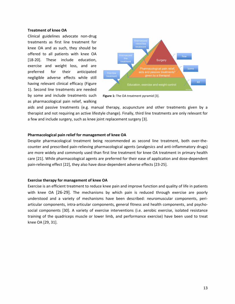
Treatment of knee OA
Clinical guidelines advocate non-drug
treatments as first line treatment for
knee OA and as such, they should be
offered to all patients with knee OA
[18-20]. These include education,
exercise and weight loss, and are
preferred for their anticipated
negligible adverse effects while still
having relevant clinical efficacy (Figure
1). Second line treatments are needed
by some and include treatments such
as pharmacological pain relief, walking
aids and passive treatments (e.g. manual therapy, acupuncture and other treatments given by a
therapist and not requiring an active lifestyle change). Finally, third line treatments are only relevant for
a few and include surgery, such as knee joint replacement surgery [3].
Pharmacological pain relief for management of knee OA
Despite pharmacological treatment being recommended as second line treatment, both over-the-
counter and prescribed pain-relieving pharmacological agents (analgesics and anti-inflammatory drugs)
are more widely and commonly used than first line treatment for knee OA treatment in primary health
care [21]. While pharmacological agents are preferred for their ease of application and dose-dependent
pain-relieving effect [22], they also have dose-dependent adverse effects [23-25].
Exercise therapy for management of knee OA
Exercise is an efficient treatment to reduce knee pain and improve function and quality of life in patients
with knee OA [26-29]. The mechanisms by which pain is reduced through exercise are poorly
understood and a variety of mechanisms have been described: neuromuscular components, peri-
articular components, intra-articular components, general fitness and health components, and psycho-
social components [30]. A variety of exercise interventions (i.e. aerobic exercise, isolated resistance
training of the quadriceps muscle or lower limb, and performance exercise) have been used to treat
knee OA [29, 31].
Figure 1: The OA treatment pyramid [3].
14
Neuromuscular exercise (NEMEX)
A major goal of traditional knee OA rehabilitation is to enhance muscle strength [32, 33]. The aim of
NEMEX programs is different since the intention is to improve postural control (i.e. position of the trunk
and lower limbs relative to one another) and functional performance (i.e. quality of movement
performance) by challenging lower-limb muscles in functional positions [34].
Unlike conventional strength training, neuromuscular exercise addresses the quality of movement and
emphasizes joint control in all three biomechanical/movement planes. Neuromuscular exercise has
effects on knee functional performance, knee biomechanics and muscle activation patterns of the
surrounding knee musculature. Neuromuscular exercise is used effectively for prevention and
rehabilitation of anterior cruciate ligament injury [35-38], rehabilitation in patients with a meniscal tear
with or without the combination of meniscectomy [1, 39] and in patients with moderate to severe OA
prior to total joint replacement [40-42].
15
Theoretical framework
Introduction to knee biomechanics
The primary movement (i.e. flexion/extension) of the knee occurs in the sagittal plane (Figure 2A). With
increasing knee OA severity, movements and/or malalignment (i.e. adduction/abduction or
varus/valgus) of the knee in the frontal plane can develop (Figure 2B) [43], whereas alterations to
movements (i.e. internal/external-rotation) in the transversal plane are more pronounced in ACL-
deficient knees (Figure 2C) [44].
Moments such as the external knee adduction moment is the product of the moment lever arm and
magnitude of the ground reaction force (Figure 2D), and can thus be altered by the magnitude/length of
the lever arm or the magnitude/length of the ground reaction force.
Figure 2: Planes and moments of the knee (A) sagittal plane, (B) frontal plane, (C) transversal plane, and (D) frontal plane
moment lever arm and ground reaction force. Figure 2D, adapted from Münderman et al. [45].
This thesis is positioned within the framework of the following four interactions (Figure 3) [5]: 1)
external measured knee loads during gait (Figure 3A) are related to the 2) internal knee joint forces
acting on the knee (Figure 3B), which are the sum of muscle forces, passive tissue forces, and contact
forces; 3) these internal forces determine cartilage tissue stress (Figure 3C); 4) which determine the
cartilage metabolism (Figure 3D), that again can cause alterations in gait.
Specifically, this thesis will describe and analyze a novel load measure of total knee joint load and load
modifying interventions (Figures 3A and B).
16
Figure 3: A conceptual model for biomechanical factors driving knee OA, (A) the external kinematic and kinetic signals related
to knee OA, (B) the joint internal forces acting at the knee joint, (C) the cartilage tissue stress/strain is determined by internal
force acting over an area of contact, and (D) the biological response is related to the local mechanical environment [5].
17
The role of external load on knee OA
Biomechanical factors are a driving factor for progression
During the stance phase of walking, relatively high loads are applied to the knee and loading of the
medial knee compartment especially [46] has been associated with disease progression [47, 48], severity
[49] and structural changes [50] in patients with knee OA.
Aetiology
The onset [5, 43] and progression [5, 51] of knee OA is to a large extent based on mechanical factors,
that is, excess joint load or abnormal knee joint load or a combination of both. Excess knee joint load
can be caused by an acute overload trauma (e.g. ACL rupture [52]), chronically with obesity [53, 54] or
structural degeneration related to knee OA causing meniscal tears and malalignment [43]. Abnormal
knee joint loads caused by abnormal anatomy of either congenital or acquired origin (e.g. partial
meniscectomy [55]) can lead to increased focal stress on a certain part of the joint.
External knee loads relation to internal knee joint forces
External knee moments are used as a surrogate measure of compressive internal knee joint force. This
assumption has been well established in approximately 10 subjects with load cell instrumented knee
implants [46, 56-58]. Despite large variations in correlations (R² =0.44 to 0.81), the relationship between
the external knee adduction moment (KAM) and compressive medial knee joint force is considered
robust at a group level but to a less degree at an individual level [46, 56-58]. KAM is, therefore,
considered a valid outcome in OA research. It is important to note that subjects with instrumented knee
implant [46, 56-58] have total knee joint replacements and presumably severe knee OA, and as a result,
these findings might not be applicable to subjects with mild to moderate knee OA. However, two studies
combining KAM and knee flexion moment (KFM) have improved correlation (from R2= 0.63-0.87 to 0.85-
0.91) between external moments and compressive medial knee joint forces, measured and estimated
respectively in subjects with total knee joint replacement and ACL reconstruction [57, 59]. Thus,
combining moments from more than one plane might lead to better estimates of compressive internal
knee joint force.
Knee OA severity and variations in knee loads
Several cross-sectional studies have reported a relationship between radiographic knee OA severity and
external peak KAM (Figure 4) [49, 60-65]. However, there does not seem to be a relationship between
knee OA severity and peak KFM (Figure 5) [64-66]. Since the combination of KAM and KFM is better
correlated to compressive medial knee joint forces than KAM or KFM alone, the combination is likely to
be better related to knee OA severity.
18
Figure 4: Relationship between external knee adduction moment and radiographic severity (KL grade), whiskers represents
standard deviation (SD).
Figure 5: Relationship between external Knee Flexion Moment and radiographic severity (KL grade), whiskers represents
standard deviation (SD).
A recent meta-analysis found very low evidence for a causal link between the external KAM and
structural progression of knee OA in subjects with moderate to severe knee OA [67]. Although unknown,
hypothetically it is more likely that the combination of KAM and KFM will show a causal link to knee OA
progression.
Using outcomes composed of one plane only, such as KAM or KFM, may not provide the best
assumption of medial compartment loading [68]. Thus, biomechanical outcome measures of total knee
loading incorporating all three planes (frontal, sagittal and transversal) may be beneficial in OA research.
0
1
2
3
4
5
6
0,0 1,0 2,0 3,0 4,0
pe
ak K
AM
[N
m/(
BW
×HT%
)]
Radiographic knee OA severity (KL grade)
Peak KAM and knee OA Severity
Blazek 2013 normal weight
Blazek 2013 Obese
Kean 2012
Henriksen 2011
Creaby 2010
Henriksen 2010
Henriksen 2010 Healthy w/wo pain
Schmitt 2007 first peak KAM
Thorp 2006
Sharma 1998
0
1
2
3
4
5
0,0 1,0 2,0 3,0 4,0
pe
ak K
FM [
Nm
/(B
W×H
T%)]
Radiographic knee OA severity (KL grade)
Peak KFM and knee OA Severity
Favre 2014 midstance
Blazek 2013 normal weight
Blazek 2013 Obese
Henriksen 2010
Henriksen 2010 Healthy w/wo pain
Schmitt 2007
19
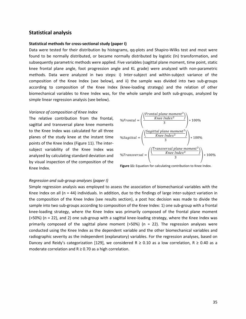
Development of a novel knee joint load measure A novel knee joint load measure, the Knee Index (also described in the literature as ‘total reaction
moment’ [69]), which includes moments from all three planes, has recently been introduced as a
surrogate measure of total load across both knee compartments [69]. In contrast to KAM, the Knee
Index is sufficiently sensitive to distinguish between pain relief induced by placebo, non-steroidal anti-
inflammatory drugs (NSAIDs) or opioids in subjects with moderate knee OA [69]. Furthermore, external
peak KAM has been shown to increase with age and knee OA severity [49, 60-63, 65], and no
relationship between peak KFM and age has been found [64-66, 69]. This indicates that middle-aged
individuals with mild to moderate knee OA use a knee-loading strategy composed of both KAM and KFM
compared to older individuals with more severe knee OA. This further indicates that changes in KAM
relate to changes in knee joint load to a limited degree only in middle-aged individuals with mild to
moderate knee OA.
The Knee Index is a functional variable, constructed from the maximal external moments affecting the
knee in the frontal, sagittal and transversal planes [70]. Although unknown, it is likely that subjects with
knee OA develop individual loading strategies to manage pain and OA severity during gait. These
strategies might vary from primarily loading the knee through the sagittal, primarily through the frontal
plane or a relatively equal distribution through both planes. Such a hypothesized inter-subject variation
in loading strategy will impact the utility of single-plane variables (e.g. KAM and KFM) in research and
clinical practice to a greater extent than multi-plane variables (e.g. the Knee Index).
Thus, to interpret the underlying biomechanical characteristics of the Knee Index, the respective
contributions of the knee moments derived from the three planes and potential inter-subject variation
during gait are important to determine. Furthermore, overall knee joint load has been shown to be
associated with altered gait strategies in the form of variation in single plane biomechanical variables
(such as KAM, KFM, knee flexion/extension angle, knee alignment, etc.) [48, 71-75]. Thus, to determine
if the Knee Index might be sensitive and responsive, it is important to determine associations between
the Knee Index and a variety of single-plane measures in a case mix of individuals with mild to moderate
knee OA. Considering the varied relationship between radiographic severity and KAM and KFM (Figures
4 and 5) [49, 60-66], it is important to determine if such a relationship also exists between the Knee
Index and radiographic severity in individuals with mild to moderate knee OA.
The Knee Index is theoretically influenced by the functional alignment of the trunk, pelvis, and lower
limb segments with respect to the knee during movement and the ground reaction force generated.
Thus, it is likely that interventions, such as neuromuscular exercise, targeting the efficiency of lower limb
movement and muscle activation patterns can be effective in improving dynamic knee joint loading [51,
76].
20
Neuromuscular exercise (NEMEX) and knee biomechanics Subjects with degenerative knees are known to have deficiencies of sensory dysfunction, lower limb
has been applied and has been shown to improve patient-reported outcomes, functional performance
and knee extensor strength in subjects with risk of knee OA [39] and with severe knee OA [40, 78, 79].
Additionally, neuromuscular exercise increases functional knee stability and in pilot studies has shown
potential to reduce knee joint loads and improve cartilage matrix quality in those at risk of, or with, mild
OA [26, 27, 80] but not in those with malaligned knees and moderate to severe knee OA [81].
NEMEX and knee joint load
Despite its use in other conditions and in more
severe stages of knee OA, there is only one study
that has investigated the load-modifying effect of
NEMEX in the early stages of knee OA. It was an
uncontrolled pilot study consisting of 13 patients
with mild knee OA [26]. This study found a -
0.8Nm/kg (95% CI -0.04;-0.16) reduction (14%) in
peak KAM during one-leg rise from a stool following
8 weeks of neuromuscular exercise. However, a
randomized controlled trial showed no between-
group difference in KAM comparing NEMEX and
quadriceps exercise for subjects with severe knee
OA and malalignment [79]. Despite conflicting results about disease severity, we hypothesize that
NEMEX can amend knee joint load in individuals with mild to moderate knee OA. NEMEX can amend
knee joint load in both the frontal and sagittal planes by improving alignment of the lower extremity
(Figure 6A and B) and reducing functional instability [34], by improving muscle activation patterns of the
surrounding knee musculature. Although unknown, we expect that NEMEX can reduce the overall knee
joint load (Knee Index). The load in the frontal plane (KAM) can be reduced by improving functional knee
alignment, thus reducing the ground reaction force-to-knee lever arm [82, 83], secondly the ground
reaction force-to-knee lever arm can be altered by normalising foot progression angle (toe-in) and this
can increase 1st peak KAM but reduce 2nd peak KAM [74, 83, 84], and normalised trunk lean (not leaning
over weight bearing leg) can increase KAM [73, 83, 84]. The load in the sagittal plane (KFM) can be
altered by NEMEX, firstly, by reducing gait speed and thereby the ground reaction force, and secondly,
by reducing the knee flexion angle and thereby a decrease in KFM by reducing the ground reaction
force-to-knee lever arm [85] and normalising the foot progression angle [74]. The transversal plane can
be altered by NEMEX via improved functional knee alignment and functional stability, although the
transversal plane is likely to play a limited role in the composition of the Knee Index due to its limited
magnitude. In summary, NEMEX will be able to reduce 1st peak KAM and KFM by reducing the ground
reaction force-to-knee lever arm and the ground reaction force by reducing gait speed. It is however,
unknown if either KAM or KFM will be reduced equally, and if they are not, this may alter walking
strategy.
Figure 6: Correct (A) functional alignment ‘Knee over foot
position’, and incorrect (B) functional alignment ‘Knee
medial to foot position’ [1, 2].
21
Pharmacological pain relief and joint load in knee OA Pain is a mechanism that helps to protect damaged tissue and tissue at risk of damage. In healthy
subjects, experimentally induced knee pain has been shown to replicate altered gait movement
strategies seen in patients with mild knee OA, that is, reduced 1st peak KAM and KFM [86]. The most
common pharmacological pain relief used in the management of knee OA is acetaminophen and
NSAIDs. Studies have shown that orally administered pharmacologically-initiated pain relief
(acetaminophen and NSAIDs) in moderate knee OA is associated with increased loads in KAM (4% to
11%) and KFM (10% to 25%) (Figure 7) [69, 87, 88]. However, treatment also resulted in increased gait
speed (7% to 16%) [69, 87], and it is possible that the increased knee loads reported are an effect of
increased gait speed [72].
Additionally, studies of stronger pharmacological pain relief, injections with lidocaine or hyaluronic-acid,
have been linked to increased 1st peak KAM, reduced KFM and increased gait speed [36, 89, 90] and
Tanezumab® (nerve growth factor inhibitor) has been linked to knee OA progression [91, 92]. Therefore,
pharmacological pain relief, by eliminating the protective mechanism of the pain itself, may be
detrimental for knee joint structures by increasing overall knee joint load and thus we investigated this
by use of the Knee Index.
Figure 7: Change in external loads from pharmacological (oral NSAIDs) pain relief in subjects with knee OA. Changes seen for
Hurwitz et al. are based on estimates. An increase in gait speed from baseline to follow-up, 6.6% and 16.3% was seen for Boyer
et al. and Schnitzer et al., respectively.
0
5
10
15
20
25
30
Ch
ange
in p
eak
pla
ne
mo
me
nts
[%
]
Frontal Sagittal
Change in external loads from orally administered pharmacological (acetaminophan and NSAIDs) pain-relief in subjects with knee OA
Boyer 2012, first peak KAM
Schnitzer 1993, peak KAM
Hurwitz 2000, peak KAM
Boyer 2012, peak flexion moment
Schnitzer 1993, max quadriceps moment
Hurwitz 2000, peak flexion moment
22
Two pain relieving treatments
Multiple meta-analyses have shown that the pain-relieving effect seen from acetaminophen and NSAIDs
is comparable with that of exercise [29, 31, 93-95]. Together with the previously described link between
pharmacological pain relief and increased knee joint loads, it is likely that these two equally effective
treatment modalities will affect the knee joint load in opposite directions [32, 47, 69, 96, 97].
23
Aims
General aims The overall aim of this thesis was to compare the effectiveness of a specific neuromuscular exercise
program with optimized analgesics and anti-inflammatory drug use on knee joint loads, as well as pain
and functional performance in individuals with mild to moderate medial tibiofemoral knee OA.
Specific aims The specific aims of this thesis were:
To describe a novel biomechanical index of total knee joint loading in patients with mild to
moderate knee OA by investigating: i) the relative contribution and inter-subject variation of
each plane to the Knee Index and ii) how other biomechanical variables and radiographic
severity relates to the Knee Index (paper I);
To provide a detailed description of a progressive NEMEX therapy program aimed at improving
postural control and functional performance in middle-aged subjects with mild to moderate
knee OA, and to investigate the subjects’ response to the program in terms of: i) progression
over time in each exercise, ii) exertion after individual sessions, iii) pain, iv) adverse events, and
v) adherence to training (paper II);
To describe the protocol for a randomized controlled trial designed to compare the
effectiveness of a specific neuromuscular exercise program with optimized analgesics and anti-
inflammatory drug use on knee loads, as well as pain and functional performance in individuals
with mild to moderate medial tibiofemoral knee OA (paper III); and
To compare the effectiveness of a specific neuromuscular exercise program with optimized
analgesics and anti-inflammatory drug use on knee loads (the 1st peak Knee Index), as well as
pain and functional performance in individuals with mild to moderate medial tibiofemoral knee
OA (paper IV).
24
Material and methods All subjects involved in this thesis were allocated through the recruitment process of the EXERPHARMA
trial (papers III and IV).
The EXERPHARMA trial has been approved by the regional Committee for Medical Research Ethics,
Project-ID: S-20110153 and the Danish Data Protecting Agency. The study is registered at
ClinicalTrials.gov, Identifier: NCT01638962. The Danish Medicines Agency has reviewed the protocol.
The procedures followed are in accordance with the ethical standards of the responsible committee on
human experimentation (institutional and national) and with the Declaration of Helsinki 1975, as revised
in 2000. Because the intervention involves advice on optimal use of analgesics instead of a prescription
of a specified dose, the study is considered a non-pharmacological study and is therefore not required to
undergo review by the Danish Medicines Agency’s external trial unit.
Subjects
A sample of 93 subjects with knee OA (including both men and
women) aged 40-70 years was recruited via primary care
general practitioners (GPs) [98] in the community of Odense
and Middelfart, Denmark, and from advertisements in local
clubs, libraries, print media, and Facebook, from July 2012 to
May 2015 (Figure 8). For a full list of inclusion and exclusion
criteria, see Table 2. In summary, the eligibility criteria were
selected to achieve high external validity of the study findings
using a pragmatic trial design [99]. Patients had to have
persistent knee pain in accordance with the ACR criteria [11]
and no contra-indication for exercise, NSAIDs or x-ray, or have
had leg surgery/trauma within the previous 6 months.
At the clinical examination, the subjects answered a
questionnaire to record gender, age, weight, height, level of
education, as well as working, smoking and civil status.
In Paper I, we used a cross-sectional design and we reported on
baseline data from the EXERPHARMA trial, from the first 44
subjects who were included in the study from September 2012 to April 2014 (Figure 8).
In paper II, we used a case-series design and reported on the first 23 subjects (12 females and 11 males),
randomized to supervised exercise therapy lasting 8 weeks with two sessions weekly in the
EXERPHARMA trial (Figure 8).
Figure 6: Subjects for this thesis. The large
circle represents the total sample of 93
subjects in the EXERPHARMA trial (paper
IV). The dark grey part represents the first
44 subjects included (paper I) and the
black circle represents the first 23 subjects
randomized to NEMEX-KOA (paper II).
Figure 8: Subjects for this thesis. The large
circle represents the total sample of 93
subjects in the EXERPHARMA trial (paper
IV). The dark grey part represents the first
44 subjects included (paper I) and the
black circle represents the first 23 subjects
randomized to NEMEX-KOA (paper II).
25
Table 2: Eligibility criteria of the EXERPHARMA trial.
Inclusion criteria 1. Compliance with the ACR criteria:
a. Risk factors: Age >40years, female, overweight, occupation, family history of OA
c. Objective examination: Crepitus during active movement, bony tenderness, bony enlargement, palpable
effusion, no palpable warmth, restricted movement, joint instability
2. Medial knee OA of KL grades 0, 1, 2 and 3
3. Willingness to participate in exercise intervention and pharmacological intervention
4. A maximum of 80/100 points in the KOOS Pain subscale
5. BMI of less than 32
Exclusion criteria 1. General:
Difficulty complying with treatment schedule; inability to fill out questionnaires; inability to ambulate without
assistive device; problems affecting the lower extremity overriding the problems from the knee
Physician-determined:
Any condition that is contraindicating use of acetaminophen, NSAIDs or exercise; already taking NSAIDs or
acetaminophen at doses similar to or higher than the study dose; diagnosis of systemic arthritis
2. X-ray:
Medial greater than lateral joint space width; medial knee OA of KL grade 4
3. Surgical intervention:
At any point in the past:
ACL reconstruction; tibial osteotomy; ankle, knee or hip total joint replacement
Within the past 6 months:
Knee surgery including arthroscopy; steroid injection; known ACL deficiency
Within the next 6 months:
Knee surgery planned
Sample size calculation In the EXERPHARMA trial (papers III and IV), the sample size calculation was based on the assumed
superiority of the exercise intervention. For a two-sample pooled t-test of a normal mean difference
with a two-sided significance level of 0.05, we assumed a common standard deviation of Knee Index of
0.8 Nm/BW×HT% [69], and therefore a sample size of 42 subjects per group was required to obtain a
power of at least 80% to detect a difference between the means of 1st peak Knee Index of 0.5
Nm/BW×HT% (corresponding to a 27% difference in means) [69]. To allow for some attrition during the
trial period, we decided to include 100 subjects in total (randomized 1:1). If the drop-out rate proved to
be lower, the number of recruited subjects was adjusted accordingly, but would not be below 84.
The EXERPHARMA trial (papers III and IV) Trial design and setting
The EXERPHARMA trial (papers III and IV) was a single center, unstratified (with balanced randomization
[1:1]), single‐blind, controlled, parallel‐group study conducted in Denmark. The study protocol
conformed to the SPIRIT Statement [100] and the subsequent reporting followed the recommendations
from the CONSORT Statement for non-pharmacological studies [101] (Figure 9).
26
Figure 9: Flow diagram for the EXERPHARMA trial.
27
Procedure, randomization and allocation concealment and blinding (papers I to IV)
Subjects were given a short introduction to the study and were assessed for eligibility by a GP. The GPs
were recruited via a letter of invitation and given an honorarium (the equivalent of €35) for each
included subject. Subjects recruited through advertisements were assessed for eligibility by a
physiotherapist. Thereafter, the subject was invited to a formal information meeting with the project
manager, during which the signing of an informed consent form and a clinical assessment took place for
every subject.
Eligible subjects were randomly allocated in permuted blocks of 4-6 generated a priori by our statistician
to either the group receiving the NEMEX therapy (NEMEX-group) or the group receiving the analgesics
and NSAIDs therapy (Pharma-group). Consecutively numbered opaque envelopes were opened after the
subject had been tested at baseline by the project manager. The subject was informed about the
allocation shortly after baseline testing.
The neuromuscular exercise program (papers II, III and IV) We have applied the principles of neuromuscular training previously described in detail (The NEMEX-
KOA (NEuroMuscular Exercise – Knee OsteoArthritis) [102]. In brief, each exercise session consisted of:
warming up, neuromuscular exercises and cooling down (Table 5).
The warming up part was performed at a ‘rather strenuous’ level on Borg’s category ratio (CR-10) scale
graded 0 to 10, where 0 is ‘no exertion at all’ and 10 is ‘maximal exertion’ [103, 104]. The neuromuscular
exercise part comprised 11 exercises (Table 5) with the following key elements: functional performance,
postural control, lower extremity muscle strength, balance and functional stability of the trunk and knee
[34]. To allow for progression, four levels of difficulty were available for each exercise (with the
exception of kettlebell swing and cable/elastic band exercises that only had three levels each) (for
examples of levels of difficulty, see Table 5). Progression was made when the subject and supervising
physiotherapist deemed that an exercise was performed with good sensorimotor control and good
quality of performance. The cooling down part included gait retraining and stretching exercises for the
lower extremity (Table 5) [39, 41, 42].
Training took place in groups, at one of two clinics under the supervision of experienced
physiotherapists specialized in the training of musculoskeletal disorders. All treating physiotherapists in
this study received education in the exercise program and the study’s data registration process. New
subjects continuously entered the training group, that is, the group held both novice subjects and those
who had participated in a number of the training sessions and, thus, were more familiar with the
training regime. The subjects were offered two supervised training sessions a week, each of 60 minutes,
and the intervention period was 8 weeks (up to a maximum of 16 sessions) [29]. In order to enhance
compliance with the program, the importance of adhering to the exercises so as to achieve a training
effect was explained to the subjects.
Rescue medication in the NEMEX-group (papers III and IV). Although we did not recommend it, subjects
were allowed over-the-counter and prescribed pharmacological pain relief as rescue medication. Use of
rescue medicine was noted by the subjects in their individual drug use diaries.
28
Table 5: Content of the 8-weeks neuromuscular exercise program (NEMEX-KOA) regarding exercises, volume and progression
(examples of low and high level of exercise difficulty).
Volume Low level of exercise difficulty High level of exercise difficulty
Warming up 10 min Ergometer cycling, treadmill or stepping exercise Neuromuscular exercises with a focus on strength gain Lunge 2 x 12 repetitions No requirements, arms can be
used for balance With upper body rotation in the crouch position
Squat 2 x 12 repetitions No requirements, arms can be used for balance
Kettlebell held in front of chest
Step-up 2 x 12 repetitions With one foot on bench. Step up and down with the other foot
Stand on one foot on bench, jump down and land on the same foot
Kettlebell swing 2 x 12 repetitions Kettlebell is held in both hands Kettlebell held in one hand Neuromuscular exercises with a focus on functional performance Weight transfer 2 x 12 repetitions With flexed knees, move body
weight from side to side With a kettlebell resting on each shoulder
Mini trampoline 2 x 12 repetitions Move body weight from side to side
Jump on one leg
Cloth under foot 2 x 12 repetitions With the non-weight bearing leg sliding in abduction
With the non-weight bearing leg sliding in large figure eights
Cable/elastic band exercise
2 x 12 repetitions Standing with both knees fully extended
Standing on a soft surface e.g. exercise mat
Neuromuscular exercises with a focus on postural stability Side lying jumping jacks 2 x 12 repetitions In side lying with weight on
forearm and hip; raise and lower pelvis controlled and slowly.
With the hip lifted from the ground, perform a full abduction in topmost arm
Pelvic lift 2 x 12 repetitions Both feet on exercise ball. Lift and lower pelvis
With one foot on ball and pelvis lifted, flex and extend the knee
Neuromuscular exercises where some levels contained jumps Limping cross, all levels 2 x 12 repetitions Hop on one leg straight forward
and back Hop on one leg around in the cross, first one way then the other
Mini trampoline, all levels
See above
Squat level 2 and 4 See above Step-up level 3-4 See above Weight transfer level 3 See above Cooling down 10 min Stretching exercises for the lower extremity muscles Gait retraining e.g. walking in various ways including backwards with emphasis on alignment
29
Data collection related to the exercise program (paper II)
At every exercise session, the physiotherapist recorded in an exercise diary the level of difficulty that all
specific exercises were performed at.
Exertion for every exercise session was recorded by the treating physiotherapist by asking the subject to
rate their exertion on the CR-10.
Pain from exercise was defined by the change in pain from ‘prior to’ to ‘after’ each exercise session and
was recorded by the treating physiotherapist by asking the subject to rate their pain on a modified visual
analog scale (VAS), graded from 0 to 10, where 0 is ‘no pain’ and 10 is ‘pain as bad it could be’. The scale
was split into three sections, pain up to 2 was considered ‘safe’ and coloured green, between 2 and 5
was considered ‘acceptable’ and coloured yellow, and pain above 5 was considered ‘avoid’ and coloured
red [41, 105]. Increase in resting pain compared to normal was accepted as long as the increase had
subsided to normal resting pain level 24 hours after the exercise session [32, 41, 105]. Finally, resting
pain over the duration of the study was calculated as the change in resting pain from ‘prior to’ the first
to ‘prior to’ the last exercise session.
Adherence to NEMEX-KOA for each subject was based upon attendance at the scheduled 16 sessions,
and subjects exercising for less than 6 weeks (of the possible 8 weeks) were afterwards interviewed
regarding reasons for low attendance.
Pharmacological pain relief (papers III and IV) Subjects in the Pharma-group received information on how to best use mild analgesics (acetaminophen)
and anti-inflammatory drugs (oral NSAIDs), in doses consistent with Danish guidelines [18]. Information
was provided by a pamphlet and a video outlining their recommended use. The Osteoarthritis Research
Society International (OARSI), the EULAR, and the Danish guidelines recommend starting treatment with
acetaminophen up to 4g/daily in 3-4 doses. If acetaminophen proved to be inadequate, the treatment
could be supplemented with an oral NSAID [18-20]. For subjects with an increased risk of gastro-
intestinal problems, a mucosal protector was recommended in addition to the NSAID. Subjects were
encouraged to take their medication according to need, and they could change their medication when
their pain level altered.
Treatment in the Pharma-group was designed to reflect recommended use of acetaminophen and over-
the-counter NSAIDs. Therefore, subjects needed to pay for their own drugs. In Denmark, where the trial
took place, the cost for full-dose (4,000mg daily for 8 weeks) use of acetaminophen would be the
equivalent of €30 (in 2013 prices), and for full-dose (2,400mg daily for 6 weeks) NSAIDs (e.g. Ibuprofen)
the cost would be the equivalent of €30. If subjects did not have sufficient pain relief from over-the-
counter acetaminophen, the information pamphlet informed them to contact their GP, who may
prescribe additional NSAIDs.
30
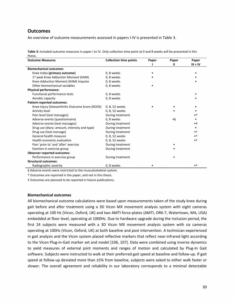
Outcomes An overview of outcome measurements assessed in papers I-IV is presented in Table 3.
§ Adverse events were restricted to the musculoskeletal system.
† Outcomes are reported in the paper, and not in this thesis.
‡ Outcomes are planned to be reported in future publications.
Biomechanical outcomes
All biomechanical outcome calculations were based upon measurements taken of the study knee during
gait before and after treatment using a 3D Vicon MX movement analysis system with eight cameras
operating at 100 Hz (Vicon, Oxford, UK) and two AMTI force-plates (AMTI, OR6-7, Watertown, MA, USA)
embedded at floor level, operating at 1000Hz. Due to hardware upgrade during the inclusion period, the
first 24 subjects were measured with a 3D Vicon MX movement analysis system with six cameras
operating at 100Hz (Vicon, Oxford, UK) at both baseline and post intervention. A technician experienced
in gait analysis and the Vicon system placed reflective markers that reflect near-infrared light according
to the Vicon Plug-in-Gait marker set and model [106, 107]. Data were combined using inverse dynamics
to yield measures of external joint moments and ranges of motion and calculated by Plug-In Gait
software. Subjects were instructed to walk at their preferred gait speed at baseline and follow-up. If gait
speed at follow-up deviated more than ±5% from baseline, subjects were asked to either walk faster or
slower. The overall agreement and reliability in our laboratory corresponds to a minimal detectable
Table 3: Included outcome measures in paper I to IV. Only collection time point at 0 and 8 weeks will be presented in this thesis.
Outcome Measures
Collection time points Paper I
Paper II
Paper III + IV
Biomechanical outcomes: Knee Index (primary outcome) 0, 8 weeks • • 1st peak Knee Adduction Moment (KAM) 0, 8 weeks • • Knee Adduction Moment (KAM) Impulse 0, 8 weeks • Other biomechanical variables 0, 8 weeks • Physical performance: Functional performance tests 0, 8 weeks • Aerobic capacity 0, 8 weeks • Patient-reported outcomes: Knee injury Osteoarthritis Outcome Score (KOOS) 0, 8, 52 weeks • • • Activity level 0, 8, 52 weeks • • Pain level (text messages) During treatment •† Adverse events (questionnaire) 0, 8 weeks •§ • Adverse events (text messages) During treatment •‡ Drug use (diary: amount, intensity and type) During treatment • Drug use (text message) During treatment •† General health measure 0, 8, 52 weeks •† Health economic evaluation 0, 8, 52 weeks •† Pain ‘prior to’ and ‘after’ exercise During treatment • Exertion in exercise group During treatment • Observer-reported outcomes: Performance in exercise group During treatment • Structural outcomes: Radiographic severity 0, 8 weeks • •†
31
change (MDC) of 19.3% and 26.7%, and intraclass correlation coefficients of 0.81 and 0.76 for Knee
Index and first peak KAM respectively.
The Knee Index – primary outcome of the randomized controlled trial (papers I, III and IV)
The primary outcome for the EXERPHARMA trial was change in knee load in the study knee during gait.
The primary outcome was between-group change in the Knee Index immediately after intervention.
External peak joint moments in the frontal, sagittal and transversal planes of the first 10-50% of the
stance phase were used to calculate the Knee Index (Figure 10) [70]. The Knee Index is a surrogate for
total load across both compartments, and has been chosen since changes in the external moment are
known to occur in the sagittal plane prior to and without accompanying changes in the frontal and/or
transversal plane [51, 69, 108]. The Knee Index will be reported normalized for height and weight.
In paper I, we investigated the properties of the Knee Index by determining the contribution from the
three planes (frontal, sagittal, transversal) and the correlation between the Knee Index and other
biomechanical outcomes.
Other biomechanical variables (paper I)
To investigate biomechanical variables that might relate to the Knee Index in this study, we tested a
number of variables (see Table 4 for a detailed description of collection and calculation of each of them).
These variables have previously been found to be related to the variability of knee joint load or disease
severity: time point [71], dynamic sagittal plane angle [48, 71], knee alignment [71, 72, 109] and gait
speed [72] Additionally, we included biomechanical variables that have been associated with alterations
in gait strategies (i.e. trunk lean [73, 84] and foot progression angle [74, 75, 110]).
Additional biomechanical outcome for the EXERPHARMA trial (paper III and IV)
The secondary biomechanical outcome for the EXERPHARMA trial (Table 3) was 1st Peak KAM during gait
normalized for height and weight. As explorative outcomes we chose KAM impulse normalized for
Percentage distribution of the contributors to Knee Index(Mean of 5 trials, study knee)
Frontal Sagittal Transversal
38
The total sample was divided into sub-groups based upon frontal or sagittal knee moment strategy. As
shown in the time-course patterns for the whole sample (Figure 13A), the sagittal strategy sub-group is
characterized with frontal plane moments close to zero for the first half of the stance phase (Figure
13B), whereas the frontal strategy sub-group is characterized with sagittal plane moments close to zero
for the first half of the stance phase (Figure 13C), and the contribution from the transversal plane is very
little in the whole sample and both sub-groups.
Figure 13: Examples of time-course patterns of the Knee Index, sagittal, frontal and transverse plane moment, according to
different loading strategies. (A) Represents the mean of all subjects, (B) represents subjects with sagittal loading strategy, and
(C) represents subjects with frontal loading strategy.
Association between Knee Index and other biomechanical variables
The sub-group analyses resulted in better associations (higher R-values) than the analyses conducted on
all individuals. The sub-group with a frontal knee-loading strategy only showed associations between the
Knee Index and frontal plane kinetics and kinematics, whereas the sub-group with a sagittal knee-
loading strategy showed associations between the Knee Index and both frontal and sagittal plane
kinetics and kinematics, and gait speed. Additionally, no associations were seen for variations in
transversal plane and for variables representing altered gait strategies, trunk lean and foot progression
angle. No association between the Knee Index and KL grade was seen for both the whole group and sub-
groups.
39
Feasibility of NEMEX-KOA (paper II)
Progression of exercises
For the first 23 subjects randomized to NEMEX-KOA, it was possible for the 18 subjects who participated
in six or more sessions to progress to the more complex levels of difficulty in half or more of the
neuromuscular exercises, and overall, those subjects who performed a greater number of exercise
sessions were able to progress to a higher level of difficulty. The neuromuscular exercises were grouped
into four categories:
- Neuromuscular exercises with a focus on strength gain including the exercises: squat, lunge,
step-up and kettlebell swing (Figures 14 A, B, C and D).
- Neuromuscular exercises with a focus on functional performance including the exercises: weight transfer, cloth under foot, mini trampoline and cable/elastic band (Figures 18 E, F, G and H).
- Neuromuscular exercises with a focus on postural stability including the exercises: pelvic lift and side lying jumping jack (Figures 14 I and J).
- Neuromuscular exercises where some levels contained jumps including: all levels of limping cross and mini trampoline, squat level 2 and 4, step-up level 3-4 and weight transfer level 3 (Figures 14 A, C, E, K and G). Less than half of the subjects were able to progress and perform exercise levels containing jumps with high impact on landing (Figures 14 A, C and K), when not performed on a trampoline or on a soft exercise mat (Figures 14 E and G).
Exertion and pain
Perceived exertion of the exercise program for the individual subjects ranged from ‘light’ to ‘very heavy’.
Overall, we found few reports of a clinically relevant increase in pain from exercise and few reports of
high pain following exercise. Four of the 23 subjects reported a short-term clinically relevant increase in
pain from exercise (defined as >2 VAS points) after 1-2 out of the 16 scheduled sessions. The increase in
pain was temporary and subjects were able to continue the exercise program.
Adverse Events and adherence
None of the 23 subjects reported any treatment-specific musculoskeletal adverse events.
Seven of the 23 subjects attended exercise for less than 6 weeks, three of these stopped due to knee
pain, and one specified that the pain was due to the original pain and disability and not necessarily
aggravated by the exercise. The remaining four subjects who attended for less than 6 weeks had low
attendance for other reasons, such as work commitments or transportation difficulties and, in one case,
a cardiovascular procedure.
40
Figure 14: Level of difficulty at which each exercise was performed at the first, halfway and last exercise sessions. Neuromuscular exercises with a focus on strength gain ((A) squat, (B) lunge, (C) step-up and (D) kettlebell swing), exercises with a focus on functional performance ((E) weight transfer, (F) cloth under foot, (G) mini trampoline, (H) cable/elastic band), exercise with a focus on postural stability ((I) pelvic lift and (J) side lying jumping jacks) and exercise where some levels contained jumps ((G) mini trampoline, (K) limping cross, (A) squat level 2 and 4, (C) step-up level 3-4 and (E) weight transfer level 3). The numbers (0-3/4) from the center to the circumference correspond to level of difficulty, 1 being the lowest. The numbers (1-23) around the circumference refer to the 23 individual subjects. The green area indicates the level of difficulty for the individual 23 subjects at the first session. The yellow line indicates the level of difficulty for the individual 23 subjects at the halfway session. The red area indicates the level of difficulty for the 23 individual subjects at the last session. Subjects with identification (ID) 1-5 attended 2-5 exercise sessions and subjects with ID 6-23 attended 6-16 sessions.
41
The EXERPHARMA trial (paper IV)
Primary outcome
At follow-up, no within-group or between-group changes in the primary outcome, Knee Index during
gait, were observed for the two groups (-0.07 [-0.17; 0.04] Nm/(BW×HT%) (Figure 15).
Secondary outcomes
No significant between-group difference was observed for KAM (-0.09 [-0.23; 0.04] Nm/(BW×HT%).
However, there was a statistically significant increase in KAM during gait after intervention for the
NEMEX-group (0.12 [95%CI, 0.03; 0.22] Nm/(BW×HT%) (Figure 15). Small to moderate effect sizes (effect
sizes: 0.05 – 0.53 ) and mostly statistically significant, but clinically non-relevant, within-group changes
were seen in the majority of the KOOS subscale scores and some of the functional tests in both
treatment groups with no statistically significant differences between groups (Figure 15).
Only 17 and 14 participants from the Pharma-group and NEMEX-group, respectively, were able to
conduct the one-leg rise from a stool test according to the description [102]. As a consequence, no
imputation on the data was performed and the statistical analyses were performed ‘as observed’ (Figure
15).
Explorative outcomes
No within nor between-group changes were observed for walking velocity. Small significant within-
group changes (effect sizes: 0.05-0.09) were observed for the remaining explorative outcomes but
no between-group differences were seen (Figure 15).
Adverse events
No between-group risk differences in adverse events were observed for the following categories;
Abdominal and intestinal, Musculoskeletal symptoms, Central nervous system and psychiatric
symptoms, Skin and subcutaneous symptoms, and Miscellaneous symptoms demonstrated (data not
shown).
42
Figure 15: The between-group changes for primary and secondary outcomes. Values are presented as between-group
difference from baseline and 95% confidence intervals (mean [95%CI]). Note that x-axes for Biomechanical outcomes of walking
and one-leg-rise from a stool are inverted. Abbreviations are: external knee adduction moment (KAM), external knee adduction
moment impulse (KAM impulse), Knee injury and Osteoarthritis Outcome Scores (KOOS) for pain, symptoms, activities of daily
living (ADL), sport and recreation (Sport/rec), knee-related quality of life (QOL), Åstrand maximal endurance capacity (Åstrand
VO2max) and the University of California at Los Angeles Activity score (UCLA).
*One-leg rise from a stool was only collected n =17 for the Pharma-group and n = 14 for the NEMEX-group, and consequently,
the statistics are performed ‘as observed’ (no imputation).
43
Discussion
Main findings The main findings of this thesis were that the Knee Index (paper I), a novel index of total knee joint load,
was primarily composed of moments in the frontal and sagittal planes. In addition, a large inter-subject
variation in composition was observed, indicating that there are two knee-loading strategies in subjects
with mild to moderate knee OA.
Secondly, the neuromuscular exercise program (NEMEX-KOA) (paper II) was found to be feasible for
subjects with mild to moderate knee OA in terms of most subjects being able to progress to more
complex levels of difficulty for most of the neuromuscular exercises with few incidents of short-term
increase in pain from exercise.
Thirdly, the EXERPHARMA trial (papers III and IV) was designed to evaluate the possible difference in
joint load-modifying effects of two common primary care interventions provided to patients with knee
OA. These two interventions, NEMEX-KOA compared with information on recommended use of
analgesics and anti-inflammatory drugs, which could potentially have opposite effects on the knee joint
load. Due to poor adherence, especially in the Pharma-group, and a similar intake of pain relief
medication in both groups, we essentially compared the addition of 8 weeks of NEMEX-KOA to ‘care as
usual’. We found a 2.6% non-significant and clinically irrelevant increase in the Knee Index in the
NEMEX-group, no change in the Pharma-group, and no significant or clinically relevant difference
between treatment groups. The results for the secondary outcomes of 1st peak KAM supported the
primary finding.
Methodological considerations
Study design
This thesis employs study designs of low to high levels of evidence. Paper I used a cross-sectional design
and paper II used a case series design, both of which result in a low level of evidence. Papers III and IV
employ the rigorous randomized controlled trial design that results in a higher level of evidence [131].
Paper I was based on a cross-sectional design and it was reported according to the STROBE Statement
[132], but due to the design, we could not infer causality. The intention was, instead, to produce
associations to indicate if the novel outcome, the Knee Index, is a sensitive and responsive outcome,
which can be used in OA research in the future [133].
Paper II was based on a case series design, reported according to the CARE Statement [134], but due to
the design, it was not possible to state or compare the effect of the NEMEX-KOA program with other
interventions or with a control group. The intention was, instead, to describe the NEMEX-KOA program
in detail and to demonstrate its feasibility (with respect to progression, exertion, pain, adverse events
and adherence) for subjects with mild to moderate knee OA.
Papers III and IV were based on a randomized controlled trial design and they were reported according
to the CONSORT Statement [101] with a rigorous study design. Inclusion criteria were kept wide to
reflect daily clinical practice. However, our study has limitations. The blinding of assessors was
44
attempted through patient discretion and restriction of access to previously obtained data. We did not
measure the success of assessor blinding; however, the analyses were masked to group allocation.
Adherence to interventions was relatively low, which may impact the internal validity, but nevertheless,
reflect daily practice and hence, a pragmatic design. Thus, in combination with wide inclusion criteria
the generalizability of the current findings is considered high.
Subjects The subjects were recruited from two settings: referral from GPs and from advertising in, for example,
the local newspaper. It is likely that we recruited from two different populations, as the numbers
needed to screen for subjects referred from GPs were 1.7 and 7.3 for subjects recruited from
advertisements. Recruitment from two populations is likely to have increased the heterogeneity of the
current sample and as a result, increased the generalizability of the results from the randomised
controlled trial.
Inclusion and exclusion criteria
Inclusion criteria were kept wide to reflect daily clinical practice. Subjects with mild to moderate clinical
signs of knee OA were included in this thesis, therefore the ACR [11] and EULAR [12] criteria for clinically
diagnosing knee OA were combined. Structural changes on radiographs were not part of the diagnosis
because generally, only about half the people with mild knee OA symptoms have structural changes on
radiographs [135]. In order to test the effect of two pain-relieving treatments for knee OA, we included
subjects with mild or stronger knee pain, corresponding to a cut-off of ≤80 on the KOOS pain subscale. In
order to reduce variation in the primary outcome, the Knee Index, we chose a cut-off of a BMI of less
than 32, to minimize skin movement, based on previous findings [136]. We chose to exclude subjects
with recent and planned surgery to the knee and ACL reconstruction at any time point, because little is
known about how these treatments influence gait kinematics and kinetics. Additionally, individuals with
joint replacement of the lower limb or high tibial osteotomy were excluded, since these people have
severe OA of the ankle, knee or hip.
Outcomes
Choice of biomechanical outcomes
Although, there is no consensus on which biomechanical variables to report from intervention studies,
and the fact that the causation between knee joint loading and the structural progression of knee OA is
limited, the current body of evidence indicates that knee joint biomechanics during walking provide
important information related to mobility limitations, and that gait analyses is a sensitive tool for
detection of even subtle changes [137].
To investigate potential changes in net knee moments, we chose the Knee Index as our primary
outcome for the EXERPHARMA trial (papers III and IV). The Knee Index was chosen as changes in the
45
external moment are known to occur in the sagittal plane before, and without accompanying, changes
in the frontal and/or transverse plane [51, 69, 108].
Other similar indices of knee joint forces in three planes include a recent randomized controlled trial
that demonstrated a 4.5% increase in knee loading during walking in persons with knee-related
limitations following a rosehip powder intervention compared with placebo by means of the total Knee
Reaction Moment [137]. The interpretation of the total Knee Reaction Moment is similar to the Knee
Index in the present study, albeit there are differences in the computational approach [137].
We have shown that the Knee Index is primarily composed of frontal and sagittal moments, and we
identified two knee-loading strategies (sagittal or frontal plane strategy) in a sub-sample of the current
study population (paper I). Why subjects with mild to moderate knee OA adopt different knee-loading
strategies is difficult to elucidate from the current data. A recent study demonstrated that the frontal
plane ground reaction force-to-knee lever arm accounted for 30% of the increase in KAM in patients
following arthroscopic partial meniscectomy [82]. This may also be the case for the current study since
both dynamic and static knee frontal plane angles (outcomes that are associated with the lever arm) in
the frontal knee-loading sub-group were associated with the Knee Index and to a higher degree than for
the sagittal strategy sub-group. Thus, knee alignment (static or dynamic) is most probably a key
determinant of knee-loading strategy and a shift from a sagittal plane-loading strategy to a medio-
lateral direction is most likely associated with knee alignment. Consequently, previous OA research that
has focused on frontal or sagittal plane outcomes alone and has demonstrated no effect after
interventions [79, 138] may have suffered from not capturing a possible response in a group who
demonstrate a loading strategy where changes will not be captured by a single plane outcome of joint
load. To improve our understanding of different knee-loading strategies, future studies should consider
reporting joint loads for the sagittal and frontal planes separately, as well as in combination.
As secondary and explorative outcomes, we reported 1st peak KAM and KAM impulse during walking for
the EXERPHARMA trial (papers III and IV). These were chosen to report biomechanical outcomes that are
widely used in OA research. Finally, the choice of combining objective and patient-reported outcomes
resulted in four domains as recommended: pain, functional performance, patient global assessment and
joint imaging [139].
Functional performance
The functional performance tests were chosen for their applicability to the population and ease of use in
a clinical setting. Despite this, only 35 of the 93 subjects (20 from the Pharma-group) performed the
maximum number of one-leg rises from a stool test successfully one or more times at both baseline
and/or follow-up. At baseline, subjects performed a median of three rises for the current study, which is
much fewer than previously reported (17-25 one-leg rises) in a younger population with chronic knee
pain [112].
Observed results from both the maximum number of knee-bends in 30 seconds and one-leg hop for
distance tests, were in line with previous results reported for a younger meniscectomized population
[113, 116] and an older population with moderate to severe knee OA [42, 140].
46
Progression in NEMEX-KOA
Progression in exercise difficulty was presented as the performed level of difficulty of each individual
exercise at first, median and last exercise session in order to illustrate the progression through the
complete intervention (8 weeks) and to indicate if an exercise held enough levels of difficulty, or
suffered from floor or ceiling effects. Traditional methods such as cardiac-output for aerobic exercise
and load for resistance training are not applicable for measuring and dictating progression in NEMEX, in
that NEMEX aims at improving sensorimotor control and obtaining functional joint stabilization.
The exercise difficulty increased with each level (3 to 4 levels), and examples of how an increase was
produced included changing to a softer more challenging surface during a weight-bearing exercise, or
increasing the load or a combination of both. Although each level was associated with increased
complexity, the increase in complexity might not be linear (i.e. the amount of increase from level one to
level two was not necessarily similar to the amount of increase from level two to level three, etc.).
3D motion analysis
The gait laboratory upgrade during the inclusion period that was mentioned earlier does not influence
our biomechanical findings. The primary reasons for variation and errors in gait laboratories are largely
related to marker placement (for this laboratory, MDC 19.3%) and within-subject variation (≈7% was
reported in paper I) and only to a small degree to hardware (error measure <2.0mm ≈0.5%) [141].
Walking speed is known to effect gait parameters (especially sagittal plane moments) [142]. The gait
trials were collected at standardized speed (close to a predetermined velocity (±5%)) to allow the
investigation of a potential altered ‘true knee-loading strategy’. Consequently, our findings should be
generalized with caution, especially in comparison with trials using no methods or other methods for
controlling for speed [142].
Unreported outcomes
There was a discrepancy between the ClinicalTrials.gov registration and the published study protocol
(paper III) [102]. As a consequence, paper IV did not report the secondary biomechanical outcomes
during one leg rise as stated in the study protocol, and the secondary outcomes (pain level measured
with text messaged, UCLA and Åstrand VO2max) were defined as ‘not pre-specified outcomes’.
The EXERPHARMA trial included biomechanical outcome variables (Knee Index, 1st peak KAM and KAM
Impulse) obtained during one-leg rise from a stool and patient-reported outcomes (text messages:
adverse events, global perceived effect, patient-acceptable symptom state and treatment since the end
of the intervention), outcomes (SF-36 and EQ-5D) collected at 8-weeks follow-up and all outcomes
collected at 52-weeks follow-up, were not reported in this thesis or the papers it is based upon. These
outcomes are planned to be reported in further studies based on follow-up data and secondary analyses
from the EXERPHARMA trial.
47
Missing observation (papers IV)
Only a few missing measurements were reported in this study. A total of 8 subjects (5 from the Pharma-
group) were unavailable for 3D motion analysis and intention to treat analysis, and a total of 11 subjects
(3 from the Pharma-group) were unavailable for patient-reported outcomes. Thus 85 subjects (41 from
the Pharma-group) were available for the primary analysis, the sample size calculation required 42
subjects in each group, and thus the current study was adequately powered.
This study had an acceptable response rate for patient-reported outcomes. The KOOS questionnaire at
follow up had a response rate of 87.3% (Pharma-group 90.4% and NEMEX-group 84.3%). The response
rate for drug use diary was ≈86% (Pharma-group 85% and NEMEX-group 87%). These indicate that our
study does not suffer from reporting bias.
Interventions
Progression in neuromuscular exercises (paper II)
As expected, the subjects progressed differently for the individual exercises in the NEMEX-KOA program.
We observed that subjects attending six or more exercise sessions progressed to more complex levels of
difficulty. Although, there are methodological differences, our findings are supported by the literature,
which reports that greater exercise attendance is positively related to physical function [31, 143].
To reduce the risk of ceiling effects with long-term adherence to NEMEX-KOA, we recommend having a
variety of levels for progression in exercises with a focus on functional performance. For the exercises
where only a few subjects were able to progress to the highest level of difficulty, adding smaller
intermediate levels may have helped subjects to progress.
Exercises that included jumping were clearly a hindrance for progression. An in vitro study has shown
that cartilage at early stage OA is most sensitive to high loading rates (comparable to jumping activities)
[144]. Consequently, exercises containing jumping should only be performed with great caution and
under supervision.
Pain related to neuromuscular exercise (paper II)
A pain level of >5 VAS points after one or more exercise sessions was reported by 3 of the 23 subjects,
which was in line with previous results reported for a younger population with degenerative meniscus
tears [145] and an older population performing NEMEX prior to total joint replacement [41] of 5% and
32%, respectively. In terms of increased pain after an individual session, NEMEX-KOA is as safe as
NEMEX for total joint replacement. For comparison, in studies of aerobic and resistance training,
increase in knee pain has been reported by 8-18% of subjects with knee OA performing aerobic [146] or
resistance [79, 147] training. In these studies, increase in knee pain was measured with a variety of
methods ranging from logbooks to reports by instructors and reports of dropouts. Even though a direct
comparison to our study is problematic due to methodological differences, NEMEX-KOA seems to be as
safe as any other exercise intervention for subjects with mild to moderate knee OA.
48
Effect of NEMEX-KOA and Pharma on Knee Index (paper IV)
Potentially, the two interventions could affect knee joint load in opposite directions. Since the
compliance was poor (only 7% had good compliance) in the Pharma-group, there was a possibility that
the lack of difference between-groups was caused by poor compliance. The within-group changes can
help shed light on this question. The Pharma-group, with poor compliance, showed no within-group
change in knee joint load (-0.01 [-0.08; 0.07]). In the NEMEX-group, where 49% had good compliance,
we found a small increase (0.06 [-0.02; 0.12]). A post hoc analysis found low correlation (r < 0.17)
between change in outcomes of knee joint loading (Knee Index, 1st peak KAM and KAM impulse) and the
number of attended exercise sessions indicating that compliance to exercise apparently did not affect
alterations in knee loading to a large extent.
It should be emphasized that the within-group changes (group mean values and 95% CI) in Knee Index
are within the estimated error margin (MDC = 19%) and much lower than a previously reported 13.5%
increase in response to NSAID (that is, Celocoxib) treatment in comparison with placebo [69] and the a
priori defined 27% change for the sample size calculation. Consequently, the present within-group
changes of 0.4% and 2.6% in biomechanical loading should be considered minute and clinically
irrelevant.
No between-group differences in KOOS sub-scores and functional tests were observed. The within-
group effect from NEMEX was not as strong as expected based on prior literature [40, 42, 46]. However,
it cannot be excluded that the NEMEX group experienced a clinically relevant difference in terms of self-
reported Sport and Recreation function and some functional performance tests.
Adherence to interventions (paper IV)
Achieving and maintaining adherence to exercise interventions is important if beneficial effects of
exercise are to be realized [148]. However, adherence to health interventions is a complex challenge,
especially for individuals with chronic conditions [148]. The current study demonstrated an adherence
to exercise that was relatively low, since only 49% fulfilled the pre-defined good compliance of ≥12
sessions. This is lower than the 74% and 93% adherence to exercise in patients scheduled for total joint
replacement, previously shown in studies from our research group [42, 149] and may be partly
explained by our subjects being younger (≈58 years), more fit to work (≈52% working full-time or part-
time) and only moderately affected by OA (≈10-20 points better KOOS scores) in comparison with the
above-mentioned studies. In addition, our study used a pragmatic approach and exercise was performed
at private clinics supervised by local physiotherapists as opposed to training performed at research
institutions by project staff. This may further have affected adherence negatively.
Only three subjects (7%) in the Pharma-group demonstrated good compliance to the drug treatment
intervention. Subjects received information on how best to use acetaminophen and oral NSAIDs, in
doses consistent with Danish guidelines [18] but were not obliged to take certain doses. Thus, the
pharmacologically-initiated pain relief was most likely not large enough to induce either increased knee
moments as previously seen in subjects with knee OA [36, 87-89, 91] or dose-dependent adverse effects
as observed in other trials [23-25].
49
Conclusions There was a large variation in the composition of the Knee Index, it was primarily composed of the
frontal and sagittal planes, indicating that some subjects with mild to moderate knee OA have a frontal,
while others have a sagittal, moment-dominated strategy. The sub-group with a sagittal strategy was
associated with both sagittal and frontal plane moments and angles, whereas the sub-group with a
frontal strategy was associated with only frontal plane moments and angles.
An 8-week neuromuscular exercise program for individuals with mild to moderate knee OA (NEMEX-
KOA) was shown to be feasible in terms of progression, exertion, pain, adverse events and adherence.
Most subjects were able to progress to more complex neuromuscular exercises. Jumping activities were
however generally not feasible. Those subjects attending a greater number of exercise sessions were
able to progress to higher levels of difficulty and reported a larger decrease in pain. There were limited
numbers of incidences of temporary increase in exercise-related pain.
The EXERPHARMA trial evaluated the possible joint load-modifying effects of a neuromuscular exercise
program compared with information on the recommended use of analgesics and anti-inflammatory
drugs. Due to poor adherence, especially in the Pharma-group, and a similar intake of pain relief
medication in both groups, we essentially compared the addition of 8 weeks of neuromuscular exercise
to ‘care as usual’, which did not result in reduced knee joint load during walking and no between-group
differences in pain, symptoms and functional performance.
50
Clinical implications and future perspectives Variation in knee OA loading strategy should be taken into consideration when evaluating OA research
in middle-aged subjects with mild to moderate knee OA. Therefore, further research on index measures
of total knee joint load such as the Knee Index, are needed. Additionally, further research should be
conducted on how well index measures of total knee joint load are associated with compressive knee
joint force and progression of knee OA.
We showed that the current neuromuscular exercise program (NEMEX-KOA) for individuals with mild to
moderate knee OA was feasible and effective on improving symptoms and physical performance. Prior
to broader use of NEMEX-KOA, substituting jumping activities with activities with a low impact on
landing should be considered.
This effectiveness trial showed that neuromuscular exercise will have limited clinical impact on reducing
knee joint load in the current setup where individuals with mild to moderate knee OA attend exercise
sessions at private clinics, where compliance is likely to be low. Therefore, it is of continued interest to
investigate if neuromuscular exercise is efficacious in reducing knee joint load in individuals with mild to
moderate knee OA, in order to improve our understanding of load-amendable interventions.
51
Summary Background
Knee osteoarthritis (OA) is a mechanically-driven disease. It is suggested that medial tibiofemoral knee-
joint load increases with pharmacological pain relief, indicating that pharmacological pain relief may be
positively associated with disease progression. Treatment modalities that can both relieve pain and
reduce knee-joint load would be preferable. The knee-joint load is influenced by functional alignment of
the trunk, pelvis, and lower-limb segments with respect to the knee, as well as the ground-reaction
force generated during movement. Neuromuscular exercise (NEMEX) can influence knee joint load and
decrease knee pain. It includes exercises to improve balance, muscle activation, functional alignment,
and functional knee stability.
The overall aim of this thesis was to compare the effectiveness of a specific neuromuscular exercise
program with optimized analgesics and anti-inflammatory drug use on knee joint loads, as well as pain
and functional performance in individuals with mild to moderate medial tibiofemoral knee OA.
A biomechanical outcome of total knee joint load has not yet been described in detail. Therefore the
first study (paper I) of this thesis assessed the Knee Index, a novel measure of total knee joint load that
incorporates all three planes, and has been shown to be sensitive to changes in pain in subjects with
moderate knee OA. However, the relative contribution and inter-subject variation of each plane to the
Knee Index has not previously been described. Additionally, associations between the Knee Index and
other biomechanical variables have not been described to indicate if the Knee Index is a sensitive and
responsive biomechanical outcome suitable as a primary outcome in a randomized controlled trial.
A NEMEX program specifically designed for subjects with mild to moderate knee OA has not previously
been described. Therefore, in a randomized controlled trial, the second study (paper II) described the
feasibility of the current NEMEX for subjects with mild to moderate knee OA (NEMEX-KOA), in terms of
progression, pain, exertion, adverse events and adherence, to indicate if NEMEX-KOA could be a suitable
intervention for subjects with mild to moderate knee OA.
The third study (papers III and IV) described and reported a pragmatic randomized controlled trial (the
EXERPHARMA trial), that was designed to investigate the effectiveness of NEMEX-KOA, compared with
optimized analgesics and anti-inflammatory drug use (Pharma), on the measures of knee-joint load
(Knee Index) in individuals with mild to moderate medial tibiofemoral knee OA.
Method We performed a randomized, single-blind, clinical trial comparing NEMEX-KOA with Pharma (papers I, II,
III and IV). Subjects with mild to moderate medial knee OA were recruited from general medical
practices in the community of Odense and Middelfart, Denmark, and from advertisements in local clubs,
libraries, print media, and Facebook, and were randomly allocated (1:1) to one of two 8-week
treatments, either to (a) exercise therapy twice a week (the NEMEX-group), or to (b) a pamphlet and
video materials with information on the recommended use of analgesics and anti-inflammatory drugs
(acetaminophen and oral NSAIDs) (the Pharma-group). The primary outcome was change in the Knee
Index during walking after 8 weeks of intervention. Secondary and exploratory outcomes included
changes in Knee Injury and Osteoarthritis Outcome Score (KOOS), UCLA Activity score and functional
52
performance tests (maximum one-leg rises from a stool, maximum number of knee-bends in 30 seconds
and one-leg hop for distance).
For the cross-sectional study (paper I), we calculated the Knee Index using 3-dimensional gait analysis in
a sample of the first 44 subjects, aged 40-70 years with mild to moderate knee OA, recruited for the
randomized controlled trial. Subjects were instructed to walk at a self-selected speed and the average of
the 1st peak Knee Index for five successful gait trials was subsequently used in the analysis.
The case series study (paper II) reported on the first 23 subjects randomized to the 8-week twice weekly
NEMEX-KOA regime, including 11 exercises with 3-4 levels of difficulty. The level of difficulty was noted
for each exercise in every session. Exertion, pain, adverse events and adherence were recorded at each
session.
Results In total, 93 subjects (57% women, 58 ± 8 years with a BMI of 27 ± 4) were randomized to the Pharma-
group (n = 46) or the NEMEX-group (n = 47) with data from 41 and 44 patients, respectively, available at
follow up.
The Knee Index was mainly composed of frontal and sagittal plane moments, with a large inter-subject
variation in the composition. This resulted in additional post-hoc association analyses dividing the
sample into sub-groups (frontal or sagittal plane dominance). The sub-group analyses resulted in better
associations than the analyses conducted on all individuals.
The 18 subjects that participated in six or more sessions progressed at least one level of difficulty in half
or more of the exercises. However, few subjects were able to progress to jumping activities. Exertion
ranged from ‘light’ to ‘very heavy’. Four subjects reported a clinically relevant increase in short-term
pain. No musculoskeletal adverse events were reported. Notably, three of 23 subjects dropped out
giving knee pain as the reason. Their pain ratings were, however, not getting worse.
The Knee Index during gait showed a statistically significant increase immediately after the 8-week
intervention for the NEMEX-group, corresponding to a statistically non-significant between-group
difference. Despite within-group improvements in most KOOS subscale scores and functional
performance in both treatment groups, no statistically significant between-group differences were
observed.
Conclusion From this thesis, I conclude that despite large variation in the composition of the Knee Index, it was
primarily composed of the frontal and sagittal planes. The Knee Index therefore holds promise as both a
sensitive and responsive biomechanical outcome for the evaluation of total knee joint load.
Furthermore, the NEMEX-KOA program was found feasible for subjects with baseline mild to severe
knee pain. Subjects were able to progress in level of difficulty with few reports of clinically relevant
increase in pain and no adverse events. Jumping activities were however not feasible. These findings
hold promise for investigating the effectiveness of NEMEX-KOA in individuals with mild to moderate OA.
Due to poor adherence, especially in the Pharma-group, and a similar intake of pain relief medication in
both groups, the EXERPHARMA trial essentially compared the addition of 8 weeks of neuromuscular
exercise to ‘care as usual’, which did not result in reduced knee joint load during walking and no
between-group differences in pain, symptoms and functional performance.
53
Resumé [Danish] Baggrund Knæartrose er en mekanisk drevet sygdom. Det formodes, at belastningen over det mediale
tibiofemorale ledkammer stiger med anvendelsen af farmakologisk smertelindring, hvilket indikerer, at
farmakologisk smertelindring kan være positivt associeret med sygdomsprogression.
Behandlingsmetoder der kan smertelindre og reducere belastningen over knæleddet vil være at
foretrække. Belastningen over knæleddet påvirkes af den funktionelle alignment af torso, bækken og
benets segmenter, i forhold til knæet, samt reaktionskraften fra underlaget dannet under bevægelse.
Neuromuskulær træning (NEMEX) kan påvirke belastningen over knæleddet og mindske smerter i
knæet. Træningen omfatter øvelser til at forbedre balance, muskel aktivering, funktionel alignment og
funktionel knæstabilitet.
Det overordnede formål med denne afhandling var at sammenligne effekten, af et specifikt
neuromuskulært træningsprogram med optimeret anvendelse af analgetika og anti-inflammatoriske
lægemidler, på den samlede belastning over knæleddet, samt smerter og fysisk funktion hos personer
med mild til moderat knæartrose.
Et biomekanisk effektmål til at måle den samlede belastning over knæleddet, er endnu ikke blevet
beskrevet i detaljer. Derfor omhandlede det første studie (artikel I) i denne afhandling, vurderingen af
Knee Index, et nyt effektmål af den totale belastning over knæleddet, som består af alle tre planer. Knee
Index har vist sig at være sensitiv nok, til at kunne detektere ændring i knæsmerter hos personer med
moderat knæartrose. Det er imidlertid uvist, i hvilket omfang hvert plan bidrager til Knee Index og hvor
stor variation der er mellem deltagere. Associationer mellem Knee Index og andre biomekaniske
variable har aldrig tidligere været beskrevet. Med denne viden kan vi få en indikator for, om Knee Index
kan være et sensitivt og svarfølsomt biomekanisk effektmål, som ville kunne anvendes som primært
effektmål i et randomiseret kontrolleret forsøg.
Et NEMEXs program designet til personer med mild til moderat knæartrose er ikke tidligere blevet
beskrevet. Hvorfor den anden undersøgelse (artikel II) i denne afhandling, indeholdt en beskrivelse af
anvendeligheden af nærværende NEMEX program til deltagere med mild til moderat knæartrose
(NEMEX-KOA). Anvendeligheden blev vurderet ud fra progression, smerte, anstrengelse, utilsigtede
hændelser og fastholdelse til programmet (compliance). Således kan det vurderes, om NEMEX-KOA ville
kunne være en passende intervention til personer med mild til moderat knæartrose i et randomiseret
kontrolleret forsøg.
Den tredje undersøgelse (artikler III og IV) beskrev og rapporterede et pragmatisk randomiseret
kontrolleret forsøg (EXERPHARMA studiet), som blev designet til at undersøge effekten af NEMEX-KOA,
sammenlignet med optimeret anvendelse af analgetika og anti-inflammatoriske lægemidler (Pharma),
på den samlede belastning over knæleddet (Knee Index) hos personer med mild til moderat tibiofemoral
knæartrose.
Metode Vi udførte et randomiseret, enkelt-blindet, klinisk studie, der sammenlignede NEMEX-KOA med Pharma
(artikler I, II, III og IV). Deltagere med mild til moderat knæartrose blev rekrutteret fra almen praksis, i
Odense og Middelfart kommune i Danmark, og via annoncer i lokale foreninger, biblioteker, trykte
medier og Facebook. Deltagerne blev tilfældigt fordelt (1:1) til en af to 8-ugers behandlinger, enten (a)
54
NEMEX-gruppen; to træninger ugentligt eller (b) Pharma-gruppen; information om den anbefalede brug
af analgetika og anti-inflammatoriske lægemidler (paracetamol og tablet NSAID) via folder og
videomateriale. Det primære effektmål var ændring i Knee Index under gang efter 8 ugers intervention.
Sekundære og eksplorative effektmål var bl.a. ændringer i symptomer målt via Knee Injury and
Osteoarthritis Outcome Scores (KOOS), UCLA Aktivitets score og tests af funktionel præstation
(maksimalt antal et-bens oprejsninger fra taburat, maksimalt antal knæbøjninger på 30 sekunder og et-
bens hop for afstand).
I tværsnitsundersøgelsen (artikel I) beregnede vi Knee Index via 3-dimensionel ganganalyse, i en
stikprøve af de første 44 personer rekrutteret til det randomiserede kontrollerede forsøg, i alderen 40-
70 år med mild til moderat knæartrose. Deltagerne blev instrueret i at gå med selvvalgt hastighed. Knee
Index blev analyseret som gennemsnittet af den første spidsbelastning efter hæl-isæt, for 5 vellykkede
gang forsøg.
I case-serie studiet (artikel II) beskrev vi de første 23 deltagere, som blev randomiseret til 8-ugers
NEMEX-KOA med to ugentlige træningssessioner, indeholdende 11 øvelser i 3-4 sværhedsgrader.
Sværhedsgraden af hver enkelt øvelse ved hver session blev registreret. Anstrengelse, smerte,
utilsigtede hændelser og compliance blev ligeledes registreret ved hver træningssession.
Resultater I alt blev 93 deltagere (57% kvinder, 58 ± 8 år med et BMI på 27 ± 4) randomiseret til enten Pharma-
gruppen (n = 46) eller NEMEX-gruppen (n = 47); data fra henholdsvis 41 og 44 deltagere var tilgængelige
ved opfølgning.
Knee Index var hovedsageligt sammensat af momenterne fra det frontale og sagittale plan, med store
inter-individuelle variationer i sammensætningen. Dette resulterede i yderligere post hoc associations-
analyser, hvor vi inddelte stikprøven i to undergrupper (domineret af henholdsvis det frontale eller
sagittale plan). Analysen af undergrupperne, resulterede i bedre associationer end for analyser baseret
på hele gruppen.
De 18 deltagere, der deltog i 6 eller flere træningssessioner, progredierede mindst én sværhedsgrad i
halvdelen eller flere af øvelserne. Hvorimod, kun få deltagere var i stand til at progrediere til aktiviteter
indeholdende hop. Anstrengelsesniveauet for træningsprogrammet varierede fra "let" til "meget tungt".
Fire deltagere rapporterede en klinisk relevant forværring af kortvarige knæsmerter. Ingen
muskuloskeletale utilsigtede hændelser blev rapporteret. Bemærkelsesværdigt er det, at 3 af 23
deltagere faldt fra i løbet af interventionen, de begrundede frafaldet med knæsmerter. Deres smerte
rapportering viste imidlertid ingen forværring.
Knee Index under gang steg statistisk signifikant lige efter 8 ugers intervention for NEMEX-gruppen
svarende til en statistisk ikke-signifikant forskel mellem grupperne. Begge grupper rapporterede
forbedringer på de fleste KOOS subskalaer og funktionelle præstationer. På trods af dette, fandtes ingen
statistisk signifikante forskelle mellem grupperne.
Konklusion Ud fra denne afhandling kan jeg konkludere, at på trods af stor variation i sammensætningen af Knee
Index, bestod det primært af det frontale og sagittale plan. Knee Index er et lovende, sensitivt og
svarfølsomt, biomekanisk effektmål, til vurderingen af den samlede belastning over knæleddet.
55
Derudover, blev NEMEX-KOA programmet fundet anvendeligt, til personer med milde til svære smerter i
knæet ved baseline. Deltagerne kunne progrediere i øvelses sværhedsgrad, med få hændelser af klinisk
relevant forværring af knæsmerter og uden utilsigtede hændelser. Aktiviteter som indeholdt hop, var
imidlertid ikke anvendelige. Disse fund indikerer, at det vil være relevant at undersøge effekten af
NEMEX-KOA som intervention til personer med mild til moderat knæartrose.
På grund af lav overholdelse af interventionsregimerne, især i Pharma-gruppen, og et sammenligneligt
lægemiddelforbrug i begge interventionsgrupper, endte EXERPHARMA studiet med reelt at undersøge
effekten af 8 ugers neuromuskulær træning i tillæg til vanlig behandling. Undersøgelsen resulterede i, at
der ikke fandtes reduktion i belastningen over knæleddet under gang. Der blev heller ikke fundet
forskelle mellem grupperne for smerter, symptomer og funktionel præstation.
56
Acknowledgements I would like to express my thanks to several people for supporting me on accomplishing this thesis. With
great gratitude I thank:
Supervisors
Ewa M. Roos, my main supervisor, for being my solid yet evolving reference point in OA research
methodology.
Anders Holsgaard-Larsen, my co-supervisor, for amongst other things providing me with sound collegial
advice on handling life as a researcher.
Co-authors
Jens Søndergaard and Robin Christensen, co-authors, for inspirational talks on the pragmatic
randomized controlled trial design.
Colleagues
Mark W. Creaby and his wife Jen Kneen, for housing and introducing me and my wife to life as a
researcher in Brisbane, Australia.
Research unit for Musculoskeletal Function and Physiotherapy (FOF): for insightful meetings, discussions
and teachings on research methodology, strategy and much more.
My fellow PhD-students: Louise, Inge, Henrik, Kim, Camilla, Lotte, Allan and Kristoffer, for sharing my ups
and downs, in both work and private life.
Dennis B. Nielsen and Rasmus S. Sørensen, motion laboratory test personnel, for the endless data
processing and testing of subjects.
Ann-Sofie Jensen, Rasmus Holst, Søren H Jensen and Connie L Hansen, physiotherapists, for taking good
care of subjects in the exercise group.
Anne Marie Rosager, for co-ordinating the final parts of the EXERPHARMA trial.
Department of physiotherapy at Slagelse Hospital, for patiently housing and employing me while I
finished this thesis.
Suzanne Capell, for proof reading of manuscripts and thesis.
Friends and family
Most importantly my wife Anne Clausen, for endless support and an open heart when I needed it the
most.
57
References 1. Ericsson YB, Dahlberg LE, Roos EM: Effects of functional exercise training on performance and
muscle strength after meniscectomy: a randomized trial. Scand J Med Sci Sports 2009, 19:156-
165.
2. Ageberg E, Link A, Roos EM: NEMEX-TJR training program. BMC Musculoskelet Disord 2010,
17:126.
3. Roos EM, Juhl CB: Osteoarthritis 2012 year in review: rehabilitation and outcomes.
Osteoarthritis Cartilage 2012, 20:1477-1483.
4. Martel-Pelletier J, Boileau C, Pelletier JP, Roughley PJ: Cartilage in normal and osteoarthritis
conditions. Best Pract Res Clin Rheumatol 2008, 22:351-384.
5. Andriacchi TP, Favre J: The nature of in vivo mechanical signals that influence cartilage health
and progression to knee osteoarthritis. Curr Rheumatol Rep 2014, 16.
6. Bitton R: The economic burden of osteoarthritis. Am J Mang Care 2009, 15:S230-S235.
7. Global, regional, and national incidence, prevalence, and years lived with disability for 301
acute and chronic diseases and injuries in 188 countries, 1990–2013: a systematic analysis for
the Global Burden of Disease Study 2013. Lancet 2015, 22:743-800.
8. Cross M, Smith E, Hoy D, Nolte S, Ackerman I, Fransen M, Bridgett L, Williams S, Guillemin F, Hill
CL, et al: The global burden of hip and knee osteoarthritis: estimates from the global burden of
disease 2010 study. Ann Rheum Dis 2014, 73:1323-1330.
9. Lachance L, Sowers MF, Jamadar D, Hochberg M: The natural history of emergent osteoarthritis
of the knee in women. Osteoarthritis Cartilage 2002, 10:849-854.
10. Nicholls E, Thomas E, Van der Windt D, Peat G: The long-term course of knee pain severity in
middle and old age: distinct trajectories from latent class growth modelling in population
cohort data (the CAS-K study and the osteoarthritis initiative (OAI)). In Osteoarthritis Cartilage,