Neuron, Vol. 28, 499–510, November, 2000, Copyright 2000 by Cell Press Cerebellar Long-Term Depression Requires PKC-Regulated Interactions between GluR2/3 and PDZ Domain–Containing Proteins cGMP and cGMP-dependent protein kinase (PKG) may also be important (see Daniel et al., 1998; Bear and Linden, 2000, for review). Progress has also been made toward a molecular understanding of the cerebellar LTD expression mecha- Jun Xia,* Hee Jung Chung,* Cornelia Wihler,* Richard L. Huganir,* and David J. Linden* ‡ * Department of Neuroscience Howard Hughes Medical Institute Johns Hopkins University School of Medicine nism. Cerebellar LTD may be detected using test pulses 725 North Wolfe Street of AMPA receptor agonists such as glutamate, quisqua- Baltimore, Maryland 21205 late or AMPA in intact (Ito et al., 1982), slice (Crepel and Krupa, 1988), and cell culture preparations (Linden et al., 1991, 1993). Furthermore, cerebellar LTD may be Summary seen with AMPA receptor agonist test pulses in reduced preparations that lack functional presynaptic terminals Cerebellar LTD requires activation of PKC and is ex- (Linden, 1994) including dendritic macropatches and pressed, at least in part, as postsynaptic AMPA recep- acutely dissociated Purkinje cells (Linden, 1996; Nara- tor internalization. Recently, it was shown that AMPA simhan and Linden, 1996; Narasimhan et al., 1998). Thus, receptor internalization requires clathrin-mediated it appears as if cerebellar LTD expression involves some endocytosis and depends upon the carboxy-terminal form of postsynaptic AMPA receptor downregulation. region of GluR2/3. Phosphorylation of Ser-880 in this This could potentially involve changes in AMPA receptor region by PKC differentially regulates the binding of unitary conductance, kinetics, glutamate affinity, or the PDZ domain–containing proteins GRIP/ABP and number/distribution of receptors in the postsynaptic PICK1. Peptides, corresponding to the phosphory- membrane. lated and dephosphorylated GluR2 carboxy-terminal Support for this last idea has come from experiments PDZ binding motif, were perfused in cerebellar Pur- showing that postsynaptic manipulations that interfere kinje cells grown in culture. Both the dephospho form with clathrin-mediated endocytosis block the induction (which blocks binding of GRIP/ABP and PICK1) and of cerebellar LTD in culture, while manipulations that the phospho form (which selectively blocks PICK1) induce postsynaptic internalization of AMPA receptors attenuated LTD induction by glutamate/depolarization (such as bath application of insulin or IGF-1) produce pairing, as did antibodies directed against the PDZ an LTD-like effect that mutually occludes LTD induced domain of PICK1. These findings indicate that expres- by synaptic pairing (Wang and Linden, 2000). Interest- sion of cerebellar LTD requires PKC-regulated interac- ingly, similar results have been found in studies of hip- tions between the carboxy-terminal of GluR2/3 and pocampal LTD. Treatments that interfered with post- PDZ domain–containing proteins. synaptic clathrin-mediated endocytosis prevented both pairing-induced LTD at the Schaffer collateral/commis- Introduction sural synapse (Luscher et al., 1999; Man et al., 2000) and persistent internalization of AMPA (but not NMDA) Cerebellar long-term depression (LTD) is a cellular receptors induced by brief exposure to AMPA (Carroll model system of memory that has been suggested to et al., 1999b; Luscher et al., 1999) or insulin (Man et al., comprise a portion of the engram for some forms of 2000) in cultured hippocampal neurons. These findings motor learning (see Ito, 1989; Mauk, 1997; Bear and suggest that cerebellar and at least one form of hippo- Linden, 2000, for review). It is a persistent, input-specific campal LTD may share a common expression mecha- attenuation of the parallel fiber–Purkinje cell synapse nism (see Luscher et al., 2000; Turrigiano, 2000, for that is produced when parallel fiber and climbing fiber review). inputs to a Purkinje cell are briefly coactivated at low How might PKC activation be linked to clathrin-medi- frequency, and, in recent years, considerable progress ated internalization of AMPA receptors to result in the has been made toward defining the molecular events expression of cerebellar LTD? Several lines of evidence necessary for its induction. These include three initial are suggestive. First, experiments using a heterologous postsynaptic events: mGluR1 activation, AMPA receptor expression system have shown that insulin-induced in- activation, and Ca 21 influx via voltage-gated Ca 21 chan- ternalization of AMPA receptors requires the presence nels. These three signals appear to converge upon the of GluR2/3, specifically, the carboxyl terminus (Man et activation of protein kinase C (PKC) as inhibition of PKC al., 2000). This region contains a conserved PDZ binding blocks cerebellar LTD induction and application of an motif that serves as a binding site for glutamate receptor exogenous PKC activator produces an LTD-like phe- interacting protein/AMPA receptor binding protein (GRIP/ nomenon (Crepel and Krupa, 1988; Linden and Connor, ABP) and protein interacting with C-kinase 1 (PICK1) as 1991; Hartell, 1994; Narasimhan and Linden, 1996; De well as a separate site that binds N-ethylmaleimide- Zeeuw et al., 1998; Freeman et al., 1998; Matsuda et sensitive factor (NSF; see Kim and Huganir, 1999; Braith- al., 2000). In addition, inhibition of postsynaptic protein waite et al., 2000, for review). Second, a specific residue phosphatase activity through a cascade involving NO, within this PDZ binding motif, GluR2 Ser-880, is phos- phorylated by PKC, and this results in a strong decrease in the binding of GRIP/ABP (Matsuda et al., 1999; Chung ‡ To whom correspondence should be addressed (e-mail: dlinden@ jhmi.edu). et al., 2000) but not PICK1 (Chung et al., 2000). Third,
Cerebellar Long-Term Depression RequiresPKC-Regulated Interactions between GluR2/3and PDZ Domain–Containing Proteins
cGMP and cGMP-dependent protein kinase (PKG) mayalso be important (see Daniel et al., 1998; Bear andLinden, 2000, for review).
Progress has also been made toward a molecularunderstanding of the cerebellar LTD expression mecha-
Jun Xia,*† Hee Jung Chung,*† Cornelia Wihler,*†
Richard L. Huganir,*† and David J. Linden*‡
*Department of Neuroscience†Howard Hughes Medical InstituteJohns Hopkins University School of Medicine
nism. Cerebellar LTD may be detected using test pulses725 North Wolfe Streetof AMPA receptor agonists such as glutamate, quisqua-Baltimore, Maryland 21205late or AMPA in intact (Ito et al., 1982), slice (Crepel andKrupa, 1988), and cell culture preparations (Linden etal., 1991, 1993). Furthermore, cerebellar LTD may beSummaryseen with AMPA receptor agonist test pulses in reducedpreparations that lack functional presynaptic terminalsCerebellar LTD requires activation of PKC and is ex-(Linden, 1994) including dendritic macropatches andpressed, at least in part, as postsynaptic AMPA recep-acutely dissociated Purkinje cells (Linden, 1996; Nara-tor internalization. Recently, it was shown that AMPAsimhan and Linden, 1996; Narasimhan et al., 1998). Thus,receptor internalization requires clathrin-mediatedit appears as if cerebellar LTD expression involves someendocytosis and depends upon the carboxy-terminalform of postsynaptic AMPA receptor downregulation.region of GluR2/3. Phosphorylation of Ser-880 in thisThis could potentially involve changes in AMPA receptorregion by PKC differentially regulates the binding ofunitary conductance, kinetics, glutamate affinity, orthe PDZ domain–containing proteins GRIP/ABP andnumber/distribution of receptors in the postsynapticPICK1. Peptides, corresponding to the phosphory-membrane.lated and dephosphorylated GluR2 carboxy-terminal
Support for this last idea has come from experimentsPDZ binding motif, were perfused in cerebellar Pur-showing that postsynaptic manipulations that interferekinje cells grown in culture. Both the dephospho formwith clathrin-mediated endocytosis block the induction(which blocks binding of GRIP/ABP and PICK1) andof cerebellar LTD in culture, while manipulations thatthe phospho form (which selectively blocks PICK1)induce postsynaptic internalization of AMPA receptorsattenuated LTD induction by glutamate/depolarization(such as bath application of insulin or IGF-1) producepairing, as did antibodies directed against the PDZan LTD-like effect that mutually occludes LTD induceddomain of PICK1. These findings indicate that expres-by synaptic pairing (Wang and Linden, 2000). Interest-sion of cerebellar LTD requires PKC-regulated interac-ingly, similar results have been found in studies of hip-tions between the carboxy-terminal of GluR2/3 andpocampal LTD. Treatments that interfered with post-PDZ domain–containing proteins.synaptic clathrin-mediated endocytosis prevented bothpairing-induced LTD at the Schaffer collateral/commis-Introductionsural synapse (Luscher et al., 1999; Man et al., 2000)and persistent internalization of AMPA (but not NMDA)Cerebellar long-term depression (LTD) is a cellularreceptors induced by brief exposure to AMPA (Carrollmodel system of memory that has been suggested toet al., 1999b; Luscher et al., 1999) or insulin (Man et al.,comprise a portion of the engram for some forms of2000) in cultured hippocampal neurons. These findingsmotor learning (see Ito, 1989; Mauk, 1997; Bear andsuggest that cerebellar and at least one form of hippo-Linden, 2000, for review). It is a persistent, input-specificcampal LTD may share a common expression mecha-
attenuation of the parallel fiber–Purkinje cell synapsenism (see Luscher et al., 2000; Turrigiano, 2000, for
that is produced when parallel fiber and climbing fiberreview).
inputs to a Purkinje cell are briefly coactivated at low How might PKC activation be linked to clathrin-medi-frequency, and, in recent years, considerable progress ated internalization of AMPA receptors to result in thehas been made toward defining the molecular events expression of cerebellar LTD? Several lines of evidencenecessary for its induction. These include three initial are suggestive. First, experiments using a heterologouspostsynaptic events: mGluR1 activation, AMPA receptor expression system have shown that insulin-induced in-activation, and Ca21 influx via voltage-gated Ca21 chan- ternalization of AMPA receptors requires the presencenels. These three signals appear to converge upon the of GluR2/3, specifically, the carboxyl terminus (Man etactivation of protein kinase C (PKC) as inhibition of PKC al., 2000). This region contains a conserved PDZ bindingblocks cerebellar LTD induction and application of an motif that serves as a binding site for glutamate receptorexogenous PKC activator produces an LTD-like phe- interacting protein/AMPA receptor binding protein (GRIP/nomenon (Crepel and Krupa, 1988; Linden and Connor, ABP) and protein interacting with C-kinase 1 (PICK1) as1991; Hartell, 1994; Narasimhan and Linden, 1996; De well as a separate site that binds N-ethylmaleimide-Zeeuw et al., 1998; Freeman et al., 1998; Matsuda et sensitive factor (NSF; see Kim and Huganir, 1999; Braith-al., 2000). In addition, inhibition of postsynaptic protein waite et al., 2000, for review). Second, a specific residuephosphatase activity through a cascade involving NO, within this PDZ binding motif, GluR2 Ser-880, is phos-
phorylated by PKC, and this results in a strong decreasein the binding of GRIP/ABP (Matsuda et al., 1999; Chung‡ To whom correspondence should be addressed (e-mail: dlinden@
jhmi.edu). et al., 2000) but not PICK1 (Chung et al., 2000). Third,
Neuron500
activation of PKC is also associated with a translocationof Ser-880-phosphorylated GluR2 and PICK1, but notGRIP/ABP immunoreactivity from dendritic shafts tospines in cultured hippocampal neurons (Chung et al.,2000). Fourth, PKC activating phorbol esters result inGluR2 internalization in neocortical neurons (Chung etal., 2000) and mutation of GluR2 Ser-880 to alanine pre-vents this in a heterologous expression system (H. J.Chung et al., submitted).
While these biochemical experiments are suggestive,their relevance for synaptic plasticity remains undeter-mined. Thus, we have performed whole-cell patch clamprecordings and microfluorimetric Ca21 imaging of cere-bellar Purkinje cells in culture together with manipula-tions to modify the interactions between GluR2/3, GRIP/ABP, and PICK1. In so doing, we sought to test thehypothesis that LTD expression is achieved by phos-phorylation of GluR2 at Ser-880 (or GluR3 at the analo-gous residue, Ser-885) by PKC, which regulates the in-teraction of GluR2 with GRIP/ABP and PICK1, therebypriming AMPA receptors for clathrin-mediated endocy-tosis.
Results
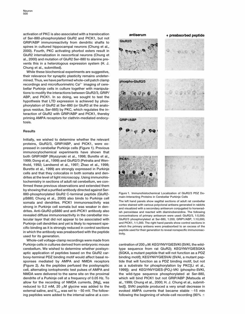
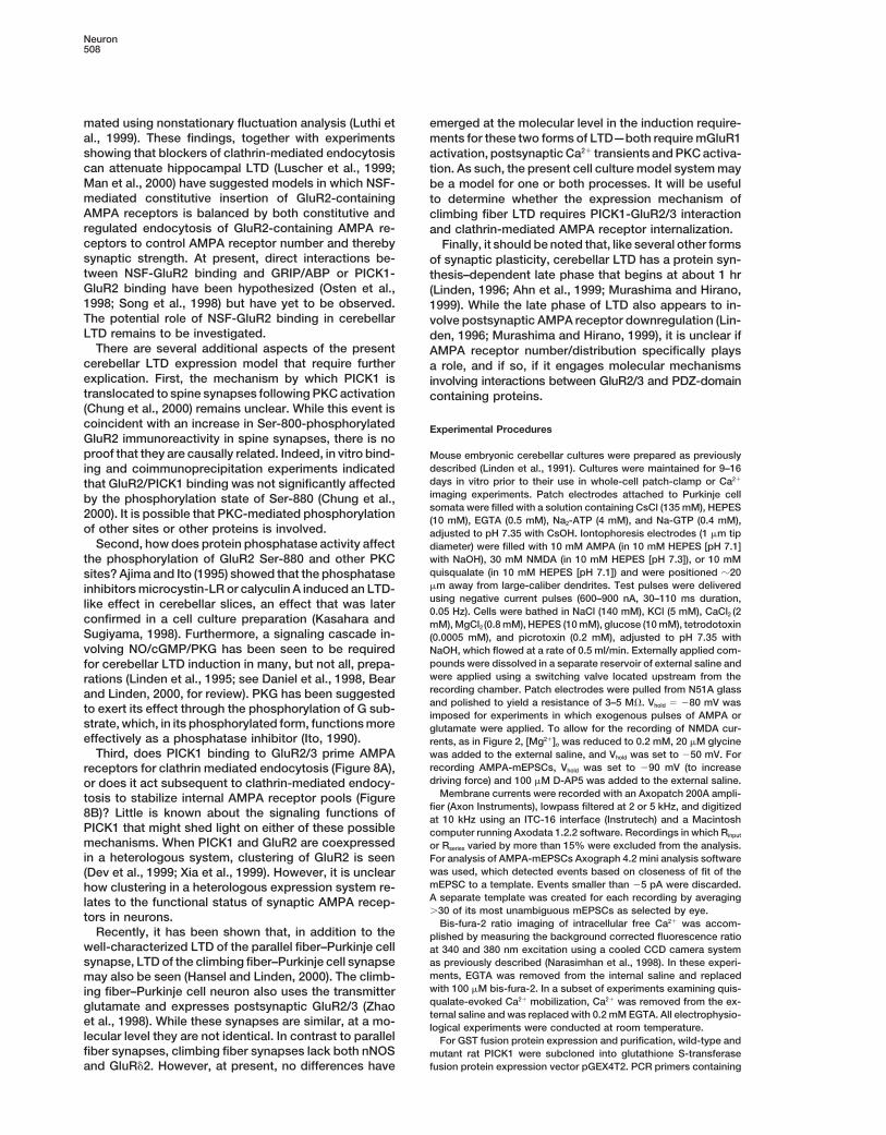
Initially, we wished to determine whether the relevantproteins, GluR2/3, GRIP/ABP, and PICK1, were ex-pressed in cerebellar Purkinje cells (Figure 1). Previousimmunocytochemical experiments have shown thatboth GRIP/ABP (Wyszynski et al., 1998; Burette et al.,1999; Dong et al., 1999) and GluR2/3 (Petralia and Wen-thold, 1992; Landsend et al., 1997; Zhao et al., 1998;Burette et al., 1999) are strongly expressed in Purkinjecells and that they colocalize in both somata and den-drites at the level of light microscopy. Using immunohis-tochemistry in sections of adult rat cerebellum, we con-firmed these previous observations and extended themby showing that a purified antibody directed against Ser-
Figure 1. Immunohistochemical Localization of GluR2/3 PDZ Do-880-phosphorylated GluR2 C-terminal peptide (GluR2-main–Interacting Proteins in Cerebellar Purkinje CellspS880; Chung et al., 2000) also binds to Purkinje cellThe left hand panels show sagittal sections of adult rat cerebellarsomata and dendrites. PICK1 immunoreactivity wascortex stained with various polyclonal antisera generated in rabbits
strong in Purkinje cell somata but was weaker in den- and visualized with a secondary antiserum conjugated to horserad-drites. Anti-GluR2-pS880 and anti-PICK1 antibody also ish peroxidase and reacted with diaminobenzidine. The followingrevealed diffuse immunoreactivity in the cerebellar mo- concentrations of primary antiserum were used: GluR2/3, 1:5,000;
GluR2/3 phosphorylated at Ser-880, 1:200; GRIP1/ABP, 1:10,000;lecular layer that did not appear to be associated withand PICK1, 1:1,000. The right hand panels show control sections inPurkinje cell dendrites and yet is likely to represent spe-which the primary antisera were preabsorbed to an excess of thecific binding as it is strongly reduced in control sectionspeptide used for their generation to reveal nonspecific immunoreac-
in which the antibody was preabsorbed with the peptide tivity.used for its generation.
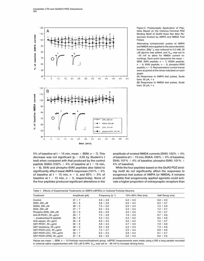
Whole-cell voltage-clamp recordings were made fromPurkinje cells in cultures derived from embryonic mouse centration of 200 mM: KEGYNVYGIESVKI (SVKI, the wild-
type sequence from rat GluR2); KEGYNVYGIESGKAcerebellum. We wished to determine whether postsyn-aptic application of peptides based on the GluR2 car- (SGKA, a mutant peptide that will not function as a PDZ
binding motif); KEGYNVYGIESVAI (SVAI, a mutant pep-boxy-terminal PDZ binding motif would affect basal re-sponses mediated by AMPA and NMDA receptors tide that will function as a PDZ binding motif, but not
as a substrate for phosphorylation by PKC[Li et al.,(Figure 2). As the peptides perfused the postsynapticcell, alternating iontophoretic test pulses of AMPA and 1999]); and KEGYNVYGIES-(PO4)-VKI (phospho-SVKI,
the wild-type sequence phosphorylated at Ser-880,NMDA were delivered to the same site on the proximaldendrite of a Purkinje cell at a frequency of 0.05 Hz. To which will bind PICK1 but not GRIP/ABP [Matsuda et
al., 1999; Chung et al., 2000; H. J. Chung et al., submit-allow for the recording of NMDA currents, [Mg]o wasreduced to 0.2 mM, 20 mM glycine was added to the ted]). SVKI peptide produced a very small decrease in
evoked AMPA currents that became stable z15 minexternal saline, and Vhold was set to 250 mV. The follow-ing peptides were added to the internal saline at a con- following the beginning of whole-cell recording (90% 6
Cerebellar LTD and GluR2/3 PDZ Interactions501
Figure 2. Postsynaptic Application of Pep-tides Based on the Carboxy-Terminal PDZBinding Motif of GluR2 Does Not Alter Re-sponses Evoked by AMPA and NMDA TestPulses
Alternating iontophoretic pulses of AMPAand NMDA were applied to the same dendriticlocation. [Mg21]o was reduced to 0.2 mM, 20mM glycine was added, and Vhold was set to250 mV to allow for NMDA current re-cordings. Each point represents the mean 6
SEM. SVKI peptide, n 5 7; SGKA peptide,n 5 6; SVAI peptide, n 5 5; phospho-SVKIpeptide, n 5 5. Representative current traceswere acquired at the times indicated on eachgraph.(A) Responses to AMPA test pulses. Scalebars: 50 pA, 1 s.(B) Responses to NMDA test pulses. Scalebars: 20 pA, 1 s.
5% of baseline at t 5 15 min, mean 6 SEM, n 5 7). This amplitude of evoked NMDA currents (SVKI: 102% 6 4%of baseline at t 5 15 min; SGKA: 100% 6 3% of baseline;decrease was not significant (p . 0.05 by Student’s t
test) when compared with that produced by the control SVAI: 101% 6 4% of baseline; phospho-SVKI: 101% 64% of baseline).peptide SGKA (102% 6 4% of baseline at t 5 15 min,
n 5 6). SVAI and phospho-SVKI peptides also failed to While the four peptides based on the GluR2 PDZ bind-ing motif do not significantly affect the responses tosignificantly affect basal AMPA responses (101% 6 4%
of baseline at t 5 15 min, n 5 5, and 95% 6 5% of exogenous test pulses of AMPA (or NMDA), it remainspossible that exogenously applied agonists could acti-baseline at t 5 15 min, n 5 5, respectively). None of
the four peptides produced significant alterations in the vate a higher proportion of extrasynaptic receptors than
Table 1. Effects of Experimental Treatments on AMPA-mEPSCs in Cultured Purkinje Neurons
Values are mean 6 SEM. n 5 10 Purkinje neurons/treatment group. mEPSC measurements were made using a 500 s long sample recordedin external saline supplemented with 100 mM D-AP5. Vhold was set to 290 mV to increase driving force.
Neuron502
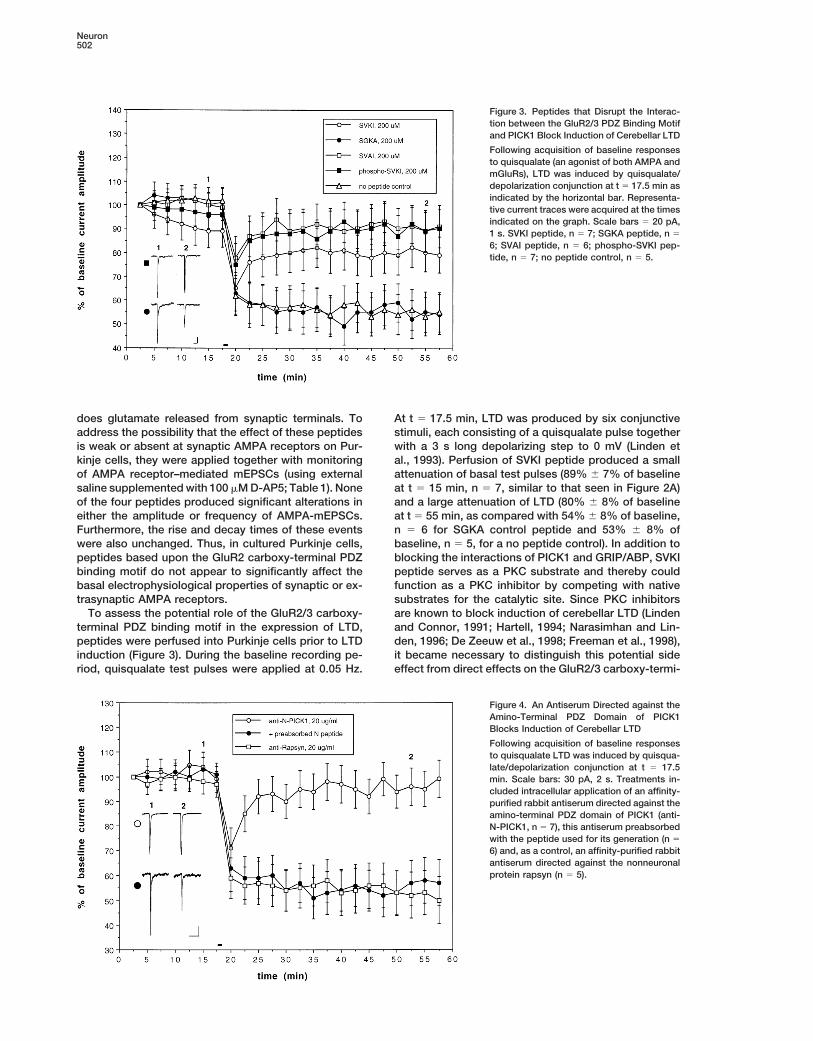
Figure 3. Peptides that Disrupt the Interac-tion between the GluR2/3 PDZ Binding Motifand PICK1 Block Induction of Cerebellar LTD
Following acquisition of baseline responsesto quisqualate (an agonist of both AMPA andmGluRs), LTD was induced by quisqualate/depolarization conjunction at t 5 17.5 min asindicated by the horizontal bar. Representa-tive current traces were acquired at the timesindicated on the graph. Scale bars 5 20 pA,1 s. SVKI peptide, n 5 7; SGKA peptide, n 5
6; SVAI peptide, n 5 6; phospho-SVKI pep-tide, n 5 7; no peptide control, n 5 5.
does glutamate released from synaptic terminals. To At t 5 17.5 min, LTD was produced by six conjunctivestimuli, each consisting of a quisqualate pulse togetheraddress the possibility that the effect of these peptides
is weak or absent at synaptic AMPA receptors on Pur- with a 3 s long depolarizing step to 0 mV (Linden etal., 1993). Perfusion of SVKI peptide produced a smallkinje cells, they were applied together with monitoring
of AMPA receptor–mediated mEPSCs (using external attenuation of basal test pulses (89% 6 7% of baselineat t 5 15 min, n 5 7, similar to that seen in Figure 2A)saline supplemented with 100 mM D-AP5; Table 1). None
of the four peptides produced significant alterations in and a large attenuation of LTD (80% 6 8% of baselineat t 5 55 min, as compared with 54% 6 8% of baseline,either the amplitude or frequency of AMPA-mEPSCs.
Furthermore, the rise and decay times of these events n 5 6 for SGKA control peptide and 53% 6 8% ofbaseline, n 5 5, for a no peptide control). In addition towere also unchanged. Thus, in cultured Purkinje cells,
peptides based upon the GluR2 carboxy-terminal PDZ blocking the interactions of PICK1 and GRIP/ABP, SVKIpeptide serves as a PKC substrate and thereby couldbinding motif do not appear to significantly affect the
basal electrophysiological properties of synaptic or ex- function as a PKC inhibitor by competing with nativesubstrates for the catalytic site. Since PKC inhibitorstrasynaptic AMPA receptors.
To assess the potential role of the GluR2/3 carboxy- are known to block induction of cerebellar LTD (Lindenand Connor, 1991; Hartell, 1994; Narasimhan and Lin-terminal PDZ binding motif in the expression of LTD,
peptides were perfused into Purkinje cells prior to LTD den, 1996; De Zeeuw et al., 1998; Freeman et al., 1998),it became necessary to distinguish this potential sideinduction (Figure 3). During the baseline recording pe-
riod, quisqualate test pulses were applied at 0.05 Hz. effect from direct effects on the GluR2/3 carboxy-termi-
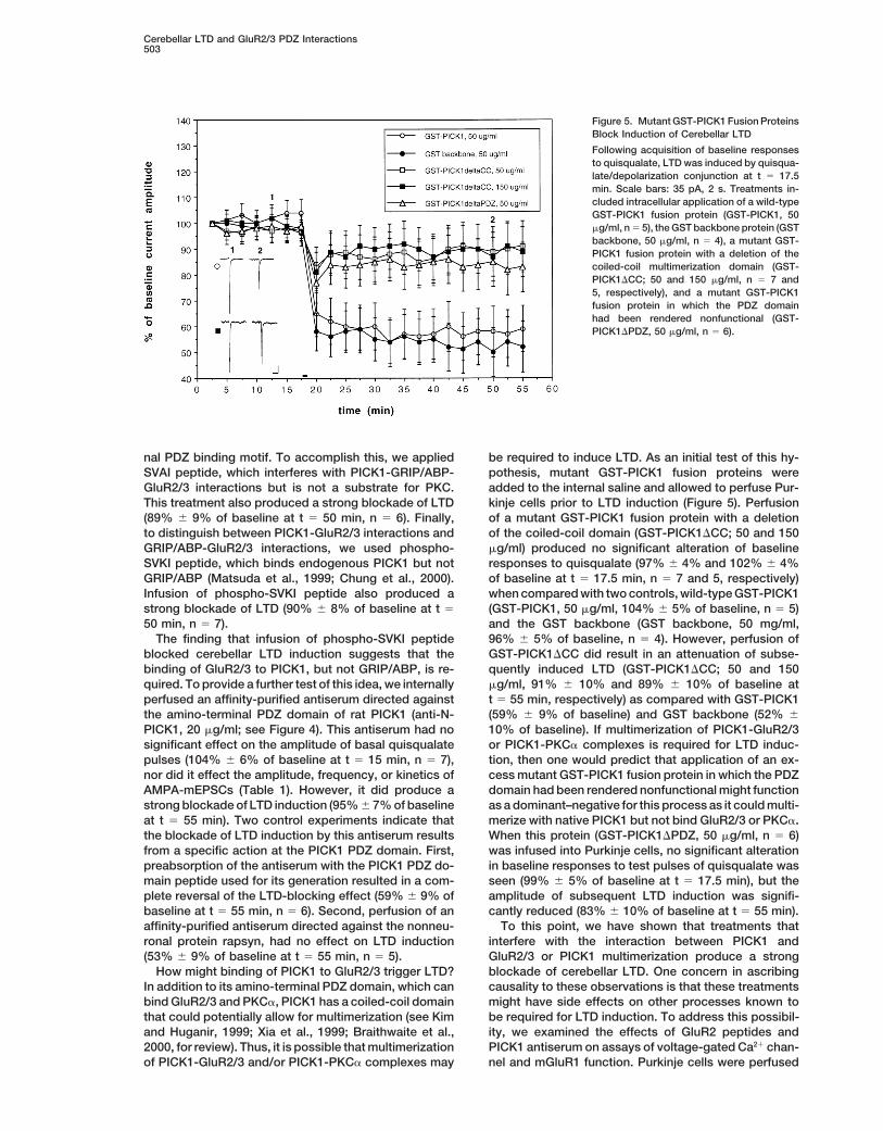
Figure 4. An Antiserum Directed against theAmino-Terminal PDZ Domain of PICK1Blocks Induction of Cerebellar LTD
Following acquisition of baseline responsesto quisqualate LTD was induced by quisqua-late/depolarization conjunction at t 5 17.5min. Scale bars: 30 pA, 2 s. Treatments in-cluded intracellular application of an affinity-purified rabbit antiserum directed against theamino-terminal PDZ domain of PICK1 (anti-N-PICK1, n 5 7), this antiserum preabsorbedwith the peptide used for its generation (n 5
6) and, as a control, an affinity-purified rabbitantiserum directed against the nonneuronalprotein rapsyn (n 5 5).
Cerebellar LTD and GluR2/3 PDZ Interactions503
Figure 5. Mutant GST-PICK1 Fusion ProteinsBlock Induction of Cerebellar LTD
Following acquisition of baseline responsesto quisqualate, LTD was induced by quisqua-late/depolarization conjunction at t 5 17.5min. Scale bars: 35 pA, 2 s. Treatments in-cluded intracellular application of a wild-typeGST-PICK1 fusion protein (GST-PICK1, 50mg/ml, n 5 5), the GST backbone protein (GSTbackbone, 50 mg/ml, n 5 4), a mutant GST-PICK1 fusion protein with a deletion of thecoiled-coil multimerization domain (GST-PICK1DCC; 50 and 150 mg/ml, n 5 7 and5, respectively), and a mutant GST-PICK1fusion protein in which the PDZ domainhad been rendered nonfunctional (GST-PICK1DPDZ, 50 mg/ml, n 5 6).
nal PDZ binding motif. To accomplish this, we applied be required to induce LTD. As an initial test of this hy-pothesis, mutant GST-PICK1 fusion proteins wereSVAI peptide, which interferes with PICK1-GRIP/ABP-
GluR2/3 interactions but is not a substrate for PKC. added to the internal saline and allowed to perfuse Pur-kinje cells prior to LTD induction (Figure 5). PerfusionThis treatment also produced a strong blockade of LTD
(89% 6 9% of baseline at t 5 50 min, n 5 6). Finally, of a mutant GST-PICK1 fusion protein with a deletionof the coiled-coil domain (GST-PICK1DCC; 50 and 150to distinguish between PICK1-GluR2/3 interactions and
GRIP/ABP-GluR2/3 interactions, we used phospho- mg/ml) produced no significant alteration of baselineresponses to quisqualate (97% 6 4% and 102% 6 4%SVKI peptide, which binds endogenous PICK1 but not
GRIP/ABP (Matsuda et al., 1999; Chung et al., 2000). of baseline at t 5 17.5 min, n 5 7 and 5, respectively)when compared with two controls, wild-type GST-PICK1Infusion of phospho-SVKI peptide also produced a
strong blockade of LTD (90% 6 8% of baseline at t 5 (GST-PICK1, 50 mg/ml, 104% 6 5% of baseline, n 5 5)and the GST backbone (GST backbone, 50 mg/ml,50 min, n 5 7).
The finding that infusion of phospho-SVKI peptide 96% 6 5% of baseline, n 5 4). However, perfusion ofGST-PICK1DCC did result in an attenuation of subse-blocked cerebellar LTD induction suggests that the
binding of GluR2/3 to PICK1, but not GRIP/ABP, is re- quently induced LTD (GST-PICK1DCC; 50 and 150mg/ml, 91% 6 10% and 89% 6 10% of baseline atquired. To provide a further test of this idea, we internally
perfused an affinity-purified antiserum directed against t 5 55 min, respectively) as compared with GST-PICK1(59% 6 9% of baseline) and GST backbone (52% 6the amino-terminal PDZ domain of rat PICK1 (anti-N-
PICK1, 20 mg/ml; see Figure 4). This antiserum had no 10% of baseline). If multimerization of PICK1-GluR2/3or PICK1-PKCa complexes is required for LTD induc-significant effect on the amplitude of basal quisqualate
pulses (104% 6 6% of baseline at t 5 15 min, n 5 7), tion, then one would predict that application of an ex-cess mutant GST-PICK1 fusion protein in which the PDZnor did it effect the amplitude, frequency, or kinetics of
AMPA-mEPSCs (Table 1). However, it did produce a domain had been rendered nonfunctional might functionas a dominant–negative for this process as it could multi-strong blockade of LTD induction (95% 6 7% of baseline
at t 5 55 min). Two control experiments indicate that merize with native PICK1 but not bind GluR2/3 or PKCa.When this protein (GST-PICK1DPDZ, 50 mg/ml, n 5 6)the blockade of LTD induction by this antiserum results
from a specific action at the PICK1 PDZ domain. First, was infused into Purkinje cells, no significant alterationin baseline responses to test pulses of quisqualate waspreabsorption of the antiserum with the PICK1 PDZ do-
main peptide used for its generation resulted in a com- seen (99% 6 5% of baseline at t 5 17.5 min), but theamplitude of subsequent LTD induction was signifi-plete reversal of the LTD-blocking effect (59% 6 9% of
baseline at t 5 55 min, n 5 6). Second, perfusion of an cantly reduced (83% 6 10% of baseline at t 5 55 min).To this point, we have shown that treatments thataffinity-purified antiserum directed against the nonneu-
ronal protein rapsyn, had no effect on LTD induction interfere with the interaction between PICK1 andGluR2/3 or PICK1 multimerization produce a strong(53% 6 9% of baseline at t 5 55 min, n 5 5).
How might binding of PICK1 to GluR2/3 trigger LTD? blockade of cerebellar LTD. One concern in ascribingcausality to these observations is that these treatmentsIn addition to its amino-terminal PDZ domain, which can
bind GluR2/3 and PKCa, PICK1 has a coiled-coil domain might have side effects on other processes known tobe required for LTD induction. To address this possibil-that could potentially allow for multimerization (see Kim
and Huganir, 1999; Xia et al., 1999; Braithwaite et al., ity, we examined the effects of GluR2 peptides andPICK1 antiserum on assays of voltage-gated Ca21 chan-2000, for review). Thus, it is possible that multimerization
of PICK1-GluR2/3 and/or PICK1-PKCa complexes may nel and mGluR1 function. Purkinje cells were perfused
Neuron504
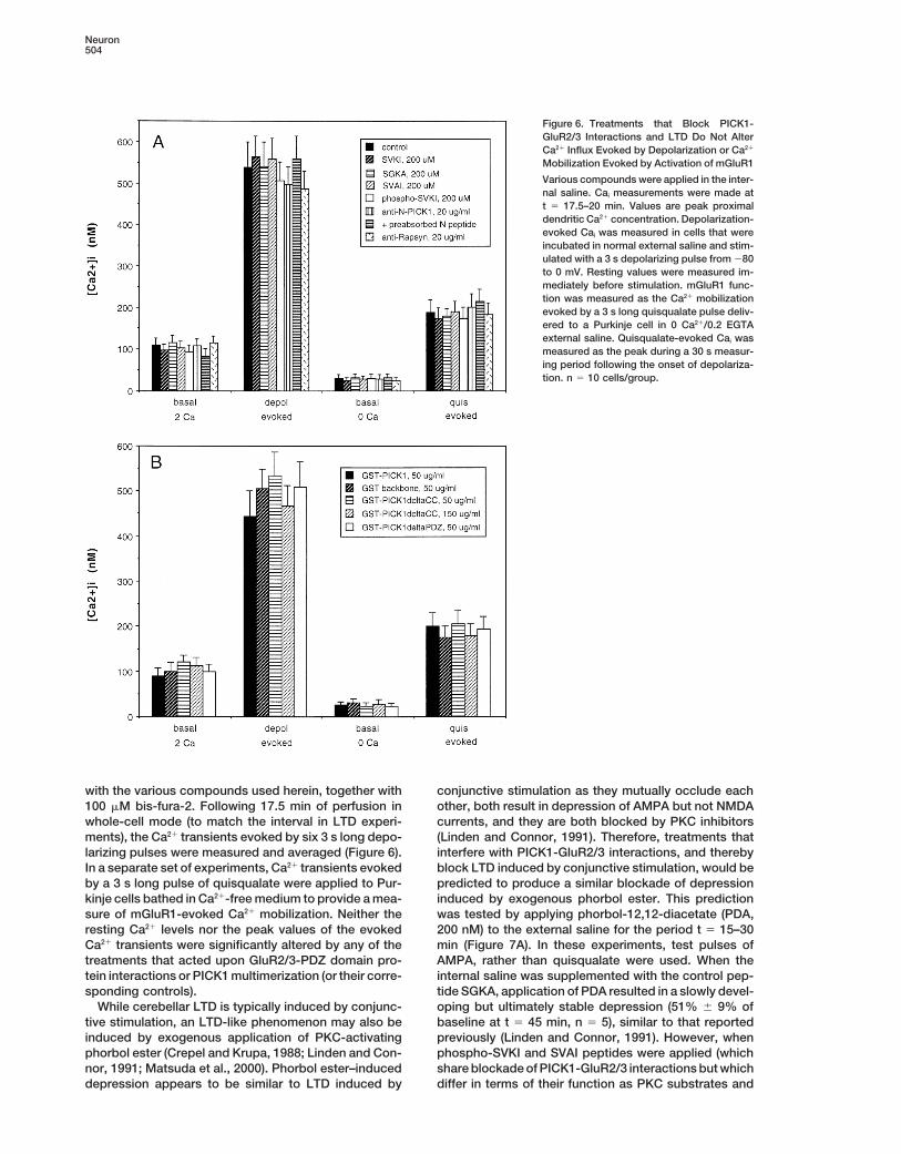
Figure 6. Treatments that Block PICK1-GluR2/3 Interactions and LTD Do Not AlterCa21 Influx Evoked by Depolarization or Ca21
Mobilization Evoked by Activation of mGluR1
Various compounds were applied in the inter-nal saline. Cai measurements were made att 5 17.5–20 min. Values are peak proximaldendritic Ca21 concentration. Depolarization-evoked Cai was measured in cells that wereincubated in normal external saline and stim-ulated with a 3 s depolarizing pulse from 280to 0 mV. Resting values were measured im-mediately before stimulation. mGluR1 func-tion was measured as the Ca21 mobilizationevoked by a 3 s long quisqualate pulse deliv-ered to a Purkinje cell in 0 Ca21/0.2 EGTAexternal saline. Quisqualate-evoked Cai wasmeasured as the peak during a 30 s measur-ing period following the onset of depolariza-tion. n 5 10 cells/group.
with the various compounds used herein, together with conjunctive stimulation as they mutually occlude eachother, both result in depression of AMPA but not NMDA100 mM bis-fura-2. Following 17.5 min of perfusion in
whole-cell mode (to match the interval in LTD experi- currents, and they are both blocked by PKC inhibitors(Linden and Connor, 1991). Therefore, treatments thatments), the Ca21 transients evoked by six 3 s long depo-
larizing pulses were measured and averaged (Figure 6). interfere with PICK1-GluR2/3 interactions, and therebyblock LTD induced by conjunctive stimulation, would beIn a separate set of experiments, Ca21 transients evoked
by a 3 s long pulse of quisqualate were applied to Pur- predicted to produce a similar blockade of depressioninduced by exogenous phorbol ester. This predictionkinje cells bathed in Ca21-free medium to provide a mea-
sure of mGluR1-evoked Ca21 mobilization. Neither the was tested by applying phorbol-12,12-diacetate (PDA,200 nM) to the external saline for the period t 5 15–30resting Ca21 levels nor the peak values of the evoked
Ca21 transients were significantly altered by any of the min (Figure 7A). In these experiments, test pulses ofAMPA, rather than quisqualate were used. When thetreatments that acted upon GluR2/3-PDZ domain pro-
tein interactions or PICK1 multimerization (or their corre- internal saline was supplemented with the control pep-tide SGKA, application of PDA resulted in a slowly devel-sponding controls).
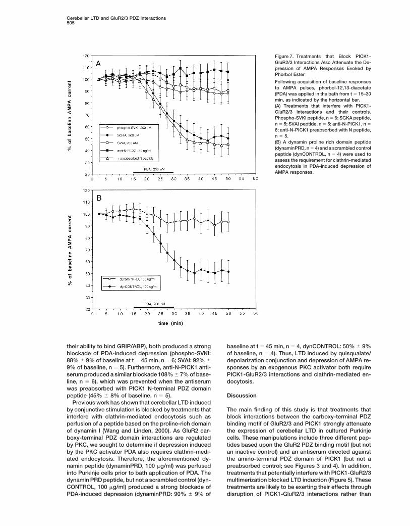
While cerebellar LTD is typically induced by conjunc- oping but ultimately stable depression (51% 6 9% ofbaseline at t 5 45 min, n 5 5), similar to that reportedtive stimulation, an LTD-like phenomenon may also be
induced by exogenous application of PKC-activating previously (Linden and Connor, 1991). However, whenphospho-SVKI and SVAI peptides were applied (whichphorbol ester (Crepel and Krupa, 1988; Linden and Con-
nor, 1991; Matsuda et al., 2000). Phorbol ester–induced share blockade of PICK1-GluR2/3 interactions but whichdiffer in terms of their function as PKC substrates anddepression appears to be similar to LTD induced by
Cerebellar LTD and GluR2/3 PDZ Interactions505
Figure 7. Treatments that Block PICK1-GluR2/3 Interactions Also Attenuate the De-pression of AMPA Responses Evoked byPhorbol Ester
Following acquisition of baseline responsesto AMPA pulses, phorbol-12,13-diacetate(PDA) was applied in the bath from t 5 15–30min, as indicated by the horizontal bar.(A) Treatments that interfere with PICK1-GluR2/3 interactions and their controls.Phospho-SVKI peptide, n 5 6; SGKA peptide,n 5 5; SVAI peptide, n 5 5; anti-N-PICK1, n 5
6; anti-N-PICK1 preabsorbed with N peptide,n 5 5.(B) A dynamin proline rich domain peptide(dynaminPRD, n 5 4) and a scrambled controlpeptide (dynCONTROL, n 5 4) were used toassess the requirement for clathrin-mediatedendocytosis in PDA-induced depression ofAMPA responses.
their ability to bind GRIP/ABP), both produced a strong baseline at t 5 45 min, n 5 4, dynCONTROL: 50% 6 9%of baseline, n 5 4). Thus, LTD induced by quisqualate/blockade of PDA-induced depression (phospho-SVKI:
88% 6 9% of baseline at t 5 45 min, n 5 6; SVAI: 92% 6 depolarization conjunction and depression of AMPA re-sponses by an exogenous PKC activator both require9% of baseline, n 5 5). Furthermore, anti-N-PICK1 anti-
serum produced a similar blockade 108% 6 7% of base- PICK1-GluR2/3 interactions and clathrin-mediated en-docytosis.line, n 5 6), which was prevented when the antiserum
was preabsorbed with PICK1 N-terminal PDZ domainpeptide (45% 6 8% of baseline, n 5 5). Discussion
Previous work has shown that cerebellar LTD inducedby conjunctive stimulation is blocked by treatments that The main finding of this study is that treatments that
block interactions between the carboxy-terminal PDZinterfere with clathrin-mediated endocytosis such asperfusion of a peptide based on the proline-rich domain binding motif of GluR2/3 and PICK1 strongly attenuate
the expression of cerebellar LTD in cultured Purkinjeof dynamin I (Wang and Linden, 2000). As GluR2 car-boxy-terminal PDZ domain interactions are regulated cells. These manipulations include three different pep-
tides based upon the GluR2 PDZ binding motif (but notby PKC, we sought to determine if depression inducedby the PKC activator PDA also requires clathrin-medi- an inactive control) and an antiserum directed against
the amino-terminal PDZ domain of PICK1 (but not aated endocytosis. Therefore, the aforementioned dy-namin peptide (dynaminPRD, 100 mg/ml) was perfused preabsorbed control; see Figures 3 and 4). In addition,
treatments that potentially interfere with PICK1-GluR2/3into Purkinje cells prior to bath application of PDA. Thedynamin PRD peptide, but not a scrambled control (dyn- multimerization blocked LTD induction (Figure 5). These
treatments are likely to be exerting their effects throughCONTROL, 100 mg/ml) produced a strong blockade ofPDA-induced depression (dynaminPRD: 90% 6 9% of disruption of PICK1-GluR2/3 interactions rather than
Neuron506
side effects because they did not affect the basal func- (H. J. Chung et al., submitted), and (3), in a heterologousexpression system, mutation of GluR2 Ser-880 to ala-tion of AMPA receptors (Figure 2 and Table 1), voltage-
gated Ca21 channels or mGluR1 (Figure 6). Thus, block- nine prevented phorbol ester–induced GluR2 internal-ization (H. J. Chung et al., submitted). However, thereade of PICK1-GluR2/3 interactions attenuates cerebellar
LTD expression in a manner that is independent of the are several caveats that also should be mentioned. First,the present experiments were done using immature Pur-initial signals known to be required for its induction.
While the GluR2/3 carboxy-terminal PDZ binding motif kinje cells in culture and so one must always be con-cerned that the cellular mechanisms underlying cerebel-can interact with both PICK1 and GRIP/ABP, the finding
that two different treatments (phospho-SVKI peptide lar LTD might be different in intact, adult tissue. Second,the prior observation that phorbol ester treatment ofand anti-N-PICK1 antiserum) that block PICK1 but not
GRIP/ABP interactions attenuate LTD (Figures 3 and 4) cerebellar cultures results in GluR2 internalization (Mat-suda et al., 2000) is potentially complicated by that factsuggests that it is the binding of the former that is re-
quired. Blockade of the PICK1-GluR2/3 interaction at- that these measures were made using immunoblotsfrom cultures in which only about 2.5% of the neuronstenuates LTD induced by quisqualate/depolarization
conjunction (which has previously been shown to re- are Purkinje cells (Furuya et al., 1998). Third, while thereis now reason to believe that AMPA receptor internaliza-quire PKC activation; Linden and Connor, 1991) and an
LTD-like phenomenon produced by exogenous PKC- tion plays a role in LTD expression, it would be prema-ture to conclude that it can wholly account for it. Injec-activating phorbol ester (Figure 7A). Both of these forms
of depression require clathrin-mediated internalization tion of a catalytically active fragment of PKC intocultured hippocampal neurons modulated the decayof AMPA receptors (Wang and Linden, 2000; Figure 7B).
While the present data indicate a role for interactions rate of AMPA-mEPSCs in hippocampal neurons (Wanget al., 1994) and it is not impossible to imagine thatbetween GluR2/3 and PICK1 in LTD induction, it should
be cautioned that this does not mean that GRIP/ABP a similar mechanism or another utilizing alterations inAMPA receptor unitary conductance (Benke et al., 1998)is not involved. A recent report has shown that when
cultured hippocampal neurons are transfected with mu- might be operative in Purkinje cells.Prior work using other model systems has implicatedtant GluR2 subunits that can bind PICK1 but not GRIP/
ABP, these mutants subunits do not accumulate at syn- GluR2/3 interactions in use-dependent synaptic plastic-ity. In spinal dorsal horn neurons, serotonin can produceapses to the same degree as wild-type GluR2 (Osten
et al., 2000). Furthermore, the observation that PKC- a form of LTP that appears to involve the recruiting ofAMPA receptors to previously silent synapses (Li andmediated phosphorylation of GluR2 Ser-880 results in
a strong decrease in the binding of GRIP/ABP (Matsuda Zhuo, 1998). Like cerebellar LTD, this potentiation canbe blocked by postsynaptic PKC inhibitors or mimickedet al., 1999; Chung et al., 2000) suggests a scheme in
which the release of GluR2/3 from GRIP/ABP is required by PKC activators (Li et al., 1999). In addition, peptidesbased upon the GluR2 carboxy-terminal PDZ bindingfor LTD induction. Unfortunately, it is difficult to test this
idea directly without a method of stabilizing the GRIP/ motif were used (they used 10 amino acid peptides asopposed to 15 in the present study). Postsynaptic appli-ABP-GluR2/3 interaction in living Purkinje cells.
A model of cerebellar LTD induction and expression cation of SVKI peptide did not produce basal alterationsin glutamatergic EPSCs but blocked LTP induced bybased upon these observations is shown in Figure 8A.
Conjoint activation of mGluR1, AMPA receptors and serotonin or phorbol ester. However, in contrast to thepresent results, Li et al. (1999) found that EVKI peptidevoltage-gated Ca21 channels results in stimulation of
PKC. PKC phosphorylates Ser-880 of GluR2 (and possi- (which should be functionally equivalent to our phos-pho-SVKI) failed to block serotonin-induced LTP. Thus,bly other sites as well) resulting in both unbinding of
GRIP/ABP and, through an unknown mechanism, the serotonin-induced LTP in spinal dorsal horn appears torequire GRIP/ABP-GluR2/3 rather than PICK1-GluR2/3recruitment of PICK1 to synaptic AMPA receptors.
PICK1 recruitment together with GRIP/ABP unbinding interactions supporting the idea that GRIP/ABP-GluR2/3interactions may stabilize AMPA receptors in the post-then primes AMPA receptors for clathrin-mediated inter-
nalization. Alternatively, GRIP/ABP unbinding is suffi- synaptic plasma membrane.GluR2/3 carboxy-terminal PDZ domain interactionscient to prime synaptic AMPA receptors for internaliza-
tion but synaptic strength is not depressed unless PICK1 have also been implicated in hippocampal LTD. H. J.Chung et al. (submitted) show that internal applicationbinding to GluR2/3 also occurs, thereby stabilizing the
internal pool and attenuating compensatory insertion of of SVKI and phospho-SVKI GluR2 peptides, but not anSVKA control, attenuates LTD at the Schaffer collateral-AMPA receptors (see Figure 8B). Importantly, in both of
these models, application of reagents that block either CA1 synapse in a slice preparation. Furthermore, in sep-arate experiments using field potential recording, LTDPICK1-GluR2/3 or GRIP/ABP-GluR2/3 interactions (or
both) would not be expected to alter the basal response induction was associated with an increase in Ser-880-phosphorylated GluR2 as measured by Western blot.of Purkinje cells to AMPA test pulses.
These models are consistent with certain recent ob- This increase was blocked by pretreatments that pre-vent this form of LTD such as an NMDA receptor antago-servations including: (1) activation of PKC resulted in an
increase in Ser-880-phosphorylated GluR2 in spines and nist or a protein phosphatase inhibitor. Taken togetherwith previous results showing that both hippocampalthe translocation of PICK1 immunoreactivity from den-
dritic shafts to spines in cultured hippocampal neurons and cerebellar LTD require clathrin-mediated endocyto-sis (Luscher et al., 1999; Man et al., 2000, Wang and(Chung et al., 2000), (2) PKC activation evoked GluR2
internalization in cerebellar (Matsuda et al., 2000), neo- Linden, 2000) and are associated with GluR2 internaliza-tion (Carroll et al., 1999a; Luscher et al., 1999; Man etcortical (Chung et al., 2000), and hippocampal neurons
Cerebellar LTD and GluR2/3 PDZ Interactions507
Figure 8. Two Models for Expression of LTD in Cultured Purkinje Cells
(A) Synaptic AMPA receptors containing at least one GluR2/3 subunit are bound to GRIP/ABP in the basal state. Activation of PKC duringLTD induction results in the phosphorylation of GluR2 Ser-880, This causes the unbinding of GRIP/ABP and the recruitment of PICK1 to thesynapse. PICK1 can multimerize through its coiled-coil domain. Synaptic GluR2/3 containing AMPA receptors in the GRIP/ABP-unbound,PICK1-bound state are then primed for internalization by clathrin-mediated endocytosis.(B) A variant of the model above. In this scheme, GRIP/ABP unbinding is sufficient to prime synaptic AMPA receptors for internalization.However, PICK1 binds AMPA receptors in the internal pool, thereby stabilizing them and attenuating compensatory AMPA receptor insertion.
al., 2000; Matsuda et al., 2000), the present findings manipulations to disrupt this interaction and measurethe consequences for synaptic transmission in the hip-suggest that cerebellar and at least one form of hippo-
campal LTD may share aspects of a common expression pocampus (see Kullmann, 1999; Luscher et al., 2000;Turrigiano, 2000, for review). While there have beenmechanism. However, it should be cautioned that there
are important differences as well. PKC-activating phor- some differences between experiments, in general, pep-tides designed to block the NSF-GluR2 interaction pro-bol esters do not typically induce either LTD or a de-
crease in mEPSC amplitude at hippocampal synapses duce an attenuation of evoked AMPA receptor-medi-ated EPSCs as well as a reduction in the both the(Carroll et al., 1998). Likewise, while there are varieties
of hippocampal LTD that require activation of an mGluR amplitude and frequency of AMPA-mEPSCs (Nishimuneet al., 1998; Song et al., 1998; Luthi et al., 1999; Luschercascade (Stanton et al., 1991; Bolshakov and Siegel-
baum, 1994; Yang et al., 1994) and consequent activa- et al., 1999; Noel et al., 1999). The latter presumablyrepresents the complete “silencing” of a fraction of syn-tion of PKC (Oliet et al., 1997; Otani and Connor, 1998;
Wang et al., 1998), the more commonly studied form of apses and in consistent with the finding that this treat-ment causes a reduction in the surface expression ofhippocampal LTD requires activation of NMDA recep-
tors and protein phosphatases and is independent of AMPA receptors as determined immunocytochemically(Luscher et al., 1999; Noel et al., 1999). Furthermore, themGluR or PKC activation (see Bear and Linden, 2000,
for review). attenuation of synaptic transmission produced by thesepeptides occludes subsequent LTD induction at the hip-In addition to the PDZ binding motif at the extreme
carboxyl terminus of GluR2, there is a binding site for pocampal Schaffer collateral-CA1 synapse (Luscher etal., 1999; Luthi et al., 1999), and neither LTD inductionthe protein NSF between residues 844 and 853 (see Kim
and Huganir, 1999; Braithwaite et al., 2000, for review). nor NSF peptide application appeared to alter EPSCkinetics or AMPA receptor unitary conductance as esti-In recent years, several laboratories have used various
Neuron508
mated using nonstationary fluctuation analysis (Luthi et emerged at the molecular level in the induction require-al., 1999). These findings, together with experiments ments for these two forms of LTD—both require mGluR1showing that blockers of clathrin-mediated endocytosis activation, postsynaptic Ca21 transients and PKC activa-can attenuate hippocampal LTD (Luscher et al., 1999; tion. As such, the present cell culture model system mayMan et al., 2000) have suggested models in which NSF- be a model for one or both processes. It will be usefulmediated constitutive insertion of GluR2-containing to determine whether the expression mechanism ofAMPA receptors is balanced by both constitutive and climbing fiber LTD requires PICK1-GluR2/3 interactionregulated endocytosis of GluR2-containing AMPA re- and clathrin-mediated AMPA receptor internalization.ceptors to control AMPA receptor number and thereby Finally, it should be noted that, like several other formssynaptic strength. At present, direct interactions be- of synaptic plasticity, cerebellar LTD has a protein syn-tween NSF-GluR2 binding and GRIP/ABP or PICK1- thesis–dependent late phase that begins at about 1 hrGluR2 binding have been hypothesized (Osten et al., (Linden, 1996; Ahn et al., 1999; Murashima and Hirano,1998; Song et al., 1998) but have yet to be observed. 1999). While the late phase of LTD also appears to in-The potential role of NSF-GluR2 binding in cerebellar volve postsynaptic AMPA receptor downregulation (Lin-LTD remains to be investigated. den, 1996; Murashima and Hirano, 1999), it is unclear if
There are several additional aspects of the present AMPA receptor number/distribution specifically playscerebellar LTD expression model that require further a role, and if so, if it engages molecular mechanismsexplication. First, the mechanism by which PICK1 is involving interactions between GluR2/3 and PDZ-domaintranslocated to spine synapses following PKC activation containing proteins.(Chung et al., 2000) remains unclear. While this event iscoincident with an increase in Ser-800-phosphorylated Experimental ProceduresGluR2 immunoreactivity in spine synapses, there is noproof that they are causally related. Indeed, in vitro bind- Mouse embryonic cerebellar cultures were prepared as previously
described (Linden et al., 1991). Cultures were maintained for 9–16ing and coimmunoprecipitation experiments indicateddays in vitro prior to their use in whole-cell patch-clamp or Ca21that GluR2/PICK1 binding was not significantly affectedimaging experiments. Patch electrodes attached to Purkinje cellby the phosphorylation state of Ser-880 (Chung et al.,somata were filled with a solution containing CsCl (135 mM), HEPES2000). It is possible that PKC-mediated phosphorylation (10 mM), EGTA (0.5 mM), Na2-ATP (4 mM), and Na-GTP (0.4 mM),
of other sites or other proteins is involved. adjusted to pH 7.35 with CsOH. Iontophoresis electrodes (1 mm tipSecond, how does protein phosphatase activity affect diameter) were filled with 10 mM AMPA (in 10 mM HEPES [pH 7.1]
with NaOH), 30 mM NMDA (in 10 mM HEPES [pH 7.3]), or 10 mMthe phosphorylation of GluR2 Ser-880 and other PKCquisqualate (in 10 mM HEPES [pH 7.1]) and were positioned z20sites? Ajima and Ito (1995) showed that the phosphatasemm away from large-caliber dendrites. Test pulses were deliveredinhibitors microcystin-LR or calyculin A induced an LTD-using negative current pulses (600–900 nA, 30–110 ms duration,like effect in cerebellar slices, an effect that was later0.05 Hz). Cells were bathed in NaCl (140 mM), KCl (5 mM), CaCl2 (2
confirmed in a cell culture preparation (Kasahara and mM), MgCl2 (0.8 mM), HEPES (10 mM), glucose (10 mM), tetrodotoxinSugiyama, 1998). Furthermore, a signaling cascade in- (0.0005 mM), and picrotoxin (0.2 mM), adjusted to pH 7.35 withvolving NO/cGMP/PKG has been seen to be required NaOH, which flowed at a rate of 0.5 ml/min. Externally applied com-
pounds were dissolved in a separate reservoir of external saline andfor cerebellar LTD induction in many, but not all, prepa-were applied using a switching valve located upstream from therations (Linden et al., 1995; see Daniel et al., 1998, Bearrecording chamber. Patch electrodes were pulled from N51A glassand Linden, 2000, for review). PKG has been suggestedand polished to yield a resistance of 3–5 MV. Vhold 5 280 mV wasto exert its effect through the phosphorylation of G sub-imposed for experiments in which exogenous pulses of AMPA or
strate, which, in its phosphorylated form, functions more glutamate were applied. To allow for the recording of NMDA cur-effectively as a phosphatase inhibitor (Ito, 1990). rents, as in Figure 2, [Mg21]o was reduced to 0.2 mM, 20 mM glycine
Third, does PICK1 binding to GluR2/3 prime AMPA was added to the external saline, and Vhold was set to 250 mV. Forrecording AMPA-mEPSCs, Vhold was set to 290 mV (to increasereceptors for clathrin mediated endocytosis (Figure 8A),driving force) and 100 mM D-AP5 was added to the external saline.or does it act subsequent to clathrin-mediated endocy-
Membrane currents were recorded with an Axopatch 200A ampli-tosis to stabilize internal AMPA receptor pools (Figurefier (Axon Instruments), lowpass filtered at 2 or 5 kHz, and digitized8B)? Little is known about the signaling functions of at 10 kHz using an ITC-16 interface (Instrutech) and a Macintosh
PICK1 that might shed light on either of these possible computer running Axodata 1.2.2 software. Recordings in which Rinput
mechanisms. When PICK1 and GluR2 are coexpressed or Rseries varied by more than 15% were excluded from the analysis.in a heterologous system, clustering of GluR2 is seen For analysis of AMPA-mEPSCs Axograph 4.2 mini analysis software
was used, which detected events based on closeness of fit of the(Dev et al., 1999; Xia et al., 1999). However, it is unclearmEPSC to a template. Events smaller than 25 pA were discarded.how clustering in a heterologous expression system re-A separate template was created for each recording by averaginglates to the functional status of synaptic AMPA recep-.30 of its most unambiguous mEPSCs as selected by eye.
tors in neurons. Bis-fura-2 ratio imaging of intracellular free Ca21 was accom-Recently, it has been shown that, in addition to the plished by measuring the background corrected fluorescence ratio
well-characterized LTD of the parallel fiber–Purkinje cell at 340 and 380 nm excitation using a cooled CCD camera systemsynapse, LTD of the climbing fiber–Purkinje cell synapse as previously described (Narasimhan et al., 1998). In these experi-
ments, EGTA was removed from the internal saline and replacedmay also be seen (Hansel and Linden, 2000). The climb-with 100 mM bis-fura-2. In a subset of experiments examining quis-ing fiber–Purkinje cell neuron also uses the transmitterqualate-evoked Ca21 mobilization, Ca21 was removed from the ex-glutamate and expresses postsynaptic GluR2/3 (Zhaoternal saline and was replaced with 0.2 mM EGTA. All electrophysio-
et al., 1998). While these synapses are similar, at a mo- logical experiments were conducted at room temperature.lecular level they are not identical. In contrast to parallel For GST fusion protein expression and purification, wild-type andfiber synapses, climbing fiber synapses lack both nNOS mutant rat PICK1 were subcloned into glutathione S-transferase
fusion protein expression vector pGEX4T2. PCR primers containingand GluRd2. However, at present, no differences have
Cerebellar LTD and GluR2/3 PDZ Interactions509
the desired PICK1 mutations were synthesized using full-length rat K. Davies, eds. (Baltimore, MD: Johns Hopkins University Press),pp. 455–517.PICK1 cDNA as template for mutants generation. PCR mutagenesis
were performed using a Quikchange site-directed mutagenesis kit Benke, T.A., Luthi, A., Isaac, J.T., and Collingridge, G.L. (1998). Mod-(Stratagene). All mutations were subsequently confirmed by se- ulation of AMPA receptor unitary conductance by synaptic activity.quencing. To create the DPDZ mutation, lysine and aspartic acid at Nature 393, 793–797.amino acids 27 and 28 of PICK1 were replaced with alanine. The
Bolshakov, V.Y., and Siegelbaum, S.A. (1994). Postsynaptic induc-DCC mutation was made by deleting amino acids 139–166 of PICK1.
tion and presynaptic expression of hippocampal long-term depres-All constructs were then transformed to E. coli BL21 cells to produce
sion. Science 264, 1148–1152.GST fusion proteins. After IPTG induction, GST fusion proteins were
Braithwaite, S.P., Meyer, G., and Henley, J.M. (2000). Interactionsliberated by sonication and solubilized by 1% Triton X-100. Glutathi-between AMPA receptors and intracellular proteins. Neuropharma-one Sepharose 4B (Pharmacia Biotech) was added to the clearedcology 39, 919–930.cell lysates. After washing, GST fusion proteins were eluted by re-
duced Glutathione. Purified GST fusion proteins were then dialyzed Burette, A., Wyszynski, M., Valtschanoff, J.G., Sheng, M., and Wein-against internal solution. Fusion protein concentration was deter- berg, R.J. (1999). Characterization of glutamate receptor interactingmined by reading of O.D. 280 nm against the standard curve. protein-immunopositive neurons in cerebellum and cerebral cortex
The dynamin I proline-rich domain peptide (QVPSRPNRAP) and of the albino rat. J. Comp. Neurol. 411, 601–612.the dynamin scrambled control peptide (QPPASNPRVR) were syn- Carroll, R.C., Nicoll, R.A., and Malenka, R.C. (1998). Effects of PKAthesized and purified at the Biosynthesis and Sequencing Facility, and PKC on miniature excitatory postsynaptic currents in CA1 pyra-Department of Biological Chemistry, Johns Hopkins University midal cells. J. Neurophysiol. 80, 2797–2800.School of Medicine. The GluR2 peptides (KEGYNVYGIESVKI, KEG
Carroll, R.C., Lissin, D.V., von Zastrow, M., Nicoll, R.A., and Malenka,YNVYGIESGKA, KEGYNVYGIESVAI, and KEGYNVYGIES-[PO4]-VKI)R.C. (1999a). Rapid redistribution of glutamate receptors contributeswere synthesized and purified at the Howard Hughes Medical Insti-to long-term depression in hippocampal cultures. Nat. Neurosci. 2,tute Biopolymer Facility at Johns Hopkins University School of Medi-454–460.cine. The PICK1 antiserum was generated in rabbits against anCarroll, R.C., Beattie, E.C., Xia, H., Luscher, C., Altschuler, Y., Nicoll,N-terminal peptide as previously described (Xia et al., 1999). Bis-R.A., Malenka, R.C., and von Zastrow, M. (1999b). Dynamin-depen-fura-2 was from Molecular Probes, tetrodotoxin from Alexis Bio-dent endocytosis of ionotropic glutamate receptors. Proc. Natl.chemicals, AMPA, NMDA, and D-AP5 from Tocris, and phorbol-Acad. Sci. USA 96, 14112–14117.12,13-diacetate was from LC Laboratories. All other reagents were
from Sigma. Chung, H.J., Xia, J., Scannevin, R.H., Zhang, X., and Huganir R.L.Immunohistochemistry was performed as described in Dong et (2000). Phosphorylation of the AMPA receptor subunit GluR2 differ-
al. (1999) with small variations. Adult male Sprague-Dawley rats entially regulates its interaction with PDZ domain–containing pro-were anesthetized with pentobarbital and transcardially perfused teins. J. Neurosci. 20, 7258–7267.with phosphate buffered saline (PBS), followed by cold 4% para- Crepel, F., and Krupa, M. (1988). Activation of protein kinase Cformaldehyde in PBS. Following removal, brains were postfixed for induces a long-term depression of glutamate sensitivity of cerebellar2 days and cryoprotected overnight at 48C in 30% sucrose/PBS. Purkinje cells. An in vitro study. Brain Res. 458, 397–401.Sagittal sections (40 mm thick) were washed four times and then
Daniel, H., Levenes, C., and Crepel, F. (1998). Cellular mechanismsblocked for 1 hr at room temperature in a PBS solution containingof cerebellar LTD. Trends Neurosci. 21, 401–407.0.2%–0.3% Triton and 2%–8% normal goat serum. Blocking solutionDev, K.K., Nishimune, A., Henley, J.M., and Nakanishi, S. (1999). Thefor sections to be stained for GluR2/3, phospho-GluR2/3, or GRIP1protein kinase Ca binding protein PICK1 interacts with short butcontained 2%–3% bovine serum albumin, while that for PICK1 con-not long form alternative splice variants of AMPA receptor subunits.tained 5% nonfat dry milk. Sections were incubated with primaryNeuropharmacology 38, 635–644.polyclonal antisera from rabbits for 2–3 days at 48C. The generation
of antisera directed against GluR2/3 (Dong et al., 1997), phospho- De Zeeuw, C.I., Hansel, C., Bian, F., Koekkoek, S.K.E., van Alphen,GluR2/3 (Chung et al., 2000), GRIP1/ABP (Dong et al., 1999), and A.M., Linden, D.J., and Oberdick, J. (1998). Expression of a proteinPICK1 (Xia et al., 1999) have previously been described. Control kinase C inhibitor in Purkinje cells blocks cerebellar long-term de-sections were incubated with antisera preabsorbed with an excess pression and adaptation of the vestibulo-ocular reflex. Neuron 20,of the peptide originally used for their generation. Sections were 495–508.rinsed three times in PBS and then incubated in biotinylated goat
Dong, H., O’Brien, R.J., Fung, E.T., Lanahan, A.A., Worley, P.F.,anti-rabbit IgG 1:200 in PBS with 1.5% normal goat serum and
and Huganir, R.L. (1997). GRIP: a synaptic PDZ domain–containingprocessed with a Vectastain IgG Elite Kit for rabbit (Vector Labs).
protein that interacts with AMPA receptors. Nature 386, 279–284.Horseradish peroxidase immunostaining was visualized using di-
Dong, H., Zhang, P., Song, I., Petralia, R.S., Liao, D., and Huganir,aminobenzidine as a substrate. Sections were viewed using a ZeissR.L. (1999). Characterization of the glutamate receptor-interactingAxioskop and images were acquired with a digital CCD camera andproteins GRIP1 and GRIP2. J. Neurosci. 19, 6930–6941.Metamorph software (Universal Imaging).Freeman, J.H., Shi, T., and Schreurs, B.G. (1998). Pairing-specificlong-term depression prevented by blockade of PKC or intracellularAcknowledgmentsCa. Neuroreport 9, 2237–2241.
Thanks to D. Gurfel who provided skillful technical assistance and Furuya, S., Makino, A., and Hirabayashi, Y. (1998). An improvedto C. Hansel, S. Morris, and Y. Shen for helpful suggestions. This method for culturing cerebellar Purkinje cells with differentiated den-work was supported by USPHS MH51106 and MH01590 (D. J. L.), drites under a mixed monolayer setting. Brain Res. Protocols 3,NS36715 (R. L. H.), the Develbiss Fund (D. J. L.), and HHMI (R. L. H.). 192–198.
Hansel, C., and Linden, D.J. (2000). Long-term depression of theReceived July 31, 2000; revised September 18, 2000. cerebellar climbing fiber–Purkinje neuron synapse. Neuron 26,
473–482.References
Hartell, N.A. (1994). cGMP acts within cerebellar Purkinje cells toproduce long-term depression via mechanisms involving PKC andAhn, S., Ginty, D.D., and Linden, D.J. (1999). A late phase of cerebel-PKG. Neuroreport 5, 833–836.lar long-term depression requires activation of CaMKIV and CREB.Ito, M. (1989). Long-term depression. Ann. Rev. Neurosci. 12,Neuron 23, 559–568.85–102.Ajima, A., and Ito, M. (1995). A unique role of protein phosphatasesIto, M. (1990). Long-term depression in the cerebellum. Seminarsin cerebellar long-term depression. Neuroreport 6, 297–300.Neurosci. 2, 381–390.Bear, M.F., and Linden, D.J. (2000). The mechanisms and meaning
of long-term synaptic depression. In The Synapse, W.M. Cowan and Ito, M., Sakurai, M., and Tongroach, P. (1982). Climbing fibre induced
Neuron510
depression of both mossy fiber responsiveness and glutamate sen- cerebellar long-term depression in reduced preparations. J. Neuro-physiol. 80, 2963–2974.sitivity of cerebellar Purkinje cells. J. Physiol. 324, 113–134.
Nishimune, A., Isaac, J.T., Molnar, E., Noel, J., Nash, S.R., Tagaya,Kasahara, J., and Sugiyama, H. (1998). Modulation of glutamateM., Collingridge, G.L., Nakanishi, S., and Henley, J.M. (1998). NSFsensitivities by inhibitors of protein kinase and a protein phospha-binding to GluR2 regulates synaptic transmission. Neuron 21, 87–97.tase in cultured rat Purkinje cells. Neurosci. Lett. 247, 139–142.
Noel, J., Ralph, G.S., Pickard, L., Williams, J., Molnar, E., Uney, J.B.,Kim, J.H., and Huganir, R.L. (1999). Organization and regulation ofCollingridge, G.L., and Henley, J.M. (1999). Surface expression ofproteins at synapses. Curr. Opin. Cell Biol. 11, 248–254.AMPA receptors in hippocampal neurons is regulated by an NSF-Kullmann, D.M. (1999). AMPA receptor attrition in long-term depres-dependent mechanism. Neuron 23, 365–376.sion. Neuron 24, 288–290.Oliet, S.H., Malenka, R.C., and Nicoll, R.A. (1997). Two distinct formsLandsend, A.S., Amiry-Moghaddam, M., Matsubara, A., Bergersen,of long-term depression coexist in CA1 hippocampal pyramidalL., Usami, S.-I., Wenthold, R.J., and Ottersen, O.P. (1997). Differentialcells. Neuron 18, 969–982.localization of d glutamate receptors in the rat cerebellum: coexpres-Osten, P., Srivastava, S., Inman, G.J., Vilim, F.S., Khatri, L., Lee,sion with AMPA receptors in parallel fiber-spine synapses and ab-L.M., States, B.A., Einheber, S., Milner, T.A., Hanson, P.I., andsence from climbing fiber-spine synapses. J. Neurosci. 17, 834–842.Ziff, E.B. (1998). The AMPA receptor GluR2 C terminus can mediateLi, P., and Zhou, M. (1998). Silent glutamatergic synapses and noci-a reversible, ATP-dependent interaction with NSF and a- andception in mammalian spinal cord. Nature 393, 695–698.b-SNAPs. Neuron. 21, 99–110.
Li, P., Kerchner, G.A., Sala, C., Wei, F., Huettner, J.E., Sheng, M.,Osten, P., Khatri, L., Perez, J.L., Kohr, G., Giese, G., Daly, C., Schulz,and Zhou, M. (1999). AMPA receptor-PDZ interactions in facilitationT.W., Wensky, A., Lee, L.M., and Ziff, E.B. (2000). Mutagenesis re-of spinal sensory synapses. Nat. Neurosci. 2, 972–977.veals a role for ABP/GRIP binding to GluR2 in synaptic surface
Linden, D.J. (1994). Input-specific induction of cerebellar long-term accumulation of the AMPA receptor. Neuron 27, 313–325.depression does not require presynaptic alteration. Learn. Mem. 1,
Otani, S., and Connor, J.A. (1998). Requirement of rapid Ca21 entry121–128.and synaptic activation of metabotropic glutamate receptors for
Linden, D.J. (1996). A protein synthesis-dependent late phase of the induction of long-term depression in adult rat hippocampus. J.cerebellar long-term depression. Neuron 17, 483–490. Physiol. 511, 761–770.Linden, D.J., and Connor, J.A. (1991). Participation of postsynaptic Petralia, R.S., and Wenthold, R.J. (1992). Light and electron immuno-PKC in cerebellar long-term depression in culture. Science 254, cytochemical localization of AMPA-selective glutamate receptors1656–1659. in the rat brain. J. Comp. Neurol. 318, 329–354.Linden, D.J., Dickinson, M.H., Smeyne, M., and Connor, J.A. (1991). Song, I., Kamboj, S., Xia, J., Dong, H., Liao, D., and Huganir, R.L.A long-term depression of AMPA currents in cultured cerebellar (1998). Interaction of the N-ethylmaleimide-sensitive factor withPurkinje neurons. Neuron 7, 81–89. AMPA receptors. Neuron. 21, 393–400.Linden, D.J., Smeyne, M., and Connor, J.A. (1993). Induction of Stanton, P.K., Chattarji, S., and Sejnowski, T.J. (1991). 2-Amino-cerebellar long-term depression in culture requires postsynaptic 3-phosphonopropionic acid, an inhibitor of glutamate-stimulatedaction of sodium ions. Neuron 11, 1093–1100. phosphoinositide turnover, blocks induction of homosynaptic long-
term depression, but not potentiation, in rat hippocampus. Neurosci.Linden, D.J., Dawson, T.M., and Dawson, V.L. (1995). An evaluationLett. 127, 61–66.of the nitric oxide/cGMP/ cGMP-dependent protein kinase cascade
in the induction of cerebellar long-term depression in culture. J. Turrigiano, G.G. (2000). AMPA receptors unbound: membrane cy-Neurosci. 15, 5098–5105. cling and synaptic plasticity. Neuron 26, 5–8.Luscher, C., Xia, H., Beattie, E.C., Carroll, R.C., von Zastrow, M., Wang, Y.-T., and Linden, D.J. (2000). Expression of cerebellar long-Malenka, R.C., and Nicoll, R.A. (1999). Role of AMPA receptor cycling term depression requires postsynaptic clathrin-mediated endocyto-in synaptic transmission and plasticity. Neuron 24, 649–658. sis. Neuron 25, 635–664.Luscher, C., Nicoll, R.A., Malenka, R.C., and Muller, D. (2000). Synap- Wang, L.-Y., Dudek, E.M., Browning, M.D., and MacDonald, J.F.tic plasticity and dynamic modulation of the postsynaptic mem- (1994). Modulation of AMPA/kainate receptors in cultured murinebrane. Nat. Neurosci. 3, 545–550. hippocampal neurons by protein kinase C. J. Physiol. 475, 431–437.
Luthi, A., Chittajallu, R., Duprat, F., Palmer, M.J., Benke, T.A., Kidd, Wang, Y., Wu, J., Rowan, M.J., and Anwyl, R. (1998). Role of proteinF.L., Henley, J.M., Isaac, J.T., and Collingridge, G.L. (1999). Hippo- kinase C in the induction of homosynaptic long-term depressioncampal LTD expression involves a pool of AMPARs regulated by by brief low frequency stimulation in the dentate gyrus of the ratthe NSF-GluR2 interaction. Neuron. 24, 389–399. hippocampus in vitro. J. Physiol. 513, 467–475.
Man, H.Y., Lin, J.W., Ju, W.H., Ahmadian, G., Liu, L., Becker, L.E., Wyszynski, M., Kim, E., Yang, F.-C., and Sheng, M. (1998). Biochemi-Sheng, M., and Wang, Y.T. (2000). Regulation of AMPA receptor- cal and immunocytochemical characterization of GRIP, a putativemediated synaptic transmission by clathrin-dependent receptor in- AMPA receptor anchoring protein, in the rat brain. Neuropharmacol-ternalization. Neuron 25, 649–662. ogy 37, 1335–1344.
Xia, J., Zhang, X., Staudinger, J., and Huganir, R.L. (1999). ClusteringMatsuda, S., Mikawa, S., and Hirai, H. (1999). Phosphorylation ofof AMPA receptors by the synaptic PDZ domain–containing proteinserine-880 in GluR2 by protein kinase C prevents its C terminusPICK1. Neuron 22, 179–187.from binding with glutamate receptor-interacting protein. J. Neuro-
chem. 73, 1765–1768. Yang, X.D., Connor, J.A., and Faber, D.S. (1994). Weak excitationand simultaneous inhibition induce long-term depression in hippo-Matsuda, S., Launey, T., Mikawa, S., and Hirai, H. (2000). Disruptioncampal CA1 neurons. J. Neurophysiol. 71, 1586–1590.of AMPA receptor GluR2 clusters following long-term depression
induction in cerebellar neurons. EMBO J. 19, 2765–2774. Zhao, H.M., Wenthold, W., and Petralia, R.S. (1998). Glutamate re-ceptor targeting to synaptic populations on Purkinje cells is develop-Mauk, M.D. (1997). Roles of cerebellar cortex and nuclei in motormentally regulated. J. Neurosci. 18, 5517–5528.learning: contradictions or clues? Neuron 18, 343–346.
Murashima, M., and Hirano, T. (1999). Entire course and distinctphases of day-lasting depression of miniature EPSC amplitudes incultured Purkinje neurons. J. Neurosci. 19, 7326–7333.
Narasimhan, K., and Linden, D.J. (1996). Defining a minimal compu-tational unit for cerebellar long-term depression. Neuron 17,333–341.
Narasimhan, K., Pessah, I.N., and Linden, D.J. (1998). Inositol-1,4,5-trisphosphate receptor-mediated Ca mobilization is not required for