Proc. Nati. Acad. Sci. USA Vol. 80, pp. 2390-2394, April 1983 Neurobiology Neuronal production, migration, and differentiation in a vocal control nucleus of the adult female canary brain (learning/neurogenesis/neuronal death/glial cells/endothelial cells) STEVEN A. GOLDMAN AND FERNANDO NOTTEBOHM The Rockefeller University, 1230 York Avenue, New York, New York 10021 Communicated by Viktor Hamburger, December 10, 1982 ABSTRACT The vocal control nucleus designated HVc (hy- perstriatum ventrale, pars caudalis) of adult female canaries ex- pands in response to systemic testosterone administration, which also induces the females to sing in a male-like manner. We became interested in the possibility of neurogenesis as a potential basis for this phenomenon. Intact adult female canaries were injected with [3H]thymidine over a 2-day period. Some birds were given tes- tosterone implants at various times before thymidine. The birds were sacrificed 5 wk after hormone implantation, and their brains were processed for autoradiography. In parallel control experi- ments, some birds were given implants of cholesterol instead of testosterone. All birds showed considerable numbers of labeled neurons, glia, endothelia, and ventricular zone cells in and around HVc. Ultrastructural analysis confirmed the identity of these la- beled neurons. Cholesterol- and testosterone-treated birds had similar neuronal labeling indices, which ranged from 1.8% to 4.0% in HVc. Thus, neurogenesis occurred in these adults indepen- dently of exogenous hormone treatment. Conversely, both glial and endothelial proliferation rates were markedly stimulated by exogenous testosterone treatment. We determined the origin of the thymidine-incorporating neurons by sacrificing two thymi- dine-treated females soon after their thymidine injections, pre- cluding any significant migration of newly labeled cells. Analysis of these brains revealed no cells of neuronal morphology present in HVc but a very heavily labeled ventricular zone overlying HVc. We conclude that neuronal precursors exist in the HVc ventricular zone that incorporate tritiated thymidine during the S phase pre- ceding their mitosis; after division these cells migrate into, and to some extent beyond, HVc. This ventricular zone neurogenesis seems to be a normally occurring phenomenon in intact adult female ca- naries. The primary telencephalic song-control nucleus, HVc (hyper- striatum ventrale, pars caudalis) (1, 2), of adult female canaries doubles in size in response to androgen treatment as these birds develop male-like song (3). We became interested in the pos- sibility that addition of neurons might contribute to this in- crease in HVc volume. We report here that new neurons are added at relatively high rates to nucleus HVc of adult intact fe- male canaries. This occurs as ventricular zone stem cells pro- liferate, and then some of the daughter cells migrate into HVc and differentiate into young neurons. This adult neurogenesis occurs independently of exogenous testosterone treatment; however, both glial and endothelial proliferation can be stim- ulated by testosterone administration. MATERIALS AND METHODS On day 0 of this study (June 10, 1981) 18 intact, 1-yr-old female canaries were implanted with Silastic tubes (4) containing either testosterone (14 birds), cholesterol (3 birds), or nothing (1 bird). For successive 2-day periods beginning with day 0, indi- vidual birds received intramuscular (M. pectoralis) injections of [3H]thymidine ([methyl-3H]thymidine, 6.7 Ci/mmol; 1 Ci = 3.7 X I010 Bq; New England Nuclear), a marker of DNA syn- thesis and inferentially of cellular replication (5-7). Each bird received six injections of 50 ,uCi each (2.5 p.Ci/g of body weight), spaced 8 hr apart. On days 0 and 1 after testosterone implantation, the first bird was injected; subsequent birds were injected on days 2-3, 4-5, 6-7, 8-9, and 18-19. Duplicates of three of these birds (2-3, 4-5, and 6-7) were also treated with [3H]thymidine and separately used for subsequent ultrastruc- tural analysis. Matched cholesterol-implanted controls were also injected with [3H]thymidine on days 2-3, 4-5, and 6-7. Fur- ther controls consisted of one bird given an empty Silastic im- plant (injected on days 4-5 after implantation) and one bird given [3H]thymidine for a 2-day period ending 14 days before tes- tosterone implantation. All of these birds were sacrificed 37 days after their Silastic implantation (i.e., 3-5 wk after they were injected with [3H]thymidine). Finally, two birds were injected with [3H]thymidine on days 2-3 and 4-5 after testosterone but were killed within 48 hr after the last injection, as opposed to the longer survival times allowed the other 16 birds. All birds were kept on a natural photoperiod for the duration of this study. On day 37 after hormone implantation, the birds were sac- rificed under pentobarbital anesthesia. Most of the canaries were perfused with 40 ml of phosphate-buffered saline (pH 7.4), fol- lowed by 60 ml of buffered 10% (vol/vol) formalin; their brains were removed and postfixed for 2 wk. The brains were embed- ded into paraffin and cut transversely into 6-gm sections. The sections were mounted onto glass slides, dewaxed, and dipped into Kodak NTB-3 emulsion, which was then exposed at 40C for 3-7 wk. The slides were then developed (Kodak D-19 at 17°C for 3 min), and the sections were dehydrated and counter- stained with cresyl violet. All slides were examined for the pres- ence of labeled cells. For each bird, 5 nonadjacent sections con- taining the left HVc were chosen randomly for quantification. Each left HVc was photographed in black and white at a mag- nification of x 50 with a Zeiss standard microscope. At this mag- nification, nucleus HVc fills about half of a 5 X 7 inch (13 X 18 cm) print. [3H]Thymidine-labeled cells were marked on these prints by using a colored code to identify different types of la- beled cells. The person marking the labeled cells on the pho- tographs did not know what treatment each bird had received. Meanwhile, the three testosterone-treated birds to be used for electron microscopic analysis were perfused with ice-cold phosphate-buffered saline (40 ml), followed by one-fourth strength phosphate-buffered Karnovsky's fixative (1.25% glu- taraldehyde/1% paraformaldehyde/0. 1 M phosphate/6% su- crose) (8), and their brains were removed and postfixed for 4 Abbreviation: HVc, hyperstriatum ventrale, pars caudalis. 2390 The publication costs of this article were defrayed in part by page charge payment. This article must therefore be hereby marked "advertisement" in accordance with 18 U. S. C. §1734 solely to indicate this fact.
Transcript
Proc. Nati. Acad. Sci. USAVol. 80, pp. 2390-2394, April 1983Neurobiology
Neuronal production, migration, and differentiation in a vocalcontrol nucleus of the adult female canary brain
STEVEN A. GOLDMAN AND FERNANDO NOTTEBOHMThe Rockefeller University, 1230 York Avenue, New York, New York 10021
Communicated by Viktor Hamburger, December 10, 1982
ABSTRACT The vocal control nucleus designated HVc (hy-perstriatum ventrale, pars caudalis) of adult female canaries ex-pands in response to systemic testosterone administration, whichalso induces the females to sing in a male-like manner. We becameinterested in the possibility of neurogenesis as a potential basis forthis phenomenon. Intact adult female canaries were injected with[3H]thymidine over a 2-day period. Some birds were given tes-tosterone implants at various times before thymidine. The birdswere sacrificed 5 wk after hormone implantation, and their brainswere processed for autoradiography. In parallel control experi-ments, some birds were given implants of cholesterol instead oftestosterone. All birds showed considerable numbers of labeledneurons, glia, endothelia, and ventricular zone cells in and aroundHVc. Ultrastructural analysis confirmed the identity of these la-beled neurons. Cholesterol- and testosterone-treated birds hadsimilar neuronal labeling indices, which ranged from 1.8% to 4.0%in HVc. Thus, neurogenesis occurred in these adults indepen-dently of exogenous hormone treatment. Conversely, both glialand endothelial proliferation rates were markedly stimulated byexogenous testosterone treatment. We determined the origin ofthe thymidine-incorporating neurons by sacrificing two thymi-dine-treated females soon after their thymidine injections, pre-cluding any significant migration of newly labeled cells. Analysisof these brains revealed no cells of neuronal morphology presentin HVc but a very heavily labeled ventricular zone overlying HVc.We conclude that neuronal precursors exist in the HVc ventricularzone that incorporate tritiated thymidine during the S phase pre-ceding their mitosis; after division these cells migrate into, and tosome extent beyond, HVc. This ventricular zone neurogenesis seemsto be a normally occurring phenomenon in intact adult female ca-naries.
The primary telencephalic song-control nucleus, HVc (hyper-striatum ventrale, pars caudalis) (1, 2), of adult female canariesdoubles in size in response to androgen treatment as these birdsdevelop male-like song (3). We became interested in the pos-sibility that addition of neurons might contribute to this in-crease in HVc volume. We report here that new neurons areadded at relatively high rates to nucleus HVc of adult intact fe-male canaries. This occurs as ventricular zone stem cells pro-liferate, and then some of the daughter cells migrate into HVcand differentiate into young neurons. This adult neurogenesisoccurs independently of exogenous testosterone treatment;however, both glial and endothelial proliferation can be stim-ulated by testosterone administration.
MATERIALS AND METHODSOn day 0 of this study (June 10, 1981) 18 intact, 1-yr-old femalecanaries were implanted with Silastic tubes (4) containing either
testosterone (14 birds), cholesterol (3 birds), or nothing (1 bird).For successive 2-day periods beginning with day 0, indi-vidual birds received intramuscular (M. pectoralis) injections of[3H]thymidine ([methyl-3H]thymidine, 6.7 Ci/mmol; 1 Ci =3.7 X I010 Bq; New England Nuclear), a marker of DNA syn-thesis and inferentially of cellular replication (5-7). Each birdreceived six injections of 50 ,uCi each (2.5 p.Ci/g of bodyweight), spaced 8 hr apart. On days 0 and 1 after testosteroneimplantation, the first bird was injected; subsequent birds wereinjected on days 2-3, 4-5, 6-7, 8-9, and 18-19. Duplicates ofthree of these birds (2-3, 4-5, and 6-7) were also treated with[3H]thymidine and separately used for subsequent ultrastruc-tural analysis. Matched cholesterol-implanted controls were alsoinjected with [3H]thymidine on days 2-3, 4-5, and 6-7. Fur-ther controls consisted of one bird given an empty Silastic im-plant (injected on days 4-5 after implantation) and one bird given[3H]thymidine for a 2-day period ending 14 days before tes-tosterone implantation. All of these birds were sacrificed 37 daysafter their Silastic implantation (i.e., 3-5 wk after they wereinjected with [3H]thymidine). Finally, two birds were injectedwith [3H]thymidine on days 2-3 and 4-5 after testosterone butwere killed within 48 hr after the last injection, as opposed tothe longer survival times allowed the other 16 birds. All birdswere kept on a natural photoperiod for the duration of this study.On day 37 after hormone implantation, the birds were sac-
rificed under pentobarbital anesthesia. Most of the canaries wereperfused with 40 ml of phosphate-buffered saline (pH 7.4), fol-lowed by 60 ml of buffered 10% (vol/vol) formalin; their brainswere removed and postfixed for 2 wk. The brains were embed-ded into paraffin and cut transversely into 6-gm sections. Thesections were mounted onto glass slides, dewaxed, and dippedinto Kodak NTB-3 emulsion, which was then exposed at 40C for3-7 wk. The slides were then developed (Kodak D-19 at 17°Cfor 3 min), and the sections were dehydrated and counter-stained with cresyl violet. All slides were examined for the pres-ence of labeled cells. For each bird, 5 nonadjacent sections con-taining the left HVc were chosen randomly for quantification.Each left HVc was photographed in black and white at a mag-nification of x 50 with a Zeiss standard microscope. At this mag-nification, nucleus HVc fills about half of a 5 X 7 inch (13 X18 cm) print. [3H]Thymidine-labeled cells were marked on theseprints by using a colored code to identify different types of la-beled cells. The person marking the labeled cells on the pho-tographs did not know what treatment each bird had received.
Meanwhile, the three testosterone-treated birds to be usedfor electron microscopic analysis were perfused with ice-coldphosphate-buffered saline (40 ml), followed by one-fourthstrength phosphate-buffered Karnovsky's fixative (1.25% glu-taraldehyde/1% paraformaldehyde/0.1 M phosphate/6% su-crose) (8), and their brains were removed and postfixed for 4
Abbreviation: HVc, hyperstriatum ventrale, pars caudalis.
2390
The publication costs of this article were defrayed in part by page chargepayment. This article must therefore be hereby marked "advertisement"in accordance with 18 U. S. C. §1734 solely to indicate this fact.
Proc. Natl. Acad. Sci. USA 80 (1983) 2391
hr at 40C. They were then cut transversely into 200-gm sec-tions on a vibratome, and both right and left HVcs were man-ually excised from the relevant sections while viewed at x25magnification under a Zeiss dissecting microscope. The re-sultant slides of HVc were osmium-fixed, dehydrated, andembedded into Araldite. Adjacent 1-gm semithin and 100- to150-nm thin sections were then cut and mounted onto slides or200-mesh copper grids, respectively (9). The I-nm sections weredipped into Kodak NTB-3 emulsion, exposed for 6-7 wk at 40C,and then developed as above and counterstained with meth-ylene blue/azure. These sections were examined for the pres-ence of labeled cells, which were then more precisely identi-fied in the corresponding thin sections. The thin sections werestained with lead citrate and uranyl acetate, and then viewedand photographed at 80 kV in a Philips 300 electron micro-scope. For both the 6-gm paraffin and the 1-gm Araldite sec-tions, a cell was considered labeled if it had five grains over thenucleus; background labeling was virtually nonexistent.
RESULTSThe birds, both testosterone-treated and controls, showed con-siderable numbers of labeled cells in and around HVc (Fig. 1).The labeled cells (see Fig. 2) fell into several broad classes whenviewed in 6-gm paraffin sections (10, 11): (i) large (10- to 18-,m soma diameter) cells with clear nuclei, large basophilic cen-tral nucleoli, and scant cytoplasm, which cells we tentativelyidentified as neurons; (ii) smaller cells (6-10 Mm) containing lightnuclei with stippled heterochromatin, small eccentric nucleoli,and scant cytoplasm, which we considered astrocytes; (iii) smallcells (5-8 Am) with deeply basophilic cytoplasm, small nuclei,and dark karyoplasm, considered to be oligodendrocytes; (iv)thin, fusiform, perivascular cells, clearly endothelial; (v) verysmall (4-6 ,um) cells of variable morphology lining the ventricleover HVc (Figs. 1-3), which are presumed to be ependymal andsubependymal cells and henceforth referred to as ventricularzone cells (12); and (vi) cells of uncertain identity.
In order to substantiate our impression that many of the cellsincorporating [3H]thymidine were neurons, we next examinedthe i-Am plastic sections, which yielded much better morpho-logic resolution than the 6-gm sections. The 1-gm sectionsshowed labeling over many nuclei clearly belonging to neurons.
4c 4
~:jQ
444
I~~~~~~~~~~~~~~~~M
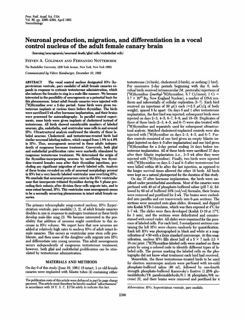
FIG. 1. HVc is the magnocellular region ventral to the collapsedlateral ventricle. The collapsed ventricle is delimited by the two ap-posed ventricular zones, revealed-here as a thin brow of small, darklystaining cells. Notice the presence of labeled cells inside HVc. In thiscase the labeled cells are all either endothelial or glia. The ventricularzone overlying HVc also shows labeled cells. This section-was takenfrombird 13 (Table 1), which had been sacrificed two days afterthe last[5H~thymidine injection. HP, hippocampus. (Bar = 200 gm.)
4 .w4
..; 4,4.,
LA k_,--.
Is~*4 A!
mak
Or
1rtt
Ws
4 a.J!8'
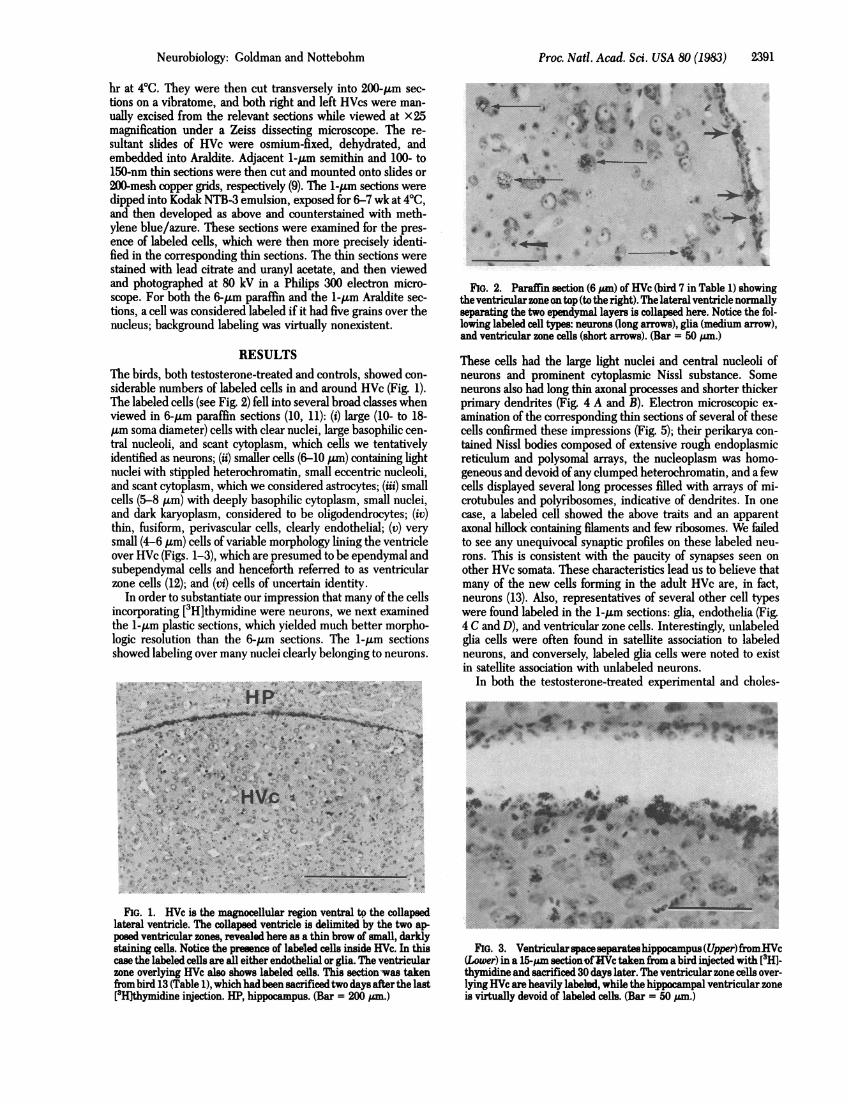
FIG. 2. Paraffin section (6 bum) of HVc (bird 7 in Table 1) showingthe ventricular zone on top (to the right). The lateral ventricle normallyseparating the two ependymal layers is collapsed here. Notice the fol-lowing labeled cell types: neurons (long arrows), glia (medium arrow),and ventricular zone cells (short arrows). (Bar = 50 gm.)
These cells had the large light nuclei and central nucleoli ofneurons and prominent cytoplasmic Nissl substance. Someneurons also had long thin axonal processes and shorter thickerprimary dendrites (Fig. 4 A and B). Electron microscopic ex-amination of the corresponding thin sections of several of thesecells confirmed these impressions (Fig. 5); their perikarya con-tained Nissl bodies composed of extensive rough endoplasmicreticulum and polysomal arrays, the nucleoplasm was homo-geneous and devoid of any clumped heterochromatin, and a fewcells displayed several long processes filled with arrays of mi-crotubules and polyribosomes, indicative of dendrites. In onecase, a labeled cell showed the above traits and an apparentaxonal hillock containing filaments and few ribosomes. We failedto see any unequivocal synaptic profiles on these labeled neu-rons. This is consistent with the paucity of synapses seen onother HVc somata. These characteristics lead us to believe thatmany of the new cells forming in the adult HVc are, in fact,neurons (13). Also, representatives of several other cell typeswere found labeled in the i-Mim sections: glia, endothelia (Fig.4 C and D), and ventricular zone cells. Interestingly, unlabeledglia cells were often found in satellite association to labeledneurons, and conversely, labeled glia cells were noted to existin satellite association with unlabeled neurons.
In both the testosterone-treated experimental and choles-
;E
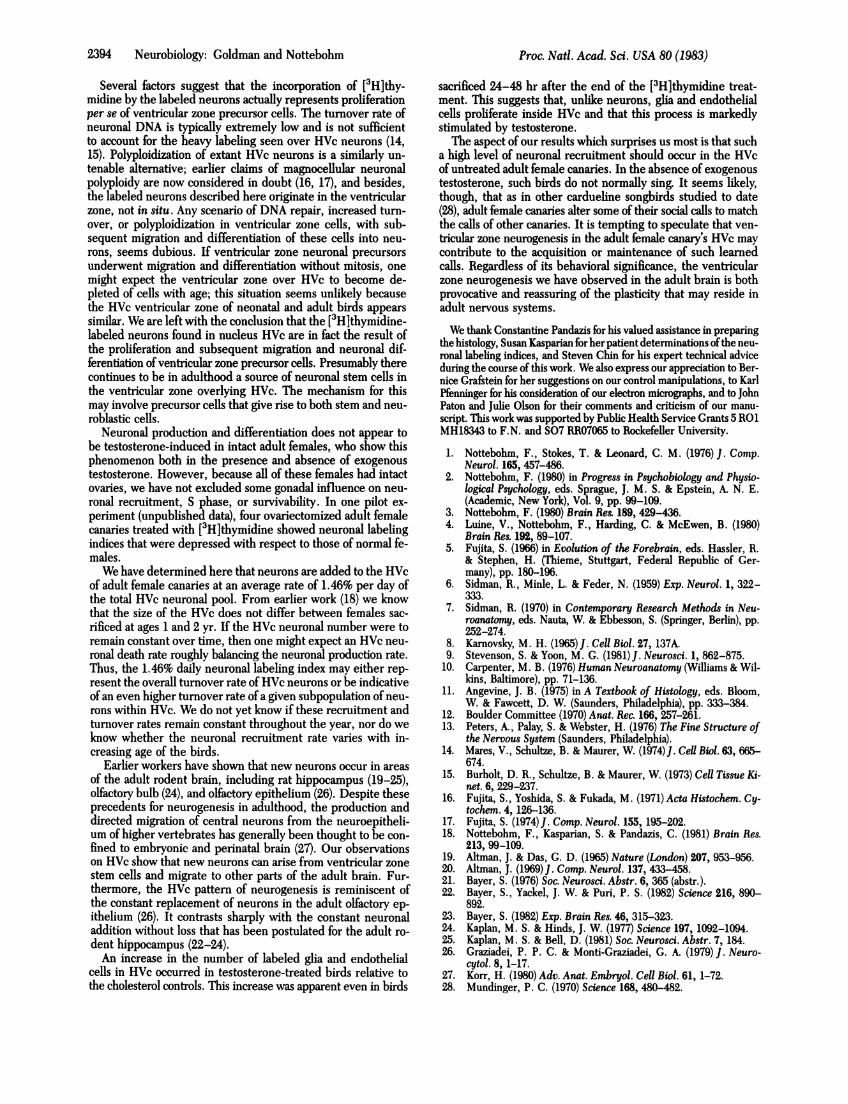
FIG. 3. Ventricularspaceseparateshippocampus(Upper)fromiHVc(Lower) in a 15-am sectionofHVctaken from a bird injected with [3H1-thymidine and sacrificed 30 days later. The ventricular zone cells over-lying HVc are-heavily labeled, while the hippecampal ventricular zoneis virtually devoid of labeled cells. (Bar = 50 pm.)
Neurobiology: Goldman and Nottebohm
P.
2392 Neurobiology: Goldman and Nottebohm
0*efi
9t A
FIG. 4. (A and B) Two neurons labeled with [3H]thymidine in 1-jim Araldite sections counterstained with methylene blue. Araldite is harderthan paraffin; as a result, fewer silver grains are exposed during autoradiography, yielding a seemingly lighter degree of labeling. (C) Two labeledastrocytes displaying circumferential nucleoli, one of which shows a probable astrocytic end-foot. (D) Several labeled endothelial cells, whose thin,fusiform morphology and pericapillary placement is obvious. These sections were processed for ultrastructural analysis; they were taken from theHVc from a bird sacrificed 32 days after its last [3H]thymidine injection. (Bar = 50 ,um.)
terol-treated control birds, sacrificed 37 days after onset of ste-roid treatment, 1.8-4.0% of the neurons in HVc were labeledby the 2-day [3H]thymidine injection protocol (see Table 1). This-represents a neuronal production rate of at least 0.9-2.0%.perday in HVc. A labeling index of 1.5% per day was noted in theone bird in which thymidine was given before the testosteronetreatment, well within the range observed when steroid treat-
ment preceded the [3H]thymidine injections. However, the twobirds that had been sacrificed shortly (24 and 48 hr, respec-tively) after their last [3H]thymidine injections displayed no la-beling of neurons in HVc itself. In these two cases, however,the ventricular zone over HVc was blanketed with labeled cellnuclei of various morphologies, whereas within HVc only en-dothelial and glia cells were labeled (Fig. 1).
Table 1. Labeling indices after various hormone and thymidine regimensTime of sacrifice,t
days aftertreatment
Treatment* LastSteroid Time, Silastic [3H]dTd Neurons, Number of labeled cells per 100 neurons§
Bird type days implant injection totalno. Neurons Glia Endothelia Ambiguous1 C 4-5 36 31 1,082 3.70 1.02 1.66 1.202 C 6-7 36 29 1,610 2.05 1.42 0.99 0.563 N 4-5 36 31 1,862 1.34 1.02 0.32 0.3241 T -14-1a. 19 33 1,159 3.02 2.33 1.81 2.425 T 0-1 36 35 1,596 3.01 7.46 18.86 1.826 T 2-3 36 33 1,122 3.47 3.30 6.86 2.237 T 4-5 36 31 1,635 2.20 9.79 21.28 3.798 T 4-5 40 35 830 1.92 11.80 26.63 2.059 T 6-7 36 29 1,399 2.93 5.36 9.01 1.8610 T 8-9 36 27 1,281 3.82 8.74 6.01 3.6711 T 17-18 36 18 2,369 4.01 1.60 0.80 1.3512 T 4-5 6 1 2,085 0.10 3.98 2.69 0.8613 T 6-7 9 2 1,093 0.20 5.85 18.57 3.48
* Silastics of the same size had been filled with cholesterol (C), testosterone (T), or nothing (N). Time spec-ifies the days after Silastic implant when [3H]thymidine was given.tNumber of days elapsed since the day of Silastic implant or since the last 13H]thymidine injection.tCounts of all neurons in the five left HVc sections analyzed for each bird.§ This conversion was done so that the number of labeled cells of each type could be related to a commondenominator. Only in the ease of labeled neurons does this constitute a proper labeling index.
sNotice that bird 4 received its [3H]thymidine starting 14 days before the Silastic implant.
Proc. Natl. Acad. Sci. USA 80 (1983)
Proc. Natl. Acad. Sci. USA 80 (1983) 2393
FIG. 5. Electron micrograph of a thin (100-150 pm) section adjacent to that shown as a 1-,um section in Fig. 4B. This cell has a large nucleus(N) with dispersed chromatin, prominent Golgi apparatus, an abundant cytoplasm with extensive rough endoplasmic reticulum, axonal hillock (A),and in this section a primary dendrite (D) cut transversely, whose connection to the cell body is indicated by the arrow. All of these characteristicsare indicative of this cell's neuronal identity. This cell also contains several multivesicular bodies (MV), which are common in HVc neurons, andwhat appears to be an invaginated cilium, perhaps related to this cell's ventricular zone origin. (Bar = 4.4 am.)
Labeled neurons also occurred in neostriatum adjacent toHVc. None were found in nucleus robustus archistriatalis, aforebrain vocal control nucleus to which HVc projects (1). Labelin ventricular zone cells, as found over HVc, was restricted toa few parts of the brain (Fig. 3). The occurrence of labeled cellsin non-HVc parts of the brain will be reported elsewhere.The total number of labeled glia and endothelial cells in HVc
was larger in the testosterone-treated birds than in the cho-lesterol controls (Table 2). This effect retains its sign and mag-
Table 2. Effect of testosterone on labeling indices of neurons,glia, and endothelial cells (from Table 1)
Number of labeled cells per 100 neuronsTreatment Birds Neurons* Glia* Endothelia*
P>0.20 P< 0.01 P<0.05* [3H]Thymidine-labeled cells; values shown correspond to the mean ±SD. P values were obtained by using a two-tailed t test.
tBirds with cholesterol-filled and empty Silastic tubes, pooled;N = 3.t [3H]Thymidine injections given sometime during the 10 days aftertestosterone implant, with a survival of 27-35 days after the last in-jection; N = 6.
nitude when the data are expressed either as total counts or interms ofnumber of labeled glia or endothelial cells per 100 neu-rons. It was noticeable in birds sacrificed 24-48 hr or 17-35days after the end of the [3H]thymidine treatment (Table 1).
DISCUSSIONNeurons in nucleus HVc of adult female canaries incorporatesystemically administered [3H]thymidine. We have attemptedto determine the origin of labeled HVc neurons, by noting wherethis label first appears. As noted above, two of the birds in thisstudy were sacrificed within 48 hr after their [3H]thymidineinjections to reduce the significant migration of any cells thathad incorporated label. Whereas long (3-5 wk) survival timesafter [3H]thymidine administration resulted in many labeledneurons and glia cells in HVc, the shorter survival times yieldedHVcs that were devoid of labeled neurons. Instead, in theseearly-sacrificed birds, the HVc manifested a heavily labeledventricular zone, along with many labeled endothelial and gliacells. Therefore, we conclude that a population of ventricularzone precursor cells normally proliferates in the adult femalecanary brain, with subsequent migration of these cells into HVcand their differentiation therein into neurons and perhaps ad-ditional glia.
Neurobiology: Goldman and Nottebohm
2394 Neurobiology: Goldman and Nottebohm
Several factors suggest that the incorporation of [3H]thy-midine by the labeled neurons actually represents proliferationper se of ventricular zone precursor cells. The turnover rate ofneuronal DNA is typically extremely low and is not sufficientto account for the heavy labeling seen over HVc neurons (14,15). Polyploidization of extant HVc neurons is a similarly un-tenable alternative; earlier claims of magnocellular neuronalpolyploidy are now considered in doubt (16, 17), and besides,the labeled neurons described here originate in the ventricularzone, not in situ. Any scenario of DNA repair, increased turn-over, or polyploidization in ventricular zone cells, with sub-sequent migration and differentiation of these cells into neu-rons, seems dubious. If ventricular zone neuronal precursorsunderwent migration and differentiation without mitosis, onemight expect the ventricular zone over HVc to become de-pleted of cells with age; this situation seems unlikely becausethe HVc ventricular zone of neonatal and adult birds appearssimilar. We are left with the conclusion that the [3Hlthymidine-labeled neurons found in nucleus HVc are in fact the result ofthe proliferation and subsequent migration and neuronal dif-ferentiation of ventricular zone precursor cells. Presumably therecontinues to be in adulthood a source of neuronal stem cells inthe ventricular zone overlying HVc. The mechanism for thismay involve precursor cells that give rise to both stem and neu-roblastic cells.
Neuronal production and differentiation does not appear tobe testosterone-induced in intact adult females, who show thisphenomenon both in the presence and absence of exogenoustestosterone. However, because all of these females had intactovaries, we have not excluded some gonadal influence on neu-ronal recruitment, S phase, or survivability. In one pilot ex-periment (unpublished data), four ovariectomized adult femalecanaries treated with [3H]thymidine showed neuronal labelingindices that were depressed with respect to those of normal fe-males.We have determined here that neurons are added to the HVc
of adult female canaries at an average rate of 1.46% per day ofthe total HVc neuronal pool. From earlier work (18) we knowthat the size of the HVc does not differ between females sac-rificed at ages 1 and 2 yr. If the HVc neuronal number were toremain constant over time, then one might expect an HVc neu-ronal death rate roughly balancing the neuronal production rate.Thus, the 1.46% daily neuronal labeling index may either rep-resent the overall turnover rate of HVc neurons or be indicativeof an even higher turnover rate of a given subpopulation of neu-rons within HVc. We do not yet know if these recruitment andturnover rates remain constant throughout the year, nor do weknow whether the neuronal recruitment rate varies with in-creasing age of the birds.
Earlier workers have shown that new neurons occur in areasof the adult rodent brain, including rat hippocampus (19-25),olfactory bulb (24), and olfactory epithelium (26). Despite theseprecedents for neurogenesis in adulthood, the production anddirected migration of central neurons from the neuroepitheli-um of higher vertebrates has generally been thought to be con-fined to embryonic and perinatal brain (27). Our observationson HVc show that new neurons can arise from ventricular zonestem cells and migrate to other parts of the adult brain. Fur-thermore, the HVc pattern of neurogenesis is reminiscent ofthe constant replacement of neurons in the adult olfactory ep-ithelium (26). It contrasts sharply with the constant neuronaladdition without loss that has been postulated for the adult ro-dent hippocampus (22-24).An increase in the number of labeled glia and endothelial
cells in HVc occurred in testosterone-treated birds relative tothe cholesterol controls. This increase was apparent even in birds
sacrificed 24-48 hr after the end of the [3H]thymidine treat-ment. This suggests that, unlike neurons, glia and endothelialcells proliferate inside HVc and that this process is markedlystimulated by testosterone.The aspect of our results which surprises us most is that such
a high level of neuronal recruitment should occur in the HVcof untreated adult female canaries. In the absence of exogenoustestosterone, such birds do not normally sing. It seems likely,though, that as in other cardueline songbirds studied to date(28), adult female canaries alter some of their social calls to matchthe calls of other canaries. It is tempting to speculate that ven-tricular zone neurogenesis in the adult female canary's HVc maycontribute to the acquisition or maintenance of such learnedcalls. Regardless of its behavioral significance, the ventricularzone neurogenesis we have observed in the adult brain is bothprovocative and reassuring of the plasticity that may reside inadult nervous systems.
We thank Constantine Pandazis for his valued assistance in preparingthe histology, Susan Kasparian for her patient determinations of the neu-ronal labeling indices, and Steven Chin for his expert technical adviceduring the course of this work. We also express our appreciation to Ber-nice Grafstein for her suggestions on our control manipulations, to KarlPfenninger for his consideration of our electron micrographs, and to JohnPaton and Julie Olson for their comments and criticism of our manu-script. This work was supported by Public Health Service Grants 5 RO1MH18343 to F.N. and S07 RR07065 to Rockefeller University.
1. Nottebohm, F., Stokes, T. & Leonard, C. M. (1976) J. Comp.Neurol. 165, 457-486.
2. Nottebohm, F. (1980) in Progress in Psychobiology and Physio-logical Psychology, eds. Sprague, J. M. S. & Epstein, A. N. E.(Academic, New York), Vol. 9, pp. 99-109.
3. Nottebohm, F. (1980) Brain Res. 189, 429-436.4. Luine, V., Nottebohm, F., Harding, C. & McEwen, B. (1980)
Brain Res. 192, 89-107.5. Fujita, S. (1966) in Evolution of the Forebrain, eds. Hassler, R.
& Stephen, H. (Thieme, Stuttgart, Federal Republic of Ger-many), pp. 180-196.
6. Sidman, R., Minle, L. & Feder, N. (1959) Exp. Neurol. 1, 322-333.
7. Sidman, R. (1970) in Contemporary Research Methods in Neu-roanatomy, eds. Nauta, W. & Ebbesson, S. (Springer, Berlin), pp.252-274.
8. Karnovsky, M. H. (1965)J. Cell Biol. 27, 137A.9. Stevenson, S. & Yoon, M. G. (1981)J. Neurosci. 1, 862-875.
10. Carpenter, M. B. (1976) Human Neuroanatomy (Williams & Wil-kins, Baltimore), pp. 71-136.
11. Angevine, J. B. (1975) in A Textbook of Histology, eds. Bloom,W. & Fawcett, D. W. (Saunders, Philadelphia), pp. 333-384.
12. Boulder Committee (1970) Anat. Rec. 166, 257-261.13. Peters, A., Palay, S. & Webster, H. (1976) The Fine Structure of
the Nervous System (Saunders, Philadelphia).14. Mares, V., Schultze, B. & Maurer, W. (1974)1. Cell Biol. 63, 665-
674.15. Burholt, D. R., Schultze, B. & Maurer, W. (1973) Cell Tissue Ki-
net. 6, 229-237.16. Fujita, S., Yoshida, S. & Fukada, M. (1971) Acta Histochem. Cy-
tochem. 4, 126-136.17. Fujita, S. (1974) J. Comp. Neurol. 155, 195-202.18. Nottebohm, F., Kasparian, S. & Pandazis, C. (1981) Brain Res.
213, 99-109.19. Altman, J. & Das, G. D. (1965) Nature (London) 207, 953-956.20. Altman, J. (1969)J. Comp. Neurol. 137, 433-458.21. Bayer, S. (1976) Soc. Neurosci. Abstr. 6, 365 (abstr.).22. Bayer, S., Yackel, J. W. & Puri, P. S. (1982) Science 216, 890-
892.23. Bayer, S. (1982) Exp. Brain Res. 46, 315-323.24. Kaplan, M. S. & Hinds, J. W. (1977) Science 197, 1092-1094.25. Kaplan, M. S. & Bell, D. (1981) Soc. Neurosci. Abstr. 7, 184.26. Graziadei, P. P. C. & Monti-Graziadei, G. A. (1979) J. Neuro-
cytol. 8, 1-17.27. Korr, H. (1980) Adv. Anat. Embryol. Cell Biol. 61, 1-72.28. Mundinger, P. C. (1970) Science 168, 480-482.