ORIGINAL PAPER Georg NorthoffNeuropsychiatry An old discipline in a new gestalt bridging biological psychiatry, neuropsychology, and cognitive neurology Received: 24 July 2007 / Accepted: 30 October 2007 / Published online: 23 February 2008 j Abstract The recent develop ments of psych iatrygetting bett er insi ght into the bi ol og ical basis of psychiatric disorders questions the old division be- tween psychiatry and neurology. The present paper focus on the concept of neuropsychiatry, its historical antecedents and closely ass oci ated dis ciplines like biological psychiatry, behavioral neurology and neu- rops yc ho logy . A spec ia l emph asis is put on the question of function and localization; the suggestions are mad e tha t the concept of neuronal integration may bridge the often discussed gap between locali- zation and holism in the relation between function and brain regions. Examples of different mechanisms of neuronal integration are discussed and applied to specifi c neurop sychia tric disorders. It is conclu ded that the concept of neuronal integration may offer an appropriate conceptual tool to establish the concept of neuropsychiatry in a new and meaningful gestalt at the inter face between biologica l psychi atry, neuropy- cholog y and cognit ive neurol ogy. Introduction: the concept of ‘‘Neuropsychiatry’’ A cli nic ian taking care of a pat ient suf fer ing from Parki nson’s di sea se does not ha ve to treat motor symptoms alone (tremor, akinesia, and rigor), which bel ong to the fiel d of neurol ogy , but bey ond he als o has do diagnose and treat affective (e.g., depressions) or cognitive symptoms ( e.g., bradyphrenia, retardation ofthi nki ng) , whi ch bel ong to the fiel d of psy chi atry . However, it is quite unli kel y that he wi ll meet al l requirements of both fields because either he is a neu- rologist or a psychiatrist, and he therefore has learned different ways of thinking and different methods. This reflects the paradox situation in contemporary rela- tionship between neurology and psychiatry: On one hand, neurology and psychiatry are regarded as two different disciplines with different content concerning met hod ology, dis eas es, dia gno stics and the rapy. On the other hand, the boundaries of separation between both disciplines melt away by applying neurological meth- odology and diagnostics in psychiatric diseases (CT, Spect,andPET)aswellasbytheinterestofneurologyin complex mental funct ions and psych iatric symptoms in neurol ogi cal dis eases. Par tic ula rly in the Ang lo- American region this resulted in the foundation of the disciplin e of ‘‘neurops ych iatry’’: ‘‘Although hal f a century ago neurology and psychiatry seemed to be diverging from a common purpose and sanding as two stools apart, these recent advances have not only seen the stools bridged by a plank, but the whole structure has come gradually to resemble a bench, which for some seems quite comfor table. The las t dec ade has seen not only an exponential growth of knowledge in the field of the neurosci ences, but al so a resolution of some interdisciplinary rivalry, and laid the foundations ofneuropsychiatry for at least the rest of this century.’’ [117] (Tables 1 , 2) Neurology, psychiatry and neuropsychiatry j Historic development of the separation of neurology and psychiatry Neurol ogy as an ind epe nde nt dis ciplin e has dev el- oped at the beginnin g of the nineteent h century when Parkinson’s disease and Multiple sclerosis were de- fined as neural diseases [97] (see also Ref. [ 4]). Fol- lowing advances in ana tomy and pathol ogy in the G. Northoff, MD, PhD (&) Department of PsychiatryOtto-von-Guericke University of Magdeburg Leipziger Strasse 44 39120 Magdeburg, GermanyTel.: +49-391/6714234 Fax: +49-391/6715223 E-Mail: [email protected]Eur Arch Psychiatry Clin Neurosci (2008) DOI 10.1007/s00406-007-0783-6 EA PCN 783
Transcript
7/27/2019 Neuropsychiatry– an old discipline in a new gestalt bridging biological psychiatry, neuropsychology, and cognitive …
An old discipline in a new gestalt bridging biological psychiatry,
neuropsychology, and cognitive neurology
Received: 24 July 2007 / Accepted: 30 October 2007 / Published online: 23 February 2008
j Abstract The recent developments of psychiatry getting better insight into the biological basis of
psychiatric disorders questions the old division be-tween psychiatry and neurology. The present paperfocus on the concept of neuropsychiatry, its historicalantecedents and closely associated disciplines likebiological psychiatry, behavioral neurology and neu-ropsychology. A special emphasis is put on thequestion of function and localization; the suggestionsare made that the concept of neuronal integrationmay bridge the often discussed gap between locali-zation and holism in the relation between functionand brain regions. Examples of different mechanismsof neuronal integration are discussed and applied tospecific neuropsychiatric disorders. It is concluded
that the concept of neuronal integration may offer anappropriate conceptual tool to establish the conceptof neuropsychiatry in a new and meaningful gestalt atthe interface between biological psychiatry, neuropy-chology and cognitive neurology.
Introduction: the concept of ‘‘Neuropsychiatry’’
A clinician taking care of a patient suffering fromParkinson’s disease does not have to treat motorsymptoms alone (tremor, akinesia, and rigor), which
belong to the field of neurology, but beyond he also hasdo diagnose and treat affective (e.g., depressions) orcognitive symptoms (e.g., bradyphrenia, retardation of thinking), which belong to the field of psychiatry.
However, it is quite unlikely that he will meet allrequirements of both fields because either he is a neu-
rologist or a psychiatrist, and he therefore has learneddifferent ways of thinking and different methods. Thisreflects the paradox situation in contemporary rela-tionship between neurology and psychiatry: On onehand, neurology and psychiatry are regarded as twodifferent disciplines with different content concerningmethodology, diseases, diagnostics and therapy. On theother hand, the boundaries of separation between bothdisciplines melt away by applying neurological meth-odology and diagnostics in psychiatric diseases (CT,Spect,andPET)aswellasbytheinterestofneurologyincomplex mental functions and psychiatric symptomsin neurological diseases. Particularly in the Anglo-
American region this resulted in the foundation of thediscipline of ‘‘neuropsychiatry’’: ‘‘Although half acentury ago neurology and psychiatry seemed to bediverging from a common purpose and sanding as twostools apart, these recent advances have not only seenthe stools bridged by a plank, but the whole structurehas come gradually to resemble a bench, which forsome seems quite comfortable. Thelast decade has seennot only an exponential growth of knowledge in thefield of the neurosciences, but also a resolution of someinterdisciplinary rivalry, and laid the foundations of neuropsychiatry for at least the rest of this century.’’[117] (Tables 1, 2)
Neurology, psychiatry and neuropsychiatry
j Historic development of the separationof neurology and psychiatry
Neurology as an independent discipline has devel-oped at the beginning of the nineteenth century whenParkinson’s disease and Multiple sclerosis were de-fined as neural diseases [97] (see also Ref. [4]). Fol-lowing advances in anatomy and pathology in the
G. Northoff, MD, PhD (&)Department of Psychiatry Otto-von-Guericke University of MagdeburgLeipziger Strasse 4439120 Magdeburg, Germany Tel.: +49-391/6714234Fax: +49-391/6715223E-Mail: [email protected]
Eur Arch Psychiatry Clin Neurosci (2008) DOI 10.1007/s00406-007-0783-6
E AP C N
7 8 3
7/27/2019 Neuropsychiatry– an old discipline in a new gestalt bridging biological psychiatry, neuropsychology, and cognitive …
nineteenth century correlations between the clinicalappearance and the pathological–anatomical sub-strate became possible to an increasing degree; thishas been successful in numerous neurological dis-eases and symptoms (e.g., aphasia) and consequently established the field of neurology as an independentdiscipline of medicine. On the contrary clinical–pathological correlations in psychiatry did not show
significant success, which strengthened the separationof medicine/neurology on the one hand and psychi-atry on the other hand. The previously apparentconnection between psychiatry and philosophy/humanities [97], the letter dealing with mental statesas implicated in psychiatric diseases as mental dis-orders, additionally supported the separation betweenneurology and psychiatry.
The application of scientific principles in psychiatry has later been established by Griesinger, Kahlbaum,Kraepelin and Maudsely, who created a consistentnosology and who regarded psychiatric diseases asdiseases of the brain. Nevertheless, correlations be-
tween the clinical appearance and the pathological–anatomical substrate still remained unsuccessful.Whereas in the field of neurology, such correlationsshowed rising success, since neurological diseasescould be localized anatomically–morphologically in thebrain implying that they were regarded as ‘‘structural’’diseases. The failure of structural localization of psy-chiatric diseases, in contrast, resulted in their accep-tance as ‘‘functional’’ diseases with the meaning‘‘psychological’’ and thus mental origin as distin-guished from ‘‘structural’’ describing the ‘‘organic’’andthus neuronal origin [97]. This opposition of ‘‘structure
versus function’’ and ‘‘localization versus non-locali-zation’’ accounted for the separation of neurology andpsychiatry in a decisive manner; the neurologist dealtwith structural diseases of the brain, the psychiatristfocused on functional diseases of the mind. This lead tothe development of neurology and psychiatry as inde-pendent disciplines in the Anglo-American regionwhereas in the German-speaking regions both disci-
plines have been kept together for a long time in thecommon discipline of ‘‘Nervenheilkunde’’.
j Changes in the relationship between neurologyand psychiatry
Different developments in the last decades questionthe comparison of neurology and psychiatry. Thesedevelopments mounted from both the fields: neurol-ogy and psychiatry; in the following they are shortly described. By the discovery of variability of thebrain’s neuronal structures, i.e., the plasticity, and theintroduction of new imaging techniques (MRI, PET;
see below) neurology changed its appearance: Solely static-structural observations have been replaced by adynamic–functional anatomy [63]. This ‘‘functionalneuroanatomy’’ not only examines different staticstructures but tries to point out the connections be-tween these structures, to show plasticity of connec-tions and structures as well as the influence of function on structure [117]. The boundaries betweenanatomy and physiology melt away because of this‘‘functional neuroanatomy’’, where the contrast of anatomical-static structure and physiological-dy-namic function is dismantled and rather regarded as arelationship of mutual complementation: ‘‘The
boundaries between anatomy and physiology,between form and function, break down at an ultra-structural level. Anatomy is not static. Pharmaco-therapy may alter structure as well as function.’’ [63].The new imaging techniques on the one hand allow toimage anatomic structures more exactly and moredetailed. On the other hand they make it possible todraw a connection between functional changes andanatomic structure—they are a kind of ‘‘window forthe brain function’’ [11]. These developments make itpossible, to look at psychiatric diseases, too. Thephysiologic-functional examination, which is still
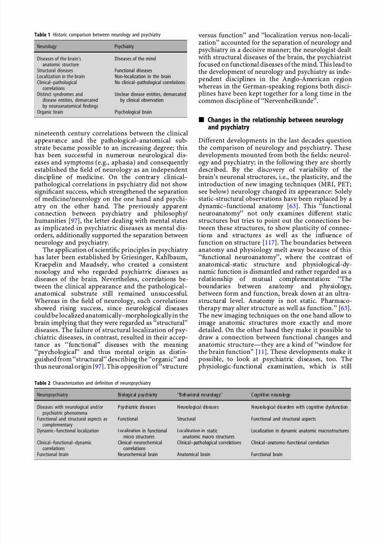
Table 2 Characterization and definition of neuropsychiatry
getting off the ground, could shed new light on thepathophysiology of psychiatric diseases.
The development of psychopharmacology sup-ported the hypothesis already set by Griesinger thatmental diseases are diseases of the brain. The ques-tion of the mechanisms of the pharmacological effectsin the brain by psychopharmacological drugs resultedin an intensive exploration of neurotransmitters,
synapses and receptors in the brain, which itself re-sulted in a better and extended understanding of physiological brain functions. Out of this the disci-pline of ‘‘biological psychiatry’’ emerged, which triesto correlate psychopathological phenomena withfunctional and structural changes in the brain [118].Initially ‘‘Biological psychiatry’’ mainly dealt withsynapses, neurotransmitters and receptors; by exam-ination of the mechanisms of effectiveness one tries togain insight into the structural and functional eventsin the brain in psychiatric symptoms and diseases.
Not only in psychiatry changes have happened, butalso in neurosciences and in neurology. Over the last
10–20 years, neuroscience shifted its interested moreand more on affective and cognitive functions likeworking memory [57], emotional perception and
judgment [32, 74, 88], attention [84, 85], and evenoriginally philosophical notions like consciousness[44] and self [124]. This has lead to the developmentof novel disciplines like cognitive neuroscience andaffective neuroscience [81] which, because of theirinterest in more complex affective and cognitivefunctions, focus on those neural processes that allow to integrate and coordinate neural activity acrossdifferent regions implicating complex neural net-works in predominantly functional terms. Such
coordination and organization of neural activity may for instance be achieved by plastic changes allowingneural activity to adapt to the respective challengesand tasks required. Another such mechanism recently discussed is the synchronization of neural activity across different brain regions by coordination of theirfrequency as for instance by gamma-band oscillationsthat are currently discussed to be possibly crucial inconstituting consciousness [44, 113]. Based on thesedevelopments, neurology showed a rising interest incomplex phenomena and behavior which could notclearly be traced back to reflexes [10]. Here theinsufficiency of classic neurology became clear[52]—because of this mainly in America the disci-pline of ‘‘behavioral neurology’’ developed. It dealswith complex phenomena like aphasia and amnesiaand tries to localize these phenomena neurologically and neuroanatomically, respectively [18]. The oldmethod of clinical–pathological correlations (seeabove) is here applied in a new field of interest, thefield of higher-order cognitive phenomena and com-plex behavior,—in doing so the focus is still onstructure and the aim of localization [63]. This,however, is changing and the focus on ‘‘anatomicstatic macrostructures’’ becomes more and more
complemented by considering ‘‘functional dynamicmacrostructures’’ which in turn allows to account forhigher-order cognitive phenomena like attention,theory of mind, etc. (see for instance Refs. [84, 85]).This is the focus of a recently developed specialbranch of neurology, Cognitive Neurology, whichtherefore may be regarded as paradigmatic subset of aneuropsychiatry in a current gestalt. Cognitive Neu-
rology investigates cognitive dysfunctions for instanceattention or theory of mind in neurological disorderslike Parkinson’s disorder and is therefore able to draw interesting parallels with psychiatric disorders likecatatonia [69].
The discipline of ‘‘neuropsychology’’, constantly developing in the last years, too, examines the ‘‘cor-relations between brain function and psychologicalprocesses’’ [91] closely following classical neuropa-thology [41, 91]. The separation between neuropsy-chology on one hand and ‘‘behavioral neurology’’ onthe other hand is mainly in the USA pursued. In‘‘behavioral neurology’’ mainly aphasia and amnesia
are investigated. The main topic of neuropsychology is to provide objective methods for the examination of psychological function, which then can be applied onthe clinical problems as dealt with in ‘‘behavioralneurology’’ and set into relationship to brain struc-ture and function [3]. The ‘‘behavioral neurologist’’primarily looks at the structures of the brain andsecondarily their relation to complex psychologicalfunctions. The neuropsychologist, on the contrary,regards primarily the psychological functions andsecondarily their relation to the structures of thebrain [18]. The neuropsychologist captures affectiveand cognitive alterations in the Parkinson’s disease
using standardized tests in an objective manner—thecorrelation of these results with structures und func-tions of the brain is left to the neurologist to a largeextent. The quantitative, operationalized measure-ment of psychological functions, the so-called psy-chometrics [41], is increasingly used in psychiatry,too, where psychopathological phenomena are cap-tured quantitatively and objectively by operational-ization of the psychopathological symptoms. Thus thepsychopathology which previously often has beencalled non-scientific becomes affiliated with an ‘‘em-piric–scientific methodology’’ [64] thereby gainingscientific status.
The above described developments of differentdisciplines in the border area between neurology andpsychiatry aim to bridge the contrasts between bothfields from different directions (biological psychiatry,behavioral neurology, neuropsychology, and cognitiveneurology) [63]. All these disciplines would explainthe above described example of Parkinson’s diseasedifferently: The biological psychiatrist localizes theaffective and cognitive symptoms in the microstruc-ture of the transmitters, synapses and receptors; the‘‘behavioral neurologist’’ localizes the same symptomsin the macro structures of the brain; the neuropsy-
7/27/2019 Neuropsychiatry– an old discipline in a new gestalt bridging biological psychiatry, neuropsychology, and cognitive …
chologist objectivizes and standardizes psychologicalfunctions; and the cognitive neurologist would pointout the anatomo-functional networks of the cognitivesymptoms. All of them (with probably the exceptionof the cognitive neurologist) place motor/neurologicaland psychiatric symptoms next to each other andexplain them more or less independently from eachother—the internal connection of motor and psy-
chological alterations in the Parkinson’s disease asexperienced by the patient gets lost. Because of this inthe following we want to show that a neuropsychiatry could be able to demonstrate these internal connec-tions between psyche and motor activity.
j Neuropsychiatry: characterization and definition
The discipline of neuropsychiatry tries to bridge thegap between neurology on the one hand and psychi-atry on the other hand—in doing so psychologicalfunctions and neurological structures are not only tobe observed in the same time but are to be connected
internally coherently: ‘‘This new orientation of whichJellife spoke, and of which he himself was a notableexemplar, did not involve merely combining neuro-logical and psychiatric knowledge (as every neurolo-gist and psychiatrist does to some extent), butconjoining them seeing them as inseparable, seeinghow psychiatric phenomena might emerge from thephysiological, or how, conversely, they might betransformed into it—...’’ [63]. The static-structural,strictly localizing observation of the classic neurology and the ‘‘behavioral neurology’’ will be contrasted by a dynamic-functional approach of the neuropsychia-try. Psychological and motor alterations in the Par-
kinson’s disease are no longer explained separately and independently but are regarded as two expres-sions of a uniform dynamic-functional structure—thealteration of this structure, and not of the twodifferent symptom complexes as two differentphenomena, have to be explained. Thus the ‘‘neuro-logilization’’ of psychiatric functions [63] is impossi-ble—behavior can not only be observed withneurological methodology, but has to be assessed by integration of neurological and psychiatric knowledge[63].
Biological psychiatry can not be identified with andreduced to neuropsychology, because it does not
exclusively deal with complex macro phenomena of behavior and psychological functions, but with microphenomena of the synapses, transmitters and recep-tors—psychological functions mainly remain beyondobservation [13]. While biological psychiatry usesfunctional neuroanatomy, restricting it to micro-structures (synapses), behavioral neurology focuseson macrostructures of the brain though consideringthem solely in a static, anatomic-structural sense.Neuropsychiatry should aim to combine the dynamic-functional approach with the observation of macro-structures in the phenomena of behavior and higher-
order psychological functions. In other terms, neu-ropsychiatry in this sense would take a middle posi-tion between a static-structural, localizing neurology on the one hand and a dynamic-functional, holistic/anti-localizing psychiatry on the other. As a bridgebetween neuroanatomy and psychopathology neuro-psychiatry has to examine functional and dynamicprocesses being positioned in between strictly local-
izable neurological functions and strictly holisticpsychological processes [79]. This middle level of neuropsychiatry undermines the traditional opposi-tion of structure versus function and localizationversus non-localization.
Function, localization and neuronal integrationin neuropsychiatry
j Function and localization
I already mentioned the contrast of structure versus
function and characterized the latter as crucial for thediscipline of neuropsychiatry. What does the term‘‘function’’ mean? ‘‘Functional’’ can be understood intwo senses [99]: Firstly, ‘‘functional’’ means just non-organic and thus psychological, as it has beenunderstood in psychiatry. Secondly, ‘‘functional’’ canbe understood in a physiological sense in contrast toanatomic—here ‘‘functional’’ describes dynamic,plastic and variable physiological processes, being incontrast to the static, anatomic structure; this secondmeaning of the word ‘‘functional’’ has been used inthe characterization of functional neuroanatomy (seeabove). I here want to follow the second and original
meaning of the word ‘‘functional’’ [118] and regardthe physiologic-functional description as middle levelbetween static-structural pathology and dynamic-functional psychiatry as the specific neuropsychiatriclevel [79] which is now more and more taken intoaccount in empirical and explanatory regard in bothcurrent neurology and psychiatry when consideringaffective and cognitive dysfunction in their respectivedisorders [69, 109, 113, 120, 123, 126]. Physiologicfunctions may not correlate with a specific staticanatomic structure any more, but they develop in so-called functional systems [51, 52]. These systemsproduce distinct functions by a dynamic constellation
of changes between different parts of the brainreflecting what may be called neuronal integration(see below). As demonstrated by current investiga-tions of neuroplasticity in both healthy and psychi-atric subjects (see Ref. [125]), they are plastic, show asystemic (and not a concrete) structure, and operateby dynamic auto regulation [51]. The realization of function depends then on dynamic systems that in-clude different brain regions which Luria character-ized as ‘‘functional systems’’: ‘‘According to this view a function is, in fact, a functional system (...) directedtowards the performance of a particular biological
7/27/2019 Neuropsychiatry– an old discipline in a new gestalt bridging biological psychiatry, neuropsychology, and cognitive …
task and consisted of a group of interconnected actsthat produce the corresponding biological effect. Themost significant feature of a functional system is that,as a rule, it is based on a complex dynamic ‘‘con-stellation’’ of connections, situated at different levelsof the nervous system, that in the performance of theadaptive task, may be changed with the task itself may be unchanged.’’ [51].
The brain here is regarded as a network consistingof different, overlapping functional systems with aninternal dynamic, a so-called ‘‘neurodynamic’’ [63],which have already been demonstrated in the form of ‘‘resonant oscillator circuits’’ for the brain [108]. Forour example of Parkinson’s disease this would mean,that the functional interaction of the functional sys-tems of motor action, affect/emotion and cognitionand their ‘‘interfunctional relation’’ [121] are altered.The motor action cannot be observed separated fromaffect/emotion and cognition because of the mutually overlapping systems—between the different func-tional systems there are so-called ‘‘functional knots’’
[52], enabling the motor action to influence affect/emotion and cognition directly, and reverse. This is,for instance, a model of Parkinson’s disorder that iscurrently pursued in Cognitive Neurology.
What does this neuropsychiatric network with itsinterconnections between the different functionalsystems imply for the problem of localization versusnon-localization and consecutively for the separationbetween neurology and psychiatry? Historically andcurrently, the discussion of mental processes is oftencharacterized by the opposition of localizationists,who claim for exact localizability of mental processesin structures of the brain, and holists or aquipoten-
tionalists, who believed that all structures of the brainare necessary for mental processes [36]. Both ap-proaches can be considered ‘‘psycho morphologicalattempts’’ [52] which give priority to either thestructural–functional differentiation/specialization of the brain (localizationists) or to the plasticity of thebrain (holists) as visible in functional restitution fol-lowing structural lesions. A dynamic-functional neu-ropsychiatry regards both positions as differentaspects of the organization of the neuronal networkwithout either aspect prevailing or dominating. Neu-ropsychiatry aims to combine both positions in aconcept of ‘‘systemic–dynamic localization.’’ [52] Inthis concept a function cannot be localized in a dis-tinct anatomic structure but in a functional systemwith its functional interconnections. On the otherhand no anatomic structure of the brain can be as-signed to only one function, but it is always involvedin different functional systems simultaneously orsuccessively—Luria calls this ‘‘functional pluripoten-tialism.’’ [52]
This ‘‘functional pluripotentialism’’ the functionaloverlaps, the ‘‘functional knots’’, of the functionalsystems of affection, cognition and motor action aswell as their connection in the case of the Parkinson’s
disease. This can be localized neither in a distinctanatomic structure (localizationists) nor in the wholebrain (holists)—a distinct kind of alteration of thefunctional interaction of the functional systems of motor action, affect/emotion and cognition is repre-sented by the dynamic-functional localization in theParkinson’s disease. This makes clear, that traditionalcontrasts of structure versus function and localization
versus non-localization, which resulted in the sepa-ration of neurology and psychiatry (see above), canno longer be maintained in the present form andcould be bridged by a dynamic-functional neuropsy-chiatry. The middle level between structural localiza-tion and functional holism of mental states may becharacterized dynamic-functional which may berealized by what can be called neuronal integration.This is well apparent in current psychiatry, particu-larly biological psychiatry, and cognitive neurology that both consider affective and cognitive dysfunc-tions in their respective disorders in such dynamic-functional terms thereby revealing different mecha-
nisms of neuronal integration and their specific waysof alterations in these patients [69, 90, 92, 113, 120].
j Neuronal integration
Neuronal integration describes the coordination andadjustment of neuronal activity across multiple brainregions. The interaction between distant and remotebrain areas is considered necessary for a complexfunction to occur, such as emotion or cognition [21,92]. Neuronal integration focusing on the interactionbetween two or more brain regions must be distin-guished from neuronal segregation [21, 92]. Here a
particular cognitive or emotional function or pro-cessing capacity is ascribed to neural activity in asingle area that is both necessary and sufficient; onecan subsequently speak of neuronal specialization andlocalization. We assume that higher psychologicalfunctions as complex emotional–cognitive interac-tions cannot be localized in specialized or segregatedbrain regions. Instead, we assume that higher psy-chological functions require interaction between dif-ferent brain regions and thus neuronal integration asit is currently emphasized in both biological psychi-atry and cognitive neurology in their focus on brainimaging of affective and cognitive dysfunction.
For neuronal integration to be possible, distant andremote brain regions have to be linked together whichis provided by connectivity. Connectivity describesthe relation between neural activity in different brainareas. There is anatomical connectivity for which wewill use the term connections in order to clearly dis-tinguish it from functional connectivity. In addition,Friston and Price [26] distinguish between functionaland effective connectivity: Functional connectivity describes the ‘‘correlation between remote neuro-physiological events’’ which might be due to eitherdirect interaction between the events or other factors
7/27/2019 Neuropsychiatry– an old discipline in a new gestalt bridging biological psychiatry, neuropsychology, and cognitive …
mediating both events. A correlation can either indi-cate a direct influence of one brain area on another ortheir indirect linkage via other factors. In the first casethe correlation is due to the interaction itself whereasin the second the correlation might be due to otherrather indirect factors like for example stimuli basedon common inputs. In contrast, effective connectivity describes the direct interaction between brain areas, it
‘‘refers explicitly to the (direct) influence that oneneural system exerts over another, either at a synapticor population level.’’ [26] Here, effective connectivity is considered on the population level because thiscorresponds best to the level of different brain regionsinvestigated here. For example, the prefrontal cortexmight modulate its effective connectivity with sub-cortical regions thereby influencing specific functionslike for example interoceptive processing. Based uponconnectivity, neural activity between distant and re-mote brain regions has to be adjusted, coordinated,and harmonized. Coordination and adjustment of neural activity might not be arbitrarily but guided by
certain principles of neuronal integration [72]. Theseprinciples describe functional mechanisms accordingto which the neural activity between remote anddistant brain regions is organized and coordinated asfor instance in top-down modulation (see also [68]).Another mechanism of how to integrate and coordi-nate neural across different brain regions is the syn-chronization of their frequency ranges as it has forinstance been demonstrated in the case of gamma-band oscillations that are supposed to allow for thosecomplex integrational processes that may underlieconsciousness [44, 113].
Examples of neuronal integration
In the following, I briefly want to discuss someexamples of neuronal integration that might be par-ticularly relevant for neuropsychiatric disorders.
j Top-down modulation and posttraumatic stressdisorder
Top-down modulation might be described as modu-lation of hierarchically lower regions by those beinghigher in the hierarchy. Often top-down modulationconcerns modulation of neural activity in subcorticalregions by cortical regions. For example, premotor/motor cortical regions might modulate neural activity in subcortical basal ganglia like the caudate andstriatum [54, 68]. Yet another example is top-downmodulation of primary visual cortex by prefrontalcortical regions which has been shown to be essentialin visual processing [44]. Top-down modulationmight be related to the concepts of ‘‘re-entrant cir-cuitry’’ [113] and feedback modulation [43]. Theseconcepts allow for circuiting of information and re-adjustment of neural activity in one area according to
another rather distant area. This provides the possi-bility of adjusting, filtering, and tuning neural activity in the lower area according to the one in the higherarea. For example, top-down modulation allows forattentional modulation of visual input which makesselective visual perception possible [43].
I here want to focus on the medial prefrontalcortex. Neural activity in both medial prefrontal
cortex and amygdala has been shown to be involvedin emotional processing [65, 87]. Their functionalrelationship is supposed to be characterized by top-down modulation of the amygdala by the medialprefrontal cortex [14, 84, 85, 104]. Medial prefrontalcortical regions seem to exert also top-down controlof neural activity in the insula [66] that is densely andreciprocally connected with subcortical medial re-gions like the hypothalamus, the periaquaeductal grey (PAG), the substantia nigra, and various brain stemnuclei such as the raphe nuclei and the locus coeru-leus [81, 82].
Both the amygdala and the subcortical medial re-
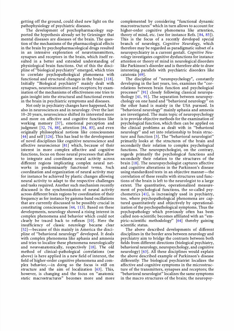
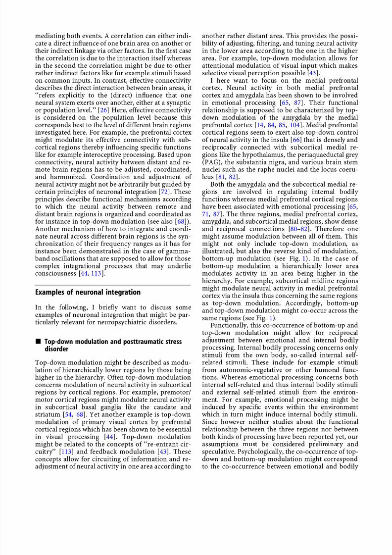
gions are involved in regulating internal bodily functions whereas medial prefrontal cortical regionshave been associated with emotional processing [65,71, 87]. The three regions, medial prefrontal cortex,amygdala, and subcortical medial regions, show denseand reciprocal connections [80–82]. Therefore onemight assume modulation between all of them. Thismight not only include top-down modulation, asillustrated, but also the reverse kind of modulation,bottom-up modulation (see Fig. 1). In the case of bottom-up modulation a hierarchically lower areamodulates activity in an area being higher in thehierarchy. For example, subcortical midline regions
might modulate neural activity in medial prefrontalcortex via the insula thus concerning the same regionsas top-down modulation. Accordingly, bottom-upand top-down modulation might co-occur across thesame regions (see Fig. 1).
Functionally, this co-occurrence of bottom-up andtop-down modulation might allow for reciprocaladjustment between emotional and internal bodily processing. Internal bodily processing concerns only stimuli from the own body, so-called internal self-related stimuli. These include for example stimulifrom autonomic-vegetative or other humoral func-tions. Whereas emotional processing concerns bothinternal self-related and thus internal bodily stimuliand external self-related stimuli from the environ-ment. For example, emotional processing might beinduced by specific events within the environmentwhich in turn might induce internal bodily stimuli.Since however neither studies about the functionalrelationship between the three regions nor betweenboth kinds of processing have been reported yet, ourassumptions must be considered preliminary andspeculative. Psychologically, the co-occurrence of top-down and bottom-up modulation might correspondto the co-occurrence between emotional and bodily
7/27/2019 Neuropsychiatry– an old discipline in a new gestalt bridging biological psychiatry, neuropsychology, and cognitive …
awareness. We are aware of the emotions associatedwith certain events in the environment. This co-oc-curs with awareness of one’s own body which usually remains in the background. Such co-occurrence Suchco-occurrence might account for our predominantoutward focus, directing our attention towards otherpersons and events in our environment whereas theinward focus, directing our attention towards our ownbody is not as central and predominant and seems toremain in the background.
Posttraumatic stress disorder (PTSD) can becharacterized by a constellation of symptoms in theaftermath of a severe emotionally traumatic event.The cardinal triad of clinical features includes: re-experiencing phenomena, e.g., flashbacks, which canoccur spontaneously or in response to reminders of the traumatic event; hyperarousal, e.g., exaggeratedstartle response; and avoidance, e.g., avoiding situa-tions that remind the individual of the traumaticevent (see Ref. [96] for an overview). The symptomsin PTSD can thus be characterized by a combinationof emotional and vegetative disturbances. Recentimaging studies observed abnormalities in the ante-rior cortical midline structures including the VMPFCand the supragenual anterior cingulate cortex (seeRefs. [47, 96]. These regions have been associatedwith the inability of PTSD patients to extinct threat-ening stimuli and their inability to suppress attentionto trauma-related stimuli. Since the very same regionsare also involved in mediating self-related processing,Liberzon and Martis [47] assume that self-relatedprocessing may also be altered in PTSD which how-ever remains to be shown. Another region that hasbeen shown to be abnormal is the amygdala thatshows exaggerated responses which may mediate theabnormal hyperarousal in PTSD patients [96]. Finally,the hippocampus has also been shown to be deficient
in neural activity. One may consequently assume al-tered balance and thus top-down modulation betweencortical and subcortical midline regions in PTSD (seeRef. [96]) during emotional and self-related process-ing. Studies investigating functional and effectiveconnectivity indicate that cortical control and topdown-modulation of subcortical neural activity in theamygdala may be altered in PTSD [47, 96] for whichhowever further empirical support is needed. Alteredcortical–subcortical balance may psychologically re-sult in ‘‘exuberant acquisition of conditioned fear andexaggerated fear responses, as well as deficientextinction recall and an incapacity to appreciate safecontexts.’’ [96]
j Reciprocal modulation and depression
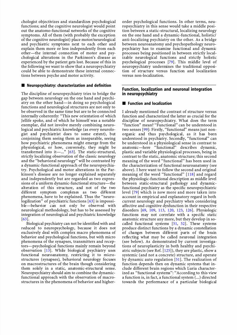
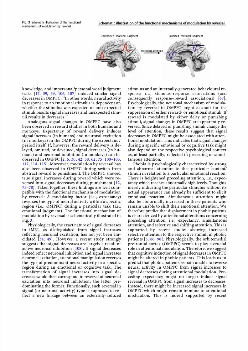
Recent studies [27, 28, 71, 72] demonstrate a patternof opposite signal changes in medial and lateral pre-frontal cortex during emotional–cognitive interaction.These results are compatible with the assumption of functional mechanisms of reciprocal modulation andreciprocal attenuation during emotional–cognitiveinteraction. Reciprocal modulation can be defined by signal changes in opposite directions (i.e., signal in-creases and decreases) in different regions. Forexample, emotional picture viewing is known to leadto signal increases in medial prefrontal cortical re-gions and concurrent signal decreases in lateral pre-frontal cortex [65, 72, 87]. In contrast, cognitive taskslike judgment or evaluation induce the reverse patternwith signal increases in lateral prefrontal cortex andsignal decreases in medial prefrontal cortex. This iscompatible with the functional mechanism of re-ciprocal modulation (see Fig. 2 and Ref. [72]). Inter-estingly, analogous patterns of reciprocal modulationhave been observed in other cortical regions including
Processing of cognitive and emotional
stimuli: Top-down modulation
Bottom-up modulation
Processing of bodily stimuli:
Schematic illustration of bottom-up and top-down
modulation between subcortical and cortical networks
Fig. 1 Schematic illustration of bottom-up and top-down modulation between subcortical and corticalnetworks
7/27/2019 Neuropsychiatry– an old discipline in a new gestalt bridging biological psychiatry, neuropsychology, and cognitive …
medial and lateral orbitofrontal cortex [71–74, 77, 78]right and left motor cortex [1], striate and extrastriatevisual cortex [40], subgenual anterior cingulate andright prefrontal cortex [48], sub/pre- and supragenualanterior cingulate [9] as well as visual and auditory cortex [45, 46]. Emotional–cognitive interaction isthen associated with the functional mechanism of reciprocal attenuation: Inclusion of an emotional
component into a cognitive task resulting in forexample emotional judgment leads to smaller signaldecreases in medial prefrontal cortical regions and, atthe same time, smaller signal increases in lateralprefrontal cortical regions; this has been calledattenuation [72]. Since attenuation concerned bothmedial and lateral prefrontal cortical regions inopposite directions (i.e., smaller signal decreases/in-creases, respectively), one can speak of reciprocalattenuation.
Depression, e.g., major depressive disorder(MDD), can be characterized by co-occurrence of affective and cognitive symptoms. Affectively de-
pressed patients can be characterized by the inability to experience and obtain pleasure, e.g., anhedonia,resulting an abnormal sadness. Cognitively, depressedpatients are no longer able to evaluate their ownemotional and bodily experience appropriately; the
judgments of their own states are ‘‘subjectively’’ dis-torted and decoupled from ‘‘objective’’ reality. Sub-
jective distortion is manifest in the extreme negativity of their judgments concerning either their own emo-tions and their own body or emotions in other per-sons and events in their environment. This extremenegativity corresponds to what psychologically has
been described as the ‘‘negative bias’’ [16, 29]. Func-tional imaging studies in depression [55, 56, 88] show hyperactivity in medial prefrontal cortex and hypo-activity in lateral prefrontal cortex during emotionalstimulation [16, 48, 55, 56]. This corresponds indeedto abnormal reciprocal modulation between medialand lateral prefrontal cortex. What however remainsto be shown is that this abnormal neural activity in
medial and lateral prefrontal cortex is related toemotional and cognitive dysfunction (see Ref. [32]).Furthermore, abnormal reciprocal attenuation duringemotional–cognitive interaction has not been dem-onstrated yet in depressed patients.
j Modulation by reversal and phobia
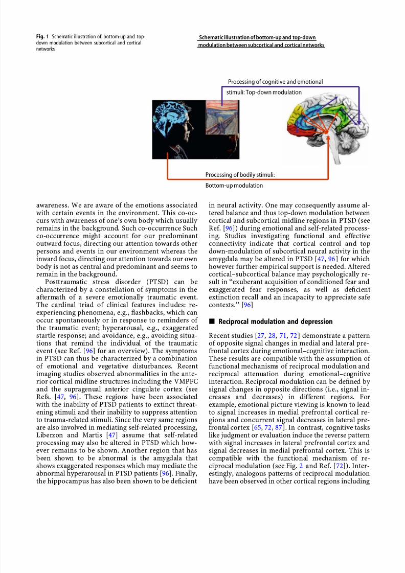
Several studies demonstrated reversal of signalchanges in the opposite direction within the sameregion. Signal changes within the OMPFC, were, forexample, reversed (from signal increases to signaldecreases) by either preceding (like expectancy) or
simultaneous (like distraction or increased focus)attentional manipulation of emotional stimulation. Toaccount for attentional modulation in the paradigm,emotional pictures were either preceded by anexpectancy period or by a simultaneous flickering [38,90, 95, 106, 107]. A recent study [73] observedreversion of signal increases to signal decreases inboth OMPFC and posterior cingulate cortex whenemotional judgment was preceded by an expectancy period. Similar changes in signal direction were alsoobserved in cognitive tasks during attentional mod-ulation. For example, noun generation, object
Emotional task Cognitive task
Emotional - cognitive task
B
A
Schematic illustration of opposite neural modulation between medial
and lateral prefrontal cortex during emotional and cognitive processing
‘Reciprocal Attenuation’
‘Reciprocal Modulation’
Fig. 2 Schematic illustration of opposite neuralmodulation between medial and lateral prefrontalcortex during emotional and cognitive processing
7/27/2019 Neuropsychiatry– an old discipline in a new gestalt bridging biological psychiatry, neuropsychology, and cognitive …
knowledge, and impersonal/personal word judgmenttasks [17, 39, 59, 106, 107] induced similar signaldecreases in OMPFC. ‘‘In other words, neural activity in response to an emotional stimulus is dependent onwhether the stimulus was expected or not; expectedstimuli results signal increases and unexpected stim-uli results in decreases.’’
Analogous signal changes in OMPFC have alsobeen observed in reward studies in both humans andmonkeys. Expectancy of reward delivery inducessignal increases (in humans) and neuronal excitation(in monkeys) in the OMPFC during the expectancy period itself. If, however, the reward delivery is de-layed, omitted, or devalued, signal decreases (in hu-mans) and neuronal inhibition (in monkeys) can beobserved in OMPFC [2, 6, 30, 42, 58, 62, 75, 100–103,112, 114, 115]. Moreover, modulation by reversal hasalso been observed in OMPFC during switch fromabstract reward to punishment. The OMPFC showedtrue signal increases during reward which were re-
versed into signal decreases during punishment [12,75–78]. Taken together, these findings are well com-patible with the functional mechanism of modulationby reversal: A modulating factor (i.e., expectancy)reverses the type of neural activity within a specificregion (i.e., OMPFC) during a particular task (i.e.,emotional judgment). The functional mechanism of modulation by reversal is schematically illustrated inFig. 3.
Physiologically, the exact nature of signal decreasesin fMRI, as distinguished from signal increasesreflecting neuronal excitation, has not yet been elu-cidated [34, 49]. However, a recent study strongly
suggests that signal decreases are largely a result of active neuronal inhibition [108]. If signal decreasesindeed reflect neuronal inhibition and signal increasesneuronal excitation, attentional manipulation reversesthe type of predominant neural activity in a specificregion during an emotional or cognitive task. Thetransformation of signal increases into signal de-creases would then correspond to reversal of neuronalexcitation into neuronal inhibition; the latter pre-dominating the former. Functionally, such reversal insignal (or neuronal activity) type is supposed to re-flect a new linkage between an externally-induced
stimulus and an internally-generated behavioural re-sponse, i.e., stimulus–response associations (andconsequently response–reward associations) [67].Psychologically, the neuronal mechanism of modula-tion by reversal in OMPFC might account for thesuppression of either reward- or emotional stimuli. If reward is modulated by either delay or punishing
stimuli, signal changes in OMPFC are apparently re-versed. Since delayed or punishing stimuli change thelevel of attention, these results suggest that signaldecreases in OMPFC might be associated with atten-tional modulation. This indicates that signal changesduring a specific emotional or cognitive task mightalso depend on the respective psychological contextas, at least partially, reflected in preceding or simul-taneous attention.
Phobia is psychologically characterized by strongand abnormal attention to that particular content/stimuli in relation to a particular emotional reaction.There is heightened preceding attention, i.e., expec-
tancy which reaches abnormally high levels. Thoughtsmerely indicating the particular stimulus without itsactual appearance can already be sufficient to elicitemotional reaction. Simultaneous attention mightalso be abnormally increased in these patients whoremain unable to shift their emotional attention. Wetherefore predict that displacement in phobic patientsis characterized by attentional alterations concerningpreceding attention, i.e., expectancy, simultaneousattention, and selective and shifting attention. This issupported by recent studies showing increasedselective attention to the respective stimuli in phobicpatients [5, 86, 98]. Physiologically, the orbitomedial
prefrontal cortex (OMPFC) seems to play a crucialrole in attentional modulation. Therefore, we suggestthat cognitive induction of signal decreases in OMPFCmight be altered in phobic patients. This leads us topredict that phobic patients remain unable to reverseneural activity in OMPFC from signal increases tosignal decreases during attentional modulation. Pre-ceding expectancy might no longer induce signalreversal in OMPFC from signal increases to decreases.Instead, there might be increased signal increases inOMPFC which might remain immune to attentionalmodulation. This is indeed supported by recent
Schematic illustration of the functional mechanisms of modulation by reversalFig. 3 Schematic illustration of the functionalmechanisms of modulation by reversal
7/27/2019 Neuropsychiatry– an old discipline in a new gestalt bridging biological psychiatry, neuropsychology, and cognitive …
imaging studies with phobic patients showing stron-ger signal increases in medial cortical regions like theOMPFC and the anterior cingulate cortex as well as inclosely connected regions like the amygdala and theinsula during exposure to the respective stimuli [15,50, 89, 110, 111, 119]. However, abnormal attentionalmodulation of neural activity in these regions by thedifferent forms of attention remains to be shown.
j Modulation by functional unity
Another example of a possible functional mechanismof emotional–cognitive interaction is the constitutionof functional unities. Functional unity can be de-scribed the coordination of the neural activity over alimited time period by means of which different re-gions are linked together with respect to a particularfunction. Such transient functional unities might beidentified based upon the psychophysiological char-acteristics or the functional connectivity of therespective regions [20–25]. The medial regions in our
brain’s cortex, the so-called cortical midline struc-tures (CMS) can be considered a functional unity [71]which is supported by different lines of evidences.First, one can often observe co-involvement and co-activation of different midline regions. For example,the above describe mechanisms of modulation by reversal cannot only be observed in the OMPFC butalso in posterior cingulate [73]. Other studies onemotions and cognitions show similar co-involvementof anterior and posterior midline regions [37, 71, 72].Second, unlike other more lateral cortical regions andsubcortical regions, the CMS show a continuous highlevel of neural activity during resting conditions such
as passive viewing of a fixation of a cross [33, 34, 57,93, 94]. Third, regions in the CMS are characterizedby close anatomical connections and tight functionalconnectivity. For example, Greicius et al. [31] inves-tigated the functional connectivity among CMS re-gions in both resting and activation state. They observed increased functional connectivity betweenanterior and posterior CMS regions in the restingstate whereas it was decreased during active cognitivetasks. Taken together these findings are compatiblewith the functional mechanisms of modulation by functional unity. The described data provide com-pelling evidence for the existence of CMS as func-
tional unity which seems to be particularly active andcohesive in the resting state [31, 122–124].Catatonia is a psychomotor syndrome showing a
unique constellation of affective, behavioral and mo-tor symptoms [7, 8, 19, 68, 69]. Most impressively,acute catatonic patients are totally immobilized,posturing in bizarre positions, and becoming totally mute which may be associated with an uncontrollableoverflow of anxieties, i.e., ‘‘immobilization by anxi-eties’’ [68, 69, 83]. Imaging studies during emotionalstimulation showed altered pattern of signal changesin medial and lateral orbitofrontal cortex (MOFC and
LOFC) in catatonic patients compared to non-cata-tonic psychiatric and healthy controls. Specifically, weobserved reduced signal changes in the MOFC andenhanced signal changes in the LOFC during negativestimulus presentation (see Refs. [69, 72] for details).Correlation analysis of functional connectivity be-tween OFC, MPFC, and premotor and motor cortex incatatonic patients as compared to non-catatonic
psychiatric and healthy controls revealed the follow-ing differences: Catatonic patients showed signifi-cantly lower scores for functional connectivity fromthe orbitofrontal cortex to the medial prefrontal andthe premotor/motor cortex when compared to non-catatonic psychiatric controls and healthy subjects.These findings of abnormal connectivity fromorbitofrontal cortex over medial prefrontal cortex topremotor/motor cortex suggest abnormal modulationby functional unity in these patients. The functionalunity across anterior cortical midline structuresseems to be less coherent than in healthy subjects.This less coherent functional unity might in turn
facilitate abnormal transformation of emotionalsymptoms into motor symptoms. Interestingly, pa-tients with conversion symptoms that show a more orless analogous combination of emotional and motorsymptoms seems to show similar neural abnormali-ties. Imaging studies in acute paralytic patients re-vealed deficits in various regions of the anterior CMSincluding the orbitofrontal and the premotor/motorcortex [35, 53, 109, 120]. Why however is there asymptomatic difference n between hysterical andcatatonic patients the former showing conversion andthe latter catatonia? It should first be noted thathysterical patients can show a catatonic-like picture
and that, conversely, catatonic patients can appearstrongly hysterical [60, 61, 68–70]. Such symptomaticoverlap suggests that both catatonia and hysteriaoverlap in the neuronal mechanisms. They mightshare the abnormal functional unity of anterior CMSresulting in abnormal motor behavior. However,modulation by functional unity might not only con-cern overlapping regions but also different regions;this in turn might explain the symptomatic differ-ences between catatonia and hysterical conversion.Future studies specifically targeting single catatonic orhysterical symptoms might reveal those neuronalmechanisms specifically associated with hystericalconversion as distinguished from those related tocatatonia.
References
1. Allison JD, Meador KJ et al (2000) Functional MRI cerebralactivation and deactivation during finger movement. Neurol-ogy 54(1):135–142
2. Anderson AK, Sobel N (2003) Dissociating intensity fromvalence as sensory inputs to emotion. Neuron 39(4):581–583
3. Beaumont JG (1987) Einfuhrung in die Neuropsychologie.Psychologie Verlags-Union, Munchen, Weinheim
7/27/2019 Neuropsychiatry– an old discipline in a new gestalt bridging biological psychiatry, neuropsychology, and cognitive …
4. Berrios GE, Markova IS (2002) The concept of neuropsychi-atry: a historical overview. J Psychosom Res 53(2):629–638
5. Bogels SM, Mansell W (2004) Attention processes in themaintenance and treatment of social phobia: hypervigilance,avoidance and self-focused attention. Clin Psychol Rev 24(7):827–856
6. Braver TS, Brown JW (2003) Principles of pleasure prediction:specifying the neural dynamics of human reward learning.Neuron 38(2):150–152
7. Bush G, Fink M et al (1996) Catatonia. I. Rating scale and
standardized examination. Acta Psychiatr Scand 93(2):129–1368. Bush G, Fink M et al (1996) Catatonia. II. Treatment with
lorazepam and electroconvulsive therapy. Acta PsychiatrScand 93(2):137–143
9. Bush G, Luu P et al (2000) Cognitive and emotional influencesin anterior cingulate cortex. Trends Cogn Sci 4(6):215–222
11. Churchland PA (1986) Neurophilosophy. Toward a unifiedscience of the mind/brain. MIT Press, Bradford, Cambridge
12. Critchley HD, Mathias CJ et al (2001) Neural activity in thehuman brain relating to uncertainty and arousal duringanticipation. Neuron 29(2):537–545
13. Cummings JL (1985) Clinical neuropsychiatry. Grund andStratton, New York
14. Davidson RJ (2002) Anxiety and affective style: role of pre-frontal cortex and amygdala. Biol Psychiatry 51(1):68–8015. Dilger S, Straube T et al (2003) Brain activation to phobia-
related pictures in spider phobic humans: an event-relatedfunctional magnetic resonance imaging study. Neurosci Lett348(1):29–32
16. Elliott R, Rubinsztein JS et al (2002) The neural basis of mood-congruent processing biases in depression. Arch Gen Psy-chiatry 59(7):597–604
17. Ferstl EC, von Cramon DY (2002) What does the frontome-dian cortex contribute to language processing: coherence ortheory of mind? Neuroimage 17(3):1599–1612
18. Filskow SB, Boll TJ (1986) Handbook of clinical Neuropsy-chology, vol 2. Wiley, New York, pp 45–81
19. Fink M (1993) Catatonia and psychotic (delusional) depres-sion, distinct syndromes in DSM-IV. Am J Psychiatry 150(7):1130–1131
20. Friston KJ (1998) Imaging neuroscience: principles or maps?Proc Natl Acad Sci USA 95(3):796–802
21. Friston K (2003) Learning and inference in the brain. NeuralNetw 16(9):1325–1352
22. Friston KJ, Fletcher P et al (1998) Event-related fMRI: char-acterizing differential responses. Neuroimage 7(1):30–40
23. Friston KJ, Harrison L et al (2003) Dynamic causal modelling.Neuroimage 19(4):1273–1302
24. Friston KJ, Josephs O et al (1998) Nonlinear event-relatedresponses in fMRI. Magn Reson Med 39(1):41–52
25. Friston KJ, Penny W (2003) Posterior probability maps andSPMs. Neuroimage 19(3):1240–1249
26. Friston KJ, Price CJ (2001) Dynamic representations andgenerative models of brain function. Brain Res Bull 54(3):275–285
27. Goel V, Dolan RJ (2003) Explaining modulation of reasoningby belief. Cognition 87(1):B11–22
28. Goel V, Dolan RJ (2003) Reciprocal neural response withinlateral and ventral medial prefrontal cortex during hot andcold reasoning. Neuroimage 20(4):2314–2321
29. Gotlib IH, Krasnoperova E et al (2004) Attentional biases fornegative interpersonal stimuli in clinical depression. J Ab-norm Psychol 113(1):121–135
30. Gottfried JA, O’Doherty J et al (2003) Encoding predictivereward value in human amygdala and orbitofrontal cortex.Science 301(5636):1104–1107
31. Greicius MD, Krasnow B et al (2003) Functional connectivity in the resting brain: a network analysis of the default modehypothesis. Proc Natl Acad Sci USA 100(1):253–258
32. Grimm S, Beck J, Schuepach D, Hell D, Boesinger P, NiehausL, Boeker H, Northoff G (2007) Imbalance between left andright dorsolateral prefrontal cortex in major depression islinked to negative emotional judgement. An fMRI study insevere major depressive disorder. Biological Psychiatry (inpress)
33. Gusnard DA, Akbudak E et al (2001) Medial prefrontal cortexand self-referential mental activity: relation to a default modeof brain function. Proc Natl Acad Sci USA 98(7):4259–4264
34. Gusnard DA, Raichle ME (2001) Searching for a baseline:
functional imaging and the resting human brain. Nat Rev Neurosci 2(10):685–69435. Halligan PW, Athwal BS et al (2000) Imaging hypnotic
paralysis: implications for conversion hysteria. Lancet355(9208):986–987
36. Harrington A (1989) Psychiatrie und die Geschichte der Lo-kalisation geistiger Funktion. Nervenarzt 60:603–611
37. Iacoboni M, Lieberman MD, Knowlton BJ, Molnar-Szakacs I,Moritz M, Throop CJ, Fiske AP (2004) Watching socialinteractions produces dorsomedial prefrontal and medialparietal BOLD fMRI signal increases compared to a restingbaseline. Neuroimage 21(3):1167–1173
38. Keightley ML, Winocur G et al (2003) An fMRI study inves-tigating cognitive modulation of brain regions associated withemotional processing of visual stimuli. Neuropsychologia41(5):585–596
39. Kelley WM, Macrae CN et al (2002) Finding the self? An event-related fMRI study. J Cogn Neurosci 14(5):785–79440. Kleinschmidt A, Buchel C et al (1998) Human brain activity
during spontaneously reversing perception of ambiguousfigures. Proc R Soc Lond B Biol Sci 265(1413):2427–2433
41. Kolb B, Whishaw JQ (1985) Fundamentals of human neuro-psychology, 2nd edn. W. Freeman Company, New York
42. Kringelbach ML, Rolls ET (2004) The functional neuroanatomy of the humanorbitofrontal cortex: evidence from neuroimagingand neuropsychology. Prog Neurobiol 72(5):341–372
43. Lamme VA (2001) Blindsight: the role of feedforward andfeedback corticocortical connections. Acta Psychol (Amst)107(1–3):209–228
44. Lamme VA (2004) Separate neural definitions of visual con-sciousness and visual attention; a case for phenomenalawareness. Neural Netw 17(5–6):861–872
45. Laurienti PJ, Burdette JH et al (2002) Deactivation of sensory-specific cortex by cross-modal stimuli. J Cogn Neurosci14(3):420–429
46. Laurienti PJ, Field AS et al (2002) Dietary caffeine consump-tion modulates fMRI measures. Neuroimage 17(2):751–757
47. Liberzon I, Martis B (2006) Neuroimaging studies of emo-tional responses in PTSD. Ann N Y Acad Sci 1071:87–109
48. Liotti M, Mayberg HS et al (2002) Unmasking disease-specificcerebral blood flow abnormalities: mood challenge in patientswith remitted unipolar depression. Am J Psychiatry 159(11):1830–1840
49. Logothetis NK, Pauls J et al (2001) Neurophysiologicalinvestigation of the basis of the fMRI signal. Nature412(6843):150–157
50. Lorberbaum JP, Kose S et al (2004) Neural correlates of speechanticipatory anxiety in generalized social phobia. Neuroreport15(18):2701–2705
51. Luria AR (1966) Higher cortical functions in man [Translatedby B. Haigh]. Basic Books, New York
52. Luria AR (1973) The working brain. An introduction toneuropsychology [Translated by B. Haigh]. Basic Books, New York
53. Marshall JC, Halligan PW et al (1997) The functional anatomy of a hysterical paralysis. Cognition 64(1):B1–8
54. Masterman DL, Cummings JL (1997) Frontal–subcortical cir-cuits: the anatomic basis of executive, social and motivatedbehaviors. J Psychopharmacol 11(2):107–114
55. Mayberg HS, Silva JA, Brannan SK, Tekell JL, Mahurin RK,McGinnis S, Jerabek PA (2002) The functional neuroanatomy of the placebo effect. Am J Psychiatry 159(5):728–737
7/27/2019 Neuropsychiatry– an old discipline in a new gestalt bridging biological psychiatry, neuropsychology, and cognitive …
56. Mayberg HS (2003) Positron emission tomography imaging indepression: a neural systems perspective. Neuroimaging ClinN Am 13(4):805–815
57. Mazoyer B, Zago L et al (2001) Cortical networks for workingmemory and executive functions sustain the conscious restingstate in man. Brain Res Bull 54(3):287–298
58. McClure SM, Berns GS et al (2003) Temporal prediction errorsin a passive learning task activate human striatum. Neuron38(2):339–346
59. Mitchell DG, Colledge E et al (2002) Risky decisions and re-
sponse reversal: is there evidence of orbitofrontal cortexdysfunction in psychopathic individuals? Neuropsychologia40(12):2013–2022
60. Modestin J, Bachmann KM (1992) Is the diagnosis of hyster-ical psychosis justified?: clinical study of hysterical psychosis,reactive/psychogenic psychosis, and schizophrenia. ComprPsychiatry 33(1):17–24
61. Modestin J, Bachmann KM (1992) A third kind of psychosis.Analysis of the literature. Schweiz Arch Neurol Psychiatr143(4):307–323
62. Montague PR, Berns GS (2002) Neural economics and thebiological substrates of valuation. Neuron 36(2):265–284
63. Mueller J (ed) (1989) Neurology and psychiatry. A meeting of minds. Karger, New York
64. Mundt C (1989) Psychopathologie heute. In: Kister KP et al(eds) Brennpunkte der Psychiatrie. Psychiatrie der Gegenwart
9, Springer, Heidelberg, pp 147–18565. Murphy FC, Nimmo-Smith I et al (2003) Functional neuro-anatomy of emotions: a meta-analysis. Cogn Affect Behav Neurosci 3(3):207–233
66. Nagai Y, Critchley HD et al (2004) Activity in ventromedialprefrontal cortex covaries with sympathetic skin conductancelevel: a physiological account of a ‘‘default mode’’ of brainfunction. Neuroimage 22(1):243–251
67. Nobre AC, Coull JT et al (1999) Orbitofrontal cortex is acti-vated during breaches of expectation in tasks of visualattention. Nat Neurosci 2(1):11–12
68. Northoff G (2002) Catatonia and neuroleptic malignant syn-drome: psychopathology and pathophysiology. J NeuralTransm 109(12):1453–1467
69. Northoff G (2002) What catatonia can tell us about ‘‘top-downmodulation’’: a neuropsychiatric hypothesis. Behav Brain Sci25(5):555–577; discussion 578–604
70. Northoff G, Eckert J et al (1997) Glutamatergic dysfunction incatatonia? Successful treatment of three acute akinetic cata-tonic patients with the NMDA antagonist amantadine. JNeurol Neurosurg Psychiatry 62(4):404–406
71. Northoff G, Bermpohl F (2004) Cortical midline structuresand the self. Trends Cogn Sci 8(3):102–107
72. Northoff G, Heinzel A et al (2004) Reciprocal modulation andattenuation in the prefrontal cortex: an fMRI study on emo-tional–cognitive interaction. Hum Brain Mapp 21(3):202–212
73. Northoff G, Richter A et al (2005) NMDA hypofunction in theposterior cingulate as a model for schizophrenia: an explor-atory ketamine administration study in fMRI. Schizophr Res72(2–3):235–248
74. Northoff G, Witzel T et al (2002) GABA-ergic modulation of prefrontal spatio-temporal activation pattern during emo-tional processing: a combined fMRI/MEG study with placeboand lorazepam. J Cogn Neurosci 14(3):348–370
75. O’Doherty JP, Dayan P et al (2003) Temporal differencemodels and reward-related learning in the human brain.Neuron 38(2):329–337
76. O’Doherty JP, Deichmann R et al (2002) Neural responsesduring anticipation of a primary taste reward. Neuron33(5):815–826
77. O’Doherty J, Kringelbach ML et al (2001) Abstract reward andpunishment representations in the human orbitofrontal cor-tex. Nat Neurosci 4(1):95–102
78. O’Doherty J, Rolls ET et al (2001) Representation of pleasantand aversive taste in the human brain. J Neurophysiol85(3):1315–1321
79. Oepen G (Hrsg.) (1988) Psychiatrie des rechten und linkenGehirns. Neuropsychologische Ansatze zum Verstandnis von,‘Personlichkeit’, Depression’ und ‘Schizophrenie’. DeutscherArzte Verlag, Koln
80. Ongur D, Price JL (2000) The organization of networks withinthe orbital and medial prefrontal cortex of rats, monkeys andhumans. Cereb Cortex 10(3):206–219
81. Panksepp J (1998) Affective neuroscience: the foundations of human and animal emotions. Oxford University Press, New York
82. Panksepp J (1998) The periconscious substrates of con-sciousness: affective states and the evolutionary origins of theself. J Conscious Stud 5(5–6):566–582
83. Perkins K (1982) Catatonia as an immobilization reflex. AustN Z J Psychiatry 23:282–288
84. Pessoa L, McKenna M et al (2002) Neural processing of emotional faces requires attention. Proc Natl Acad Sci USA99(17):11458–11463
85. Pessoa L, Ungerleider LG (2004) Neuroimaging studies of attention and the processing of emotion-laden stimuli. ProgBrain Res 144:171–182
86. Pflugshaupt T, Mosimann UP et al (2005) Hypervigilance-avoidance pattern in spider phobia. J Anxiety Disord19(1):105–116
87. Phan KL, Wager T et al (2002) Functional neuroanatomy of emotion: a meta-analysis of emotion activation studies in PET
and fMRI. Neuroimage 16(2):331–34888. Phillips ML, Drevets WC, Rauch SL, Lane R (2003) Neurobi-ology of emotion perception II: Implications for major psy-chiatric disorders. Biol Pychiatry 54(5):515–528
89. Pissiota A, Frans O et al (2003) Amygdala and anterior cin-gulate cortex activation during affective startle modulation: aPET study of fear. Eur J Neurosci 18(5):1325–1331
90. Ploghaus A, Becerra L et al (2003) Neural circuitry underlyingpain modulation: expectation, hypnosis, placebo. TrendsCogn Sci 7(5):197–200
91. Poeck K (1982) Klinische neuropsychologie. Thieme, Stuttgart92. Price CJ, Friston KJ (2002) Degeneracy and cognitive anat-
Nature 412(6843):128–13094. Raichle ME, MacLeod AM et al (2001) A default mode of brain
function. Proc Natl Acad Sci USA 98(2):676–68295. Ramnani N, Owen AM (2004) Anterior prefrontal cortex: in-
sights into function from anatomy and neuroimaging. Nat Rev Neurosci 5(3):184–194
96. Rauch SL, Shin LM, Phelps EA (2006) Neurocircuitry modelsof posttraumatic stress disorder and extinction: human neu-roimaging research—past, present, and future. Biol Psychiatry 60(4):376–382
97. Reynolds EH, Trimble MR (1989) The bridge betweenneurology and psychiatry. Churchill Livingstone, Edinbor-ough
98. Rinck M, Becker ES (2005) A comparison of attentionalbiases and memory biases in women with social phobiaand major depression. J Abnorm Psychol 114(1):62–74
99. Rogers D (1987) Neuropsychiatry. Br J Psychiatry 150:425–427100. Rolls ET (2000) The orbitofrontal cortex and reward. Cereb
Cortex 10(3):284–294101. Rolls ET, Tovee MJ et al (1999) The neurophysiology of
backward visual masking: information analysis. J Cogn Neu-rosci 11(3):300–311
102. Schultz W (2000) Multiple reward signals in the brain. Nat Rev Neurosci 1(3):199–207
103. Schultz W, Tremblay L et al (2000) Reward processing inprimate orbitofrontal cortex and basal ganglia. Cereb Cortex10(3):272–284
104. Shin LM, Wright CI et al (2005) A functional magnetic reso-nance imaging study of amygdala and medial prefrontal cor-tex responses to overtly presented fearful faces inposttraumatic stress disorder. Arch Gen Psychiatry 62(3):273–281
7/27/2019 Neuropsychiatry– an old discipline in a new gestalt bridging biological psychiatry, neuropsychology, and cognitive …
105. Shmuel A, Yacoub E et al (2002) Sustained negative BOLD,blood flow and oxygen consumption response and its cou-pling to the positive response in the human brain. Neuron36(6):1195–1210
106. Simpson JR Jr, Drevets WC et al (2001) Emotion-inducedchanges in human medial prefrontal cortex: II. During antic-ipatory anxiety. Proc Natl Acad Sci USA 98(2):688–693
107. Simpson JR Jr, Snyder AZ et al (2001) Emotion-inducedchanges in human medial prefrontal cortex: I. During cog-nitive task performance. Proc Natl Acad Sci USA 98(2):683–
687108. Singer W (1989) Search for coherence: a basic principle of cortical self-organization. Concepts of neuroscience, first is-sue, 1
109. Spence SA, Crimlisk HL et al (2000) Discrete neurophysio-logical correlates in prefrontal cortex during hysterical andfeigned disorder of movement. Lancet 355(9211):1243–1244
110. Straube T, Kolassa IT et al (2004) Effect of task conditions onbrain responses to threatening faces in social phobics: anevent-related functional magnetic resonance imaging study.Biol Psychiatry 56(12):921–930
111. Straube T, Mentzel HJ et al (2004) Brain activation to phobia-related words in phobic subjects. Neurosci Lett 372(3):204–208
112. Tobler PN, Dickinson A et al (2003) Coding of predicted re-ward omission by dopamine neurons in a conditioned inhi-
bition paradigm. J Neurosci 23(32):10402–10410113. Tononi G, Edelman GM (2000) Schizophrenia and the mech-anisms of conscious integration. Brain Res Brain Res Rev 31(2–3):391–400
115. Tremblay L, Schultz W (2000) Modifications of rewardexpectation-related neuronal activity during learning in pri-mate orbitofrontal cortex. J Neurophysiol 83(4):1877–1885
116. Tremblay L, Schultz W (2000) Reward-related neuronalactivity during go-nogo task performance in primate orbito-frontal cortex. J Neurophysiol 83(4):1864–1876
117. Trimble MR (1981) Neuropsychiatry. Wiley, Clichester118. Trimble MR (1988) Biological psychiatry. Wiley, Clichester119. Veltman DJ, Tuinebreijer WE et al (2004) Neurophysiological
correlates of habituation during exposure in spider phobia.
Psychiatry Res 132(2):149–158120. Vuilleumier P, Chicherio C et al (2001) Functional neuro-anatomical correlates of hysterical sensorimotor loss. Brain124(Pt 6):1077–1090
121. Vygotsky LS (1978) Mind in society. The development of higher psychological processes. Harvard University Press,Cambridge
122. Wicker B, Keysers C et al (2003) Both of us disgusted in My insula: the common neural basis of seeing and feeling disgust.Neuron 40(3):655–664
123. Wicker B, Perrett DI et al (2003) Being the target of another’semotion: a PET study. Neuropsychologia 41(2):139–146
124. Wicker B, Ruby P et al (2003) A relation between rest and theself in the brain? Brain Res Brain Res Rev 43(2):224–230
125. Vollmayr B, Mahlstedt MM, Henn FA (2007) urogenesis anddepression: what animal models tell us about the link. Eur
Arch Psychiatry Clin Neurosci 257(5):300–303126. Yamasaki S, Yamasue H, Abe O, Yamada H, Iwanami A,Hirayasu Y, Nakamura M, Furukawa S, Rogers MA, Tanno Y,Aoki S, Kato N, Kasai K (2007) Reduced planum temporalevolume and delusional behaviour in patients with schizo-phrenia. Eur Arch Psychiatry Clin Neurosci 257(6):318–324