100
10915 Lake Ridge Drive Knoxville, TN 37934 NSALabs.com [email protected] Sales 865-966-1266 Lab 865-675-2245 2017 CATALOG OF NEUROHISTOLOGY SERVICES NEUROSCIENCE ASSOCIATES
| Date post: | 28-May-2018 |
| Category: |
Documents |
| Upload: | truongkiet |
| View: | 216 times |
| Download: | 0 times |

10915 Lake Ridge Drive Knoxville, TN 37934 NSALabs.com [email protected] Sales 865-966-1266 Lab 865-675-2245
2017 CATALOG OF NEUROHISTOLOGY SERVICES
NeuroScieNce ASSociAteS

Who are the clients of NeuroScieNce ASSociAteS?The clients of NSA range in size and scope—from small academic research labs to major pharmaceutical companies. Organizations choose us to perform their neurohistology for a variety of reasons. Some clients completely outsource neurohistology to NSA, while others use us as an extension of their own capabilities. Our high throughput times allow time-conscious customers to rapidly respond to gaps in their internal capacity and meet critical deadlines. Clients often select NSA to perform the neurohistology for their biggest and most important studies. Regardless of the reason you choose NSA, we can assist you with your neurohistology needs and provide you with the most cost-effective, high-quality services available.
Read what our clients have to say about us:
“I have been a customer of NSA for several years and they have been critical for the success of several of our projects. Specifically, NSA provided high-quality sections for our stereology-based cell counting studies and saved us considerable time and money. Immunohistochemical studies we have completed on sections provided by NSA have been of outstanding quality and due to the MultiBrain® Technology and Large Format processing have led to significantly reduced variability in our data. NSA is responsive and collaborative and the staff are a pleasure to work with.”
Dr. Elizabeth Head, Sanders-Brown Center on Aging, University of Kentucky
“By virtue of our collaboration with Dr. Robert Switzer and NeuroScience Associates, and the use of their MultiBrain® Technology, we have been able to map out, using disintegrative staining and immunohistochemical methods, a detailed time course of post-traumatic neurodegeneration in rodent models after traumatic brain injury. Consequently, we have demonstrated definitive neuroprotective effects of a novel and exciting neuroprotective compound. This work, carried out over the past four years, has resulted in three peer-reviewed papers. Dr. Switzer and his colleagues at NSA have consistently provided extremely high-quality work with a rapid turnaround and even assisted in the interpretation of results. Their creativity, exquisite attention to detail and professional manner has been a real joy. There is no way that my laboratory could have duplicated the work they have done for us. Even if we could, the cost-effectiveness and reliability of their histological services has made them the logical choice. We will continue to turn to them.”
Dr. Ed Hall, Director, Spinal Cord & Brain Injury Research Center, University of Kentucky
“UCB started collaborating with NSA a few months ago, and I have already been very impressed by their MultiBrain sectioning technology and the high-quality of their immunostaining. This technology is particularly attractive for large studies including multiple experimental conditions. Because the variability of the immunohistochemistry procedure is reduced, it is possible to obtain reliable data and in a very short time frame. The technical staff from NSA have strong expertise in immunohistochemistry and are responsive to our requests. So far so good, we are happy with their quality and will continue working with them.
Georges Mariet-Coello, UCB Biopharma SPRL
“Having collaborated with NSA over the last few years ,I am glad to state the following: Interaction with NSA has always been very fruitful. The scientific input from NSA has been of very high standard and the company has always been very open-minded, so that novel approaches, proposed by either NSA or Lundbeck, to our studies have been implemented in the analysis phase of the different studies. This very interactive approach has proven intellectually very stimulative and has contributed significantly to the momentum of the project. The technical quality of the obtained sections and the analysis of the histology obviously is of first class, so I have a strong interest in continued collaboration with NSA.”
Dr. Bjarke Ebert, Adjunct Professor in Molecular PharmacologyHead of Electrophysiology, H. Lundbeck A/S
(More testimonials on the inside back cover)Sales 865-966-1266 NSALabs.com [email protected] Lab 865-675-2245

NeuroScieNce ASSociAteSTABLE OF CONTENTS
Letter from the President . . . . . . . . . . . . . . . . . . . . . . . . . . . . . . . . . . . . . . . . . . . . . . . . . . . . . . . . . . . . .2 NeuroScience Associates (NSA) Introduction . . . . . . . . . . . . . . . . . . . . . . . . . . . . . . . . . . . . . . . . 3 NSA Services: Comprehensive Overview . . . . . . . . . . . . . . . . . . . . . . . . . . . . . . . . . . . . . . . . . . . 4 NSA NeuroTechnologies™: Embedding and Sectioning . . . . . . . . . . . . . . . . . . . . . . . . . . . . 5 MultiBrain® Technology . . . . . . . . . . . . . . . . . . . . . . . . . . . . . . . . . . . . . . . . . . . . . . . . .5 MultiCord® Technology . . . . . . . . . . . . . . . . . . . . . . . . . . . . . . . . . . . . . . . . . . . . . . . . .8 Large Format Technology™ . . . . . . . . . . . . . . . . . . . . . . . . . . . . . . . . . . . . . . . . . . . . . 9 Variety of Species . . . . . . . . . . . . . . . . . . . . . . . . . . . . . . . . . . . . . . . . . . . . . . . . . . . .11 Stains . . . . . . . . . . . . . . . . . . . . . . . . . . . . . . . . . . . . . . . . . . . . . . . . . . . . . . . . . . . . . . . . . .12 Specialty Stains . . . . . . . . . . . . . . . . . . . . . . . . . . . . . . . . . . . . . . . . . . . . . . . . . . . . . .12 Classic Stains . . . . . . . . . . . . . . . . . . . . . . . . . . . . . . . . . . . . . . . . . . . . . . . . . . . . . . .13 Immunohistochemistry/Antibody Stains . . . . . . . . . . . . . . . . . . . . . . . . . . . . . . . . . . . 15 Fluorescent Immunohistochemistry Stains . . . . . . . . . . . . . . . . . . . . . . . . . . . . . . . . . 22 Quantitative Analysis . . . . . . . . . . . . . . . . . . . . . . . . . . . . . . . . . . . . . . . . . . . . . . . . . . . . . .23 Image Analysis . . . . . . . . . . . . . . . . . . . . . . . . . . . . . . . . . . . . . . . . . . . . . . . . . . . . . .23 Stereology . . . . . . . . . . . . . . . . . . . . . . . . . . . . . . . . . . . . . . . . . . . . . . . . . . . . . . . . . .33 Applying NSA Services in Research . . . . . . . . . . . . . . . . . . . . . . . . . . . . . . . . . . . . . . . . . . . . . . 34 Disease Research Overview . . . . . . . . . . . . . . . . . . . . . . . . . . . . . . . . . . . . . . . . . . . . . . . .34 Alzheimer Disease (AD) . . . . . . . . . . . . . . . . . . . . . . . . . . . . . . . . . . . . . . . . . . . . . . . 35 Amyotrophic Lateral Sclerosis (ALS) . . . . . . . . . . . . . . . . . . . . . . . . . . . . . . . . . . . . . 50 Huntington Disease (HD) . . . . . . . . . . . . . . . . . . . . . . . . . . . . . . . . . . . . . . . . . . . . . . 53 Multiple Sclerosis (MS) . . . . . . . . . . . . . . . . . . . . . . . . . . . . . . . . . . . . . . . . . . . . . . . .54 Parkinson Disease (PD) . . . . . . . . . . . . . . . . . . . . . . . . . . . . . . . . . . . . . . . . . . . . . . . 59 Stroke . . . . . . . . . . . . . . . . . . . . . . . . . . . . . . . . . . . . . . . . . . . . . . . . . . . . . . . . . . . . .64 Detection of Defined Targets Acetylcholinesterase Enzyme Detection . . . . . . . . . . . . . . . . . . . . . . . . . . . . . . . . . . . 66 Metal Detection: Autometallography . . . . . . . . . . . . . . . . . . . . . . . . . . . . . . . . . . . . . . 67 ß-Galactosidase (ß-Gal) Detection . . . . . . . . . . . . . . . . . . . . . . . . . . . . . . . . . . . . . . . 68 Blood Brain Barrier Detection . . . . . . . . . . . . . . . . . . . . . . . . . . . . . . . . . . . . . . . . . . . 69 Electrode Tract Detection . . . . . . . . . . . . . . . . . . . . . . . . . . . . . . . . . . . . . . . . . . . . . . 72 Stem Cell Detection . . . . . . . . . . . . . . . . . . . . . . . . . . . . . . . . . . . . . . . . . . . . . . . . . .73 NSA NeuroSafety™ Testing . . . . . . . . . . . . . . . . . . . . . . . . . . . . . . . . . . . . . . . . . . . . . . . . . . . . .75 Approach . . . . . . . . . . . . . . . . . . . . . . . . . . . . . . . . . . . . . . . . . . . . . . . . . . . . . . . . . . . . . . .75 Chemistry Changes or Other Changes from Normal . . . . . . . . . . . . . . . . . . . . . . . . . . . . . . 76 Perturbations/Inflammation . . . . . . . . . . . . . . . . . . . . . . . . . . . . . . . . . . . . . . . . . . . . . . . . .77 Permanent Damage . . . . . . . . . . . . . . . . . . . . . . . . . . . . . . . . . . . . . . . . . . . . . . . . . . . . . . .80 Approach to Neonate and Juvenile Safety Studies . . . . . . . . . . . . . . . . . . . . . . . . . . . . . . . 84 Predefined Safety Protocols . . . . . . . . . . . . . . . . . . . . . . . . . . . . . . . . . . . . . . . . . . . . . . . .86 Client Resources . . . . . . . . . . . . . . . . . . . . . . . . . . . . . . . . . . . . . . . . . . . . . . . . . . . . . . . . . . . . .88 Planning your Study . . . . . . . . . . . . . . . . . . . . . . . . . . . . . . . . . . . . . . . . . . . . . . . . . . . . .89 Tissue Preparation for Optimal Processing . . . . . . . . . . . . . . . . . . . . . . . . . . . . . . . . . . . 89 MultiBrain® and MultiCord® Block Maps . . . . . . . . . . . . . . . . . . . . . . . . . . . . . . . . . . . . . . 90 Quote Requests . . . . . . . . . . . . . . . . . . . . . . . . . . . . . . . . . . . . . . . . . . . . . . . . . . . . . . . .91 Shipping ABC’s . . . . . . . . . . . . . . . . . . . . . . . . . . . . . . . . . . . . . . . . . . . . . . . . . . . . . . . . .92 Free Floating Tissue Storage and Handling . . . . . . . . . . . . . . . . . . . . . . . . . . . . . . . . . . . 93 Index . . . . . . . . . . . . . . . . . . . . . . . . . . . . . . . . . . . . . . . . . . . . . . . . . . . . . . . . . . . . . . . . . . . . . . .94
Sales 865-966-1266 NSALabs .com -1- Info@NSALabs .com Lab 865-675-2245

Sales 865-966-1266 NSALabs .com -2- Info@NSALabs .com Lab 865-675-2245
LETTER FROM THE PRESIDENT
It is likely that many readers of this catalog share the same passion for neuroscience that I have. My beginnings in comparative neuroanatomy seeded a fascination for the known and unknown of the brain, which is perhaps the “final frontier” of human anatomy. I have always enjoyed developing and learning new and innovative processes that can further advance this exciting field and as an explorer of this pioneering discipline, I continuously strive to learn more every day. The creation of NeuroScience Associates (NSA) has provided me with an opportunity to collaborate with others and contribute to the advancement of neuroscience.
A strong driving force in creating NSA was to make available to the neuroscience community the expertise that I had gained with the Disintegrative Degeneration stains to detect neurotoxicity as well as the high throughput capabilities that I had developed with MultiBrain® and Multicord® Technologies. In 1989, I began to provide these unique neurohistologic services and consultation through the creation of NeuroScience Associates. Over the past 28 years, the MultiBrain® process has proven to be an invaluable tool in helping our clients to reduce the throughput times for histologic processing. It has been very gratifying to witness the positive impact that NSA has had on neurologic research resulting from the mass production benefits of MultiBrain® Technology, the collaborative advice and guidance we provide and our expertise in the application of all varieties of neurohistology stains. I consider myself privileged to have had the opportunity to partake in and contribute to numerous advances and discoveries, and I look forward to being a part of many more breakthroughs.
This catalog will describe the principles, technologies and mechanisms by which NSA empowers researchers to become more efficient and more productive and may enable them to entertain questions that would otherwise not be possible. I hope that you will find the information contained in this catalog informative, thought-provoking and ultimately useful. It is the objective of NeuroScience Associates to increase the productivity and enhance the success of our clients by providing superior histologic services in a cost-effective manner. I invite you to contact us today to discuss your scientific endeavors and determine how NSA can assist you with your histology and research needs.
Dr. Bob SwitzerPresident and Chief Scientific Officer
Sales 865-966-1266 NSALabs .com -3- Info@NSALabs .com Lab 865-675-2245
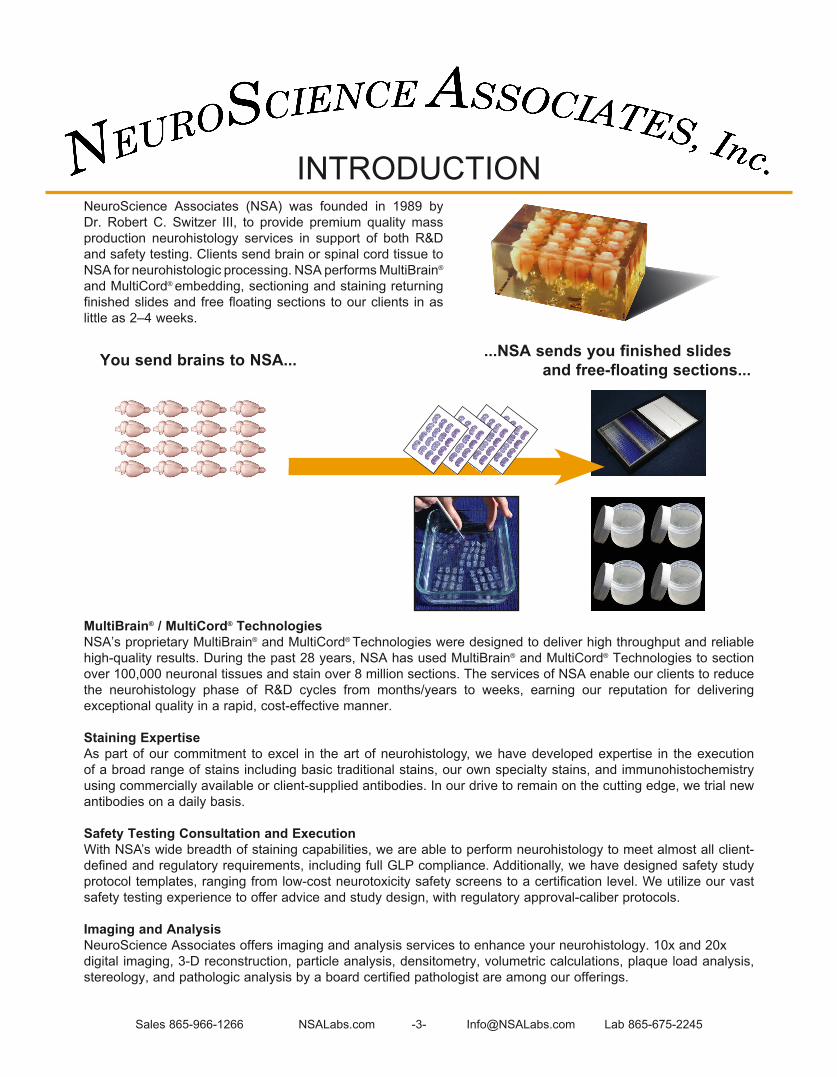
NeuroScience Associates (NSA) was founded in 1989 by Dr . Robert C . Switzer III, to provide premium quality mass production neurohistology services in support of both R&D and safety testing . Clients send brain or spinal cord tissue to NSA for neurohistologic processing . NSA performs MultiBrain® and MultiCord® embedding, sectioning and staining returning finished slides and free floating sections to our clients in as little as 2–4 weeks .
INTRODUCTION
You send brains to NSA... ...NSA sends you finished slides and free-floating sections...
MultiBrain® / MultiCord® TechnologiesNSA’s proprietary MultiBrain® and MultiCord® Technologies were designed to deliver high throughput and reliable high-quality results . During the past 28 years, NSA has used MultiBrain® and MultiCord® Technologies to section over 100,000 neuronal tissues and stain over 8 million sections . The services of NSA enable our clients to reduce the neurohistology phase of R&D cycles from months/years to weeks, earning our reputation for delivering exceptional quality in a rapid, cost-effective manner .
Staining ExpertiseAs part of our commitment to excel in the art of neurohistology, we have developed expertise in the execution of a broad range of stains including basic traditional stains, our own specialty stains, and immunohistochemistry using commercially available or client-supplied antibodies . In our drive to remain on the cutting edge, we trial new antibodies on a daily basis .
Safety Testing Consultation and ExecutionWith NSA’s wide breadth of staining capabilities, we are able to perform neurohistology to meet almost all client-defined and regulatory requirements, including full GLP compliance. Additionally, we have designed safety study protocol templates, ranging from low-cost neurotoxicity safety screens to a certification level. We utilize our vast safety testing experience to offer advice and study design, with regulatory approval-caliber protocols .
Imaging and AnalysisNeuroScience Associates offers imaging and analysis services to enhance your neurohistology . 10x and 20xdigital imaging, 3-D reconstruction, particle analysis, densitometry, volumetric calculations, plaque load analysis, stereology, and pathologic analysis by a board certified pathologist are among our offerings .

Sales 865-966-1266 NSALabs .com -4- Info@NSALabs .com Lab 865-675-2245
NSA provides Neurohistology Services, as well as a rapidly expanding repertoire of Supplemental Services that add tremendous value to our mass production neurohistology . Check our website for the latest information!
NSA SERVICES: COMPREHENSIVE OVERVIEWN
SA
Ser
vice
s: C
ompr
ehen
sive
Ove
rvie
w
NEUROHISTOLOGY
Brain and Spinal Cord Removal by experienced lab personnel Hemisectioning, returning the unused hemisphere Embedding and Sectioning NeuroTechnologiesTM the cornerstone of our services, uses proprietary Technologies to achieve mass production neurohistology . . . . . . . . . . . . . . . . . . . . . . . . . . . . . . . . . . . . . . . . . . . . . . . . . . . . 5 MultiBrain® . . . . . . . . . . . . . . . . . . . . . . . . . . . . . . . . . . . . . . . . . . . . . . . . . . . . . . . . . . . . . . . . . . . . . . . . . . . . . . . . . . . . . . . . . . . . . . . . . . . . . . . . . . . . . . . . . . . . . . . . . . . . . . . . . . . . . . . . . . . . 5 MultiCord® . . . . . . . . . . . . . . . . . . . . . . . . . . . . . . . . . . . . . . . . . . . . . . . . . . . . . . . . . . . . . . . . . . . . . . . . . . . . . . . . . . . . . . . . . . . . . . . . . . . . . . . . . . . . . . . . . . . . . . . . . . . . . . . . . . . . . . . . . . . . . . 8 Large FormatTM . . . . . . . . . . . . . . . . . . . . . . . . . . . . . . . . . . . . . . . . . . . . . . . . . . . . . . . . . . . . . . . . . . . . . . . . . . . . . . . . . . . . . . . . . . . . . . . . . . . . . . . . . . . . . . . . . . . . . . . . . . . . . . . . . . . . . 9 Specialty Stains . . . . . . . . . . . . . . . . . . . . . . . . . . . . . . . . . . . . . . . . . . . . . . . . . . . . . . . . . . . . . . . . . . . . . . . . . . . . . . . . . . . . . . . . . . . . . . . . . . . . . . . . . . . . . . . . . . . . . . . . . . . . . . . . . . . . . . 12 Classic Stains . . . . . . . . . . . . . . . . . . . . . . . . . . . . . . . . . . . . . . . . . . . . . . . . . . . . . . . . . . . . . . . . . . . . . . . . . . . . . . . . . . . . . . . . . . . . . . . . . . . . . . . . . . . . . . . . . . . . . . . . . . . . . . . . . . . . . . . . . 13 Immunohistochemistry services utilize any commercially available antibodies or client-supplied proprietary antibodies . . . . . . . . . . . . . . . . . . . . . . . . . . . . . . . . . . . . . . . . . . . . . . . . . . . . . . . . . . . . . . . . . . . . . . . . . . . . . . . . . . . . . . . . . . . . . . . . . . . . . . . . . . . . . . . . . . . . . . . . . . . . . 15 Titration Series are routinely performed on new antibodies to determine the optimal dilutions, continually expanding our capabilities .
SUPPLEMENTAL SERVICES
Experimental Design, Method Development and Consulting: NSA’s staff, powered by decades of neurohistology experience, routinely assist clients with study design for a wide range of research interests . Applying NSA Services in Research . . . . . . . . . . . . . . . . . . . . . . . . . . . . . . . . . . . . . . . . . . . . . . . . . . . . . . . . . . . . . . . . . . . . . . . . . . . . . . . . . . . . . . . . . . . . . . 34 NeuroSafety™ . . . . . . . . . . . . . . . . . . . . . . . . . . . . . . . . . . . . . . . . . . . . . . . . . . . . . . . . . . . . . . . . . . . . . . . . . . . . . . . . . . . . . . . . . . . . . . . . . . . . . . . . . . . . . . . . . . . . . . . . . . . . . . . 75 GLP: NSA has extensive experience performing all services compliant with GLP . . . . . . . . . . . . . . . . . . . . . . . . . . . . . . . . . . . . 87 Block Face Imaging: Captured during sectioning, these perfectly aligned images facilitate registration and understanding of other derived images (e .g . MRI scans or histology) . The visible white matter tracts supply a surprising amount of useful anatomical information . On their own, 3D reconstructions of the block face provide a valuable reference volume for data visualization purposes, and may be digitally ‘sliced’ in any plane (coronal, sagittal, horizontal, or random oblique cut) . Digital Imaging Enables faster review and team analysis . State -of-the-art instrumentation assures seamless image production of the entire section in sharp focus, at 10x or 20x . Image Analysis offerings are rapidly expanding and currently include: Intensity of Staining . . . . . . . . . . . . . . . . . . . . . . . . . . . . . . . . . . . . . . . . . . . . . . . . . . . . . . . . . . . . . . . . . . . . . . . . . . . . . . . . . . . . . . . . . . . . . . . . . . . . . . . . . . . . . . . . . . . . . . . 24 Percent of Ischemic Area in Stroke Model . . . . . . . . . . . . . . . . . . . . . . . . . . . . . . . . . . . . . . . . . . . . . . . . . . . . . . . . . . . . . . . . . . . . . . . . . . . . . . . . . . . . 25 Quantifying Degenerated Cell vs Survivors . . . . . . . . . . . . . . . . . . . . . . . . . . . . . . . . . . . . . . . . . . . . . . . . . . . . . . . . . . . . . . . . . . . . . . . . . . . . . . . . . . 26 Alzheimer Plaque Burden: Particle Count & Densitometry . . . . . . . . . . . . . . . . . . . . . . . . . . . . . . . . . . . . . . . . . . . . . . . . . . . . . . . . . . . 27 Alzheimer Disease Plaque Quantification.................................................................................... 29 Volumetric Calculations . . . . . . . . . . . . . . . . . . . . . . . . . . . . . . . . . . . . . . . . . . . . . . . . . . . . . . . . . . . . . . . . . . . . . . . . . . . . . . . . . . . . . . . . . . . . . . . . . . . . . . . . . . . . . . . . 30 3D Reconstructions . . . . . . . . . . . . . . . . . . . . . . . . . . . . . . . . . . . . . . . . . . . . . . . . . . . . . . . . . . . . . . . . . . . . . . . . . . . . . . . . . . . . . . . . . . . . . . . . . . . . . . . . . . . . . . . . . . . . . . 31 Volume Rendering . . . . . . . . . . . . . . . . . . . . . . . . . . . . . . . . . . . . . . . . . . . . . . . . . . . . . . . . . . . . . . . . . . . . . . . . . . . . . . . . . . . . . . . . . . . . . . . . . . . . . . . . . . . . . . . . . . . . . . . . 32 Unbiased Stereology: Provides the reference standard for quantification of cytoarchitecture, such as cell number, process length, cell volume, and structure volume . In providing a statistically accurate assessment of these endpoints, unbiased stereology serves as the “gold standard” when benchmarking other methods, or as the definitive measure in critical studies....................................... 33 Storage of Free-Floating Sections: Clients who plan for NSA to perform future stains, as well as those who may be lacking in space, take advantage of our on-site storage . Pathology: Pathologic analyses are performed by board-certified pathologists.

Sales 865-966-1266 NSALabs .com -5- Info@NSALabs .com Lab 865-675-2245
MULTIBRAIN® TECHNOLOGY
MultiBrain® Technology is the cornerstone of the efficient, high-quality neurohistologic processing performed by NSA, the ONLY source for MultiBrain® services in the world .
NSA has revolutionized the execution of neurohistology services . With MultiBrain® Technology, up to 40 brains are embedded together in a solid gelatin matrix and processed as a single unit . This technique achieves uniformity of section thickness and staining quality across cases while providing “built-in” quality control, making subsequent analysis more efficient and efficacious .
-----NSA is able to perform neurohistology up to 40X faster than traditional methods-----
Mass Production NeurohistologyWith MultiBrain® Technology, the economic and qualitative benefits of “mass production” are now available in the neurohistologic field . We section, and we stain, up to 40 brains simultaneously .
Control
Dose Level A
Dose Level B
Dose Level C
MultiBrain® services enable researchers to more efficiently
design experiments and significantly accelerate neuroanalysis. Making
valid comparisons across treatment groups and controls has never been
easier or faster!
MultiBrain® Features:• Unified processing of multiple
brains• Uniform thickness of sections• Uniform staining across all
sections• Variety of possible layouts, and orientations
MultiBrain® Advantages:• Accelerated histology up to 40X
faster than traditional methods• Simpler, more rapid comparative
analysis• Flexibility to retroactively stain
adjacent sections• Inherent quality control
16 Rat Brains Coronal Nissl-Stained
20 Mouse Brains Sagittal Campbell-Switzer AD-Stained
25 Mouse Brains Coronal Nissl-Stained
Analysis of slides is accelerated through MultiBrain® Technology
MultiBrain® principles can be applied to a variety of tissue types, orientations and numbers
40 Mouse Brains Coronal Nissl-Stained
NSA NEUROTECHNOLOGIES™: EMBEDDING AND SECTIONING
Mul
tiB
rain
® T
echn
olog
y
16 rat brains were sectioned together in a single block, and stained with the blood brain barrier compromise stain. Note the differences that can be easily observed across groups and doses by having all of the sections on the same MultiBrain® slide.

Sales 865-966-1266 NSALabs .com -6- Info@NSALabs .com Lab 865-675-2245
MULTIBRAIN® TECHNOLOGY: HOW DOES IT WORK? Clients send brains to NSA. Ideally the brains are stored in preferred solution found on our website
(under Client Resources > Tissue Prep > Shipping) and shipped overnight . NSA will perform the brain removal when requested .
Multiple brains are embedded into each block. NSA embeds up to 40 brains per block . The researcher can specify
the location of each brain in the block using the appropriate “Block Map” template available on our website . This enables efficiencies in subsequent neuroanalysis . Specimens are encased by the gelatin matrix, not infiltrated; therefore, the matrix has no effect on staining .
The block is freeze-sectioned. Sectioning is performed with a sliding microtome, producing
MultiBrain® sheets of sections, in thickness ranging 30–80µ based on the researcher’s specifications .
A resource of sections is created. With MultiBrain® processing, every free-floating sheet that is cut is
collected into a series of 24 cups (NSA default) containing antigen preserve solution . Each succeeding sheet is placed into the next cup in the series of 24, and the process cycles back to cup 1 for the 25th section . Therefore, each cup contains 1 of every 24th cut section; adjacent cups contain adjacent serial sets of sections . The result is a valuable “resource of sections” that provides ample material for several initial stains as well as subsequent stains as needed .
The designated sections are stained. For example, when processing mouse brains, we typically stain every
sixth section with each stain selected by our client . To accomplish this, we would stain tissues from cups 1, 7, 13 and 19 with Stain “A” . A second stain, Stain “B”, could then be applied to the adjacent set of sections, in cups 2, 8, 14 and 20 . The remaining cups are available, from which clients can request other stains, whether planned in advance or warranted based on results from the first set of stains . Clients may also request specific cup(s) be shipped to them for their own staining .
Finished slides are shipped back to the client. The stained slides are anatomically ordered and labeled by NSA .
The entire process from receipt of tissue at NSA to shipment of slides is usually about 3-4 weeks . Any remaining free-floating sections may be returned at the conclusion of the histology and/or stored at NSA for a fee .
1
Mul
tiB
rain
® T
echn
olog
y: E
mbe
ddin
g an
d S
ecti
onin
g
3
6
5
1
2
3
4
5
6
2
PS PP4
1
23
45 6 7 8 9 10
1112
13
14
1516
17181920212223
24
NSA NEUROTECHNOLOGIES™: EMBEDDING AND SECTIONING

Sales 865-966-1266 NSALabs .com -7- Info@NSALabs .com Lab 865-675-2245
MULTIBRAIN® TECHNOLOGYResource of Sections Allows for Differential Staining on Adjacent Sections
Example: Tissue from a human multiple sclerosis brain
Frequently Asked Questions About MultiBrain® Technology:
Q. How is the identity of each brain preserved in the MultiBrain® Block?A. At the time of embedding, only one brain at a time is outside of its container . Once the brain is in the gelatin
matrix, its position relative to the other brains is permanent . The unambiguous orientation of the MultiBrain® Block Map is provided by the black MultiBrain® Reference Marker which is comprised of one or more black dots in a designated corner of each MultiBrain® sheet of sections .
Q. If the brains I want processed by NSA are already frozen, can they be processed successfully?A. Yes, it is possible . If the tissue has not been cryoprotected (with sucrose or glycerol), there is the risk of freeze
artifact (micro tears in the tissue, often rendering it useless for microscopy) . If the tissue has been cryoprotected, then the chance for success is excellent . In either case, the risk for freeze artifact depends on the speed with which the tissue was frozen . Immersion in a slurry of 2-methyl butane (isopentane) and crushed dry ice is best . If the frozen brains were not fixed before freezing, they should be immersed in room temperature 10% phosphate-buffered formalin, with continuous stirring / agitation . Thawing and initial fixation are accomplished in one step . After overnight in the formalin, switch the tissue to buffer and the tissue is ready to be shipped to NSA for processing with MultiBrain® Technology . If the brains had been fixed, cryoprotected or not, immerse the brains in room temperature water or buffer for rapid thawing, with continuous stirring / agitation . The brains are typically able to be processed with MultiBrain® Technology successfully and the resulting sections should reveal scant or no freeze artifact .
Mul
tiB
rain
® T
echn
olog
y: E
mbe
ddin
g an
d S
ecti
onin
g
Near-adjacent sections from the same case of multiple sclerosis (MS) were stained for different features to allow comparisons of recently developed lesions (acute) versus long-standing lesions (chronic) . The hallmark lesion of MS is the loss of myelin seen to the left and high cellularity around vessels (cuffing) seen in each of the photos . This cellularity around small vessels in the acute series is absent in the chronic . Iron positivity corresponds to small vessel cellularity in the center of the lesion of the acute and in the boundary (B) zones where reactive glia are present .
Myelin stain: Weil method Nissl stain: Thionine
ACUTE
CHRONIC
Ferric iron: Perls-DAB Nissl Counterstain
B B B
B B B
NSA NEUROTECHNOLOGIES™: EMBEDDING AND SECTIONING

Sales 865-966-1266 NSALabs .com -8- Info@NSALabs .com Lab 865-675-2245
NSA NEUROTECHNOLOGIES™: EMBEDDING AND SECTIONING
Multiple Embedding of Spinal CordsMultiCord® Technology uses the same revolutionary set of technologies as MultiBrain® Technology, with as many as 40 spinal cords embedded, sectioned and stained simultaneously in the transverse/coronal plane . When there is value in viewing the same cord in different planes, multiple segments and planes of a single spinal cord can be processed together and presented on a single slide . With this configuration, up to 8 spinal cords can be processed within the same MultiCord® block . The images below depict both the “traditional” transverse option for sectioning as well as an example of multiple plane sectioning that can be accomplished through MultiCord® Technology .
40 Rodent Spinal Cords Transverse / Coronal Myelin-Stained
Rhesus spinal cord before (above) and after dividing (below) Segments are oriented during embedding to allow alternate transverse / coronal and
longitudinal sectioning and longitudinal sectioning.
7 Rhesus Monkey Spinal Cords Transverse / Coronal and Longitudinal Thionine-Stained Segments
Mul
tiC
ord®
Tec
hnol
ogy:
Em
bedd
ing
and
Sec
tion
ing
MULTICORD® TECHNOLOGY

Sales 865-966-1266 NSALabs .com -9- Info@NSALabs .com Lab 865-675-2245
Dog
Monkey
Pig
Sheep
LARGE FORMAT™ TECHNOLOGYThe embedding and sectioning of large brains is achieved utilizing many of the principles of MultiBrain® Technology . This Large Format™ Technology enables NSA to section large intact brains continuously with no interruptions . Below are images of brains from relatively small species routinely processed by NSA on 2”x 3” slides, typically sectioned at 40µ, shown with Nissl staining .
For larger brains, a specially designed stage and a modified hydraulically driven microtome are used, resulting in 3”x 5” slides for grizzly bear brains, and for human brain hemispheres . Large sections such as these are typically cut at 60–80µ and are mounted by NSA intact on a single slide .
Larg
e F
orm
at™
Neu
roT
echn
olog
y™:
Em
bedd
ing
and
Sec
tion
ing
NSA NEUROTECHNOLOGIES™: EMBEDDING AND SECTIONING NSA NEUROTECHNOLOGIES™: EMBEDDING AND SECTIONING

Sales 865-966-1266 NSALabs .com -10- Info@NSALabs .com Lab 865-675-2245
NSA NEUROTECHNOLOGIES™: EMBEDDING AND SECTIONING
LARGE FORMAT™ TECHNOLOGY HUMAN BRAIN HEMISPHERES
Key preparation elements for successfully processing human and other large brains (see NSALabs.com for more specific, up-to-date information)
• Minimize the time from death until formaldehyde fixation .
• At brain removal, perfuse fix through the vasculature .
• Store the brain in a large volume (5–10X brain volume) of buffered formaldehyde and/or change regularly .
• NEVER FREEZE the tissue .
NSA works with tissue in a variety of conditions, but best results are achieved using the guidelines above . Contact NSA to discuss your specific needs .
Advantages:• There is uniformity of staining and section
thickness across the entire intact section of the human brain hemisphere .
• For stereology, only intact, adjacent sections yield sampling that is compliant with stereologic principles .
• The inclusion of adjacent structures on the same slide provides anatomical coherence and a comprehensive perspective of the brain, even when a small area of the brain is the focus .
Pictured is a section from a human brain stained with thionine to reveal cell bodies. Magnification shown below.
Tissue Resources for ResearchersNSA can provide recommendations for organizations that bank human brain tissue with specific characteristics (disease, genetic traits, etc.). Some organizations offer specified tissue for purchase. Other organizations grant requested tissue to researchers that fulfill certain requirements, such as use for research or educational purposes .
Larg
e F
orm
at™
Neu
roT
echn
olog
y™:
Em
bedd
ing
and
Sec
tion
ing

Sales 865-966-1266 NSALabs .com -11- Info@NSALabs .com Lab 865-675-2245
SPECIES PROCESSED UTILIZING NSA NEUROTECHNOLOGIES™
In addition to the species pictured above, NSA has also processed:ArmadilloAfrican Green MonkeyBearChickenChinchillaCow
DolphinElephant ForebrainGoat Hammerhead SharkHamsterLizard
ManateeManta RayMouseMud PuppyOpossumPigeon
Marmoset Monkey
RatRabbit
Cat Dog
Grizzly Bear Guinea PigGerbil
Rhesus Monkey
Baboon
PigHuman Hemisphere
Neu
rohi
stol
ogy
of D
iffe
rent
Spe
cies
: E
mbe
ddin
g an
d S
ecti
onin
g
RaccoonSheepSnakeSquidVoleWood Rat
. . .and more!
NSA NEUROTECHNOLOGIES™: EMBEDDING AND SECTIONING NSA NEUROTECHNOLOGIES™: EMBEDDING AND SECTIONING

Sales 865-966-1266 NSALabs .com -12- Info@NSALabs .com Lab 865-675-2245
Ischemia Contrastto reveal the volume of tissue affected following ischemiasee pages 25 and 65 for more details
NSA’s Reactive Microglia Stainto reveal reactive microgliasee pages 46, and 78-79 for more details
SPECIALTY STAINS
Amino Cupric Silver (Amino CuAg)to reveal degeneration*deOlmos JS et al . Neurotox and Teratol 16:545–561,1994see pages 26, 47-48, 57, and 80-83 for more details
Campbell-Switzer Alzheimerto reveal amyloid plaques and tau abnormalities of Alzheimer pathologysee pages 5, 27-29, 36-38 and 46-47 for more details
Spe
cial
ty S
tain
s
Cupric Silver (CuAg) method*to reveal degeneration in fragile tissue*deOlmos JS et . al . Brain Res .33: 523–529,1972 see pages 63, 80 and 84-85 for more details
NSA SERVICES
Blood Brain Barrier (BBB)to reveal the locations of blood brain barrier compromise in the brain see pages 5, 69-71 and 90 for more details

Sales 865-966-1266 NSALabs .com -13- Info@NSALabs .com Lab 865-675-2245
H&Eto reveal the nuclei and cytoplasm of cell bodiessee pages 76 and 80-82 for more details
Thionine (Nissl)to reveal cell bodies in tissue . This may be applied as a counterstain against suitable stains see pages 7, 8, 10, 58, 63 and 84 for more details
Spe
cial
ty /
Cla
ssic
Sta
ins
Autometallographyfor the detection of metalssee page 67 for more details
CLASSIC STAINS (continued on following page)
X-Galto reveal β-galactosidasesee pages 68 and 74 for more details
SPECIALTY / CLASSIC STAINS
NSA SERVICES

Sales 865-966-1266 NSALabs .com -14- Info@NSALabs .com Lab 865-675-2245
Weil-Myelin (human brain)to reveal the myelinated axonssee pages 7-8, 31, 55, 57 and 58 for more details
Cla
ssic
Sta
ins
NSA SERVICESCLASSIC STAINS (continued)
Solochrometo reveal myelinsee page 55 for more details
Perls for ferric Iron (with or without DAB)to detect the presence of ferric iron in tissue (normally occurring, or from ruptured red blood cells)see pages 7, 49, 58, 63 and 72 for more details
AChEto reveal Acetylcholinesterasesee page 66 for more details
Congo Redto reveal dense fibrillar amyloid plaquessee pages 27, 36 and 37 for more details
Thioflavin Sto reveal fibrillar amyloidsee pages 36, 37 and 47 for more details

Sales 865-966-1266 NSALabs.com -15- [email protected] Lab 865-675-2245
IMMUNOHISTOCHEMISTRY / ANTIBODY STAINS
Imm
unoh
isto
chem
istr
y: L
ist
of S
tain
s
4H7H7 Huntington Disease Aggregates6E10 β-Amyloid (1-17)82E1 β-Amyloid (1-16)ApoTag 4APPARCAβ (1-40)Aβ (1-42)α-Synucleinα-Synuclein (pSer129)α-Synuclein-Human 4B12 (103-108)α-Synuclein 211 (121-125)BACE1BDNFβ-galactosidasec-Abl (pTyr412)c-Fosc-Jun (pSer63)CalbindinCalretininCaspase-3-activatedCaspase-9-activatedCathepsin-DCD11bCD11cCD163CD1B3CD3CD4CD45CD45RCD68CD8CGRPChATClaudin-1c-MycCNPase (RIP)CRFd-serineDARPP32DoublecortineGFPEGFREGR-1eIF-alphaEM-48EPOErgothioneine TransporterFerritinFGFR-1FGFR-2FLAGFrataxin (Anti-Human)
GABAGAD-65GAD-67GAP-43GAP-43 (pSer96)GAP-43 (pThr172)GDNFGephrynGFAPGFPGlutamateGPEETGSK-3β (pSer9)HNEHuman Nuclear Protein (HuNu)HuntingtinHypocretin (Orexin)Iba1IFN-gammaIGF-1IgG-2aIgG-CynoIgG-HumanIgG-Human Fc fragmentIgG-Human(H+L)IgG-MonkeyIgG-MouseIgG-RatIgG-SheepKi-67Kv3.1bLamininLAMP-1LC3BLef-1LRRK2MAGMAP-2MCA566MHCIINADPHNCAMNestinNeuNNeuroligin-2NF-1 (Neurofilament-200)nNOSNogo-ANS1 Glycoprotein [DN3]OccludinOlig-2Opioid Receptor-KappaOpioid Receptor-muOrexin (hypocretin)
ParvalbuminPBR1PDGFRbetaPhospho-Akt (Thr308)Pin-1ProcyclinPSA-NCAMPSD-95R10Z8E9RGMaRMO-14 (neurofilament-M)S100 betaSerotoninSerotonin TransporterSMASMI-31SMI-311SMI-312SMI-32SMI-71SMI-94SMI-99 myelin basic proteinSOD1SomatostatinSphingosine kinaseSTEM-101STEM-121STEM-123Substance PSynaptophysinTau 39E10 (189-205)Tau 46 (403-441)Tau 5 (210-230)Tau AT100 (pSer212/pSer214)Tau AT180 (pThr231; PHF-6)Tau AT181 (pThr181)Tau AT8 (pSer202,pThr205)Tau CP-13 (pSer202)Tau HT7 (159-163)Tau MC1 (312-322)Tau PHF-1 (pSer396,404)Tau PHF-13 (pSer396)Tau (pSer422)Tau-ALZ50 (2-10 &312-342)TDP-43TNF-αTryptophan HydroxylaseTyrosine HydroxylaseUbiquitinUSP-14VAChTVEGFYFPZona Occludens-1
Immunohistochemistry (IHC) is used to detect the presence of specific biomolecules (antigens) in tissue sections by applying an antibody to that antigen. IHC has become one of the most common tools in neurohistologic research due to the potential of high specificity when staining a target feature. NSA uses TritonX-100 to fenestrate membranes, allowing antibodies to stain the entire thickness of the section (30–80µ), removing thickness as a factor in NSA’s protocols.
We offer high-quality immunohistochemistry services utilizing commercially available antibodies and client-supplied proprietary antibodies and routinely perform titration series to expand our repertoire.
Below is a list, as of this printing, of commercial antibodies that we have successfully applied. Following the list are representative images of some of our most commonly requested immunohistochemistry. Visit our website for an up-to-date listing. (See index for photo samples throughout this catalog)
NSA SERVICES

Sales 865-966-1266 NSALabs .com -16- Info@NSALabs .com Lab 865-675-2245
Imm
unoh
isto
chem
istr
y
IMMUNOHISTOCHEMISTRY / ANTIBODY STAINS (continued)
Calbindin
Alpha-Synuclein
Didn’t find what you’re looking for? Check our website for a current listing or request a titration series for a new or custom antibody!
BrdU
NSA SERVICES
AT100 (Mouse Hippocampus) 10x
6E10 (Mouse Alzheimer Model)4G8 (Mouse Hippocampus)PS-1/APP Transgenic

Sales 865-966-1266 NSALabs .com -17- Info@NSALabs .com Lab 865-675-2245
c-fos (Mouse Hippocampus)
Doublecortin
Imm
unoh
isto
chem
istr
y
COX-2 ( Rat Hippocampus)
Didn’t find what you’re looking for? Check our website for a current listing or request a titration series for a new or custom antibody!
Calbindin (Sheep Cortex)
cGRP (Mouse Spinal Cord) 10x
EM48 Hippocampus
IMMUNOHISTOCHEMISTRY / ANTIBODY STAINS (continued)
NSA SERVICES

Sales 865-966-1266 NSALabs .com -18- Info@NSALabs .com Lab 865-675-2245
GFP (Rat Cortex)
Imm
unoh
isto
chem
istr
y
Didn’t find what you’re looking for? Check our website for a current listing or request a titration series for a new or custom antibody!
Iba1
GFPGFAP (Rat Hippocampus)Trimethyl Tin Intoxication
phospho GAP 43 pT172 (Rat Olfactory Bulb) GFAP (Mouse Amygdala)Idiopathic Astrocyte Reactivity
IMMUNOHISTOCHEMISTRY / ANTIBODY STAINS (continued)
NSA SERVICES

Sales 865-966-1266 NSALabs .com -19- Info@NSALabs .com Lab 865-675-2245
Myelin Basic Protein(Mouse Spinal Cord)
Orexin A (Rat Hypothalamus)
Imm
unoh
isto
chem
istr
y
Phospho Tau Serine 422(Human Cortex)
Phospho Tau Ser 396 (Mouse Brain)
Didn’t find what you’re looking for? Check our website for a current listing or request a titration series for a new or custom antibody!
nNOS
NeuN (Rat Hippocampus) 2x
IMMUNOHISTOCHEMISTRY / ANTIBODY STAINS (continued)
NSA SERVICES

Sales 865-966-1266 NSALabs .com -20- Info@NSALabs .com Lab 865-675-2245
SMI-312 (Mouse Striatum)
Imm
unoh
isto
chem
istr
y
Didn’t find what you’re looking for? Check our website for a current listing or request a titration series for a new or custom antibody!
Serotonin
SMI-71 (Stroke Rat)
S830, Huntington Disease Mouse ModelHippocampus, 20x
Procyclin in Mouse BrainEdinger-Westphal & Oculomotor Nuclei
IMMUNOHISTOCHEMISTRY / ANTIBODY STAINS (continued)
NSA SERVICES

Sales 865-966-1266 NSALabs .com -21- Info@NSALabs .com Lab 865-675-2245
Somatostatin (Mouse Hippocampus)
Imm
unoh
isto
chem
istr
y
Didn’t find what you’re looking for? Check our website for a current listing or request a titration series for a new or custom antibody!
TDP43 (Human Hippocampus AD) 4x
Tyrosine Hydroxylase (Rat Brain)
IMMUNOHISTOCHEMISTRY / ANTIBODY STAINS (continued)
NSA SERVICES

Sales 865-966-1266 NSALabs .com -22- Info@NSALabs .com Lab 865-675-2245
Flu
ores
cent
Im
mun
ohis
toch
emis
try NeuN Mouse Hippocampus
FLUORESCENT IMMUNOHISTOCHEMISTRY /ANTIBODY STAINS
YFP Rat Cortex
Didn’t find what you’re looking for? Check our website for a current listing or request a titration series for a new or custom antibody!
GFAP Rat Cortex Iba1 Mouse Hippocampus
Iba1 Rat Cortex CD68 ED1 Rat Cortex
NSA SERVICES

Sales 865-966-1266 NSALabs .com -23- Info@NSALabs .com Lab 865-675-2245
QUANTITATIVE ANALYSIS BY NSA
Turn histology into answers with analysis . Once your tissues are processed by one of NSA’s NeuroTechnologies™, the benefits of being stained at the same time and under the same conditions are reaped with minimized variation, increased confidence and lower analytic costs. NSA offers both Image Analysis and Stereological Analysis. We can help you plan a study to quantify most endpoints available with bright field or fluorescent microscopy.
IMAGE ANALYSIS:The first step in analyzing a high resolution image is capturing the image. Our onsite TissueScope™ platform from Huron Digital Pathology performs state-of-the-art digital imaging . These seamless, razor sharp images captured with a 10x or 20x objective allow faster review and a wide range of analytical techniques to be applied .
By applying proven image processing techniques, NSA quantifies a range of changes in stained tissue with highly reproducible results, yielding data superior to subjective assessments . Below are descriptions of the more popular image analyses performed and a summary of the examples on the following pages .
Densitometry is the relative density and intensity of staining in 2 dimensional space, used to measure the signal of a given stain in a particular Area Of Interest (AOI/ROI) . Typically all stained cellular elements are combined: cells, fibers, neuropil. Applications not described herein include assessing injection fields, enzyme staining, fiber density, AAV infection area .
Particle Counts assess the relative size, number, and distribution of stained cellular elements (2D), by focusing on isolated aspects of staining for features such as number of cells, size of deposits, and number of plaques . Densitometric analysis can be applied to particle counts to provide further information .
Volume Analysis is used to measure AOIs/ROIs and compare across animals lesions, brain structures, stained population of cells, etc .
Visualization Tools help to better understand what is going on in tissue, view in planes not available with singular plane histology, and view differences across animal groups
STEREOLOGY is a statistically rigorous means of quantifying morphological elements (2D and 3D) such as cell number, cell area/volume, nuclear area/volumes, process length, etc . See page 33 for more details.
NSA SERVICES: IMAGE ANALYSIS
Imag
e A
naly
sis
DensitometryParticleCounts Volume
VisualizationTools Page #
Intensity of Staining √ 24
% of Ischemic Area in Stroke Model √ 25
Quantifying Degenerated Cells vs Survivors √ 26Alzheimer Plaque Burden: Particle Count and Densitometry √ √ 27
Alzheimer Plaque Quantification √ √ 29
Volumetric Calculations √ √ 30
3D Reconstructions √ √ 31
Volume Rendering √ √ 32
Examples of NSA Image Analysis described on the following pages:

Sales 865-966-1266 NSALabs .com -24- Info@NSALabs .com Lab 865-675-2245
Neuroscience Associates has the capability to quantify antibody staining on tissue processed at our facilities . An example of GFAP quantification in mouse cortex is shown to the left, with corresponding data below .
A specific area of interest (AOI/ROI) is delineated followed by image processing steps to make the area more amenable for quantification. In this example, the total amount of GFAP staining can be measured as both percent area occupied of AOI/ROI and total area occupied in appropriate units (millimeters squared for example) .
Due to the gradation of light to more intense staining, the total GFAP staining can be further divided into medium (shown as green) and high (shown as red) intensity staining totals in the given AOI/ROI . These can be measured as percent area occupied of the AOI/ROI or total area of medium or high intensity staining .
NSA SERVICES: IMAGE ANALYSIS
Brain 1
Brain 2
Brain 3
Area of Medium and High Intensity GFAP Staining3.5
3
2.5
2
1.5
1
0.5
01 2 3
Total Area of GFAP Staining in Cortex3.5
3
2.5
2
1.5
1
0.5
01 2 3
Imag
e A
naly
sis:
Int
ensi
ty o
f S
tain
ing
To further reduce technical variance, expert NSA neuroanatomists ensure consistent regions of interest (AOI/ROI) .
INTENSITY OF STAINING

Sales 865-966-1266 NSALabs .com -25- Info@NSALabs .com Lab 865-675-2245
NSA SERVICES: IMAGE ANALYSISPERCENT OF ISCHEMIC AREA IN STROKE MODEL
Gerbil brain section rendered ischemic from bilateral carotid ligation. The lschemia Contrast stain sharply delineates affected areas.
Once the image of the section is digitized, boundaries of the entire section and individual ischemic zones (1–3) are drawn, using density thresholding techniques with an NIH Image. Total volumes or an “index” of the volumes affected can be calculated from analysis on additional sections.
DATA: Area (sq . units) Entire section 2 .73 Area 1 0 .02 Area 2 0 .17 Area 3 0 .11
% Ischemic area: 10 .99%
Imag
e A
naly
sis:
Per
cent
of
Isch
emic
Are
a in
Str
oke
Mod
el

Sales 865-966-1266 NSALabs .com -26- Info@NSALabs .com Lab 865-675-2245
Imag
e A
naly
sis:
Cel
l T
ype
Qua
ntif
ying
QUANTIFYING DEGENERATED CELLS VS SURVIVORS
Image Processing
Original Image
Knowing the exact degree of change between treatment groups improves therapy modeling, e .g . dose range selection in tolerance or efficacy studies. NSA quantifies the amount of damage and resistance/protection of susceptible cell populations, within any area of interest (AOI/ROI) . Using this precise, reproducible approach yields data superior to subjective assessments, and informs the study design of more rigorous stereological methods .
Below, labeled healthy and dead neurons in rat cortex (NeuN and Amino Cupric Silver degeneration stains) .
Particle Analysis parameters may include:
1 . Total profile number for healthy and degenerating cell populations
2 . A real density of the damaged population3 . Binning by size class category
Method: The original image (top) is converted into binary, false-color representations of the NeuN positive (green) and degenerating cell populations (red) for segmentation (bottom) . The NeuN positive cells are varying shades of red/brown while degenerating neurons are black . This high-contrast staining allows for robust processing and segmentation .
Results:
0
500
1000
1500
2000
2500
Cell P
rofil
e Num
ber
NeuN ACS
NeuN-positive and Degenerating cellular profiles in Rat Cingulate Cortex
MultiBrain® and MultiCord® processing minimizes technical variance, thereby increasing your ability to detect treatment or biological effects .
Expert NSA neuroanatomists ensure consistent AOI/ROIs, further reducing technical variance .
Lower Variance, Increased ConfidenceACSNeuN
Segmented original
Stains amenable to quantification include most cell phenotype and degeneration markers .
NSA SERVICES: IMAGE ANALYSIS

Sales 865-966-1266 NSALabs .com -27- Info@NSALabs .com Lab 865-675-2245
PS-1/APP Mouse Model
Traced image The area of interest is rendered totally black to get total area.
Analyze Particles: Individual plaques are automatically outlined, and the area is calculated for each.
Imag
e A
naly
sis:
Alz
heim
er P
laqu
e B
urde
n
Compilation of data from several animals yields a histogram within which comparisons of different treatment groups and controls can be made.
NSA SERVICES: IMAGE ANALYSISALZHEIMER PLAQUE BURDEN: PARTICLE COUNT AND DENSITOMETRYMouse models of Alzheimer disease are designed to display neuritic plaques similar to those found in humans with the disease . Drugs that may potentially retard plaque development or prevent the creation of plaques are then tested on these mouse models . Part of the determination of the efficacy of a drug candidate is to measure the plaque load, the percent of cortex and/or hippocampus that is occupied by plaques . Other meaningful measures that should be considered when measuring efficacy include quantification of the total number of plaques and the frequency of plaque sizes . Below is an illustration of how NSA gathers the data .
NSA Neurohistology: Neuritic plaques are revealed in freeze-cut, free-floating sections with the Campbell-Switzer staining method . Image analysis is performed on other stains, such as Aß 1-40, Aß 1-42, Thioflavin S, Congo Red, or any other stains that would reveal Alzheimer plaques .
NSA Analysis: High-resolution digital images of designated sections are captured . The percent of the area of cortex-hippocampus occupied by plaques is obtained through the following steps:• Outline cortex and hippocampus and create a separate “traced” image .• From the traced image, two other images are created in order to: 1 . Determine the total area of cortex-hippocampus . 2 . Determine the individual and total area of the plaques .

Sales 865-966-1266 NSALabs .com -28- Info@NSALabs .com Lab 865-675-2245
In addition to percent area occupied and plaque density, visualizing the number of plaques of different sizes may prove valuable . In the two images below, case #1 appears to have numerous large plaques but not many small ones . In #2, however, many small plaques accompany numerous large plaques . This qualitative difference is graphically depicted by plotting the cumulative frequency of sizes for each as shown here, from which quantitative comparisons can be derived .
Large plaques “only”
Large plaques with small “spray patterns”
#2
#1
Imag
e A
naly
sis:
Alz
heim
er P
laqu
e B
urde
n
NSA SERVICES: IMAGE ANALYSISALZHEIMER PLAQUE BURDEN: PARTICLE COUNT AND DENSITOMETRY (continued)

Sales 865-966-1266 NSALabs .com -29- Info@NSALabs .com Lab 865-675-2245
Imag
e A
naly
sis:
Alz
heim
er D
isea
se /
Pla
que
Qua
ntif
icat
ion
ALZHEIMER DISEASE PLAQUE QUANTIFICATION
Original Image: A-β (1-42)
Processed image:
Densitometry Results
0
0.05
0.1
0.15
0.2
0.25
0.3
0.35
Core Shell
Control
Treatment
Segmented original:
% A
rea
Densitometric parameters may include:
1 . Total number of plaques2 . (%) Area occupied by plaques3 . Binning by staining intensity/gradient4 . Binning by size class category
Stains amenable to quantification currently include Campbell-Switzer Alzheimer stain, A-ß(1-40), and A-ß(1-42) .
Method: The original image (top) is processed to a false-color representation of the plaques (middle) for aggregate segmentation (bottom) .
Using the gradation in staining intensity (green to red in the false color image), individual plaques are outlined (blue outline), and the plaque core may be isolated separately (yellow outline) .
Lower Variance, Increased ConfidenceMultiBrain® processing minimizes technical variance, thereby increasing your ability to detect treatment or biological effects .
NSA SERVICES: IMAGE ANALYSIS

Sales 865-966-1266 NSALabs .com -30- Info@NSALabs .com Lab 865-675-2245
NSA offers multiple methods for calculating volumes depending on the researcher’s budget and data endpoint needs . These techniques include the ellipsoid method and Simpson’s Rule method .
The ellipsoid method can be implemented if the area of interest (AOI/ROI) is roughly ellipsoidal in shape . By taking a handful of measurements, a volume estimate can be calculated with ease .
The following formula is used to calculate the volume of ellipsoid-shaped regions of interest:
V = 4/3 π x 1/2(a + b + c)Where V = Volume a = Height of AOI/ROI b = Width of AOI/ROI c = Length of AOI/ROI
The more exact measure of volume calls for using an application such as Image J/FIJI to measure the areas of interest . Eleven (or more but an odd number of) uniformly spaced measures must be made to comply with the application of Simpson’s rule . More levels yields greater accuracy . Simpson’s rule is a power series used to approximate definite integrals as well as areas under curves.
Original images are taken through a series of processing steps (abbreviated above) to allow for thresholding and area of interest (AOI/ROI) selection . Stained images are converted to binary (black and white) by applying an appropriate thresholding filter. A selection is made from these binary images (shown by the yellow outline in the third column of images) . It should be noted that the AOI/ROI selection method is dependent upon the AOI/ROI and the staining of tissue . Each analysis case presents unique challenges regarding tissue and staining, therefore different methods are implemented based on these factors. After AOI/ROI are defined, they are saved and measured (area measurements shown in the bottom right images) . These measurements are then processed using Simpson’s rule for approximating definite integrals (formula shown below).
VOLUMETRIC CALCULATIONS
Imag
e A
naly
sis:
Vol
umet
ric
Cal
cula
tion
NSA SERVICES: IMAGE ANALYSIS
Sales 865-966-1266 NSALabs .com -31- Info@NSALabs .com Lab 865-675-2245
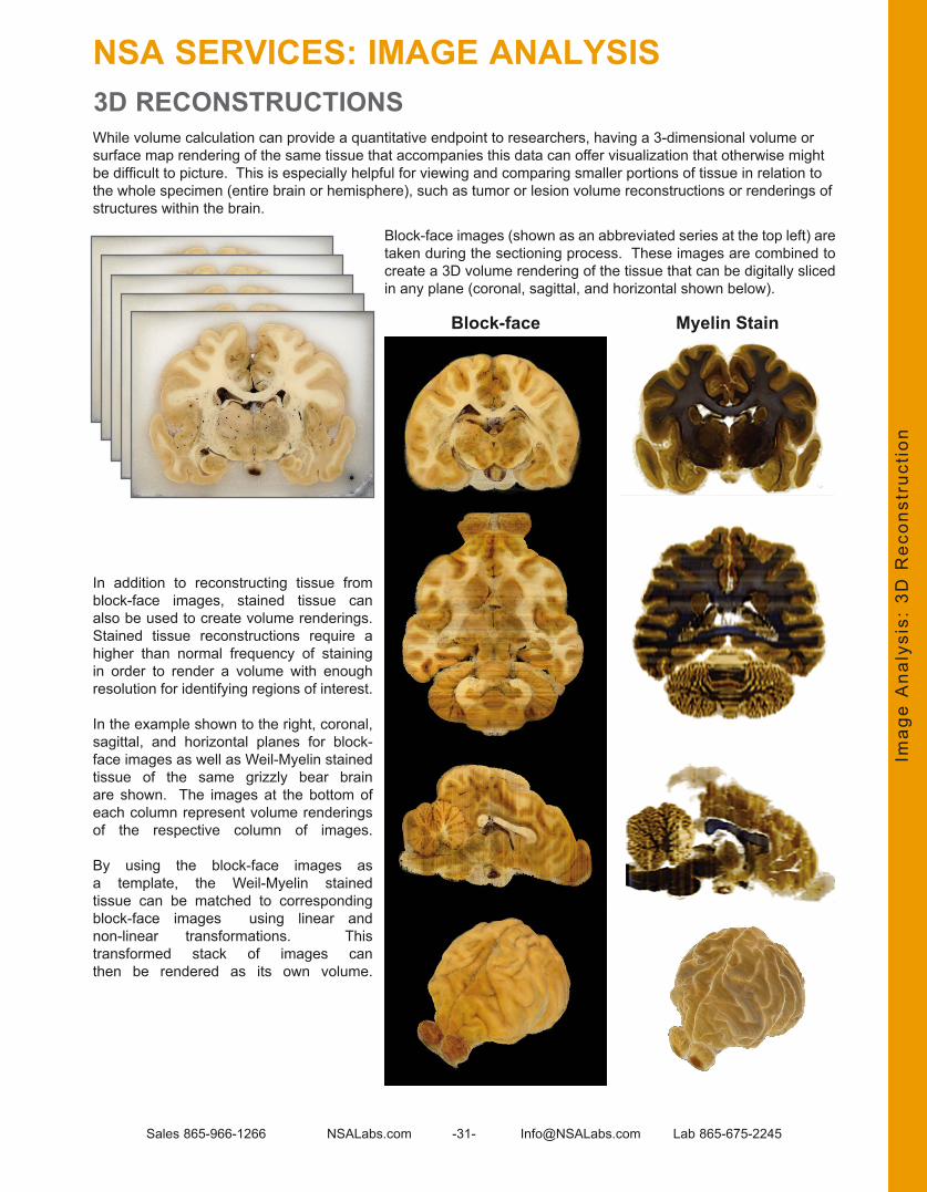
While volume calculation can provide a quantitative endpoint to researchers, having a 3-dimensional volume or surface map rendering of the same tissue that accompanies this data can offer visualization that otherwise might be difficult to picture. This is especially helpful for viewing and comparing smaller portions of tissue in relation to the whole specimen (entire brain or hemisphere), such as tumor or lesion volume reconstructions or renderings of structures within the brain .
Block-face images (shown as an abbreviated series at the top left) are taken during the sectioning process . These images are combined to create a 3D volume rendering of the tissue that can be digitally sliced in any plane (coronal, sagittal, and horizontal shown below) .
In addition to reconstructing tissue from block-face images, stained tissue can also be used to create volume renderings . Stained tissue reconstructions require a higher than normal frequency of staining in order to render a volume with enough resolution for identifying regions of interest .
In the example shown to the right, coronal, sagittal, and horizontal planes for block-face images as well as Weil-Myelin stained tissue of the same grizzly bear brain are shown . The images at the bottom of each column represent volume renderings of the respective column of images .
By using the block-face images as a template, the Weil-Myelin stained tissue can be matched to corresponding block-face images using linear and non-linear transformations . This transformed stack of images can then be rendered as its own volume .
Imag
e A
naly
sis:
3D
Rec
onst
ruct
ion
3D RECONSTRUCTIONS
Myelin StainBlock-face
NSA SERVICES: IMAGE ANALYSIS

Sales 865-966-1266 NSALabs .com -32- Info@NSALabs .com Lab 865-675-2245
In addition to whole brain or hemisphere reconstructions, segmentation of structures or cell populations within the brain can be performed to render even more informative volumes or surface maps . These renderings can be viewed as part of the whole specimen (striatum rendering within the whole brain volume, for example) .This can be especially useful for visualizing and comparing lesions . The images below (from top to bottom in the left column) show a surface map rendering of a pig brain, a surface map rendering of the white matter of the same pig brain, and finally a combination of the two renderings. The opacity of the whole brain was adjusted to view the white matter rendering inside .
Images to the right depict claustrum tracings in a mandrill brain (abbreviated tracings set) along with the reconstructed surface map below . In the bottom right image, the reconstructed claustrum can be seen within the transparent mandrill brain surface map .
Imag
e A
naly
sis:
Vol
ume
Ren
deri
ng
VOLUME RENDERING
NSA SERVICES: IMAGE ANALYSIS

Sales 865-966-1266 NSALabs .com -33- Info@NSALabs .com Lab 865-675-2245
NSA SERVICES: UNBIASED STEREOLOGY
Ste
reol
ogy
ACCURATE ANATOMICAL QUANTIFICATION
Unbiased stereology provides the reference standard for quantitation of cytoarchitecture; such as cell number, process length, cell volume, and structure volume . In providing a statistically accurate assessment of these endpoints, unbiased stereology serves as the “gold standard” when benchmarking other methods, or as the definitive measure in critical studies .
While changes in these measures will capture many experimental effects, perhaps the greatest utility of stereology is the potential to detect the absence of cells following injury, impossible with other methods; i .e . in chronic studies where weeks pass after elements of damage have been removed or disappear .
Plan: While the execution of stereological studies can seem daunting, it’s very efficient with appropriate care in study design . NSA will help you identify and control your histology variables so you can proceed with confidence . This includes the anatomical boundaries of your AOI/ROI, specimen number, section thickness, number of sections, and the number and area of sampling fields required to meet rigorous statistical requirements .
Process: The fundamental advantage of MultiBrain® and MultiCord® technology for stereological studies follows from the analytical efficiency of the MultiBrain® layout (up to 40X specimens per slide), consistency of staining and reliable tissue thickness, and exhaustive sectioning . Additionally, by increasing throughput and lowering costs per section, a project can increase specimen number and reduce the greatest source of variance in counting; inter-specimen/biological variation .
Execute: Count neurons faster with nucleolar staining using the razor sharp AgNOR silver stain, which increases decision speed while reducing fatigue error at the each critical step of identifying neurons . AgNOR may further reduce counting time by allowing lower power objective and/or numerical aperture . Similarly, quickly identify and quantify specific cell types and features with immunohistochemical labeling (e .g . tyrosine hydroxylase or NeuN), optimized for contrast to resolve cellular or subcellular boundaries . Also of note, reduced slide/section sorting and searching via MultiBrain® layout drastically improves efficiency .
Outsource: NSA partners closely with trusted contract service providers for exceptional quality and turnaround times .
AgNOR Stain
TH Stain with Counting Frame

Sales 865-966-1266 NSALabs.com -34- [email protected] Lab 865-675-2245
DISEASE RESEARCH OVERVIEW AND TABLE OF STAINSThe table below depicts some of the most commonly used stains for detecting various categories of markers in Alzheimer (AD), Parkinson (PD), Multiple Sclerosis (MS), Amyotrophic Lateral Sclerosis (ALS),Huntington (HD) and Stroke research.
App
lyin
g N
SA
Ser
vice
s in
Res
earc
h: T
able
of
Sta
ins
Stain AD PD MS ALS HD Stroke4H7H7 Huntington Disease Aggregates AG6E10 β-Amyloid (1-17) A82E1 β-Amyloid (1-16) AAChE cN cN cN cN cN cNAmino CuAg Dg Dg Dg Dg Dg DgApoTag 4 CD CD CD CD CD CDAβ (1-40) AAβ (1-42) Aα-Synuclein cNα-Synuclein (pSer129) cNα-Synuclein-Human 4B12 (103-108) cNα-Synuclein 211 (121-125) cNAutometallography MetD MetD MetD MetD MetD MetDc-Fos act act act act act actCalbindin cN cN cN cN cN cNCalretinin cN cN cN cN cN cNCampbell-Switzer A, pTCaspase-3-activated CD CD CD CD CD CDCaspase-9-activated CD CD CD CD CD CDCD11b G G G G G GCD68 rG rG rG rG rG rGChAT cN cN cN cN cN cNCongo Red A, pTCuAg Dg Dg Dg Dg Dg DgDARPP32 cNEM-48 AGFerritin cN cN cN cN cN cNGABA cNGFAP Ast Ast Ast Ast Ast AstGlutamate cNH&E cN cN cN cN cN cNHuman Nuclear Protein (HuNu) cN cN cN cN cN cNHuntingtin cNIba1 G G G G G GIschemia Contrast pInt pIntKi-67 iN iN iN iN iN iNNestin rAst rAst rAst rAst rAst rAstNeuN cN cN cN cN cN cNNissl cN cN cN cN cN cNParvalbumin cNPerls MetD MetD MetD MetD MetD MetDPSA-NCAM iN iN iN iN iN iNReactive Microglia rG rG rG rG rG rGS100 beta Ast Ast Ast Ast Ast AstSerotonin cN cN cN cN cN cNSerotonin Transporter cN cN cN cN cN cNSMI-99 myelin basic protein myl myl myl myl myl mylSolochrome myl myl myl myl myl mylSomatostatin cN cN cN cN cN cNSubstance P cN cN cN cN cN cNSynaptophysin cN cN cN cN cN cNTau 39E10 (189-205) pTTau 46 (403-441) pTTau 5 (210-230) pTTau AT100 (pSer212/pSer214) pTTau AT180 (pThr231; PHF-6) pTTau AT181 (pThr181) pTTau AT8 (pSer202,pThr205) pTTau CP-13 (pSer202) pTTau HT7 (159-163) pTTau MC1 (312-322) pTTau PHF-1 (pSer396,404) pTTau PHF-13 (pSer396) pTTau (pSer422) pTThioflavin S pT pTTryptophan Hydroxylase cNTyrosine Hydroxylase cNVAChT cNWeil-Myelin myl myl myl myl myl myl
APPLYING NSA SERVICES IN RESEARCH
Code Legend:A = amyloidact = activity markerAG = aggregates :Ast = astrocytes
CD = cell deatCode:cN = chemo-specific neuronDg = Degeneration
G = microgliaiN = immature NeuronsMetD = Metal Detectionmyl = myelin
pInt = physical integritypT = phosphorylated Tau- tangles :rAst = reactive Astrocytes
rG= reactive marker

Sales 865-966-1266 NSALabs .com -35- Info@NSALabs .com Lab 865-675-2245
ALZHEIMER DISEASESee Table of Stains appropriate for AD page 34
OverviewThe hallmark pathological features of AD in humans are amyloid plaques and tau abnormalities . An increasing number of genetically engineered mouse models for AD research have been developed to exhibit classic Alzheimer features . The time line for development of disease features ranges from a few months to ~18 months . NSA has processed hundreds of human AD tissues and tens of thousands of brains from AD mouse models .
Mouse Tissue:When processing mouse brain hemispheres coronally, NSA embeds up to 40 in each MultiBrain® Block (#1); when processing hemisphere or intact brains sagittally, 20 (#2) entire brains are embedded (25 if the area of interest is the cortex/cerebrum) . The preference of the researcher determines which alignment is chosen .
#2 Mouse Brains Sagittal#1 Mouse Brains Hemisphere Coronal
20 mouse brains are co-embedded and appear on each MultiBrain® slide
section (25 mouse brains if the area of interest is only cortex / cerebrum).
40 mouse brain hemispheres are co-embedded and appear on each
MultiBrain® slide section.
Human Tissue:NSA processes large format sections, as seen below, providing a unique opportunity to assess large contiguous cross-sections of tissue . NSA also processes multiple smaller samples from one or more brains using MultiBrain® Technology . The standard NSA practice of encasing the brain tissue in gelatin provides a significant aid in the handling of tissue sections resulting in an improved final product .
Human Brain HemisphereMultiple Embedded Human
Brain Tissues
Alz
heim
er D
isea
se (
AD
)
See pages 27-29 for a comprehensive explanation of our Image Analysis
Services and how they can benefit your Alzheimer Disease Study.
APPLYING NSA SERVICES IN RESEARCH

Sales 865-966-1266 NSALabs .com -36- Info@NSALabs .com Lab 865-675-2245
ALZHEIMER DISEASE (continued)AMYLOID PLAQUES AND TAU ABNORMALITIES
The hallmark features of AD are amyloid plaques and tau abnormalities . Amyloid-laden plaques exist as a diffuse form and as a more dense/mature form (congophilic) . Amyloid is also found as deposits in vessels . Tau abnormalities are present in cell bodies and in the neuropil as “neuropil threads .”
The choice of which stain(s) to use is based on the specific needs of the study . The Campbell-Switzer Alzheimer pathology stain, developed by NSA, shows all of the hallmark features of Alzheimer pathology, while other antibody methods reveal limited, specific features . Please note that the Campbell-Switzer stain does not show this pathology on monkey tissue .
Strategic Approach to Amyloid Detection: Different approaches yield unique featuresAt specific times during an R&D cycle, different approaches to amyloid detection may be most useful . Each of the methods described has its own strengths and specialized advantage .
Campbell-Switzer Method: Dr . Bob Switzer and his former associate, Shannon Campbell, developed the Campbell-Switzer Alzheimer Pathology stain in the early 1980’s . Over the years, this unique stain has become a “work horse” tool for researchers, particularly in the earlier phases of R&D . Diffuse plaques are stained black and the denser fibrillar amyloid forms are stained amber . Tau abnormalities typically stain black .
Immunohistochemistry for Amyloid Plaque Detection: The antibody methods are intended to be very specific and reveal amyloid peptides that have been cleaved at a specific location . Based on a particular mouse model, treatment approach and other factors, a specific antibody is more useful than others in revealing a specific measure of efficacy .
Examples of different antibodies related to AD are shown in the next several pages .
Congo Red: Congo Red specifically reveals the denser, fibrillar amyloid plaques (hence, congophilic plaques) . While the fibrillar amyloid cores stain red, other entities in the sections can also be stained. To distinguish amyloid staining from other staining, the sections can be viewed with polarized microscopy . Fibrillar amyloid stained with Congo Red is birefringent, so when viewed with crossed polarizers a ‘cross’ will be observed with one arm being red and the other ‘apple green’—often termed a ‘maltese cross’. Paired helical filaments that form the tangles in neurons will also display this phenomenon . The birefringence occurs due to the congo red molecules binding in an orderly way on the beta pleated sheet-like configuration of the fibrillar amyloid and the paired helical filaments.
Thioflavin S: This stain, when bound to beta pleated sheet-like configurations of the amyloid and paired helical filaments, will fluoresce a yellow-green. This stain is a useful option if double staining with fluorescent markers are being used .
Alz
heim
er D
isea
se (
AD
)
APPLYING NSA SERVICES IN RESEARCH

Sales 865-966-1266 NSALabs .com -37- Info@NSALabs .com Lab 865-675-2245
The following images were acquired by applying five common techniques to adjacently cut sections of the same Tg2576 mouse brain:
Congo Red Stain Thioflavin S Stain
Aβ 1-40 IHC Aβ 1-42 IHC
Congo Red and Thioflavin S reveal only fibrillar amyloid
(dense-core, congophilic)
plaques . If the goal is to detect ONLY
congophilic plaques, Congo Red and
Thioflavin S are good staining choices .
Aβ 1-40 IHC displays a
broader range of amyloid than the congophilic markers shown
above. Aβ 1-42 IHC displays a broader range of features
than Aβ 1-40.
Campbell-Switzer Alzheimer Pathology Stain
Alternatively, the Campbell-Switzer method stains the broadest range of amyloid while allowing for differentiation between congophilic and diffuse plaques: The Campbell-Switzer method appears to reveal a similar level of amyloid as Aβ 1-42 IHC. However, as can be seen in the close-up, the congophilic plaques stain a different color (amber) vs. the diffuse plaques (black), whereas with the Aβ 1-42 IHC method, both diffuse and congophilic appear the same color . The high-contrast images resulting from this stain are ideally suited for densitometric analysis of plaque loads (more details on pages 27-29) .
Alz
heim
er D
isea
se (
AD
)
APPLYING NSA SERVICES IN RESEARCHALZHEIMER DISEASE (continued)AMYLOID PLAQUES

Sales 865-966-1266 NSALabs .com -38- Info@NSALabs .com Lab 865-675-2245
Neuritic Plaques and Tau abnormalities in Striatum
6E10 Mouse Alzheimer Model
Campbell-Switzer Alzheimer Pathology Stain (Human)
Aβ 1-42 Human Alzheimer
Alz
heim
er D
isea
se (
AD
)
APPLYING NSA SERVICES IN RESEARCHALZHEIMER DISEASE (continued)AMYLOID PLAQUES AND TAU ABNORMALITIES
Early-onset, familial Alzheimer disease in temporal lobe cortex
The pathology is characterized by few neurons with tangles (black circular to tear-drop shapes) in hippocampus (right) while abundant in temporal lobe .
Neuritic plaques (amber-brown) revealed by Aß 1-28 immunohistochemistry in the hippocampus
Blue = Nissl substance (RNA) in cell bodies of neurons and glia . Black = Tau abnormalities and neuropil threads; Gallyas silver method .
Aβ 1-28 IHC
Aβ 1-28 IHC Images courtesy of Dr. Alex Osmand, University of Tennessee, Knoxville

Sales 865-966-1266 NSALabs .com -39- Info@NSALabs .com Lab 865-675-2245
AT8 (pSer202 + Thr205) Surface Cells, P301L Mouse Cortex, 20x
AT8 (pSer202 + Thr205) Unspecified AD Mouse Model, Cortex, 20x
AT8 (pSer202 + Thr205) Unspecified AD Mouse Model, Hippocampus, 20x
AT8 (pSer202 + Thr205) Unspecified AD Mouse Model, Hippocampus, 40x
Anti-Tau HT7 (Epitope 158-163) Mouse P301L AD Model
Anti-Tau HT7 (Epitope 158-163) Mouse P301L AD Model, 10x
Alz
heim
er D
isea
se (
AD
)
APPLYING NSA SERVICES IN RESEARCHALZHEIMER DISEASE (continued) TAU ABNORMALITIES

Sales 865-966-1266 NSALabs .com -40- Info@NSALabs .com Lab 865-675-2245
Anti-Human Tau (Epitope 243-441)Human AD Cortex, 20x
Anti-Human Tau (Epitope 243-441)Magnified to show neuritic plaque and Tau
abnormalities, 40x
TAU 46 (Epitope 404-441)Mouse P301L AD Model, Hemisphere
Tau 46 (Epitope 404-441)Human AD Cortex, 10x
Tau 46 (Epitope 404-441)Human AD Cortex, Magnified to show neuritic plaque
and Tau abnormalities, 20x
Tau 46 (Epitope 404-441)Mouse P301L AD Model, Hippocampus
Alz
heim
er D
isea
se (
AD
)
ALZHEIMER DISEASE TAU ABNORMALITIES (continued)
APPLYING NSA SERVICES IN RESEARCH

Sales 865-966-1266 NSALabs .com -41- Info@NSALabs .com Lab 865-675-2245
Tau pSer396p301L Mouse Model, Cortex and Hippocampus, 10x
Tau pSer396p301L Mouse Model, Hippocampus, 40x
Alz
heim
er D
isea
se (
AD
)
Tau pSer422Human Cortex, 10x
Tau pSer422Human AD Cortex, 20x
Tau pSer422p301L Mouse Model, Cortex and Hippocampus, 2x
Tau pSer422p301L Mouse Model, Cortex and Hippocampus, 4x
ALZHEIMER DISEASE TAU ABNORMALITIES (continued)
APPLYING NSA SERVICES IN RESEARCH

Sales 865-966-1266 NSALabs .com -42- Info@NSALabs .com Lab 865-675-2245
Gallyas Silver Method for revealing Tau abnormalitiesHuman AD Hippocampus 2x
Tau pThr181Human Cortex, 10x
Tau pThr181Human AD Cortex, 1x
Alz
heim
er D
isea
se (
AD
)
ALZHEIMER DISEASE TAU ABNORMALITIES (continued)
APPLYING NSA SERVICES IN RESEARCH

Sales 865-966-1266 NSALabs .com -43- Info@NSALabs .com Lab 865-675-2245
Alz
heim
er D
isea
se (
AD
)
Gallyas Silver Method Human AD Hippocampus 10x
Gallyas Silver Method Human AD Hippocampus 20x
Gallyas Silver Method Mouse Cortex 10x
Gallyas Silver Method Mouse Cortex 20x
ALZHEIMER DISEASE TAU ABNORMALITIES (continued)
APPLYING NSA SERVICES IN RESEARCH

Sales 865-966-1266 NSALabs .com -44- Info@NSALabs .com Lab 865-675-2245
ALZHEIMER DISEASE (continued)OLIGOMERS
Alz
heim
er D
isea
se (
AD
)
Amyloid Oligomer A11 Human AD Cortex, 2x
Amyloid Oligomer A11 Human AD Cortex, 40x
Amyloid Oligomer A11 Human AD Dentate-Gyrus, 4x
Amyloid Oligomer A11 Human AD Dentate-Gyrus, 40x
Beta Amyloid Oligomers
APPLYING NSA SERVICES IN RESEARCH

Sales 865-966-1266 NSALabs .com -45- Info@NSALabs .com Lab 865-675-2245
Anti-T22 Tau oligomers display neurites and tau abnormalities, Human AD cortex, 10x
Alz
heim
er D
isea
se (
AD
)
ALZHEIMER DISEASE (continued)TAU OLIGOMERS
APPLYING NSA SERVICES IN RESEARCH
Anti-T22 Tau oligomers in human AD cortex shown at higher magnification in neuron cell bodies and dendrites. 20x
Anti-Tau T22 Tau oligomers, Human AD cortex, 2x
Tau Oligomers

Sales 865-966-1266 NSALabs .com -46- Info@NSALabs .com Lab 865-675-2245
GFAP Immunoreactive Hypertrophic Astrocytes Around Neuritic Plaque
Alzheimer Mouse Model Plaque Chemoarchitecture
The commonly high density of plaques found in the subiculum is shown . Co-located with the plaques, shown by the Campbell-Switzer method, are reactive microglia (Iba1 immunohistochemistry) . The reactive microglia are more evenly distributed, rather than clustered tightly around particular plaques .
Campbell-Switzer Stain Microglia-Iba1 Antibody Stain
ALZHEIMER DISEASE (continued)INFLAMMATION
Alz
heim
er D
isea
se (
AD
)
Inflammation occurs in conjunction with the hallmark pathologic features of AD and can be detected by examining microglia (Iba1 IHC) or astrocytes (GFAP IHC) .
Iba1 Immunoreactive Microglia: Alzheimer Disease Mouse Model
APPLYING NSA SERVICES IN RESEARCH

Sales 865-966-1266 NSALabs .com -47- Info@NSALabs .com Lab 865-675-2245
Campbell-Switzer Alzheimer Stain AmCuAg Disintegrative Degeneration Stain
Numerous diffuse plaques (in black) and the fewer “mature” plaques (in amber) that contain fibrillar amyloid (congophilic, Thioflavin S positive) are depicted .
The degeneration stain above reveals degenerative globular profiles at the perimeter of the mature (amber) plaques but not with the diffuse plaques . These profiles have been identified as dystrophic neurites (Brendza et al . J Comp Neurol 456: 375–383, 2003) . These may also be engorged glia processes .
ALZHEIMER DISEASE (continued)DISINTEGRATIVE DEBRIS
Sections of the brain of an old, >12 months, PS-1/APP mouse
Alz
heim
er D
isea
se (
AD
)
APPLYING NSA SERVICES IN RESEARCH

Sales 865-966-1266 NSALabs .com -48- Info@NSALabs .com Lab 865-675-2245
Degenerated Axons Amidst Mature Plaques in Olfactory Peduncle
Degenerated Axons in the Corpus Callosum and Striatal Fiber Bundles
Degenerated Axons in Optic Tract
AmCuAg Disintegrative Degeneration Stain Reveals Axon Degeneration in APP mouse
Alz
heim
er D
isea
se (
AD
)
APPLYING NSA SERVICES IN RESEARCHALZHEIMER DISEASE DISINTEGRATIVE DEBRIS (continued)
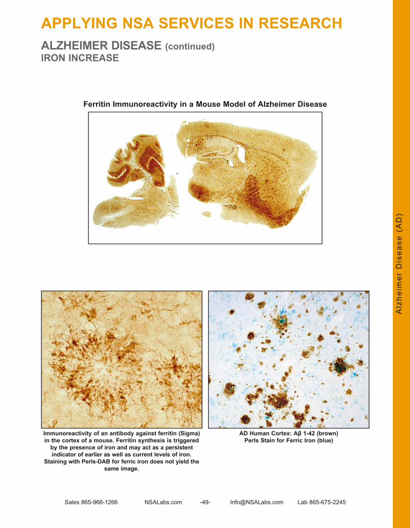
Sales 865-966-1266 NSALabs .com -49- Info@NSALabs .com Lab 865-675-2245
ALZHEIMER DISEASE (continued)IRON INCREASE
Immunoreactivity of an antibody against ferritin (Sigma) in the cortex of a mouse. Ferritin synthesis is triggered
by the presence of iron and may act as a persistent indicator of earlier as well as current levels of iron.
Staining with Perls-DAB for ferric iron does not yield the same image.
Ferritin Immunoreactivity in a Mouse Model of Alzheimer Disease
Alz
heim
er D
isea
se (
AD
)
AD Human Cortex: Aβ 1-42 (brown)Perls Stain for Ferric Iron (blue)
APPLYING NSA SERVICES IN RESEARCH

Sales 865-966-1266 NSALabs .com -50- Info@NSALabs .com Lab 865-675-2245
AMYOTROPHIC LATERAL SCLEROSIS (ALS)See Table of Stains appropriate for ALS page 34
OverviewAlso referred to as Lou Gehrig’s Disease, Amyotrophic Lateral Sclerosis (ALS) is a progressive, neurodegenerative disease affecting nerve cells in the brain and spinal cord . Motor neurons reach from the brain to the spinal cord, and from the spinal cord to the muscles throughout the body . The progressive degeneration of the motor neurons in ALS eventually leads to their death, resulting in a loss of the brain’s ability to initiate and control muscle movement .
ChAT IHC shown in coronal mouse spinal cord to detect motor neuron cells.
ChAT Motor Neuron Marker
ChAT IHC shown in longitudinal plane of mouse spinal cord, 2x.
ChAT IHC in mouse spinal cord magnified to 10x to show details.
Am
yotr
ophi
c La
tera
l S
cler
osis
(A
LS)
Rodent Spinal Cords:Spinal cord tissue of animal models is commonly used in ALS research . There are several approaches NSA employs in processing spinal cord tissue .
40 Rodent Cords Transverse/Coronal
8 Rodent Cords Transverse / Longitudinal
Up to 40 spinal cords are co-embedded and appear on each MultiBrain® slide section .
Up to 8 spinal cords are co-embedded and appear on each MultiBrain® slide section .
Human tissue:NSA processes large format sections, as seen below, providing a unique opportunity to assess large contiguous cross-sections of tissue . NSA also processes multiple smaller samples from one or more spinal cords using MultiBrain® Technology . The standard NSA practice of encasing the spinal cord tissue in gelatin provides a significant aid in the handling of tissue sections resulting in an improved final product .
Multiple-Embedded Human Spinal Cords
APPLYING NSA SERVICES IN RESEARCH

Sales 865-966-1266 NSALabs .com -51- Info@NSALabs .com Lab 865-675-2245
Ubiquitin can help identify abnormal accumulations of this protein inside the cells,
indicating a disease process.mouse spinal cord, horizontal
Ubiquitin, mouse sagittal spinal cord, 10x
Ubiquitin, mouse sagittal spinal cord, 20x Ubiquitin, mouse spinal cord, 40x
Ubiquitin in SOD1 Mouse Model
Am
yotr
ophi
c La
tera
l S
cler
osis
(A
LS)
AMYOTROPHIC LATERAL SCLEROSIS (ALS) (continued)
APPLYING NSA SERVICES IN RESEARCH

Sales 865-966-1266 NSALabs .com -52- Info@NSALabs .com Lab 865-675-2245
Amino Cupric Silver StainAmino Cupric Silver stain in SOD ALS mouse spinal cord model is shown below . The black “squiggles” are red blood cells within vessels and the fine black lines indicate bundles of degenerated axons .
90 day old SOD1 mouse model, 4x
120 day old SOD1 mouse model, 4x 120 day old SOD1 mousesagittal spinal cord model, 10x
90 day old SOD1 mouse model, 10x
Am
yotr
ophi
c La
tera
l S
cler
osis
(A
LS)
APPLYING NSA SERVICES IN RESEARCHAMYOTROPHIC LATERAL SCLEROSIS (ALS) (continued)

Sales 865-966-1266 NSALabs .com -53- Info@NSALabs .com Lab 865-675-2245
EM48 CortexEM48 Striatum
EM48 and S830 Show the Aggregates of Huntington Disease
Hun
ting
ton
Dis
ease
(H
D)
HUNTINGTON DISEASESee Table of Stains appropriate for HD page 34
APPLYING NSA SERVICES IN RESEARCH
EM48 Hippocampus S830, Huntington Disease Mouse ModelHippocampus, 20x
OverviewThe pathological features of Huntington Disease that are most often studied through neurohistology include de-myelination, axonal degeneration and inflammation involving an immune reaction including high cellularity around vessels . NSA has processed both human and animal model brains and spinal cords in support of Huntington Disease research .
Rodent Brains:Brain tissue of animal models is commonly used in Huntington Disease research . There are several approaches NSA employs in processing brain tissue .
25 Mouse Brains Coronal
Up to 25 mouse brains are co-embedded and appear on each
MultiBrain® slide section .

Sales 865-966-1266 NSALabs .com -54- Info@NSALabs .com Lab 865-675-2245
OverviewThe pathological features of Multiple Sclerosis (MS) that are most often studied through neurohistology include de-myelination, axonal degeneration and inflammation involving an immune reaction including high cellularity around vessels . NSA has processed both human and animal model brains and spinal cords in support of MS research .
Rodent Spinal Cords:Spinal cord tissue of animal models is commonly used in MS research . There are several approaches NSA employs in processing spinal cord tissue .
40 Rodent Cords Transverse/Coronal
8 Rodent Cords Transverse / Longitudinal
25 Mouse Brains Coronal 16 Rat Brains Coronal
Up to 40 spinal cords are co-embedded and appear on each MultiBrain® slide section .
Up to 25 mouse brains are co-embedded and appear on each
MultiBrain® slide section .
Up to 16 rat brains are co-embedded and appear on each
MultiBrain® slide section .
Up to 8 spinal cords are co-embedded and appear on each MultiBrain® slide section .
40 Mouse or 32 Rat Brain Hemispheres Coronal
Up to 40 mouse brains are co-embedded and appear on each
MultiBrain® slide section .
Rodent Brains:
Human tissue:NSA processes large format sections, as seen below, providing a unique opportunity to assess large contiguous cross-sections of tissue . NSA also processes multiple smaller samples from one or more brains using MultiBrain® Technology . The standard NSA practice of encasing the brain tissue in gelatin provides a significant aid in the handling of tissue sections resulting in an improved final product .
Human Brain Hemisphere Multiple-Embedded Human Brain Tissues
Mul
tipl
e S
cler
osis
(M
S)
MULTIPLE SCLEROSISSee Table of Stains appropriate for MS page 34
APPLYING NSA SERVICES IN RESEARCH

Sales 865-966-1266 NSALabs .com -55- Info@NSALabs .com Lab 865-675-2245
MULTIPLE SCLEROSIS (continued)DE-MYELINATION
A variety of methods can be used to reveal myelin, which effectively exposes areas of de-myelination . The most common techniques employed by NSA include the Weil-Myelin method and Solochrome method .
Thalamus Cingulate
Solochrome
Weil-Myelin
Mul
tipl
e S
cler
osis
(M
S)
APPLYING NSA SERVICES IN RESEARCH

Sales 865-966-1266 NSALabs .com -56- Info@NSALabs .com Lab 865-675-2245
MULTIPLE SCLEROSIS (continued)INFLAMMATION
As with other disease models, inflammation markers such as Iba1 and GFAP can be useful indicators of perturbations in conjunction with other MS pathology, as seen below .
GFAP
Iba1
Plaques in White Matter
Mul
tipl
e S
cler
osis
(M
S)
APPLYING NSA SERVICES IN RESEARCH

Sales 865-966-1266 NSALabs .com -57- Info@NSALabs .com Lab 865-675-2245
MS Monkey Spinal Cord: Adjacent Stained Sections
Myelin StainWeil Method
Neurodegeneration StainAmino-Cupric Silver
MULTIPLE SCLEROSIS (continued)AXONAL DEGENERATION
Longitudinal section (from the same cord shown above) that clearly reveals the degenerating pattern of axons. These images suggest that axon degeneration may
precede the loss of myelin.
Degenerated axons (black) are visible in the upper right, but are not in the same location
in the myelin-stained image to the left. Mul
tipl
e S
cler
osis
(M
S)
APPLYING NSA SERVICES IN RESEARCH

Sales 865-966-1266 NSALabs .com -58- Info@NSALabs .com Lab 865-675-2245
MULTIPLE SCLEROSIS (continued) LESIONS AND CUFFING
The presence of high cellularity around vessels, a characteristic of MS pathology which is often called cuffing, as seen around the large vessels in these adjacent sections .
Myelin stain: Weil Method
Nissl stain: Thionine
ACUTE CHRONIC
Ferric iron:Perls-DAB Nissl
Counterstain
Different stains provide different perspectives into the architecture of the MS lesions . For example, the margin of the plaque in the chronic plaque contains cells (microglia) rich in iron, while the corresponding zone in the acute plaques does not .
Mul
tipl
e S
cler
osis
(M
S)
APPLYING NSA SERVICES IN RESEARCH

Sales 865-966-1266 NSALabs .com -59- Info@NSALabs .com Lab 865-675-2245
PARKINSON DISEASESee Table of Stains appropriate for PD page 34
OverviewThe primary pathological features of Parkinson Disease (PD) in humans are the loss of dopamine cells, the presence of Lewy bodies in existing cells and changes in the expression of alpha-synuclein . Disease models vary and NSA employs a wide range of tools suited to the traits of specific models .
Rodent Tissue:Rodent brains are the primary tissue processed in PD Research by NSA . NSA can process up to 40 tissues in each MultiBrain® Block .
25 Mouse Brains Coronal 16 Rat Brains Coronal40 Mouse or 32 Rat Brain
Hemispheres Coronal
Up to 25 mouse brains are co-embedded and appear on
each MultiBrain® slide section.
Up to 16 rat brains are co-embedded and appear on
each MultiBrain® slide section.
Up to 40 mouse brains are co-embedded and appear on
each MultiBrain® slide section.
Human Tissue:NSA processes large format sections, as seen below, providing a unique opportunity to assess large contiguous cross-sections of tissue . NSA also processes multiple smaller samples from one or more brains using MultiBrain® Technology . The standard NSA practice of encasing the brain tissue in gelatin provides a significant aid in the handling of tissue sections resulting in an improved final product .
Human Brain HemisphereMultiple-Embedded
Human Brain Tissues
Par
kins
on D
isea
se (
PD
)
APPLYING NSA SERVICES IN RESEARCH

Sales 865-966-1266 NSALabs .com -60- Info@NSALabs .com Lab 865-675-2245
PARKINSON DISEASE (continued)TYROSINE HYDROXYLASE IHC
PD research models typically use a method for inducing the loss of dopamine cells, or create lesions that interrupt the axons projecting to striatum . Tyrosine Hydroxylase (TH) is a robust marker of elements of the dopamine system .
Rat brain normal expressing TH equally in both hemispheres.
Rat animal model treated to create the loss of the dopamine cells that produce TH.
16 rat brains of a PD model on a MultiBrain® slide at the approximate same levels. All brains have lost TH staining in the left striatum.
TH-Stained Substantia Nigra
A silver nucleolar stain (AgNOR: Ag Nucleolar Organizing Region protocol used in oncology research), when paired with Tyrosine Hydroxylase IHC staining, is an excellent tool for use in stereology in Parkinson research .
AgNOR-TH in Substantia Nigra
Tyrosine Hydroxylase IHC
Par
kins
on D
isea
se (
PD
)
APPLYING NSA SERVICES IN RESEARCH

Sales 865-966-1266 NSALabs .com -61- Info@NSALabs .com Lab 865-675-2245
PARKINSON DISEASE (continued)ALPHA-SYNUCLEIN
Alpha-Synuclein pSer129Mouse Brain Cortex, 20x
Alpha-Synuclein pSer129Synthetic A-Syn Fibril Injected
Mouse Brain Cortex, 20x
Alpha-Synuclein pSer129Mouse Brain Cortex,10x
Alpha-Synuclein pSer129Substantia nigra in a human PD brain, cytoplasmic and axonal aggregates, 60x
Alpha-Synuclein pSer129A53T mouse cortex,
pyramidal cells
Par
kins
on D
isea
se (
PD
)
APPLYING NSA SERVICES IN RESEARCH

Sales 865-966-1266 NSALabs .com -62- Info@NSALabs .com Lab 865-675-2245
Placement of a vector containing the code for human alpha-synuclein in rat brains is taken up by cells and Alpha-Synuclein is expressed.
Transgenic mouse brain expressing human Alpha-Synuclein 211 (121-125), 20x (left) and 40x (right)
Par
kins
on D
isea
se (
PD
)
APPLYING NSA SERVICES IN RESEARCHPARKINSON DISEASEALPHA-SYNUCLEIN (continued)

Sales 865-966-1266 NSALabs .com -63- Info@NSALabs .com Lab 865-675-2245
PARKINSON DISEASE (continued)DISINTEGRATIVE DEGENERATION
In PD research, models that employ an acute method of destroying the TH-producing cells, the degeneration of those cells can be witnessed using a Disintegrative Degeneration stain: Cupric Silver deOlmos method .
Degenerated neurons in substantia nigra in MPTP-treated BALB/C57 mouse
InflammationAs with other disease models, inflammation markers such as GFAP and Iba1 can be useful indicators of perturbations in conjunction with other PD pathology .
Aberrant Iron in PDIron ‘decompartmentalized’ has been implicated in the pathologic process . Useful tools for probing this include Perls-DAB for ferric iron and Ferritin IHC for the iron storage molecule .
Perls-DAB reveals iron in the substantia nigra (left) and red nucleus
(right) of this human tissue. This section was counterstained for nissl
substance with Thionine (blue).
Par
kins
on D
isea
se (
PD
)
APPLYING NSA SERVICES IN RESEARCH

Sales 865-966-1266 NSALabs .com -64- Info@NSALabs .com Lab 865-675-2245
STROKESee Table of Stains appropriate for Stroke page 34
OverviewAnimal models for stroke attempt to induce a cerebral ischemia to mimic the effects of a stroke in humans, which tends to be more difficult to replicate in smaller rodents than in larger animals . Neurohistology for stroke research primarily focuses on methods to quantify the ischemic region and characterize the margin of surviving tissue .
Animal Models:
25 Mouse Brains Coronal 16 Rat Brains Coronal Dog Brain
Up to 25 mouse brains are co-embedded and appear on
each MultiBrain® slide section.
Up to 16 rat brains are co-embedded and appear on
each MultiBrain® slide section.
Large brains such as monkey, sheep, and dog are processed
intact. If hemisected two hemispheres are co-embedded
and appear on the same MultiBrain® slide section.
SMI-71 Endothelial Barrier Protein EBPRat-normal vessels
Blood Brain Barrier (BBB) Compromise
SMI-71 Endothelial Barrier Protein EBP Rat stroke model reveals loss of EBP associated with
Blood Brain Barrier Compromise
Str
oke
See Stroke Image Analysis
page 25
APPLYING NSA SERVICES IN RESEARCH

Sales 865-966-1266 NSALabs .com -65- Info@NSALabs .com Lab 865-675-2245
STROKEISCHEMIA
Protective effects of interventions after stroke are ideally based on clinical neurologic outcomes and subsequent neurohistology . In small animal stroke models, difficulties with long survival times and neurologic evaluation prompt many investigators to perform acute studies and determine protection by histologic evaluations immediately after inducing an infarction . Many stains have been used to identify infarct areas, but it is not clear if these areas include or could delineate the penumbra, i .e ., the potentially salvageable tissue at the margin of the infarct . The triphenyltetrazolium chloride (TTC) method, for instance, is known to give variable infarct volumes depending on how long after the infarct the tissue is harvested . A fortuitous observation led to a staining method which delineates the penumbra while highlighting the infarcted volume in sharp contrast . Normal tissue is dense black, infarcted areas fail to stain and gradations of gray identify the penumbra zone . Section images are digitized and, based on optical density, the volumes of the infarct, penumbra and normal brain are calculated . Adjacent sections stained with other methods including H&E, thionine and GFAP IHC display less definition of affected areas .
Appearance of stroke in popular animal models with decreasing severity
GerbilLigation of Carotid Arteries
RatMiddle Cerebral Artery Occlusion
Str
oke
APPLYING NSA SERVICES IN RESEARCH
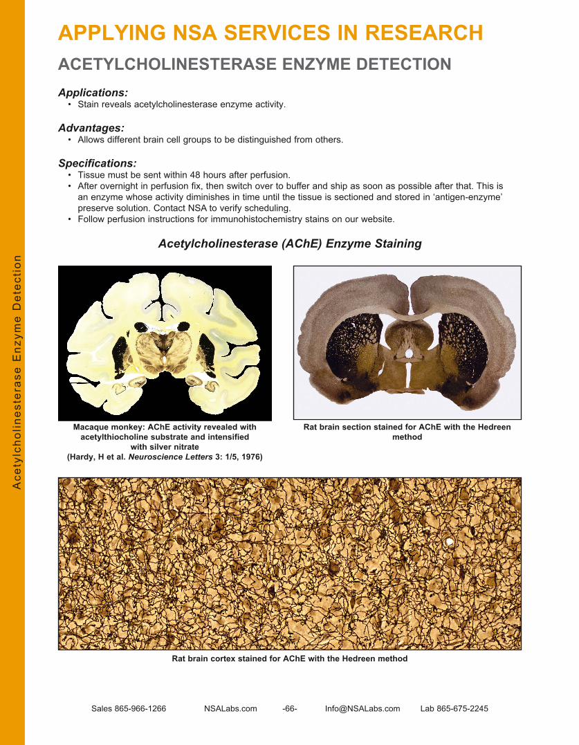
Sales 865-966-1266 NSALabs .com -66- Info@NSALabs .com Lab 865-675-2245
ACETYLCHOLINESTERASE ENZYME DETECTIONApplications:
• Stain reveals acetylcholinesterase enzyme activity .
Advantages:• Allows different brain cell groups to be distinguished from others .
Specifications:• Tissue must be sent within 48 hours after perfusion . • After overnight in perfusion fix, then switch over to buffer and ship as soon as possible after that. This is
an enzyme whose activity diminishes in time until the tissue is sectioned and stored in ‘antigen-enzyme’ preserve solution . Contact NSA to verify scheduling .
• Follow perfusion instructions for immunohistochemistry stains on our website .
Acetylcholinesterase (AChE) Enzyme Staining
Macaque monkey: AChE activity revealed with acetylthiocholine substrate and intensified
with silver nitrate(Hardy, H et al. Neuroscience Letters 3: 1/5, 1976)
Ace
tylc
holin
este
rase
Enz
yme
Det
ecti
on
Rat brain section stained for AChE with the Hedreen method
Rat brain cortex stained for AChE with the Hedreen method
APPLYING NSA SERVICES IN RESEARCH

Sales 865-966-1266 NSALabs .com -67- Info@NSALabs .com Lab 865-675-2245
METAL DETECTION: AUTOMETALLOGRAPHY
Applications:• Used for the detection of various metals in tissue, e .g ., Bi, Cu, Hg, Au and Ag .
Advantages:• A histologic means of detection for the location of certain metals .• Complements the nonanatomical detection of metals by atomic absorption (A .A) or by ICP (Inductively
Coupled Plasma) spectroscopy .
Autometallography is a histologic technique that exploits the presence of metal ions in tissue to act as a nucleation point around which silver ions are gathered to create a visible reaction product . The process is called “physical development .” The Timm’s silver sulfide method is the most well-known autometallography procedure and it favors the revelation of zinc but not other metals .
Gorm Danscher in Denmark has championed the application of other autometallography protocols for detecting various metals in tissue . Another contributor, F . Gallyas of Hungary, has developed a family of physical developers that broaden the range of metals detectable with autometallography .
Neurons in the hypoglossal nucleus of a mouse that was chronically administered bismuth sub-salicylate. The location of bismuth was revealed using an acidic
physical developer described by Gallyas.
(Ross, J .F ., Switzer, R .C . III, Poston, M .R ., and Lawhorn, G .T . Distribution of bismuth in the brain after intraperitoneal dosing of bismuth subnitrate in mice: Implications for routes of entry of xenobiotic metals into the brain . Brain Res 725: 137–154, 1996)
Met
al D
etec
tion
: A
utom
etal
logr
aphy
APPLYING NSA SERVICES IN RESEARCH

Sales 865-966-1266 NSALabs .com -68- Info@NSALabs .com Lab 865-675-2245
ß-GALACTOSIDASE (ß-GAL) DETECTION
Applications:• The X-Gal enzyme stain and the ß-Gal IHC method are tools to reveal ß-Gal presence . • These are used to identify the product of the lacZ gene, which is used as a marker for identifying cells that
have been introduced into a host animal or transfected with a viral vector .
Advantages:• The signal observable from the enzyme method is usually considered superior to that achieved with the IHC
method . • The enzyme method forms an intense blue precipitant in the presence of ß-Gal expression . • Tissue must be sent shortly after perfusion, as this is an enzyme whose activity diminishes with time . • Contact NSA to verify scheduling .
Specifications:• Tissue must be sent within 48 hours after the animal is perfused . • After perfusion and over night in fix, switch the brains to buffer (PBS) and then ship as soon as possible to
NSA . This is an enzyme whose activity diminishes with time . • Contact NSA to verify scheduling .
β-Galactosidase IHC (β-Gal)
X-Gal Enzyme Histochemistryß-G
alac
tosi
dase
(ß
-Gal
) D
etec
tion
APPLYING NSA SERVICES IN RESEARCH

Sales 865-966-1266 NSALabs .com -69- Info@NSALabs .com Lab 865-675-2245
BLOOD BRAIN BARRIER (BBB) COMPROMISE DETECTION
Applications:• Anti-IgG Stain reveals the locations of blood brain barrier compromise in the brain .
Advantages:• Allows for visualization of BBB compromise that is not accompanied by a loss in cells .• Does not require the injection of a foreign protein marker (i .e ., horseradish peroxidase (HRP)) into the
vasculature .
Specifications:• Perfusion fixation is preferred with overnight in perfusion fix then transfer to PBS. CAUTION: sacrifice
with CO2 suffocation and not immediate brain removal and placement in fix can allow serum proteins to extravasate into the brain parenchyma and yield a false positive of BBB compromise (see image bottom right on p 71) .
The locations of BBB compromise can be revealed with immunohistochemistry by using an antibody against the animal’s own IgG . Areas with “leaky” BBB allow serum proteins to pass into the brain parenchyma, e .g ., IgG’s and albumin .
BBB Compromise by Physical TraumaIn the MultiBrain® section below (derived from freeze-sectioning a MultiBrain® Block of 16 rat brains), forebrain sections are shown from rat brains that had been injected with a test article . Different amounts were injected, then the rats were sacrificed at different times following the injection . The result was different degrees of leaky BBB as indicated by the brown coloration (the blue is a Nissl counterstain) . Note the halo of BBB immunostaining extends beyond the margin of the frank lesion .
Blo
od B
rain
Bar
rier
Com
prom
ise
Det
ecti
on
APPLYING NSA SERVICES IN RESEARCH

Sales 865-966-1266 NSALabs .com -70- Info@NSALabs .com Lab 865-675-2245
BLOOD BRAIN BARRIER (BBB) COMPROMISE DETECTION (continued)LOOD BRAIN BARRIER (BBB) COMPROMISE STAIN
The images in each panel below show sections from the brains of two different rabbits that were subjected to fine air emboli to create multiple infarctions . In the top panel, the locations of BBB compromise are seen as deep amber-colored areas . In the bottom panel, the lesion sites are seen by an absence of staining of cell bodies . The arrows illustrate some of the infarction sites in each of the differently stained, near adjacent sections . In the section on the left, the rather large halo of IgG staining goes beyond the lesion site near the third ventricle, as revealed with the Nissl stain below it . On the other hand, in the section on the right, the angled arrows show two distinct sites of IgG staining that correspond in size to the unstained patches in the Nissl section .
Other zones of IgG staining do not correspond to a loss of Nissl staining, perhaps illustrating BBB compromise which is unaccompanied by the loss of cells . For example, note the large area in the right section that encompasses the left external capsule, as well as the area adjacent to the left optic nerve (vertical arrows) .
NSA staff hypothesizes that the BBB may not merely be open or shut but may be opened to varying degrees such that molecules of different sizes would accordingly penetrate different degrees. For a given time after BBB compromise, small serum proteins may be found at a greater radius from a given site than larger proteins, i.e., IgGs.
Anti-IgG Immunohistochemistry
Nissl Substance (RNA)
Blo
od B
rain
Bar
rier
Com
prom
ise
Det
ecti
on
APPLYING NSA SERVICES IN RESEARCH

Sales 865-966-1266 NSALabs .com -71- Info@NSALabs .com Lab 865-675-2245
There is evidence that sacrifice by CO2 causes the BBB to open during the sacrifice process . When conducting a BBB compromise study, the impact of this event could serve to give a false indication of brain penetration for a particular compound, as the compound may not have crossed the BBB under normal circumstances . We surmise that the extreme stress, induction of acidosis and lack of oxygen during the several minutes before death result in a breakdown of the BBB . If the brain is not removed immediately or soon after cessation of breathing, IgG serum proteins may enter the brain parenchyma . The images below depict the difference between a normal IgG antibody staining pattern of an anesthetized, perfused mouse versus abnormal IgG antibody staining seen from a mouse sacrificed by CO2 suffocation . The brain was not removed immediately after death .
NSA RECOMMENDATION: If the intactness of the BBB is to be studied, the animals should be anesthetized with a barbiturate such as Nembutal, then perfused transcardially with a suitable aldehyde fix . Note that use of gaseous anesthetics such as Fluothane tends to prevent flushing of red blood cells from the brain vasculature .
References of potential interest:Use of anti-IgG to detect BBB breakdown:Richmond, J .D ., et al . Induction of heme oxygenase-1 after hyperosmotic opening of the blood-brain barrier . Br Research 780: 108–118, 1998
References of interest within this paper:37) Rapoport, S .I ., et al . Quantitative aspects of reversible osmotic opening of the blood-brain barrier . Am J Physiol 238: R421–R431, 198045) Tanno, H ., et . al . Breakdown of the blood-brain barrier after fluid percussive head injury in the rat . Part I . Distribution and time course of protein extravasation . J Neurotrauma 9: 21–32, 1992
Other serum proteins (albumin, fibrinogen, fibronectin):Yu, W .R ., et al . Systemic hypothermia following compression injury of rat spinal cord: reduction of plasma protein extravasation demonstrated by immunohistochemistry . Acta Neuropath (Berl) 98: 15–21, 1999
Entry of other agents:Muldoon, L .L ., et al . Comparison of intracerebral inoculation and osmotic blood-brain barrier disruption for delivery of adenovirus, herpes virus, and iron oxide particles to normal rat brain . Am J Path 147: 1840–1851, 1995
The appearance of IgG staining in normal (mouse) brain shows only some tincture in periventricular
structures and other areas known to have leaky BBB, such as the median eminence and area postrema.
Brain from a mouse sacrificed by CO2 suffocation and brain was not removed and placed in fix soon
after death. Widespread IgG staining results due to extravasation of serum proteins including IgGs.
Blo
od B
rain
Bar
rier
Com
prom
ise
Det
ecti
on
APPLYING NSA SERVICES IN RESEARCHBLOOD BRAIN BARRIER (BBB) COMPROMISE DETECTION (continued) CO2 SACRIFICE

Sales 865-966-1266 NSALabs .com -72- Info@NSALabs .com Lab 865-675-2245
ELECTRODE TRACT DETECTION
Applications:• Electrodes or injection are commonly made into the brain or spinal cord . Determining the locations is
essential for the recording of brain electrical events and for injections of an experimental substance .
Advantages:• The penetration into the brain or spinal cord by even the finest electrode or needle inadvertently nicks
capillaries causes a small amount of blood to enter the parenchyma . The iron content of the red blood cells is soon released and remains in that location virtually permanently . Detection of the presence of the iron becomes an excellent means of revealing the tract and end points of electrodes and injection needles .In conjunction with the presence of the iron, the astrocytes in that locale are rendered in a persistent reactive state . Detection of this reactivity with staining with a GFAP antibody provides a means to cross check the iron date .
Specifications:• Transcardial perfusion with fix and execution of the Perls reaction histochemical procedure (for ferric iron)
ensures successful tract identification. Standard IHC methods for applying the GFAP antibody also allows for successful acquisition of data .
GFAP Stain
Perls-DAB Stain
Ele
ctro
de T
ract
Det
ecti
on
APPLYING NSA SERVICES IN RESEARCH

Sales 865-966-1266 NSALabs .com -73- Info@NSALabs .com Lab 865-675-2245
STEM CELL DETECTION
Applications:• Stem cells are introduced into a living host to often achieve changes in the host’s composition . E .g . to replace missing/lost cells; replace cells with absent or faulty protein production .
Advantages:• The success of stem cell introduction is measured by a number of possible means but the initial goal is that
the stem cells were placed in the correct location; that they survive and produce the changes intended . This assessment is accomplished by histologic examination of the hosts after different lapses in time after stem cell introduction .
Specifications:• Optimal fixation of the host brains by transcardial perfusion optimizes the conditions to ensure successful
identification of the stem cells in the host animal.
Identification of Juvenile Neurons: Doublecortin (dbx) IHC Reveals Neurons That Are <7 Days Old
Ste
m C
ell
Det
ecti
on
APPLYING NSA SERVICES IN RESEARCH

Sales 865-966-1266 NSALabs .com -74- Info@NSALabs .com Lab 865-675-2245
Cells were tagged with β-Galactosidase, revealed by X-Gal histochemistry, demonstrates a
successful localization of the newly placed cells.
The darkly stained cells (dots), mostly along the inner edge of the “arrowhead” of the dentate formation indicate DNA synthesis activity. In this manner, cell division can be witnessed and quantified. The purple staining is normal cell body background stained for anatomical reference.
STEM CELL DETECTION (continued)
Stem Cell LocationA fundamental measure of efficacy for stem cell treatments is to determine whether the stem cells reached the desired location in the central nervous system . Various immunohistochemistry probes can be used to detect signature markers of the stem cells (such as a unique human protein) . Also stem cells can be deliberately made to produce markers such as β-Galactosidase, green fluorescent protein or others so that subsequent staining is able to highlight the location of introduced stem cells .
Stem Cell Activity Example: Bromodeoxyuridine (BrdU) IHCFor some stem cell therapies, it is desirable for the cells to undergo a limited degree of division within the target organ. BrdU injection into the host animal (ip) before sacrifice (e.g. 6-12h) will allow the BrdU to be incorporated into newly formed DNA of dividing cells . BrdU masquerades for thmydine, which is only used in DNA synthesis . New cells created after the BrdU injection will contain BrdU which is detected and quantified with an antibody against BrdU .
STEM 121 (human stem cells) in mouse spinal cord
Ste
m C
ell
Det
ecti
on
APPLYING NSA SERVICES IN RESEARCH

Sales 865-966-1266 NSALabs .com -75- Info@NSALabs .com Lab 865-675-2245
NSA NEUROSAFETY™ TESTINGOVERVIEW
NeuroScience Associates was founded in 1989 with the primary goal of delivering neurohistologic safety testing services to researchers worldwide. While NSA has expanded significantly in both size and breadth of offerings since that time and provides a full suite of neurohistology services including R&D discovery and efficacy of therapies, safety testing has continued to be a primary service for over 28 years . NSA’s unique approaches to testing capitalize on the capabilities of contemporary neurohistologic techniques thereby increasing the reliability of results . Reductions in the number of stains, the number of animals, the amount of time necessary to achieve results, and the amount of time and money required to conduct the safety tests are among the many benefits of working with our staff of experts .
Safety testing can take many forms depending on the phase of research, the mechanism of the test article and the audience for which the testing is being performed . NSA recognizes these differences, works with clients to assure their specific needs are met, and accommodates the very specific needs of a client’s predefined protocol, as well as requests from regulatory agencies such as FDA and EPA .
ApproachThere is no single approach to every neurologic safety testing need . Rather, a toolset of approaches exists from which each safety assessment study can choose the tools most appropriate to the study .
Behavioral vs. Pathological AssessmentsBehavioral and pathologic tests have their own unique strengths and challenges . These are complementary approaches in the realm of safety testing and should be employed in a manner suited to each .
Spectrum of Pathologic Endpoint DetectionUnfortunately, there is not a single endpoint that is both valid and comprehensive for meeting the needs of all pathologic safety evaluations . Rather, there is a spectrum of safety considerations in the pathology of the brain . At one end of the spectrum, there are no detectable changes to the Central Nervous System (CNS) (safe), and at the other end of the spectrum are permanent changes to the CNS (unsafe) . Between these two extremes, for example, are detectable changes in chemistry, neurotransmitter responses and perturbations leading to an inflammatory response . In the illustration below, the farther a change falls to the right of the spectrum of changes, the more serious is the concern that change represents from a safety perspective . At the left end of the spectrum (i .e ., chemistry changes), there are many potential pathologic endpoints from which to choose . Moving to the right (unsafe) side of the spectrum, the number of detection methods decreases and endpoints are more specific and definitive as a safety concern . While each safety study has different approaches, the best designs include evaluation of a selected point on this spectrum as well as assessments to the right of that point . Many designs focus on merely the most conclusive endpoint of permanent damage at the far right of the spectrum .
A successful safety study must first determine the scope of changes to be evaluated, then choose the methods appropriate to evaluate those chosen endpoints.
Neu
roS
afet
y™ T
esti
ng
See NSALabs.com for our “Neurotoxicity Assessment Presentation 2015”.
Go to NSALabs.com > Reference > Papers and Reference Materialsand select the link.

Sales 865-966-1266 NSALabs .com -76- Info@NSALabs .com Lab 865-675-2245
CHEMISTRY OR OTHER CHANGES FROM NORMAL
In its most literal form, neurotoxicity could be defined as any departure from normal in the CNS . However, all departures from normal or changes in CNS are not negative . In fact, many compounds deliberately cause changes to the CNS as therapy . Judgment is required as to what changes constitute a negative impact and should be evaluated in the scope of a safety study . Most often, compounds that rely on a specific mechanism of action are evaluated with respect to their impact on the brain relative to that mechanism to make sure intended effects are not accompanied by unintended ones . There are an incredible number of endpoints that could be evaluated to include a departure from normal . H&E is an example of one of the many tools used to evaluate general changes in the brain . Another common approach is to evaluate endpoints specific to the known mechanism of a compound specifically quantifying the impact on the brain . Examples of endpoints that are evaluated to reveal chemistry changes are shown in the images below:
Calbindin Serotonin
ChAT
Neu
roS
afet
y™ T
esti
ng
NSA NEUROSAFETY™ TESTING

Sales 865-966-1266 NSALabs .com -77- Info@NSALabs .com Lab 865-675-2245
PERTURBATIONS / INFLAMMATION
The detection of perturbations in the brain is often employed as a key tool in safety assessments . The presence of a perturbation signal is an undesirable finding in safety testing and represents a degree of harm has been caused to the brain . In some approaches, perturbations are used as an indicator to search for signs of permanent damage, serving as a “canary in a coal mine .” In the context of some studies, the detection of a perturbation in itself is enough to warrant a conclusive safety concern . A perturbation represents a stress to the health of the brain, but a perturbation signal alone does not indicate the infliction of permanent damage to the brain . Other endpoints discussed in the next section are able to differentiate an injury/perturbation that resolves versus an injury that leads to permanent damage .
Astrocyte Perturbations:GFAP IHC reveals all astrocytes ‘resting’ or reactive and Nestin IHC reveals only reactive astrocytes .
GFAP
Nestin-Reactive Astrocytes
Neu
roS
afet
y™ T
esti
ng
NSA NEUROSAFETY™ TESTING

Sales 865-966-1266 NSALabs .com -78- Info@NSALabs .com Lab 865-675-2245
Iba1 IHC
PERTURBATIONS / INFLAMMATION (continued)PERTURBATIONS/INFLAMMATION
Microglia PerturbationsIba1 IHC and NSA’s internally developed Reactive Microglia stain are capable of revealing activated microglia . Reactivity in Iba1 stained sections is determined by hypertrophy of the cells . The Reactive Microglia stain shows cells that are in a reactive state due to an acute perturbation . Microglia in a reactive state due to a chronic perturbation are not visible with this method .
Iba1 Hypertrophied microglia Iba1 Normal microglia
Neu
roS
afet
y™ T
esti
ng
NSA NEUROSAFETY™ TESTING

Sales 865-966-1266 NSALabs .com -79- Info@NSALabs .com Lab 865-675-2245
NSA NEUROSAFETY™ TESTINGPERTURBATIONS / INFLAMMATION (continued)
NSA’s Reactive Microglia StainResident microglia of the brain become reactive following various forms of insult, including chemical trauma, viral infection and physical trauma . The NSA protocol stains reactive microglia but not resting microglia . After staining sections from animals that had a broad range of insults, it was realized that this ‘reactive microglia’ protocol favors the staining of microglia where the injury was acutely induced . Microglia in animals with a chronic state of perturbation, such as in EAE (experimental allergic encephalitis), failed to stain---later, using the Iba1 antibody, staining revealed abundant microglia in a state of hypertrophy . Consequently, NSA’s reactive microglia staining protocol should only be used in animals that have suffered acute injury . In acutely injured brains, NSA’s reactive microglia stain reveals the same reactive features found by using the mouse and rat specific CD68 antibodies (FA11 and ED1, respectively) and the isoB4 lectin staining .
Iba1-hypertrophied microglia in rat cortex CD68 clone ED-1 (rat)
CD68 clone FA-11 (mouse)Iba1-hypertrophied microglia in mouse cortex
Iba1 vs CD68 Not all hypertrophied microglia stain positive with CD68.
Neu
roS
afet
y™ T
esti
ng
NSA’s reactive microglia stain

Sales 865-966-1266 NSALabs .com -80- Info@NSALabs .com Lab 865-675-2245
PERMANENT DAMAGE
Many alterations to the brain can be negative and potentially long-lasting, but not all can be considered permanent . Some injuries can be treated or will resolve after time if the source of the injury is removed (i .e ., a harmful compound ceases to be administered), while other injuries lead to permanent implications . The most definitive and easy way to interpret evidence of permanent damage is the destruction of neurons . Even though the brain has amazing functional compensatory power, the loss of irreplaceable neurons creates a permanent deficit . All comprehensive safety evaluations at some point need to evaluate the risk of permanent damage and the mechanism of that evaluation is to observe neurodegeneration as it occurs during the cell death cycle .
There are timing and scope considerations in designing the safety study and in selecting a detection method.
TimingDetection of neurodegeneration requires critical timing considerations . Depending on the detection method chosen, a dying cell can be viewed for only ~2–6 days from the time it begins to disintegrate, after which there is no debris remaining to observe . Therefore, the timing of sacrifice following exposure to a compound is a critical element of study design . The graphic below shows the timeline of the detectability of degenerating neurons for some known neurotoxins . The days indicate the amount of time following a single acute dose on day 0 . The bars represent the most probable timing opportunities for detection .
Scope of ElementsThe neuron has an extended temporal and spatial footprint when the dendrites, axon terminals and axons are evaluated in addition to the neuron cell body itself . The class of stains called the disintegrative degeneration stains is able to detect disintegration of each of these elements, while other methods capable of detecting cell death only reveal pathology of the cell body itself . Disintegrative degeneration stains include various Fluoro-Jade and Cupric Silver methods . NSA routinely performs the disintegrative degeneration stains as well as methods such as H&E .
Neu
roS
afet
y™ T
esti
ng
NSA NEUROSAFETY™ TESTING

Sales 865-966-1266 NSALabs .com -81- Info@NSALabs .com Lab 865-675-2245
NSA NEUROSAFETY™ TESTINGPERMANENT DAMAGE / SCOPE OF ELEMENTS (continued)
Features visible in a Full Scope Disintegrative Degeneration Stain (Amino CuAg) compared to a Cell Body–Only Stain (H&E)
Amino CuAg Control Amino CuAg Affected
H&E Control H&E Affected
H&E Affected
Amino CuAg Affected
In the Amino Cupric Silver–stained section, the disintegrating cell bodies are clearly visible, and the degenerating synaptic terminals, dendritic debris and axons are also conspicuous . In the H&E-stained section, the damage is far less obvious and the characteristic eosinophilia of the cytoplasm and pyknotic nuclei of affected cells requires higher magnification to be distinct, as shown on the right . While it is possible to witness cell death with either category of stain, the degeneration stains provide a higher contrast signal leading to cost and time efficiencies during analysis .
Neu
roS
afet
y™ T
esti
ng

Sales 865-966-1266 NSALabs .com -82- Info@NSALabs .com Lab 865-675-2245
Degenerating Synaptic Terminals Degenerating Dendrites
Degenerating Cell Bodies Degenerating Axons
Of all four degenerated elements shown on this page, only the cell bodies in the picture below (not including the dendrites) are visible with a cell body
stain such as H&E.
PERMANENT DAMAGE / SCOPE OF ELEMENTS (continued)
Features visible in the Amino CuAg Disintegrative Degeneration–stained sections
Neu
roS
afet
y™ T
esti
ng
NSA NEUROSAFETY™ TESTING

Sales 865-966-1266 NSALabs .com -83- Info@NSALabs .com Lab 865-675-2245
NSA NEUROSAFETY™ TESTING
Beta-Amyloid Precursor Protein (ß-APP),rat TBI model, corpus callosum and cortex
ß-APP, rat hippocampus
Beta-Amyloid Precursor Protein (ß-APP)
Beta-Amyloid Precursor Protein (ß-APP) has become a useful marker for axonal injury and commonly termed: ‘diffuse axon injury’ . ß-APP is carried out by fast anterograde transport vesicles to distal sites in the axon . Upon axonal injury, ß-APP pools in areas of impaired transport .
Alternative means of detecting axon damage use the disintegrative degeneration stains (most notably, the amino cupric silver (ACS) method of deOlmos) . Adjacent sections stained with the ACS stain and for ß-APP reveal quite different degrees of staining: much more axonal injury/damage is shown by the ACS suggesting that the ß-APP staining is revealing a totally different form of damage or a subset of what is diplayed in the ACS stain .
Amino Cupric Silver Stain
Neu
roS
afet
y™ T
esti
ng
Amino Cupric Silver Stain- rat TBI model, corpus callosum and cortex
Amino Cupric Silver Stain Rat Cortex
PERMANENT DAMAGE / SCOPE OF ELEMENTS (continued)

Sales 865-966-1266 NSALabs .com -84- Info@NSALabs .com Lab 865-675-2245
NEONATE AND JUVENILE
Developmental Neurotoxicity Assessment: Neonatal Programmed Cell DeathThe assessment of permanent damage in neonates and juveniles is difficult due to the normal occurrence of programmed cell death in the developing brain, and timelines for disintegration (and the corresponding windows to view these) are shortened from days to hours . Patterns of degeneration must be compared to a baseline level to determine changes in cell death activity . Architecting a study design to account for the earlier and shorter observable opportunity of cell death becomes a critical consideration .
The disruption of the chronology of programmed cell death (apoptosis) in the developing brain is an increasing issue of concern . As an example, the Disintegrative degeneration stains, i .e . CuAg have been used to show how alcohol causes extensive cell death in neonate rats (Ikonomidou et al . Science 257: 1056, 2000) .
Adjacent sections from neonate rat brain stained by NSA
The top two images are from two adjacent sections of the anterior thalamus from a 3 day old rat in which abundant programmed cell death (apoptosis) is taking place. The upper left image is stained for nissl substance to show neuron cell bodies and the upper right with the cupric silver stain to show disintegrative degeneration-seen as black dots. The ease of detection of degeneration in the cupric silver stained section vs the cell body only nissl stain is quite clear. A more highly magnified view of both of these stains is shown in the lower two images. Numerous dark blue dots can be detected among normal cells in the nissl stained image which are the pyknotic nuclei cells undergoing apoptosis. In the lower right image the black irregular profiles of degenerating cells are quite conspicuous and easily detected.
Neu
roS
afet
y™ T
esti
ng
NSA NEUROSAFETY™ TESTING
Thionine Nissl Stain
CuAg Disintegrative degeneration stain

Sales 865-966-1266 NSALabs .com -85- Info@NSALabs .com Lab 865-675-2245
NSA NEUROSAFETY™ TESTING
Density of Degeneration vs. Age in Ventral Septum of
Rat Neonate
NEONATE AND JUVENILE (continued)
The temporal histogram below illustrates peak times of cell death in the ventral septum of rat brain . The tabulation of all neuronal populations will identify critical periods during which brain development might be perturbed .
Degeneration in the Lateral Septum; Postnatal Day 3
Neu
roS
afet
y™ T
esti
ng

Sales 865-966-1266 NSALabs .com -86- Info@NSALabs .com Lab 865-675-2245
PREDEFINED PROTOCOLS
NSA has generic protocols for meeting common safety testing needs, as well as offering assistance in the design and execution of custom safety testing protocols . NSA offers two general approaches to neuropathologic safety testing: “NeuroSafety™ Certification” and “Neurotoxicity Screening .” Additionally, NSA has developed a packaged certification protocol for meeting FDA requirements for N-methyl-D-aspartic acid (NMDA) receptor antagonists .
NeuroSafety™ CertificationCertification studies are GLP-compliant protocols designed specifically to evaluate compounds in order to meet regulatory requirements . NSA has predefined protocols that are designed to meet the needs of most clients . These study designs are then customized to meet specific regulatory requirements or to address specific safety concerns of a particular class of compound .
Neurotoxicity ScreeningWhile NeuroSafety™ Certifications are performed as a requirement intended to prove the safety of a compound, Neurotoxicity Screens are conducted by NSA using the Amino CuAg Disintegrative degeneration stain, a strategic tool during the R&D cycle with the goal of identifying high-risk candidates early . Neurotoxicity screens are rapid, inexpensive tests conducted during preclinical development stages designed to offer valuable decision-making data . Eliminating non-viable compounds early offers a huge potential for R&D cost savings:
Janet Woodcock, Former FDA Director of Center for Drug Evaluation and Research (CDER) stated: “There is not enough research in finding new methods of understanding toxicology at the basic
scientific level.... Improving any one of these issues could achieve a 10% improvement in predicting failures before clinical trials and could save $100 million in development costs per drug.”
NSA’s Approach to Neurotoxicity Screening Saves Companies Time and MoneyNSA’s screening protocols reduce development cycle times and regulatory approval timelines and enable more effective use of R&D resources . The protocols provide a new mechanism to “weed out” non-viable compounds . Resources may instead be directed toward modifying non-viable compounds, refocused on viable candidates or reassigned to new candidates .
Early Indicator for Neurotoxicity Screening NSA has developed a ‘turn key’ protocol to determine at an early stage in drug development if candidate compounds are neurotoxic . Multiple compounds can be tested simultaneously . The protocol uses a minimum number of animals; a maximally tolerable dose; the amino cupric silver stain to detect degeneration, and a pathologist’s read and report that dichotomously indicates the presence of degeneration or not . Contact NSA for more specific details and pricing.
Neu
roS
afet
y™ T
esti
ng
NSA NEUROSAFETY™ TESTING

Sales 865-966-1266 NSALabs .com -87- Info@NSALabs .com Lab 865-675-2245
PREDEFINED PROTOCOLS (continued)
NMDA Receptor Antagonist CertificationThe FDA has a testing requirement as specified in an FDA document entitled “RECOMMENDATIONS FOR THE DEVELOPMENT OF NMDA RECEPTOR ANTAGONISTS .” This is based upon an Agency letter dated January 27, 1995, describing the FDA’s current recommendations for development of NMDA receptor antagonists . The requirements in this document are based upon safety concerns discovered with MK-801, and the FDA now provides this document to sponsors that are submitting a compound for approval in this class of compounds . NSA has developed a GLP safety certification study design created specifically to meet the FDA’s latest interpretation of these requirements .
Good Laboratory Practices (GLP)NSA is proficient in performing services compliant with GLP for any of our services. GLP is defined as Good Laboratory Practices as promulgated by USFDA 21CFR Part 58, USEPA and OECD . NSA utilizes an independent auditor with over 25 years QA experience to provide services related to all GLP-regulated studies .
Services provided by the auditor include: • Inspect facility periodically, provide GLP training to NSA staff, review Standard Operating Procedures (SOPs),
review equipment records • Review protocols, inspect work in progress, audit study data, audit study reports, provide QA Statement to
Study Director • Report inspection findings to NSA management and to Study Director and testing facility QA • Host visiting QA site visits
Neu
roS
afet
y™ T
esti
ng
NSA NEUROSAFETY™ TESTING
See NSALabs.com for our “Neurotoxicity Assessment Presentation 2015”.
Go to NSALabs.com > Reference > Papers and Reference Materialsand select the link.

Sales 865-966-1266 NSALabs .com -88- Info@NSALabs .com Lab 865-675-2245
OVERVIEW
Planning your Study . . . . . . . . . . . . . . . . . . . . . . . . . . . . . . . . . . . . . . . . . . . . . . . . . . . . . . . . . . . . . . . . . . . . . . . . . . . . . . . . . . . . . . . . . . . . . . . . . . . . . . . . . . . . . . . . . . . . . . . . . . . . . . . . . . . . . . . . . . . . . .89Tissue Preparation for Optimal Processing . . . . . . . . . . . . . . . . . . . . . . . . . . . . . . . . . . . . . . . . . . . . . . . . . . . . . . . . . . . . . . . . . . . . . . . . . . . . . . . . . . . . . . . . . . . . . . . . . . . . . .89MultiBrain® and MultiCord® Block Maps . . . . . . . . . . . . . . . . . . . . . . . . . . . . . . . . . . . . . . . . . . . . . . . . . . . . . . . . . . . . . . . . . . . . . . . . . . . . . . . . . . . . . . . . . . . . . . . . . . . . . . . . . . . .90Quote Requests . . . . . . . . . . . . . . . . . . . . . . . . . . . . . . . . . . . . . . . . . . . . . . . . . . . . . . . . . . . . . . . . . . . . . . . . . . . . . . . . . . . . . . . . . . . . . . . . . . . . . . . . . . . . . . . . . . . . . . . . . . . . . . . . . . . . . . . . . . . . . . . .91Shipping ABC’s . . . . . . . . . . . . . . . . . . . . . . . . . . . . . . . . . . . . . . . . . . . . . . . . . . . . . . . . . . . . . . . . . . . . . . . . . . . . . . . . . . . . . . . . . . . . . . . . . . . . . . . . . . . . . . . . . . . . . . . . . . . . . . . . . . . . . . . . . . . . . . . . .92Free Floating Tissue Storage and Handling . . . . . . . . . . . . . . . . . . . . . . . . . . . . . . . . . . . . . . . . . . . . . . . . . . . . . . . . . . . . . . . . . . . . . . . . . . . . . . . . . . . . . . . . . . . . . . . . . . . . .93
Visit our website at NSALabs.com for up-to-date information referenced in this section as well as Frequently Asked Questions, papers, presentations, MultiBrain University™ webinar recordings, and
much more!
Clie
nt R
esou
rces
CLIENT RESOURCES
1 2 3 4
6 7 8 9
11 12 13 14
16 17 18 19
5
10
15
20
21 22 23 24 25
NSA recommendsfor both domestic and
international shipments
NSA recommendsfor both domestic and
international shipments

Sales 865-966-1266 NSALabs .com -89- Info@NSALabs .com Lab 865-675-2245
PLANNING YOUR STUDY
Contact NSA Labs® during the planning phase of your study: • Benefit from NSA’s staff, powered by decades of neurohistology experience, who routinely assist clients with study design for a wide range of research interests . From dosing, to survival times, to animal counts, to stain selections, to analysis, our staff will help shape the details that can make the difference between a successful study and an unsuccessful study . • Maximize the advantages of NSA’s high throughput NeuroTechnologies™ including MultiBrain®, MultiCord® and Large Format™ Technologies . • Gain valuable assistance in preparing grants .
TISSUE PREPARATION FOR OPTIMAL PROCESSING
NSA tissue preparation protocols are critical to maximize the results of MultiBrain® and MultiCord® processing . Review in detail the perfusion protocols and carefully select the appropriate perfusion solution . Consider the stain(s) that will be executed, including potential stains that may be executed at a later date (see resource of sections on page 6) . If your project involves spinal cords, also follow the post-perfusion spinal cord preparation . Pre-evaluations of tissue will be performed by NSA prior to processing to ensure intended results .
Visit our website NSALabs .com and select Client Resources|Tissue Preparation to view the most up-to-date protocols including:
Perfusion Instructions, Pump Calibration
Post-Perfusion Spinal Cord Preparation
Fixation Method, Solutions*, and Timing
Post-Fixative Buffer Solutions, Storage and Timing
Shipping Preparation and Instructions of Tissue
*High-quality perfusion solutions yield high-quality results . Commercially available formaldehyde solutions are NOT fresh and usually have added ingredients to “stabilize” the solution for a long shelf-life .
Pla
nnin
g Y
our
Stu
dy
CLIENT RESOURCES

Sales 865-966-1266 NSALabs .com -90- Info@NSALabs .com Lab 865-675-2245
MULTIBRAIN® AND MULTICORD® BLOCK MAPS
A Full Experiment in One BlockTo specify the location of each tissue in the block, complete the appropriate map, email to histology@nsalabs .com, and include a copy with the tissue shipment . Visit our website and choose Client Resources to access the map best suited for your experiment . Below are examples of four common maps .
Mul
tiB
rain
® a
nd M
ulti
Cor
d® B
lock
Map
s
Rat Brain Block Map:up to 16 coronally-embedded brains
1 2 3 4
5 6 7 8
9 10 11 12
13 14 15 16
1 2 3 4
6 7 8 9
11 12 13 14
16 17 18 19
5
10
15
20
21 22 23 24 25
Mouse Brain Block Map:up to 25 coronally-embedded brains
These maps represent the relative location of individual brain or cord sections on slides . Specifying where each tissue will appear on the slides facilitates comparison across treatment groups and controls . The MultiBrain® Reference Marker preserves left-right orientation . If a Block Map is not provided, the brains/cords will be embedded left to right, in an ascending alphanumeric sequence based on client ID#’s .
MultiBrain® Reference Markers
CLIENT RESOURCES
Rodent Spinal Cord Block Map:up to 40 coronally-embedded cords
Rodent Spinal Cord Block Map:up to 40 alternating coronal-horizontal embedded cord segments
Control
Dose Level A
Dose Level B
Dose Level C
Analysis of slides is accelerated through MultiBrain® Technology:
16 rat brains sectioned together in a single block, and stained with the blood brain barrier compromise stain. Note the differences that can be easily observed across groups and doses by having all of the sections on the same MultiBrain® slide.
12
34
5 76 8
9 10 11 12 13 1514 16
17 18 19 20 21 2322 24
25 26 27 28 29 3130 32
33 34 35 36 37 3938 40
1 2 3 4
9 10 11 12
17 18 19 20
25 26 27 28
5
13
21
29
6
14
22
30
7
15
23
31
8
16
24
32
33 34 35 36 37 38 39 40
MultiBrain® Reference Markers

Sales 865-966-1266 NSALabs .com -91- Info@NSALabs .com Lab 865-675-2245
Quo
te R
eque
sts
CLIENT RESOURCESQUOTE REQUESTSEach study has a unique set of parameters, requiring a custom quote . Requests can be submitted on our website at NSALabs .com, via email at info@NSALabs .com, or by phone at 865-966-1266 . The following information will be discussed:
• TIME FRAME: Less than a month; 1-6 months; 1-3 years; Over 3 Years• GRANT PROPOSAL• GLP• PROJECT DESCRIPTION In general: experiments, goals, anything that could impact the quality of the histology• SPECIES• TYPE OF TISSUE: brains, cords• STAINING FREQUENCY: Standard staining frequencies for brains are every 6th for mice, every 8th for rats, every 24th for monkeys and every 33rd for humans• ANIMAL COUNT• BRAIN/CORD REMOVAL BY NSA• TISSUE ORIENTATION: coronal, sagittal, horizontal, other• SECTION THICKNESS: typically 40µ for rat brains, 35µ for mice, 80µ for humans; no less than 25µ for any tissue; no less than 40µ for stereology• AREA OF INTEREST / REGION OF INTEREST (AOI/ROI): Frequently Requested are: Brains: Entire
Cerebrum (no olfactory bulb; no cerebellum)HippocampusStriatumStriatum through substantia nigra
Cords: EntireCTL (~10mm length each of cervical, thoracic, and lumbar)Dot-Dash (Alternating coronal-horizontal segments for area)
• ANIMAL MODEL: genotype, vendor and stock number .• POSITIVE CONTROL TISSUE organ, animal, fixation, etc .• STAINS AND IMMUNOHISTOCHEMISTRY• CHROMAGEN PREFERENCE FOR IMMUNOHISTOCHEMISTRY:
DAB (brown color)Nickel-DAB (blue-black color) Immunofluorescence: fluorophores (i .e . Alexa 488)
• CELL POPULATION OF INTEREST• CELL STRUCTURE OF INTEREST: cytoplasm, nuclei, neuropil, synapsis, axons• INJECTION SITES: Are there any? If so, in which location? include Bregma coordinates• SUPPLEMENTAL SERVICES: Digital Image Capture, Image Analysis, Stereology, Pathology• LITERATURE RELATED TO YOUR PROJECT: Provide links or email files to info@NSALabs .com .

Sales 865-966-1266 NSALabs .com -92- Info@NSALabs .com Lab 865-675-2245
Shi
ppin
g In
stru
ctio
ns
CLIENT RESOURCES: SHIPPINGSHIPPING ABCsA. Pack the Tissue to assure the tissue arrives safely and in optimal condition for NSA processing:
1 . Use enough solution in each container to minimize airspace . For each rodent tissue, use a minimum 25cc container . Shipping in PBS is safe for all cases, but is required for immunohistochemistry . Consult NSA with questions about which solution is best given your staining needs .
2 . Do NOT freeze the tissue or refrigerate it during shipping . Keep the tissue in buffer (consult NSA depending on future staining requirements) . Ship tissue at room temperature . Freeze artifact will ruin tissue. Do NOT include ANYTHING COLD in shipping container . If the tissue has already been frozen (cryoprotected or not), contact NSA for instructions on how to best handle the tissue in preparation for shipping .
3 . It is not recommended to place the tissue in a cryoprotect solution prior to shipping . NSA will use a glycerol- and DMSO-based formulation to cryoprotect the tissue prior to sectioning .
4 . Specimen container lids must have a seal on the inside .
5 . Each specimen container should be indelibly and uniquely marked . Label both the container and container lid .
6 . Wrap each specimen container with parafilm and place into zip-lock or heat-seal plastic bags, so that the solution cannot leak into the box . Line the box with a plastic bag .
7 . Use styrofoam, bubble packing or other packing material to completely cushion and protect the specimen containers, especially if using glass containers!
8 . If using glass containers, each container should be individually wrapped to prevent breakage .
B. Complete the appropriate MultiBrain® Block Map (see page 90)
C. Ship the specimens overnight when possible:
NeuroScience Associates Attn: Lab Director10915 Lake Ridge Drive Knoxville, TN 37934 USA 865-675-2245
INTERNATIONAL SHIPPERS:
To satisfy USDA Guideline #1103, type the following on your organization’s letterhead and place four copies with original signatures on the outside of the box along with the airbill:
1 . The contents are fixed, normal rat (or species being shipped) brains (or spinal cords) contained in individual containers . For example: Sixteen fixed, normal rat brains in a buffered, aqueous preservative, individually contained for tissue analysis. Non-toxic, non-infectious and non-contagious.
2 . They are derived from euthanized animals: a) that have not been exposed to, or inoculated with, any livestock or poultry disease agents exotic to the United States and b) they did not originate from a facility where work with exotic disease agents affecting livestock or avian species is conducted .
3 . No antisera is present .
4 . The contents are non-toxic, non-infectious and non-contagious .
Check with your shipping provider and/or your government to determine what paperwork and procedures are required based on current laws and the shipping origin.
NSA recommendsfor both domestic and
international shipments
NSA recommendsfor both domestic and
international shipments

Sales 865-966-1266 NSALabs .com -93- Info@NSALabs .com Lab 865-675-2245
FREE FLOATING TISSUE STORAGE AND HANDLING
Antigen Preserve SolutionAll MultiBrain® and MultiCord® sections produced by NSA are placed into antigen preserve solution during sectioning and storage, maintaining the viability of this valuable resource of sections for decades . To prevent breakage of the MultiBrain® sections during transit, the containers MUST be fully topped off with this solution . Instructions for preparing antigen preserve solution can be found under Client Resources at NSALabs .com .
Mounting Instructions, Video and SolutionsThe remaining resource of free floating sections are shipped back to you, for you to perform your own stains, or return to NSA at a later date for additional stains and supplementary services . Best practices for handling the sections, including a video, are available at NSALabs .com under Client Resources . You will also find instructions for our recommended mounting solutions used to mount the free floating sections onto slides .
HistoTools® StoreVisit our website at NSALabs .com under Client Resources to visit our HistoTools® Store for all of the products we offer to assist you with handling our free floating sections .
Fre
e F
loat
ing
Tis
sue
Sto
rage
and
Han
dlin
g
CLIENT RESOURCES

Sales 865-966-1266 NSALabs .com -94- Info@NSALabs .com Lab 865-675-2245
INDEXA-Beta (Aß) 27, 29, 38Acetylcholinesterase (AChE) 14, 66African Green Monkey 11Ag Nucleolar Organizing Region (AgNOR)
33, 60Alpha-Synuclein 16, 59-62Alzheimer Disease (AD) 4, 5, 12, 16, 21,
24, 27-29, 34-49Amino Cupric Silver (ACS) 12, 26, 52, 57,
81-83Amygdala 18Amyloid 12, 14, 34-38, 44, 47Amyotrophic Lateral Sclerosis (ALS)
50-52Analysis 3-5, 23-32, 35, 37, 64, 81, 89-92Antibody 3-4, 15-22, 24, 36, 46, 49, 69,
71-72, 74, 79Antigen 6, 15, 93Apoptosis 84APP Mouse Model 16, 27, 47-48Area of Interest (AOI) 23-24, 26, 30, 33, 91Astrocyte 18, 34, 46, 72, 77Autometallography 13, 67Axon 14, 48, 52-54, 57, 60-61, 80-83, 91Beta-Amyloid Precursor Protein (ß-APP) 83ß-Galactosidase 68BALB/C57 Mouse 63Blood Brain Barrier (BBB) 5, 12, 64, 69-71,
90Bromodeoxyuridine (BrdU) 16, 74Calbindin 16-17, 76Campbell-Switzer 5, 12, 27-29, 36-38,
46-47c-fos 17Choline Acetyltransferase (ChAT) 50, 76Chromagen 91Claustrum 32Congo Red 14, 27, 36-37Consulting 4Control 5, 27, 33, 50, 81, 90-91Coronal 4-5, 8, 31, 35, 50, 53-54, 59, 64,
91Cortex 17-19, 22, 24, 26-27, 35, 38-45, 49,
53, 61, 66, 79, 83Cuffing 7, 58Cytoplasm 13, 81, 91DAB 14, 91, 94Degeneration 2, 12, 26, 47-50, 53-54, 57,
63, 80-86De-myelination 53-55Dendrites 45, 80, 82, 84Densitometric 23, 29, 37Dentate 74deOlmos 12, 63Digital Imaging 3-4, 23Dilution 4DMSO-based 92DNA 74Dopamine 59-60Dose 5, 26, 67, 80, 87, 90Electrode 72Embedding 5-6, 8, 35, 90Endpoints 4, 23, 30-31, 33, 75-77Enzyme 23, 66, 68EPA 75
Epitope 39-40FDA 75, 86-87Ferric 7, 14, 49, 58, 63, 72Ferritin 49, 63Fibrillar 14, 36-37, 47FIJI 30Free-floating sections 3, 88, 93Fluorescent 22-23, 36, 74Fluoro-Jade 80Fluothane 71Forebrain 11, 69Formalin 7Gallyas 38, 42-43, 67GAP 18Gelatin 5-7, 35, 50, 54, 59Genetic 10, 68Genotype 91GFAP 18, 22, 24, 46, 56, 63, 65, 72, 77GFP 18Glia 7, 38, 47GLP 3-4, 87, 91Glycerol 7, 92Grizzly bear 9, 11, 31Hedreen method 66Hemisphere 4, 10-11, 31-32, 35, 40, 54, 59Histogram 27, 85Histology 2, 4-6, 23, 33, 65, 67, 73, 90-91HistoTools 93Human 2, 7, 9-11, 14, 19, 21, 35, 38,
40-45, 49-50, 53-54, 59, 61-63, 74Huntington Disease (HD) 20, 53Hypothalamus 19Iba 18, 22, 46, 56, 63, 78-79Immunoglobulin G (IgG) 69-71Imaging 3-4, 23Immunoreactive 46, 49Ischemia 4, 12, 25, 64-65Juvenile 73, 84-85Large Format Technology™ 9-10Lesion 7, 31-32, 58, 60, 69-70Lewy 59Marmoset 11Matrix 7Metals 13, 67Microglia 12, 34, 46, 58, 78-79Microscopy 7, 23, 36Microtome 6, 9Models 27, 35, 50, 53-54, 56, 59-60, 63-65Molecules 36, 63, 70Monkey 8-9, 11, 36, 57, 64, 66, 91Mounting 93 (see NSALabs .com for
mounting instruction video)MPTP 63MultiBrain® 2-9, 26, 29, 33, 35, 50, 53-54,
59-60, 64, 69, 88-90, 92-93MultiCord® 2-4, 8, 26, 33, 88-90, 93Multiple Sclerosis (MS) 7, 54, 56-58Myelin 7, 14, 19, 31, 34, 55, 57-58Needle 72Nembutal 71Neonate 84-85Nestin 77NeuN 19, 22, 26, 33Neuritic 27, 38, 40, 46Neurodegeneration 50, 57, 80
Neurohistology 2-5, 11, 27, 53-54, 64-65, 75, 89
Neurons 26, 33-34, 36, 38, 50, 63, 67, 73, 80
Neuropathologic 86Neuropil 23, 36, 38, 91NeuroSafety™ 4, 75-87NeuroTechnologies™ 4, 5-11, 23, 89Neurotoxicity 2-3, 12, 76, 80, 84-86Neurotransmitter 75Neurotrauma 71Nickel-DAB 91Nissl 7, 9, 13, 38, 58, 63, 69-70NMDA 86-87nNOS 19Nuclei 13, 20, 33, 60, 63, 67, 81, 84, 91Olfactory 18, 48, 91Oligomers 44-45Orexin 19Parenchyma 69, 71-72Parkinson Disease (PD) 59-63Particle 3-4, 23, 26-28, 71Pathology 3-4, 12, 23, 36-38, 46, 56, 58,
63, 75, 80, 91PBS 68-69, 92Penumbra 65Peptide 36Perfusion 66, 68-69, 71-73, 89Perls-DAB 7, 49, 58, 63, 72Perturbations 56, 63, 75-79Phenotype 26Plaque 3-4, 12, 14, 23, 27-29, 35-38, 40,
46-48, 56, 58Plasma 67, 71Postrema 71Potential 15, 33, 71, 75, 86, 89Procyclin 20Protein 19, 51, 64, 69, 71, 73-74, 83pSer 39, 41, 61Quantitative Analysis 23Quote 88, 91Reactivity 18, 72, 78Rhesus Monkey 8, 11RNA 38, 70Rodent 8, 50, 53-54, 59, 64, 86, 90, 92Region of Interest (ROI) 23-24, 26, 30, 33,
91Sacrifice 69, 71, 74, 80Safety 3, 33, 75-77, 80, 86-87Sagittal 4-5, 31, 35, 51-52, 91Screening 86Serine 19Serotonin 20, 76Services 2-5, 12-75, 87, 91, 93Sheep 9, 11, 17, 64Shipping 6, 88-89, 92 (See NSALabs .com
for current shipping instructions)Silver 12, 26, 33, 38, 42-43, 52, 57, 60, 63,
66-67, 80-81, 83-84Solochrome 14, 55Stereology 3-4, 10, 23, 33, 60, 91Storage 4, 63, 88-89, 93Stress 71, 77Striatum 20, 32, 38, 53, 60, 91Stroke 4, 20, 25, 64-65
Visit NSALabs.com for a downloadable catalog

Sales 865-966-1266 NSALabs .com -95- Info@NSALabs .com Lab 865-675-2245
INDEX (CONTINUED)Subiculum 46Substantia Nigra 60-61, 63, 91Sulfide 67Switzer 2-3, 36, 67Synaptic 81-82Tangles 36, 38Tau 12, 19, 34-36, 38-43, 45, 94TDP-43 21Technology 2, 5-10, 33, 35, 50, 54, 59, 90Temporal 38, 80, 85Terminals 80-82Thalamus 55, 84Thioflavin S 14, 27, 36-37, 47Thionine 7, 10, 13, 58, 63, 65, 84Tissue 3, 6, 23, 35, 54, 59Titration 4, 15-22Toxicology 86Transgenic Mouse 16, 62Traumatic Brain Injury (TBI) 83Tyrosine Hydroxylase 21, 33, 60Ubiquitin 51Volumetric 3-4, 30Weil-Myelin 7, 14, 31, 55, 57-58Xenobiotic 67X-Gal 13, 68, 74Yellow fluorescent protein (YFP) 22Zinc 67
Visit NSALabs.com for a downloadable catalog

Sales 865-966-1266 NSALabs .com -96- Info@NSALabs .com Lab 865-675-2245
Testimonials from NSA clients (continued from front cover)
“I have collaborated with Dr. Switzer and NeuroScience Associates on a number of different projects that required varied and in some instances unique approaches to achieve desired results. As a pathologist, it is very clear that NSA’s MultiBrain® and MultiCord® Technology allows morphologic evaluation of the central nervous system in a comprehensive manner that would be not be possible using other existing technologies. As it relates to toxicity testing in pharmaceutical development, this technology provides a substantial advantage for detecting lesions that would often be missed in standard evaluations of brain, such as are used in routine toxicity testing or even in specific neurotoxicity testing that doesn’t use this technology. I have found Dr. Switzer’s extensive experience and expertise and the technical excellence of the NSA staff to be invaluable in providing the creativity and precision needed to meet the demands of their clients.” Dr. Daryl Thake, Pathology Consultant, Midwest ToxPath Sciences, Inc.
“We have been collaborating with NSA for several years with excellent results. Our studies include crossing between different transgenic lines and the comparison between double transgenic mice to their single transgenic as well as non-transgenic littermates. The excellent blocking and sectioning of multiple brains enables us to treat all the brain sections in exactly the same conditions for the same length of time, resulting in dependable comparison between the mice. We plan to continue this collaboration for all our studies.”
Dr. Efrat Levy, Nathan Kline Institute
“NeuroScience Associates Services is a remarkable service. In my experience, they go the extra mile, solving histology problems which other services would run from. I wholeheartedly recommend them to anyone who needs high quality histology for their research.”
Dr. Mike Tyszka, Caltech Brain Imaging Center
“NSA was extremely helpful from my very first interactions and throughout the project. NSA’s commitment to customer satisfaction is unparalleled, and they are happy to help with study design and will even find ways to help the customer get their pathology done in the most cost-effective manner. I have worked with several pathologists/clinical labs and am very impressed with the quality of tissue preservation and immunostaining using MultiBrain® Technology. The work done by NSA is of the highest quality. I look forward to working with them in the future.”
Dr. Andrew Steele, California Polytechnic State University
‘“What we appreciated most with NSA, besides their splendid work, was not only the expertise in but also the availability for discussions around different labeling methods and protocols. NSA seems to care about the outcome of the projects they help with and do not see their clients only as a source of income.”
Dr. Martin M. Mortazavi, Neurosurgeon, Thousand Oaks, California

Sales 865-966-1266 NSALabs.com [email protected] Lab 865-675-2245
Testimonials from NSA clients (continued)
“The extraordinary technique of NSA to embed and serially section up to 25 mouse brains in a single block, such that all brains are in register, and then stain floating serial sections simultaneously in cups has provided our research with information about neural circuitry not otherwise possible.
“We image living mouse brains with MRI at high magnetic field, using genetic manipulation to create mouse models of human mental and neurological disorders. We were the first to co-register 3D MR images of multiple mouse brains (Tyzka et al 2006), and were able to correlate tractography from DTI analysis of aligned images with histologic sections from NSA, also of multiple animals and in 3-dimensions. Subsequently we have published seven more full-length peer-reviewed high impact papers in NeuroImage, PLOS ONE and Magnetic Resonance in Medicine. NSA is responsive and technically expert in immunohistology and histochemistry.”
Dr. Elaine Bearer, University of New Mexico Health Sciences Center, Albuquerque
“In 2010, my young research program had adopted a brain injury model that had drifted from contemporary use. Publications on midline fluid percussion injury were few and far between, leaving little literature to validate our results. We sent 16 brains to Neuroscience Associates for silver staining, and added IBA-1 immunohistochemistry at the last minute because microglia and inflammation were becoming central to Neurotrauma pathophysiology. Before we were ready, two elegant boxes overflowing with masterpieces of sectioned, stained, and mounted slides arrived. Fast forward six years, we have now published 10 peer-reviewed manuscripts on these sections, having described rod microglia in the pathophysiology of TBI, shown the biomechanical forces of fluid percussion, and repeatedly provided evidence that neuropathology does not equate to neurodegeneration. Every new trainee in our research group uses these slides to orient themselves to rodent neuroanatomy and the unique pathophysiology uncovered by Neuroscience Associates. This Fall, new trainees will arrive and likely see something for the first time and organically grow another publishable study.”
Dr. Jonathan Lifshitz, University of Kentucky Medical Center
“With NeuroScience Associates you not only get an on-time, reliable, high-quality histology service that you can absolutely depend on—you also get the intellectual interaction of a very experienced team of neuroscientists and neurohistologists. We consider the staff at NSA an extension of our project teams, frequently benefiting from their troubleshooting skills and involving them in the interpretation of data.”
Dr. Lisa Shafer, TEVA Pharmaceuticals
“NeuroScience Associates has been an excellent partner through several research projects. Specifically, the NSA team and the MultiBrain® Technology were instrumental in our evaluation of several treatments for spinal cord injury in a non-human primate model. NSA provided sound scientific advice on stains and immunolabels, and performed high-quality sectioning, histology, and immunohistochemistry economically and on time. I look forward to working with NSA on future projects.”
Dr. Rick Layer, Research and Development Manager, InVivo Therapeutics Corporation
“We have been very happy with the fast and efficient services provided. NSA has a wide background of neurotoxicology experience to draw on for planning work and analyzing results. We hope to continue to use their services.”
Dr. Michael Guarnieri, Johns Hopkins Medical Institute
(More testimonials on the inside front cover)

25 coronal mouse brains
20 sagittal mouse brains
7 transverse/longitudinal rhesus m
onkey spinal cordshorizontal baboon brain
coronal human brain hemisphere
coronal dog brain4 coronal rabbit brains
16 coronal rat brains40 coronal m
ouse brain hem
ispheres
40 coronal mouse spinal cords
Chris MasseiVice President Business Development
[email protected] • NSALabs.com4745 Mallard Court Warrenton, VA 20187
865-966-1266 • direct 865-218-9500 • mobile 540-219-8191fax 540-341-8192
Bob Switzer, PhDPresident and Chief Scientific Officer
[email protected] • NSALabs.com10915 Lake Ridge Drive • Knoxville, TN 37934
865-675-2245 • direct 865-218-9543 • fax 865-675-2787
Robert C. Switzer IVVice President Account Management
[email protected] • NSALabs.com13729 SW Essex Drive • Portland, OR 97223
503-579-8164 • Fax 503-579-8165 • mobile 408-839-4327
NeuroScieNce ASSociAteSNSALabs.com
10915 Lake Ridge DriveKnoxville, TN 37934
Main Line: (865) 966-1266
Jim BaunChief Neurohistologist and Laboratory Director
[email protected] • NSALabs.com10915 Lake Ridge Drive • Knoxville, TN 37934
865-675-2245 • direct 865-218-9509 • fax 865-675-2787


