j ourna l homepage: www.e lsev ie r .com/ locate /neutera
Prenatal drug exposure to illicit drugs alters working memory-relatedbrain activity and underlying network properties in adolescence
Julie B. Schweitzer a,b,⁎, Tracy Riggins c, Xia Liang d, Courtney Gallen d, Pradeep K. Kurup d, Thomas J. Ross d,Maureen M. Black e, Prasanna Nair e, Betty Jo Salmeron d
a Department of Psychiatry and Behavioral Sciences, University of California Davis School of Medicine, United Statesb MIND Institute, University of California Davis School of Medicine, United Statesc Department of Psychology, University of Maryland College Park, United Statesd Neuroimaging Research Branch, National Institute on Drug Abuse, Intramural Research Program, United Statese Department of Pediatrics, University of Maryland School of Medicine, United States
Abbreviations: PDE, prenatal drug exposure; VSWM, vWM,workingmemory; fMRI, functionalmagnetic resonannetwork; CC, community comparison group; IQ, intelligencview board; NIDA, National Institutes of Drug Abuse, Natiblood-oxygen-level-dependent; AFNI, Analysis of Functiotime; BA, Brodmann area;WASI,Wechsler Abbreviated Sca⁎ Corresponding author at: University of California, Dav
Sacramento, CA 95817, United States. Tel.: +1 916 703 04E-mail address: [email protected] (
Article history:Received 18 August 2014Received in revised form 12 January 2015Accepted 5 February 2015Available online 12 February 2015
Keywords:Prenatal drug exposureIllicit drugsWorking memoryGraph theoryfMRIAdolescenceCocaine
The persistence of effects of prenatal drug exposure (PDE) on brain functioning during adolescence is poorlyunderstood.We explored neural activation to a visuospatial workingmemory (VSWM) versus a control taskusing functional magnetic resonance imaging (fMRI) in adolescents with PDE and a community comparisongroup (CC) of non-exposed adolescents. We applied graph theory metrics to resting state data using a net-work of nodes derived from the VSWM task activation map to further explore connectivity underlying WMfunctioning. Participants (ages 12–15 years) included 47 adolescents (27 PDE and 20 CC). All analyses con-trolled for potentially confounding differences in birth characteristics and postnatal environment. Signifi-cant group by task differences in brain activation emerged in the left middle frontal gyrus (BA 6) with theCC group, but not the PDE group, activating this region during VSWM. The PDE group deactivated theculmen, whereas the CC group activated it during the VSWM task. The CC group demonstrated a significantrelation between reaction time and culmen activation, not present in the PDE group. The network analysisunderlying VSWM performance showed that PDE group had lower global efficiency than the CC group and atrend level reduction in local efficiency. The network node corresponding to the BA 6 group by task interac-tion showed reduced nodal efficiency and fewer direct connections to other nodes in the network. Theseresults suggest that adolescence reveals altered neural functioning related to response planning that mayreflect less efficient network functioning in youth with PDE.
The long-term impact of prenatal exposure to illicit drugs of abuse(PDE) on brain functioning remains poorly understood with relativelyfew published reports documenting significant effects (e.g., (Li et al.,2006; Hurt et al., 2008; Li et al., 2009b; Sheinkopf et al., 2009; Li et al.,2011; Roussotte et al., 2012; Li et al., 2013a; Li et al., 2013b)). Evidencefor altered brain functioning in children and adolescents with a historyof PDE may be difficult to detect because the effects of postnatal
isual spatial workingmemory;ce imaging; DMN, defaultmodee quotient; IRB, institutional re-onal Institutes of Health; BOLD,nal Neuroimages; RT, responsele of Intelligence.is MIND Institute, 2825 50th St.,50.J.B. Schweitzer).
environmental factors are often confounded with the effects ofthe prenatal exposure (Frank et al., 2001; Ackerman et al., 2010;Buckingham-Howes et al., 2013). Withmany of the original cohortsentering adolescence, however, there is renewed interest in thepopulation due to the recognition that cortical brain regions thatmay be affected by PDE undergo significant developmental chang-es during adolescence. Furthermore, evidence from nonhuman pri-mate models of PDE suggests that disruption of performance onlearning tasks may not emerge until adolescence (Lidow, 2003).Previous studies exploring cognitive functions, such as workingmemory (WM) demonstrate subtle differences in the PDE popula-tion (Schroder et al., 2004; Burden et al., 2005; Mayes et al., 2007;Li et al., 2009b; Ackerman et al., 2010; Buckingham-Howes et al.,2013), but not universally (e.g., Betancourt, Yan et al., 2011). Theseeffects may persist after potentially confounding variables arecontrolled (e.g., birth head circumference, alcohol and tobacco pre-natal exposure), (Li et al., 2009b) however studies vary in attemptsto control for such factors. Most functional magnetic resonanceimaging (fMRI) studies do not consider confounding environmental
70 J.B. Schweitzer et al. / Neurotoxicology and Teratology 48 (2015) 69–77
variables (Ackerman et al., 2010; Buckingham-Howes et al., 2013).Thus, the controversy regarding the contribution of the postnatalenvironment versus PDE continues for adolescent-aged individualswith PDE.
Our group (Riggins et al., 2012) showed that children with PDEdemonstrated worsememory performance in comparison to a commu-nity comparison (CC) group on standardized memory measures of listlearning and story recall using the California Verbal Learning Test –Child Version (CVLT–C) (Delis et al., 1994) and Children's MemoryScale (CMS, (Cohen, 1997)). Our results suggested intact initial learningand recall of information on the simple recall task, but difficulty withincreased task demands, such as under conditions involving interfer-ence in recall conditions. Furthermore, hippocampal volume, a structureknown to support memory, was associated with memory performance.These group differences remained, even after controlling for earlychildhood environment. This current study further examines therelation between WM performance and neural functioning usingfunctional magnetic resonance imaging (fMRI) and adjusting forearly childhood environmental variables. WM, the ability to brieflymaintain and manipulate information mentally (Baddeley, 2010),continues to develop during adolescence and into young adulthoodwith maturation and better performance associated with brain activ-ity in frontal, parietal and cerebellar brain regions (Kwon et al., 2002;Crone et al., 2006; O'Hare et al., 2008). WM is a core cognitive func-tion associated with academic performance, goal achievement andself-control (Hinson et al., 2003; Barkley, 2006; Shamosh et al.,2008; Gropper and Tannock, 2009; Alloway et al., 2010). Dopamineis related to WM functioning (Backman and Nyberg, 2013) andevidence from non-human primate models suggests that PDE canaffect dopamine functioning in adult animals (Hamilton et al., 2010).
PDE research is beginning to consider how exposure is associatedwith the integrity of brain functioning in large-scale networks viaconnectivity measures (Li et al., 2006; Li et al., 2013a; Li et al., 2013b).Li et al., (2011) investigated the relation between PDE and functionalconnectivity in the default mode network (DMN). A seed-based ap-proach demonstrated that PDE was associated with stronger functionalconnectivity during resting state and less deactivation in the DMN dur-ing WM performance than in a comparison group. This same group (Liet al., 2013a) identified a set of cortical landmarks associated with ado-lescents who experienced PDE, and used the landmarks to discoverfunctional connectomic signatures that differentiate the PDE brainfrom control subjects. They identified 10 structural landmarks thatwere altered in the adolescents with PDE that were associated withthe functional connectomic signatures. The authors noted that thestructural landmarks they identified as discrepant in PDE studies arebrain networks associated with processes thought to be affected inPDE including working memory, language, executive function, motor,attention and vision processing. Graph theory analyses have the poten-tial to further characterize network functioningbyquantifying the topo-logical organization of connectivity within the brain (He and Evans,2010); however, this method has yet to be applied to the PDE popula-tion. Networks based on functional and anatomical nodes involved incognitive functioning are related to age (Dosenbach et al., 2010; Fairet al., 2012b), IQ (Li et al., 2009a; van den Heuvel et al., 2009) and clin-ical disorders (Fair et al., 2012b; Tye and Bolton, 2013). Individual taskperformance is positively associated with topological efficiency ofbrain networks during both resting (Giessing et al., 2013; Langer et al.,2013) and task performance (Bassett et al., 2009). Graph theorymay en-hance understanding of the neuronal integrity of brain regions associat-ed with task performance as functional connectivity alterations mayimpact behavioral performance.
The present study explores whether adolescents with PDE versus anon-exposedCC group evidence differences in brain functioning associ-ated with performance during a VSWM task. We derived a network ofnodes supporting WM performance from the fMRI data and thenapplied a graph theory analysis to the resting data to test for group
differences in coherence in regions associated with WM performance.We hypothesized group differences in task-related activation withreduced measures of topological efficiency in the underlying networksupporting WM functioning, which would serve as evidence for thelong-term consequences of PDE on neural functioning in an importantcognitive domain.
2. Methods
2.1. Participants
We recruited 12 to 15-year-old participants with intrauterine expo-sure to cocaine and/or heroin from a larger longitudinal study of drug-using women and their infants at a university hospital serving a pre-dominately inner-city, African American population (Nair et al., 2008).Women were eligible in the original study if they or their infants hada urine toxicology screen at birth positive for cocaine and/or heroin ora self-reported history of cocaine and/or heroin use during pregnancy(≥2×/week), and their infants were ≥32 weeks gestational age and1750 g birth weight. The initial enrollment included 265 women whomet eligibility. Non-drug-exposed CC participants were recruited at5 years of age (n= 70) or 14 years of age (n= 24). The CC participantswere born in the same hospital as the PDE group, with negative toxicol-ogy screens, no history of drug use in the mother's medical records anddenial of drug use by the mother. At the early adolescent follow up, 76PDE and 62 CC were in the main study and approached for the imagingstudy if they met the additional criteria of no neurological or medicalillness (e.g., diabetes, HIV, endocrinopathies, epilepsy, anemia or hyper-tension) that might confound data interpretation, no regular use ofmedications that might affect the imaging results (e.g., albuterol inhalerwithin 24h of scan session) andnopregnancy or current illegal druguse(both verified by urine screens). At the time of the imaging study, noneof the participants had been diagnosed with a psychiatric disorder orwere receiving psychotropic medications. Left-handed participantswere included, as subtle differences in PDEmay affect handedness pref-erence (Olsen, 1995). Of the original sample, 45% were ineligible and68% of eligible participants were scanned.
The final cohort of 47 youths (27 PDE and 20 CC, after excludingone PDE for excessive movement) did not differ by exposure in age,gestational age, sex, IQ, birth head circumference and handedness,but differed on percent in continuous maternal care from birth to6 years old, prenatal exposure to alcohol and cigarettes and birthweight and length (see Table 1). The resting network analysisincluded one fewer PDE and CC participant due to a participant re-quest to stop the scan session early and a technical problem respec-tively. The groups remained similar in age, sex, IQ, birth headcircumference and handedness without these two participants. WMbehavioral data were missing for one CC subject due to a computer mal-function, however data from the behavioral practice suggested adequateperformance, thus the imaging data were retained for all 47 participants.In this sample 12/27 were exposed to both heroin and cocaine, 13/27were exposed to cocaine but not heroin and 2/27were exposed to heroinbut not cocaine. Our cohort is consistent with the majority of studies inPDE in that there was poly-substance abuse (e.g., (Rao et al., 2007; Liet al., 2011)),(see Buckingham-Howes et al.(2013) for a review) inmothers during pregnancy including exposure to non-illicit substances(e.g., nicotine). As Lester (1998) and Bauer et al.(2002) demonstratedpolydrug experience is much more common than use of a single drugduring pregnancy when the mother is using illicit drugs and therefore,illicit drug use is typically now considered polydrug exposure. TheBauer analysis of data from 11,811 mothers in the Maternal LifestyleStudy (Bauer et al., 2002) found that 93% of women using cocaine or opi-ates admitted to using other substances, such as alcohol or tobacco, thatare known to produce negative outcomes on a fetus.
Participants and caregivers received gift certificates to compen-sate their time. Parents/guardians gave written informed consent;
Table 1Demographics and participant characteristics.
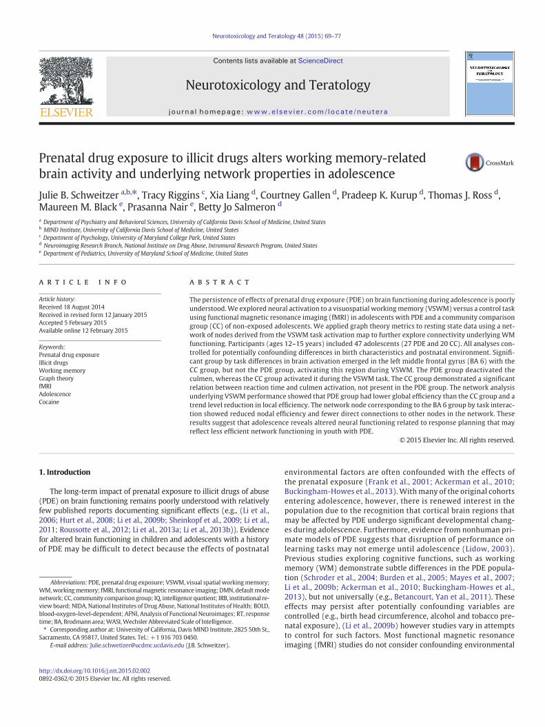
Current characteristics:Age at scan (years (SD)) 14.7, (1.1) 14.2, (1.3) F(1,45) = 2.46, p = .12Sex 13 male, 14 female 7 male, 13 female Chi square(1) = 0.81, p = .37Participant's IQ (WASI (SD)) vocabulary & matrix reasoning 88.00, (12.37) 91.80, (13.25) F(1,45) = 1.02, p = .32Discontinuous maternal care birth to 6 y.o. 56% 0% Chi square(1) = 16.32, p = .000Current caregiver IQ (WASI (SD)) vocabulary &matrix reasoning 83.26, (13.09) 89.84, (11.78)⁎ F(1,44) = 3.06, p = .09Handedness 24 right, 3 left 18 right, 2 left Chi square(1) = 0.02, p = .903Head circumference at birth (cm, (SD)) 33.47, (3.55) 35.25, (3.64)⁎ F(1,44) = 2.73, p = .105Birth weight (gm, (SD)) 2807, (480) 3449, (712)⁎ F(1,44) = 13.3, p = .001Birth length (cm, (SD)) 47.3, (4.1) 50.8, (2.8)⁎ F(1,44) = 10.49, p = .002Gestational age (weeks, Dubowitz, (SD)) 38.2, (2.5) 38.7, (1.4)⁎ F(1,44) = .642, p = .427Prenatal exposure to alcohol 56% 15% Chi square(1) = 8.00, p = .005Intensity of exposure (PDE only) N2×/week in 2nd and 3rd trimesters = 11% 1st trimester
only = 44% no exposure = 44%Prenatal exposure to cigarettes 78% 15% Chi square(1) = 18.12, p b 0.000Intensity of exposure (PDE only) N2×/week in 2nd and 3rd trimesters = 74% 1st trimester
only = 4% no exposure = 22%
⁎ n = 19.
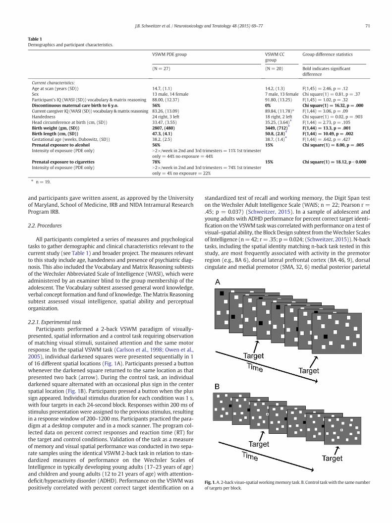
Fig. 1.A. 2-back visuo-spatial workingmemory task. B. Control taskwith the samenumberof targets per block.
71J.B. Schweitzer et al. / Neurotoxicology and Teratology 48 (2015) 69–77
and participants gave written assent, as approved by the Universityof Maryland, School of Medicine, IRB and NIDA Intramural ResearchProgram IRB.
2.2. Procedures
All participants completed a series of measures and psychologicaltasks to gather demographic and clinical characteristics relevant to thecurrent study (see Table 1) and broader project. The measures relevantto this study include age, handedness and presence of psychiatric diag-nosis. This also included the Vocabulary and Matrix Reasoning subtestsof the Wechsler Abbreviated Scale of Intelligence (WASI), which wereadministered by an examiner blind to the group membership of theadolescent. The Vocabulary subtest assessed general word knowledge,verbal concept formation and fund of knowledge. TheMatrix Reasoningsubtest assessed visual intelligence, spatial ability and perceptualorganization.
2.2.1. Experimental taskParticipants performed a 2-back VSWM paradigm of visually-
presented, spatial information and a control task requiring observationof matching visual stimuli, sustained attention and the same motorresponse. In the spatial VSWM task (Carlson et al., 1998; Owen et al.,2005), individual darkened squares were presented sequentially in 1of 16 different spatial locations (Fig. 1A). Participants pressed a buttonwhenever the darkened square returned to the same location as thatpresented two back (arrow). During the control task, an individualdarkened square alternated with an occasional plus sign in the centerspatial location (Fig. 1B). Participants pressed a button when the plussign appeared. Individual stimulus duration for each condition was 1 s,with four targets in each 24-second block. Responses within 200 ms ofstimulus presentation were assigned to the previous stimulus, resultingin a response window of 200–1200 ms. Participants practiced the para-digm at a desktop computer and in a mock scanner. The program col-lected data on percent correct responses and reaction time (RT) forthe target and control conditions. Validation of the task as a measureof memory and visual spatial performance was conducted in two sepa-rate samples using the identical VSWM 2-back task in relation to stan-dardized measures of performance on the Wechsler Scales ofIntelligence in typically developing young adults (17–23 years of age)and children and young adults (12 to 21 years of age) with attention-deficit/hyperactivity disorder (ADHD). Performance on the VSWMwaspositively correlated with percent correct target identification on a
standardized test of recall and working memory, the Digit Span teston the Wechsler Adult Intelligence Scale (WAIS; n = 22; Pearson r =.45; p = 0.037) (Schweitzer, 2015). In a sample of adolescent andyoung adults with ADHD performance for percent correct target identi-fication on the VSWM taskwas correlatedwith performance on a test ofvisual–spatial ability, the Block Design subtest from theWechsler Scalesof Intelligence (n=42; r= .35; p=0.024; (Schweitzer, 2015)). N-backtasks, including the spatial identity matching n-back task tested in thisstudy, are most frequently associated with activity in the premotorregion (e.g., BA 6), dorsal lateral prefrontal cortex (BA 46, 9), dorsalcingulate and medial premotor (SMA, 32, 6) medial posterior parietal
72 J.B. Schweitzer et al. / Neurotoxicology and Teratology 48 (2015) 69–77
Participants completed one 7-minute run containing 6 repeats of thefollowing: 12 s of rest (fixation on a smiley face), 4 s instruction screen,24 s of the control task, 4 s instruction screen, 24 s of the VSWMtask in ablock design. All participants completed the tasks in the same order. Anextra rest block ended the task. A 3-T Siemens Allegra scanner acquiredwhole-brain BOLD EPI; 39 oblique axial (30°axial to coronal), 4 mmslices; TR = 2 s; TE = 27 ms; Flip Angle = 80°; FOV = 22 × 22 cm2. Awhole-brain oblique axial T1-weighted structural image (MPRAGE)was acquired for anatomical reference (1-mm3 isotropic voxels: TR =2.5 s; TE = 4.38 ms; FA = 8°). Five minutes of eyes open resting datawere gathered using the same scan parameters after completion of theVSWM task.
categorical variables) tested for differences between groups in age,sex, IQ, caregiver IQ, birth head circumference, continuity of maternalcare to age 6, and prenatal exposure to alcohol and tobacco.
2.4.2. Behavioral data analysisA one-way ANOVA compared percent correct and RT data between
groups. Intra-individual variability (IIV) in reaction time (IIV-RT) wascalculated using an ex-Gaussian model (see Heathcote et al.(2004);Fassbender et al.(2009)) and compared between groups using ANOVA.Relationships between behavioral variables were examined with bivar-iate correlations within each group with follow-up moderated regres-sion models looking for a significant group by measure interactioneffect on the measure of interest to determine if there were significantdifferences in correlations.
2.4.3. fMRI processing and analysis
2.4.3.1. fMRI data preprocessing. fMRI data were preprocessed andanalyzed using AFNI (Cox, 1996). Both resting and task functional datawere motion corrected, aligned with anatomical images, and normalizedinto Talairach space (3mm3 voxels) and spatial smoothed to an 8mm fullwidth at half maximum (Friedman et al., 2006). The resting fMRI imageswere further processed including band-pass filtering (0.01–0.1 Hz) andremoval of nuisance signals including motion-correction parametersand the first three principal components from the time courses of whitematter and cerebrospinal fluid voxels. Furthermore, to reduce potentialmotion-related confounds in resting fMRI data (Power et al., 2012;Satterthwaite et al., 2012; Van Dijk et al., 2012), we excluded volumeswith large volume-to-volume displacement greater than 0.35 mm/°.Participants with greater than 30% of their volumes excluded (one malePDE participant) were omitted from further analysis.
2.4.3.2. fMRI data analysis. Idealwaveformswere created by convolving asquare-wave functionwith a hemodynamic response function.Multipleregression analyses generated percent signal change for the control andVSWM relative to the rest blocks for each participant. Motion parame-ters were modeled as variables of no interest. Any activation outsideof a mask of brain plus 5 mm was set to zero to avoid the possibility ofincidentally removing true brain activation.
A second-level group analysis was performed using the program3dLME within AFNI. Themodel was a 2 (Group: PDE or CC) × 2 (Condi-tion: VSWM or Control) design. LME analyses included potentiallyconfounding variables that were significantly different between thegroups, which included controlling for prenatal exposure to alcohol and
cigarettes and continuity of maternal care from birth to 6 years old. Acorrected p b 0.05 level of significance (defined as a minimum clustersize of 23 voxels (621 μl) at a voxel-wise threshold p = 0.001 as deter-mined using the program AlphaSim) was considered significant. Post-hoc analyses investigating significant Group × Condition interactionswere conducted at the whole brain level (pcorrected b 0.05) for eachgroup separately and significant clusters were examined for spatial over-lapwith the original clusters showing significantGroup×Condition inter-actions. We examined the relation between activations emerging fromsignificant Group × Condition interaction regions of interest (ROIs) andbehavioral measures (i.e., VSWM RT, percentage accuracy and IIV-RTusing bivariate correlations in each group separately, since these regionswere identified as being used differently by the two groups during thistask). Significant correlations in either groupwere followedupwith linearmodels in the whole group using the behavioral measure as the depen-dent variable and ROI activation (VSWM-Control), group as independentvariables with an interaction term included to determine if the groupsused the region in question in a significantly different way.
2.4.3.3. Network analysis of resting state fMRI data.Nodes for the networkanalysis were derived by taking the local maxima from the VSWM taskmap (WMminus control) for the whole cohort (PDE and CC combined;See Supplemental Fig. 1). Local maxima were determined by the3dmaxima command in AFNI using a minimum threshold of F N 18.16and minimum distance between peaks of 6, 3-mm isomorphic voxels,resulting in 78 nodes (see Supplemental Table 1). Mean resting fMRItime courses were extracted from 6 mm spheres centered at each ofthe 78 peak locations from the resting scan. The interregional correla-tion matrix of each subject was then obtained by calculating the partialcorrelation coefficients between the mean time courses of every pair ofnodes. We used a minimum threshold approach (resulting in networkswith a fixedminimum connectivity between nodes, but allowing differ-ent numbers of connections between participants) across a range ofthresholds as opposed to a sparcity approach in order to avoid includingnode pairswith no orminimal connectivity. Theminimumpartial corre-lation coefficient for inclusion in the network ranged between 0.2 and0.35 in increments of 0.01. The lower threshold limit was set to removeweak correlations so that only the correlations whose corresponding pvalues passed a statistical threshold (p b 0.01)were retained. The higherthreshold limit is well accepted in the literature and ensures that theresultant networks are fully connected and not arbitrarily subdivided(Lynall et al., 2010; Power et al., 2010)(Hayasaka and Laurienti, 2010).Topological metrics of local efficiency and global efficiency were deter-mined for each network, using standard calculations (Watts andStrogatz, 1998; Latora and Marchiori, 2001). Local efficiency and globalefficiencymeasure the ability of information transfer of a network at thelocal and global level, respectively, and provide a more clearly physicalmeaning for topological characterization of the networks. They arederived from each node's clustering coefficient (a measure of howwell connected its nearest neighbors are) and path length (the averageshortest path from a node to any other node in the network). To providethreshold-independent results, each network metric was integratedacross thresholds (i.e., the areas under the curves of network parame-ters across thresholds). We then compared the network metricsbetween groups using moderated regression models, with age and sexas nuisance variables including interactions of age and sex with groupin themodel. Age and sexwere included although they did not differ be-tween the groups because they have been shown to alter network func-tioning (Tian et al., 2011; Yan et al., 2011; Fair et al., 2012a, 2012b; Wuet al., 2013). Interactions between group and age and group and sexwere included because PDE may alter maturation and have differentialeffects by gender (Delaney-Black et al., 2004; Bennett et al., 2007).
Because themain imaging analysis of the task (see below) yieldeda Group by Condition interaction in an area that was a node in theVSWM network, we compared the integrated nodal degree (numberof connections of this node) and integrated nodal efficiency (inversely
Fig. 2. A. Contrast of working memory and control task across all subjects. (F-statistic iscolored by direction of task effect). B. Nodes for network analysis derived from localmaxima of taskmap inA. (For interpretation of the references to color in thisfigure legend,the reader is referred to the web version of this article.)
73J.B. Schweitzer et al. / Neurotoxicology and Teratology 48 (2015) 69–77
related to the path length of connections to the rest of the network)between groups using a moderated regression model, controlling forage and sex effects as above.
3. Results
3.1. Behavioral results
Accuracy, RT and IIV-RT did not differ between groups (see Table 2).In CC participants, percent correct on the VSWM condition was nega-tively correlated with RT (r(19) = −0.813, p b .001) and IIV-RT corre-lated negatively with accuracy (r (19) = − .673, p = .002). PDEparticipants, on the other hand, did not have a significant correlationbetween accuracy on the VSWM task and RT (r(27) = − .100, p =.621). The PDE group, however, demonstrated a similar negative corre-lation of IIV-RT to accuracy (r(27)=− .408, p = .034) as the CC group.A follow-up moderated regression model looking for a group X RTinteraction effect on accuracy, demonstrated that this relation differedsignificantly between the groups (F(1,42)= 11.39, p= .002). In regardto the relationship between accuracy and IIV-RT, the groups did notdiffer from one another (group X IIV-RT F(1,42) = 2.828, p = .100).Thus, slower responders in the CC group tended to be less accurateand more variable, while the PDE group demonstrated no relation be-tween RT and other behavioral measures, but did show a similar rela-tionship between accuracy and variability (IIV-RT) as the CC groupwith better accuracy associated with less variability in response timefor both groups.
3.2. VSWM task activation results
Significant activations from thewhole-brain analysis showedmain ef-fects of Condition within the entire group of subjects in the frontal–pari-etal attention network (see Fig. 2 below and Supplemental Fig. 1 &2) including the bilateral medial and middle frontal gyri (BA 6, 8, and9), bilateral inferior (BA 40) and superior parietal (BA 7) lobules. Thetask also activated the precuneus (BA 7), bilateral lingual gyrus (BA 17and 18), bilateral fusiform (BA 37), temporal (BA 37), occipital (BA 19)gyri and bilateral cerebellum (declive, uvula, pyramis, and culmen). Sig-nificant deactivations were observed in the parahippocampal corticesand regions of the “default network,” including bilateral superior frontalgyri (BA 9), medial frontal gyri (BA 10), anterior cingulate (BA 24, 30,31) gyrus, medial frontal gyrus (BA 6), and posterior cingulate (BA 29and 30).
Whole-brain analyses showed no main effect of group (see Supple-mental Fig. 2 & 3 for individual group VSWMvs Control taskmontages).There was a significant Group × Condition interaction in left BA 6, withonly the CC group activating this region during VSWM (see Fig. 3A, C).Further, there was a significant interaction in the right culmen, whichwas not significantly activated in either group's task map, but showeda trend toward differential activation in the PDE groupwho deactivated
Table 2Performance during VSWM⁎⁎ and control tasks.
Prenatal drug exposure group Comp
Task n = 27 n = 1
Control taskMean % correct (SD) 90.28 (11.67) 90.56Mean RT in ms (SD) 490.21 (44.42) 511.9Mean IIV⁎⁎⁎ (SD) 98.88 (31.58) 108.6
VSWM task⁎⁎Mean % correct (SD) 85.49 (10.03) 85.31Mean RT in ms (SD) 536.59 (59.13) 539.8Mean IIV⁎⁎⁎ (SD) 160.85 (40.57) 169.4
⁎ Task data missing for one subject.⁎⁎ VSWM= Visual spatial working memory task.⁎⁎⁎ IIV = Intra-individual variability.
this region during VSWM but activated it during the control condition.In addition, the CC group appeared to activate the region more thanthe PDE group during the VSWM task (see Fig. 3D, E).
Activation in left BA 6 (VSWM-control task) did not significantlycorrelatewith RT, task accuracy or IIV-RT in either group. CC did demon-strate a significant correlation between right culmen activation(VSWM-control task) and RT (r(19) = − .471, p = .042) that was notpresent in the PDE group (r(27)= .003, p= .989). Amoderated regres-sion revealed a trend level interaction effect on RT for group X rightculmen activation (F(1,42) = 3.802, p = .058). Neither group demon-strated a relation between culmen activation, accuracy or IIV-RT.
arison group Statistic
9⁎
(7.59) F (1,44) = 0.009, p = 0.9274 (72.41) F (1,44) = 1.591, p = 0.2144 (29.32) F(1.44) = .910, p = .345
(16.57) F(1,44) = 0.002, p = .9622 (80.46) F (1,44) = 0.25, p = 0.8763 (50.59) F(1,44) = .388, p = .537
Fig. 3. Group by task condition interactions: A. Left BA6. B. Corresponding node in network analysis. C. BOLD percent change versus rest by group and condition. D. Right culmen (no cor-responding node in the network analysis). E. BOLD percent change versus rest by group and condition (error bars = SEM)* p b 0.001 CC versus PDE.
74 J.B. Schweitzer et al. / Neurotoxicology and Teratology 48 (2015) 69–77
3.3. Network results on resting state data
Integrated global efficiency of the VSWM network was significantlyless in PDE than in CC controlling for age and sex (F(1,39) = 6.206,p = 0.017). Integrated local efficiency showed a trend level reductionin PDE (F(1,39) = 3.711, p = 0.061). Integrated nodal efficiency anddegree for the node corresponding to the BA6 Group × Condition inter-action were significantly reduced in PDE (Integrated node efficiency:F(1,39) = 4.139, p = 0.049; Integrated node degree: F(1,39) = 4.568,p = 0.039).
4. Discussion
The behavioral data demonstrated the expected coupling betweenRT, IIV-RT and accuracy measures in the CC group for the VSWM taskthat was not seen in the PDE group, even though their overall perfor-mance was equivalent. The behavioral findings may reflect subtle indi-cations of altered attentional and response preparatory skills in the PDEgroup. We found a reduced extent of activation associated with WMperformance in left BA 6 in the PDE adolescents compared to a well-matched CC group. The culmen also appears affected by PDE as wefound significant group by task differences in culmen activity. The CCactivated this region while the PDE group showed deactivation duringthe VSWM task and activation during the control condition. Additionalanalyses showed that in a network derived from this population's mapof task-related activity, the PDE group demonstrated significantly less
global efficiency in comparison to the CC group and a trend towardreduced local efficiency. Further, the node corresponding to the GroupX Task interaction in the left BA6, showed significantly reduced nodaldegree and efficiency at rest.
WM-related activity in BA 6 is consistent with other pediatric studies(e.g., Kwon et al., 2002; Ciesielski et al., 2006; Fassbender et al., 2011) andmay reflect the continuous updating that is required inperformingn-backtasks (Wager and Smith, 2003). Decreased frontal activity during aVSWM may not be specific to prenatal cocaine or heroin exposure, asother groups have found decreased frontal activity during a VSWM taskfor children prenatally exposed to methamphetamine (Roussotte et al.,2011). The culmen, a region in which we found more activation for theVSWM task in the CC group compared to the PDE group, is associatedwith response preparation and selection in Go/No-Go tasks (Simmondset al., 2007). A previous study found that better attention, as evidencedthrough lower RT variability, was associated with greater activity in theculmen (Simmonds et al., 2007). The CC group appears to be able tomodulate brain activity in the culmen in response to task demands asactivity in the culmen was associated with RT for the CC, but not thePDE group. Group differences in premotor activity found in BA 6 may belinked to group differences in activity in the culmen, as they appear tobe part of an interacting cerebro-cerebellar circuit (Kelly and Strick,2003). Our findings are also consistent with a recent study (Li et al.,2013b), that suggested that PDE is identifiedwith functional connectomicsignatures and processes involved in working memory, language, execu-tive function, motor and attention.
75J.B. Schweitzer et al. / Neurotoxicology and Teratology 48 (2015) 69–77
The 2-back VSWM task used in this study was relatively easy,with both groups achieving over 85% correct. Task activation differ-ences in the face of comparable performance can be interpreted indifferent ways: reduced activation may be interpreted as a reflec-tion of a less well-developed neural system (e.g., (Jolles et al.,2011)) but is often, especially in WM studies, taken to be indicativeof increased neural efficiency (e.g., (Basten et al., 2012)). Whereasthe reduced extent of activation in the PDE group in left BA6 mayrepresent improved efficiency, it may also represent failure to uti-lize the traditional network supporting WM and response prepara-tion that might result in performance deficits in a more demandingtask. Our graph theory based analysis of the network underlyingthe current VSWM task suggests multiple reduced efficiency mea-sures reflecting altered functioning in VSWM-related regions(i.e., BA6) corresponding to our Group X Task interaction. Thebehavioral data, with the absence of the traditional relationshipbetween RT and accuracy in the PDE group, provide further supportthat the PDE group is not optimally using regions associated withresponse preparation and attention. Future studies employingevent-related designs with tasks resulting in group differences inbehavioral performance may further reveal cognitive vulnerabil-ities associated with PDE.
Findings from this project add to growing literature suggesting thatthere are long-term neural effects of PDE onmemory functioning. Nota-bly, our group (Riggins et al., 2012) demonstrated in a larger samplefrom which this study was drawn that PDE participants who performworse on behavioral memory tasks have larger hippocampal volumesin comparison to the CC group. The hippocampal volumes are negative-ly correlated with memory performance, with lower memory scoresassociated with larger hippocampal volumes.
Both the frontal region (i.e., BA 6) and the culmen are likely tocontinue maturing into young adulthood (Kwon et al., 2002;Tiemeier et al., 2010). It is possible that these regions are typicallyinvolved in a cerebro-cerebellar circuit that is used to subserve theWM processes in typically-developing individuals (Kirschen et al.,2010; Bostan et al., 2013), that is atypical or underdeveloped inthe PDE group. The decreased nodal efficiency and degreeevidenced in the BA6 node by the PDE group in our study and im-paired DMN brain activity (i.e.,(Li et al., 2011)) suggest that PDEbrain functioning is more consistent with a younger maturationalstate in development. These findings support theories suggestingthat some of the effects of PDE may not be detectable until middleadolescence or young adulthood is reached, a period associatedwith the development of sophisticated cognitive and emotionalregulation. Furthermore, the use of connectivity measures, whichassess how well brain regions work together, may be superior atdetecting subtle differences in brain functioning related to devel-opment over region-by-region comparisons. Measures of function-al connectivity may someday be used to predict vulnerability formental illness and/or cognitive functioning.
5. Strengths and limitations
Strengths of this study on PDE include the use of multiple imageanalysis methods and inclusion of multiple covariates (e.g., age,sex, IQ, prenatal exposure to alcohol and tobacco, and caregiverchanges). The overall findings suggest that PDE produces subtle,but lasting impact on brain functioning. Similar to the majority oflongitudinal studies of PDE, our sample had subjects with poly-substance exposure and frequent caregiver changes. Thus, ourfindings have high ecological validity and are relevant and general-izable to the majority of PDE children as poly-substance abuse isthe norm (e.g., (Bauer et al., 2002; Lester et al., 1998)) and childrenof women engaging in polysubstance abuse experience commonenvironmental risks, regardless of the specific substance abusedby the mother. We acknowledge, though, that a limitation of this
sample, is that we cannot specifically identify which substance ofabuse (i.e., heroin or cocaine) may be responsible for the alter-ations present in the PDE group, although we did control for ciga-rette and alcohol exposure. While we were able to statisticallycontrol for caregiver changes we cannot totally rule out the effectsof environment on our participants' functioning or link the findingsto a specific drug. We recognize that stimulants, such as cocaine,versus opiates likely have different effects on the developingbrain (Lu et al., 2012). Animal model studies suggest that prenatalexposure to both cocaine and heroin can lead to impairments inspatial memory, but that the mechanisms underlying those impair-ments may be different (Lu et al., 2012). As noted in Riggins et al.,(Riggins et al., 2012) it is also likely that there are other nondirectPDE factors that impact brain functioning in our PDE participantsthat were not controlled for, such as the mental health of the par-ent or other aspects of the caregiving environment. For example,in Riggins et al., (Riggins et al., 2012) we found that maternaldepression was a significant predictor of recall ability.
Another potential concern is that we did not control for brain activa-tions associated with saccades given that the target in the control taskwas fixed, rather than moving, as it was in our VSWM task. We choseto present the control target in a fixed spot due to concerns that the par-ticipants would inadvertently practice where the target was if it wasmoving and thus, perform the control task as a WM task. We recom-mend that future studies collect saccadic information during the tasksto identify brain activation related to the saccadic movement ratherthan WM. The regions in which we found activation (e.g., BA 6) arehistorically associated with WM performance (Kwon et al., 2002;Ciesielski et al., 2006; Fassbender et al., 2011) and thus, we think it isunlikely that the activation is due solely to eye movements.
An additional methodological limitation was our use of a block-design. With an event-related task we would be able to specificallyrelate brain functioning to performance on a trial by trial basis. An ad-vantage of the block design is the statistical power and speed at whichblock design data can be acquired and both factors are important incollecting data from pediatric populations.
5.1. Conclusions and future directions
This study found that childrenwith PDE experience subtle attention-al and response preparation challenges that are related to reduced activ-ity and network-related functioning in frontal (i.e., BA 6) and cerebellarregions. Future studies should continue to follow these adolescents intoadulthood as additional exposure effectsmaynot be detectable until theadolescent–young adult period begins. Subsequent research shouldattempt to increase our ability to understand how neural–behavioralfindings may suggest specific prevention and intervention strategiesfor children who have experienced prenatal insults to reduce potentiallearning problems. This includes the need to develop targeted interven-tions that can address issues such as impaired neural connectivity andworking memory that are informed by the neuroscience findings forthis at-risk population.
Supplementary data to this article can be found online at http://dx.doi.org/10.1016/j.ntt.2015.02.002.
Funding and Disclosure
This research was funded by the National Institutes of Health grantsR01 DA07432 (Nair) and R01 DA021059 (Black) and the IntramuralResearch Program of the NIH, NIDA. The funders had no involvementin the collection, analysis or interpretation of data; in the writing ofthe report; or in the decision to submit the manuscript for publication.The authors have no competing financial interests in relation to thework described.
76 J.B. Schweitzer et al. / Neurotoxicology and Teratology 48 (2015) 69–77
Transparency Document
The Transparency document associated with this article can befound, in the online version.
Acknowledgments
Wewould like to thank Elliot Stein, Ph.D., Kim Slater, and the Neuro-imaging Research Branch of NIDA-IRP for their support with data collec-tion and analysis; Stacy Buckingham-Howes, Ph.D. and the F.U.T.U.R.E.S.team for the participant recruitment and testing, and the families fortheir participation.
References
Ackerman JP, Riggins T, Black MM. A review of the effects of prenatal cocaine exposureamong school-aged children. Pediatrics 2010;125(3):554–65.
Alloway TP, Gathercole SE, Elliott J. Examining the link between working memory behav-iour and academic attainment in children with ADHD. Dev Med Child Neurol 2010;52(7):632–6.
Backman L, Nyberg L. Dopamine and training-related working-memory improvement.Neurosci Biobehav Rev 2013;37:2209–19.
Baddeley A. Working memory. Curr Biol 2010;20(4):R136–40.Barkley R. Attention deficit hyperactivity disorder: a handbook for diagnosis and treat-
ment. New York: Guilford Press; 2006.Bassett DS, Bullmore ET, Meyer-Lindenberg A, Apud JA, Weinberger DR, Coppola R. Cog-
nitive fitness of cost-efficient brain functional networks. Proc Natl Acad Sci U S A2009;106(28):11747–52.
Basten U, Stelzel C, Fiebach CJ. Trait anxiety and the neural efficiency of manipulation inworking memory. Cogn Affect Behav Neurosci 2012;12(3):571–88.
Bauer CR, Shankaran S, Bada HS, Lester B, Wright LL, Krause-Steinrauf H, et al. The mater-nal lifestyle study: drug exposure during pregnancy and short-term maternal out-comes. Am J Obstet Gynecol 2002;186(3):487–95.
Bennett D, Bendersky M, LewisM. Preadolescent health risk behavior as a function of pre-natal cocaine exposure and gender. J Dev Behav Pediatr 2007;28(6):467–72.
Betancourt LM, et al. Adolescents with and without gestational cocaine exposure: Longi-tudinal analysis of inhibitory control, memory and receptive language. NeurotoxicolTeratol 2011;33(1):36–46.
Bostan AC, Dum RP, Strick PL. Cerebellar networks with the cerebral cortex and basalganglia. Trends Cogn Sci 2013;17(5):241–54.
Buckingham-Howes S, Berger SS, Scaletti LA, Black MM. Systematic review of prenatalcocaine exposure and adolescent development. Pediatrics 2013;131(6):e1917–36.
Burden MJ, Jacobson SW, Sokol RJ, Jacobson JL. Effects of prenatal alcohol exposure onattention and working memory at 7.5 years of age. Alcohol Clin Exp Res 2005;29(3):443–52.
Carlson S, Martinkauppi S, Rama P, Salli E, Korvenoja A, Aronen HJ. Distribution of corticalactivation during visuospatial n-back tasks as revealed by functional magnetic reso-nance imaging. Cereb Cortex 1998;8(8):743–52.
Cohen M. Children's Memory Scale. Psych Corp; 1997.Cox RW. AFNI: software for analysis and visualization of functional magnetic resonance
neuroimages. Comput Biomed Res 1996;29(3):162–73.Crone EA, Wendelken C, Donohue S, van Leijenhorst L, Bunge SA. Neurocognitive devel-
opment of the ability to manipulate information in working memory. Proc NatlAcad Sci U S A 2006;103(24):9315–20.
Delaney-Black V, Covington C, Nordstrom B, Ager J, Janisse J, Hannigan JH, et al. Prenatalcocaine: quantity of exposure and gender moderation. J Dev Behav Pediatr 2004;25(4):254–63.
Delis DC, Kramer JH, Kaplan E, Ober BA. The California Verbal Learning Test–Children'sVersion. San Antonio: The Psychological Corporation, Harcourt Brace & Company;1994.
Dosenbach NU, Nardos B, Cohen AL, Fair DA, Power JD, Church JA, et al. Prediction ofindividual brain maturity using fMRI. Science 2010;329(5997):1358–61.
Fair DA, Bathula D, Nikolas MA, Nigg JT. Distinct neuropsychological subgroups in typical-ly developing youth inform heterogeneity in children with ADHD. Proc Natl Acad SciU S A 2012a;109(17):6769–74.
Fair DA, Nigg JT, Iyer S, Bathula D, Mills KL, Dosenbach NU, et al. Distinct neural signaturesdetected for ADHD subtypes after controlling for micro-movements in resting statefunctional connectivity MRI data. Front Syst Neurosci 2012b;6:80.
Fassbender C, Zhang H, Buzy WM, Cortes CR, Mizuiri D, Beckett L, et al. A lack of defaultnetwork suppression is linked to increased distractibility in ADHD. Brain Res 2009;1273:114–28.
Fassbender C, Schweitzer JB, Cortes CR, Tagamets MA, Windsor TA, Reeves GM, et al.Working memory in attention deficit/hyperactivity disorder is characterized by alack of specialization of brain function. PLoS One 2011;6(11):e27240.
Frank DA, Augustyn M, Knight WG, Pell T, Zuckerman B. Growth, development, andbehavior in early childhood following prenatal cocaine exposure: a systematicreview. JAMA 2001;285(12):1613–25.
Friedman L, Glover GH, Krenz D, Magnotta V. Reducing inter-scanner variability of activa-tion in a multicenter fMRI study: role of smoothness equalization. Neuroimage 2006;32(4):1656–68.
Giessing C, Thiel CM, Alexander-Bloch AF, Patel AX, Bullmore ET. Human brainfunctional network changes associated with enhanced and impaired atten-tional task performance. J Neurosci 2013;33(14):5903–14.
Gropper RJ, Tannock R. A pilot study of working memory and academic achievement incollege students with ADHD. J Atten Disord 2009;12(6):574–81.
Hamilton LR, Czoty PW, Gage HD, Nader MA. Characterization of the dopamine receptorsystem in adult rhesus monkeys exposed to cocaine throughout gestation. Psycho-pharmacology (Berl) 2010;210(4):481–8.
Hayasaka S, Laurienti PJ. Comparison of characteristics between region-and voxel-basednetwork analyses in resting-state fMRI data. Neuroimage 2010;50(2):499–508.
He Y, Evans A. Graph theoretical modeling of brain connectivity. Curr Opin Neurol 2010;23(4):341–50.
Heathcote A, Brown S, Cousineau D. QMPE: estimating Lognormal, Wald, and Weibull RTdistributions with a parameter-dependent lower bound. Behav Res Methods InstrumComput 2004;36(2):277–90.
Hinson JM, Jameson TL, Whitney P. Impulsive decision making and working memory. JExp Psychol Learn Mem Cogn 2003;29(2):298–306.
Hurt H, Giannetta JM, Korczykowski M, Hoang A, Tang KZ, Betancourt L, et al. Functionalmagnetic resonance imaging and working memory in adolescents with gestationalcocaine exposure. J Pediatr 2008;152(3):371–7.
Jolles DD, Kleibeuker SW, Rombouts SARB, Crone EA. Developmental differences in pre-frontal activation during working memory maintenance and manipulation for differ-ent memory loads. Dev Sci 2011;14:713–24.
Kelly RM, Strick PL. Cerebellar loops with motor cortex and prefrontal cortex of a nonhu-man primate. J Neurosci 2003;23(23):8432–44.
Kirschen MP, Chen SH, Desmond JE. Modality specific cerebro-cerebellar activations inverbal working memory: an fMRI study. Behav Neurol 2010;23(1–2):51–63.
Kwon H, Reiss AL, Menon V. Neural basis of protracted developmental changes in visuo-spatial working memory. Proc Natl Acad Sci U S A 2002;99(20):13336–41.
Langer N, von Bastian CC, Wirz H, Oberauer K, Jancke L. The effects of working memorytraining on functional brain network efficiency. Cortex 2013;49:2424–38.
Latora V, Marchiori M. Efficient behavior of small-world networks. Phys Rev Lett 2001;87(19):198701.
Lester BM. The Maternal Lifestyles Study. Ann N Y Acad Sci 1998;846:296–305.Lester BM, LaGasse LL, Bigsby R. Prenatal cocaine exposure and child development: what
do we know and what do we do? Semin Speech Lang 1998;19(2):123–46.Li V, Milivojevic K, Kemp K Hong, Sinha R. Performance monitoring and stop signal inhi-
bition in abstinent patients with cocaine dependence. Drug Alcohol Depend 2006;85(3):205–12.
Li Y, Liu Y, Li J, Qin W, Li K, Yu C, et al. Brain anatomical network and intelligence. PLoSComput Biol 2009a;5(5):e1000395.
Li Z, Coles CD, Lynch ME, Hamann S, Peltier S, LaConte S, et al. Prenatal cocaine exposurealters emotional arousal regulation and its effects on working memory. NeurotoxicolTeratol 2009b;31(6):342–8.
Li Z, Santhanam P, Coles CD, Lynch ME, Hamann S, Peltier S, et al. Increased “defaultmode” activity in adolescents prenatally exposed to cocaine. Hum Brain Mapp2011;32(5):759–70.
Li P, Santhanam CD, Coles M Ellen, Lynch S, Hamann S Peltier, Hu X. Prenatal cocaineexposure alters functional activation in the ventral prefrontal cortex and its structuralconnectivity with the amygdala. Psychiatry Res 2013a;213(1):47–55.
Li D, Zhu L, Guo Z, Li ME, Lynch C, Coles XHu, et al. Connectomics signatures of prenatalcocaine exposure affected adolescent brains. Hum Brain Mapp 2013b;34(10):2494–510.
Lidow MS. Consequences of prenatal cocaine exposure in nonhuman primates. Brain ResDev Brain Res 2003;147(1–2):23–36.
Lu R, Liu X, Long H, Ma L. Effects of prenatal cocaine and heroin exposure on neuronaldendrite morphogenesis and spatial recognition memory in mice. Neurosci Lett2012;522(2):128–33.
Lynall ME, Bassett DS, Kerwin R, McKenna PJ, Kitzbichler M, Muller U, et al. Functionalconnectivity and brain networks in schizophrenia. J Neurosci 2010;30(28):9477–87.
Mayes L, Snyder PJ, Langlois E, Hunter N. Visuospatial working memory in school-agedchildren exposed in utero to cocaine. Child Neuropsychol 2007;13(3):205–18.
Nair P, Black MM, Ackerman JP, Schuler ME, Keane VA. Children's cognitive–behavioralfunctioning at age 6 and 7: prenatal drug exposure and caregiving environment.Ambul Pediatr 2008;8(3):154–62.
O'Hare ED, Lu LH, Houston SM, Bookheimer SY, Sowell ER. Neurodevelopmental changesin verbal working memory load-dependency: an fMRI investigation. Neuroimage2008;42(4):1678–85.
Olsen J. Is left-handedness a sensitive marker of prenatal exposures or indicators of fetalgrowth? Scand J Soc Med 1995;23(4):233–5.
Owen AM, McMillan KM, Laird AR, Bullmore E. N-back working memory paradigm: ameta-analysis of normative functional neuroimaging studies. Hum Brain Mapp2005;25(1):46–59.
Power JD, Fair DA, Schlaggar BL, Petersen SE. The development of human functional brainnetworks. Neuron 2010;67(5):735–48.
Power JD, Barnes KA, Snyder AZ, Schlaggar BL, Petersen SE. Spurious but systematiccorrelations in functional connectivity MRI networks arise from subject motion.Neuroimage 2012;59(3):2142–54.
Rao H, Wang J, Giannetta J, Korczykowski M, Shera D, Avants BB, et al. Altered restingcerebral blood flow in adolescents with in utero cocaine exposure revealed by perfu-sion functional MRI. Pediatrics 2007;120(5):e1245–54.
Riggins T, Cacic K, Buckingham-Howes S, Scaletti LA, Salmeron BJ, Black MM. Memoryability and hippocampal volume in adolescents with prenatal drug exposure.Neurotoxicol Teratol 2012;34(4):434–41.
Roussotte FF, Bramen JE, Nunez SC, Quandt LC, Smith L, O'Connor MJ, et al. Abnormalbrain activation during working memory in children with prenatal exposure to
77J.B. Schweitzer et al. / Neurotoxicology and Teratology 48 (2015) 69–77
drugs of abuse: the effects of methamphetamine, alcohol, and polydrug exposure.Neuroimage 2011;54(4):3067–75.
Roussotte FF, Rudie JD, Smith L, O'Connor MJ, Bookheimer SY, Narr KL, et al. Frontostriatalconnectivity in children duringworkingmemory and the effects of prenatal metham-phetamine, alcohol, and polydrug exposure. Dev Neurosci 2012;34(1):43–57.
Satterthwaite TD, Wolf DH, Loughead J, Ruparel K, Elliott MA, Hakonarson H, et al. Impactof in-scanner head motion on multiple measures of functional connectivity: rele-vance for studies of neurodevelopment in youth. Neuroimage 2012;60(1):623–32.
Schroder MD, Snyder PJ, Sielski I, Mayes L. Impaired performance of children exposed inutero to cocaine on a novel test of visuospatial working memory. Brain Cogn 2004;55(2):409–12.
Schweitzer JB. ; 2015 [Unpublished data].Shamosh NA, Deyoung CG, Green AE, Reis DL, Johnson MR, Conway AR, et al. Individual
differences in delay discounting: relation to intelligence, working memory, and ante-rior prefrontal cortex. Psychol Sci 2008;19(9):904–11.
Sheinkopf SJ, Lester BM, Sanes JN, Eliassen JC, Hutchison ER, Seifer R, et al. Functional MRIand response inhibition in children exposed to cocaine in utero. Preliminary findings.Dev Neurosci 2009;31(1–2):159–66.
Simmonds DJ, Fotedar SG, Suskauer SJ, Pekar JJ, Denckla MB, Mostofsky SH. Functionalbrain correlates of response time variability in children. Neuropsychologia 2007;45(9):2147–57.
Tian L,Wang J, YanC,HeY.Hemisphere- andgender-relateddifferences in small-world brainnetworks: a resting-state functional MRI study. Neuroimage 2011;54(1):191–202.
Tiemeier H, Lenroot RK, Greenstein DK, Tran L, Pierson R, Giedd JN. Cerebellum develop-ment during childhood and adolescence: a longitudinal morphometric MRI study.Neuroimage 2010;49(1):63–70.
Tye C, Bolton P. Neural connectivity abnormalities in autism: insights from the tuberoussclerosis model. BMC Med 2013;11(1):55.
van den Heuvel MP, Stam CJ, Kahn RS, Hulshoff Pol HE. Efficiency of functional brainnetworks and intellectual performance. J Neurosci 2009;29(23):7619–24.
Van Dijk KR, SabuncuMR, Buckner RL. The influence of headmotion on intrinsic function-al connectivity MRI. Neuroimage 2012;59(1):431–8.
Wager T, Smith E. Neuroimaging studies of working memory: a meta-analysis. CognAffect Behav Neurosci 2003;3(4):255–74.
Wu K, Taki Y, Sato K, Hashizume H, Sassa Y, Takeuchi H, et al. Topological organization offunctional brain networks in healthy children: differences in relation to age, sex, andintelligence. PLoS One 2013;8(2):e55347.
Yan C, Gong G, Wang J, Wang D, Liu D, Zhu C, et al. Sex- and brain size-related small-world structural cortical networks in young adults: a DTI tractography study. CerebCortex 2011;21(2):449–58.