JOURNAL OF VIROLOGY, July 2002, p. 6873–6881 Vol. 76, No. 14 0022-538X/02/$04.000 DOI: 10.1128/JVI.76.14.6873–6881.2002 Neutralizing Anti-F Glycoprotein and Anti-Substance P Antibody Treatment Effectively Reduces Infection and Inflammation Associated with Respiratory Syncytial Virus Infection Lia M. Haynes, 1 Joelyn Tonkin, 1,2 Larry J. Anderson, 1 and Ralph A. Tripp 1 * Respiratory and Enteric Virus Branch, Division of Viral and Rickettsial Diseases, National Center for Infectious Diseases, Centers for Disease Control and Prevention, 1 and Rollins School of Public Health, Emory University, 2 Atlanta, Georgia 30333 Received 24 January 2002/Accepted 12 April 2002 Respiratory syncytial virus (RSV) is the most important virus mediating lower respiratory tract illness in infants and young children. RSV infection is associated with pulmonary inflammation and increased levels of substance P (SP), making the airways and leukocytes that express SP receptors susceptible to the proinflam- matory effects of this peptide. This study examines combining neutralizing anti-F glycoprotein and anti-SP antibody treatment of RSV-infected BALB/c mice to inhibit RSV replication and inflammation associated with infection. BALB/c mice were prophylactically treated with antibody prior to RSV infection or were therapeu- tically treated at day 2 or 6 post-RSV infection. Prophylactic or therapeutic treatment with anti-SP antibodies promptly reduced pulmonary inflammatory cell infiltration and decreased the number of cells expressing proinflammatory cytokines, while anti-F antibody treatment reduced virus titers. The results suggest that combined anti-viral and anti-SP antibody treatment may be effective in treating RSV disease. Respiratory syncytial virus (RSV) is the leading cause of lower respiratory tract disease in infants and young children worldwide. A member of the Paramyxoviridae family, RSV is an enveloped virus containing a negative-sense single-stranded RNA genome. The protective immune response to RSV infec- tion is primarily directed against the two major surface viral glycoproteins, i.e., the G (attachment) and F (fusion) glyco- proteins. The F glycoprotein appears to be most important for induction of protective immunity and is associated with a high serum neutralizing antibody response (6, 37) and activation of CD14 and Toll-like receptor-4 (21). Some monoclonal anti- bodies against the F glycoprotein provide passive protection against RSV disease (8, 13, 18, 42); therefore, the F glycopro- tein has been the focus for therapeutic intervention in RSV disease. At present, there is no RSV vaccine available, and the only options to address disease are prophylactic administration of enriched anti-RSV human immune globulin (Respigam) or anti-F glycoprotein monoclonal antibodies (palivizumab [Syna- gis]), both of which are recommended only for young children at high risk for RSV disease. In addition, ribavirin (Virazole), the only specific antiviral agent approved for RSV infection, has limited efficacy (10, 19, 41; M. I. Marks and J. McBride, abstract from Ribavirin Therapy for Respiratory Syncytial Vi- rus Infections: a Scientific Workshop, Sept., 1989, Pediatr. Infect. Dis. J. 9:S84, 1990), and its use is limited for treatment of RSV infection in immune-compromised patients (10, 43). Treatment with anti-RSV human immune globulin or anti-F glycoprotein neutralizing antibodies is effective in decreasing the titer of virus but does not appear to ameliorate the disease process, suggesting that a substantial portion of disease is as- sociated with the host response to infection (27). The impor- tance of the host response to infection is also suggested by the prominence of obstructed-airway disease and wheezing during RSV infection (reminiscent of asthma), the fact that serious disease can occur with repeated infections, and the occurrence of enhanced disease in younger children vaccinated with for- malin-inactivated vaccine during subsequent RSV infection. One inflammatory mediator associated with inflammation is the tachykinin neuropeptide substance P (SP) (40). SP is pro- duced by afferent neurons and a variety of immune cells, in- cluding eosinophils, monocytes, macrophages (17, 30), lym- phocytes (9), and dendritic cells (23). Numerous studies have directly associated SP with exacerbated inflammation (22, 25, 26, 29, 31, 32, 44). SP has been shown to affect inflammation by mediating vasodilation, thereby enhancing cell trafficking, as well as by affecting the cellular events involved in proliferation and cytokine and growth factor synthesis (3–5, 7, 11, 24, 28, 36). A recent study from our laboratory showed that RSV infection of BALB/c mice increases pulmonary SP levels, and these increased levels of SP exacerbated pulmonary inflamma- tion (40). In that study, treatment of RSV-infected mice with anti-SP antibody decreased pulmonary inflammatory cells and proinflammatory cytokine expression (40). Similarly, RSV-in- fected rats have been shown to upregulate SP receptors in the lungs, an effect that was associated with increased pulmonary inflammation (20, 33). These findings suggest that SP might be important for RSV pathogenesis, and inhibiting SP might re- duce RSV-associated inflammation. In this study, we examine the effectiveness of combining antiviral treatment with a neutralizing anti-F glycoprotein monoclonal antibody with anti-SP antibody. The results show that prophylactic or therapeutic treatment with anti-SP mark- edly reduces pulmonary inflammation, suggesting that anti-SP * Corresponding author. Mailing address: Respiratory Enteric Vi- rology Branch, Division of Viral and Rickettsial Diseases, National Centers for Infectious Disease, 1600 Clifton Rd., NE, Mailstop G-09, Atlanta, GA 30333. Phone: (404) 639-0753. Fax: (404) 639-4005. E- mail: [email protected]. 6873 on June 13, 2018 by guest http://jvi.asm.org/ Downloaded from
Transcript
JOURNAL OF VIROLOGY, July 2002, p. 6873–6881 Vol. 76, No. 140022-538X/02/$04.00�0 DOI: 10.1128/JVI.76.14.6873–6881.2002
Neutralizing Anti-F Glycoprotein and Anti-Substance P AntibodyTreatment Effectively Reduces Infection and Inflammation Associated
with Respiratory Syncytial Virus InfectionLia M. Haynes,1 Joelyn Tonkin,1,2 Larry J. Anderson,1 and Ralph A. Tripp1*
Respiratory and Enteric Virus Branch, Division of Viral and Rickettsial Diseases, National Center for Infectious Diseases, Centersfor Disease Control and Prevention,1 and Rollins School of Public Health, Emory University,2 Atlanta, Georgia 30333
Received 24 January 2002/Accepted 12 April 2002
Respiratory syncytial virus (RSV) is the most important virus mediating lower respiratory tract illness ininfants and young children. RSV infection is associated with pulmonary inflammation and increased levels ofsubstance P (SP), making the airways and leukocytes that express SP receptors susceptible to the proinflam-matory effects of this peptide. This study examines combining neutralizing anti-F glycoprotein and anti-SPantibody treatment of RSV-infected BALB/c mice to inhibit RSV replication and inflammation associated withinfection. BALB/c mice were prophylactically treated with antibody prior to RSV infection or were therapeu-tically treated at day 2 or 6 post-RSV infection. Prophylactic or therapeutic treatment with anti-SP antibodiespromptly reduced pulmonary inflammatory cell infiltration and decreased the number of cells expressingproinflammatory cytokines, while anti-F antibody treatment reduced virus titers. The results suggest thatcombined anti-viral and anti-SP antibody treatment may be effective in treating RSV disease.
Respiratory syncytial virus (RSV) is the leading cause oflower respiratory tract disease in infants and young childrenworldwide. A member of the Paramyxoviridae family, RSV isan enveloped virus containing a negative-sense single-strandedRNA genome. The protective immune response to RSV infec-tion is primarily directed against the two major surface viralglycoproteins, i.e., the G (attachment) and F (fusion) glyco-proteins. The F glycoprotein appears to be most important forinduction of protective immunity and is associated with a highserum neutralizing antibody response (6, 37) and activation ofCD14 and Toll-like receptor-4 (21). Some monoclonal anti-bodies against the F glycoprotein provide passive protectionagainst RSV disease (8, 13, 18, 42); therefore, the F glycopro-tein has been the focus for therapeutic intervention in RSVdisease.
At present, there is no RSV vaccine available, and the onlyoptions to address disease are prophylactic administration ofenriched anti-RSV human immune globulin (Respigam) oranti-F glycoprotein monoclonal antibodies (palivizumab [Syna-gis]), both of which are recommended only for young childrenat high risk for RSV disease. In addition, ribavirin (Virazole),the only specific antiviral agent approved for RSV infection,has limited efficacy (10, 19, 41; M. I. Marks and J. McBride,abstract from Ribavirin Therapy for Respiratory Syncytial Vi-rus Infections: a Scientific Workshop, Sept., 1989, Pediatr.Infect. Dis. J. 9:S84, 1990), and its use is limited for treatmentof RSV infection in immune-compromised patients (10, 43).
Treatment with anti-RSV human immune globulin or anti-Fglycoprotein neutralizing antibodies is effective in decreasing
the titer of virus but does not appear to ameliorate the diseaseprocess, suggesting that a substantial portion of disease is as-sociated with the host response to infection (27). The impor-tance of the host response to infection is also suggested by theprominence of obstructed-airway disease and wheezing duringRSV infection (reminiscent of asthma), the fact that seriousdisease can occur with repeated infections, and the occurrenceof enhanced disease in younger children vaccinated with for-malin-inactivated vaccine during subsequent RSV infection.
One inflammatory mediator associated with inflammation isthe tachykinin neuropeptide substance P (SP) (40). SP is pro-duced by afferent neurons and a variety of immune cells, in-cluding eosinophils, monocytes, macrophages (17, 30), lym-phocytes (9), and dendritic cells (23). Numerous studies havedirectly associated SP with exacerbated inflammation (22, 25,26, 29, 31, 32, 44). SP has been shown to affect inflammation bymediating vasodilation, thereby enhancing cell trafficking, aswell as by affecting the cellular events involved in proliferationand cytokine and growth factor synthesis (3–5, 7, 11, 24, 28,36). A recent study from our laboratory showed that RSVinfection of BALB/c mice increases pulmonary SP levels, andthese increased levels of SP exacerbated pulmonary inflamma-tion (40). In that study, treatment of RSV-infected mice withanti-SP antibody decreased pulmonary inflammatory cells andproinflammatory cytokine expression (40). Similarly, RSV-in-fected rats have been shown to upregulate SP receptors in thelungs, an effect that was associated with increased pulmonaryinflammation (20, 33). These findings suggest that SP might beimportant for RSV pathogenesis, and inhibiting SP might re-duce RSV-associated inflammation.
In this study, we examine the effectiveness of combiningantiviral treatment with a neutralizing anti-F glycoproteinmonoclonal antibody with anti-SP antibody. The results showthat prophylactic or therapeutic treatment with anti-SP mark-edly reduces pulmonary inflammation, suggesting that anti-SP
* Corresponding author. Mailing address: Respiratory Enteric Vi-rology Branch, Division of Viral and Rickettsial Diseases, NationalCenters for Infectious Disease, 1600 Clifton Rd., NE, Mailstop G-09,Atlanta, GA 30333. Phone: (404) 639-0753. Fax: (404) 639-4005. E-mail: [email protected].
antibodies should be considered as an adjunct to antiviraltreatment to reduce RSV disease.
MATERIALS AND METHODS
Animals. Six- to 8-week-old, specific-pathogen-free female BALB/c mice(Jackson Laboratory, Bar Harbor, Maine) were used in all experiments. Themice were housed in microisolator cages and were fed sterilized water and foodad libitum. All studies were performed in accordance with the guidelines of theInstitutional Animal Care and Use Committee.
Virus and infection. The A2 strain of RSV was used in all experiments andpropagated in Vero cells (ATCC CCL 881) as previously described (39). Micewere anesthetized by intraperitoneal administration of avertin (2,2,2-tribromo-
ethanol; 0.2 ml/g of body weight; Sigma-Aldrich, St. Louis, Mo.), and intranasallychallenged with 106 PFU of RSV in Dulbecco’s PBS (GIBCO Laboratories,Grand Island, N.Y.). No fewer than three mice per treatment were examined pertime point.
Antibodies and treatment. On day �1 prior to infection or day 2 or 6 postin-fection (p.i.), mice were intraperitoneally treated with 150 �g of anti-F glyco-protein monoclonal antibody (anti-F)/mouse (1), rabbit anti-SP F(ab)2 antibody(Accurate Chemical and Scientific Corp., Westbury, N.Y.), or both anti-SPF(ab)2 and anti-F glycoprotein antibodies (anti-SP/F; 150 �g of each antibody/mouse) as previously described (40). Control mice were treated with 150 �g ofnormal rabbit F(ab)2 immunoglobulin (nIg) (Jackson ImmunoResearch Labo-ratories, Inc., West Grove, PA)/mouse, purified mouse IgG1 isotype antibody(S1-68.1) (Pharmingen, San Diego, Calif.), or a combination of both control
FIG. 1. Prophylactic treatment. Shown are flow cytometry results following prophylactic treatment (day �1 prior to infection) of mice withanti-SP and anti-F antibodies. (A) Total pulmonary leukocyte trafficking. (B to F) BAL were stained with antibodies against CD8� (B), CD4� (C),B220� (D), and CD11b� (E) cells and PMN (RB6-8C5�) (F). The data are expressed as the mean number (103) of BAL/lung (� SEM) on days3, 5, and 7 p.i. from three independent experiments. Asterisks indicate a significant difference (P � 0.05) between nIg-treated and antibody-treatedmice.
antibodies. The anti-F antibody (clone 143-6C) used in these studies is similar topalivizumab, i.e., it recognizes the same region on the F glycoprotein and bothneutralize and inhibit RSV infection (1).
Quantitation of SP. SP levels in cell-free bronchoalveolar lavage fluid wereanalyzed using a competitive enzyme-linked immunoassay kit (Cayman Chemi-cal, Ann Arbor, Mich.) in accordance with the manufacturer’s instructions asdescribed previously (40). The assay is based on the competition between free SPand an SP tracer for a limited number of SP-specific binding sites. The percentsample bound per maximum bound was calculated, and the SP concentration ofeach sample was determined based on the percent standard bound per maximumbound versus the standard SP concentration. The intra- and interassay coeffi-cients of variation were �10%. The rabbit anti-SP F(ab2) antibody used in vivodoes not interfere with the antibodies used to detect SP in the competitiveenzyme-linked immunoassay.
Cell collection and analysis. Mice were anesthetized with Avertin and exsan-guinated by severing the right caudal artery. Bronchoalveolar leukocytes (BAL)were harvested by lavaging the lungs with PBS. The procedure used for extra-cellular staining of BAL was modified for microculture staining as describedpreviously (39). Briefly, BAL were washed in Dulbecco’s PBS (GIBCO) contain-ing 1% bovine serum albumin and then stained (4°C; 30 min) with an appropriatedilution of the fluorescein isothiocyanate-conjugated or phycoerythrin-conju-gated antibodies anti-CD3ε (145-2C11), anti-CD45R/B220 (RA3-6B2), anti-CD8 (Ly-2), anti-neutrophil (polymorphonuclear leukocyte [PMN]) (RB6-8C5),and anti-CD11b (M1/70) and isotype antibody controls (Pharmingen). Intracel-lular cytokine staining was modified for microculture staining as described pre-viously (39). Briefly, BAL were incubated in PBS containing Golgi Stop (Pharm-ingen) for 3 h at 37°C to allow accumulation of intracellular cytokines. The cellswere washed in PBS and stained (4°C; 30 min) with an appropriate dilution of
FIG. 2. Early therapeutic treatment. Shown are flow cytometry results following early (day 2 p.i.) therapeutic treatment of mice with anti-SPand anti-F antibodies. (A) Total pulmonary leukocyte trafficking. (B to F) BAL were stained with antibodies against CD8� (B), CD4� (C), B220�
(D), and CD11b� (E) cells and PMN (RB6-8C5�) (F). The data are expressed as the number (103) of BAL/lung (� SEM) on days 3, 5, and 7 p.i.A representative experiment from two independent experiments is shown. Asterisks indicate a significant difference (P � 0.05) between nIg-treatedand antibody-treated mice.
VOL. 76, 2002 COMBINATION THERAPY FOR RSV DISEASE 6875
anti-CD3 antibody, fixed, and permeabilized in Cytofix/Cytoperm (Pharmingen).The cells were washed in Cytofix/Cytoperm buffer and stained (4°C; 30 min) withappropriate dilutions of anti-interleukin 2 (IL-2) (JES6-5H4), anti-IL-4 (BVD4-1D11), anti-IL-5 (TRFK5), anti-IL-10 (JES3-16E3), anti-gamma interferon(IFN-�) (XMG1.2), or anti-tumor necrosis factor alpha (TNF-�; MP6-XT22)antibody (Pharmingen) diluted in PBS containing Cytofix/Cytoperm as describedpreviously (39). Extra- and intracellular staining was analyzed using a FACScanand Cell Quest software (Becton Dickinson, San Diego, Calif.). The total num-ber of CD3� BAL expressing a particular cytokine was determined by multiply-ing the total number of BAL by the percent CD3� cells expressing that particularcytokine.
Virus titers. Virus titers in the lungs of RSV-infected mice were determined aspreviously described (39). Briefly, lungs were aseptically removed from three tofive mice per group on days 3, 5, 7, and 9 p.i. and stored at �70°C until they wereassayed. Identical weights (0.1 g) of individual lung samples were homogenizedin 1 ml of Dulbecco’s PBS, and 10-fold serial dilutions of the lung homogenateswere added to confluent Vero cell monolayers. Following adsorption (2 h; 37°C),the cell monolayers were overlaid with Dulbecco’s modified Eagle’s medium(GIBCO) containing 10% fetal bovine serum (HyClone, Logan, Utah) and
incubated at 37°C for 3 to 4 days. Plaques were enumerated by immunostainingthem with monoclonal antibodies against the G and F glycoproteins (130-2G and131-2A, respectively).
Statistical analysis. Statistical significance was determined using Student’s ttest, where a P value of �0.05 was considered statistically significant.
RESULTS
Prophylactic treatment of RSV. Prophylactic treatment 1day prior to RSV infection with anti-SP or anti-SP/F antibodywas associated with a substantial decrease in pulmonary in-flammatory cells compared to nIg-treated mice (Fig. 1). Forexample, treatment with anti-SP or anti-SP/F antibody de-creased the total number of BAL in the lung by �50% on day3 p.i. and by �75% on day 5 p.i. (the peak of virus replication[see Fig. 5]) compared to nIg antibody treatment (Fig. 1A). By
FIG. 3. Late therapeutic treatment. Shown are flow cytometry results following late (day 6 p.i.) therapeutic treatment of mice with anti-SP andanti-F antibodies. (A) Total pulmonary leukocyte trafficking. (B to F) BAL were stained with antibodies against CD8� (B), CD4� (C), B220� (D),and CD11b� (E) cells and PMN (RB6-8C5�) (F). The data are expressed as the number (103) of BAL/lung (� SEM) on days 3, 5, 7, and 9 p.i.A representative experiment from two independent experiments is shown. Asterisks indicate a significant difference (P � 0.05) between nIg-treatedand antibody-treated mice.
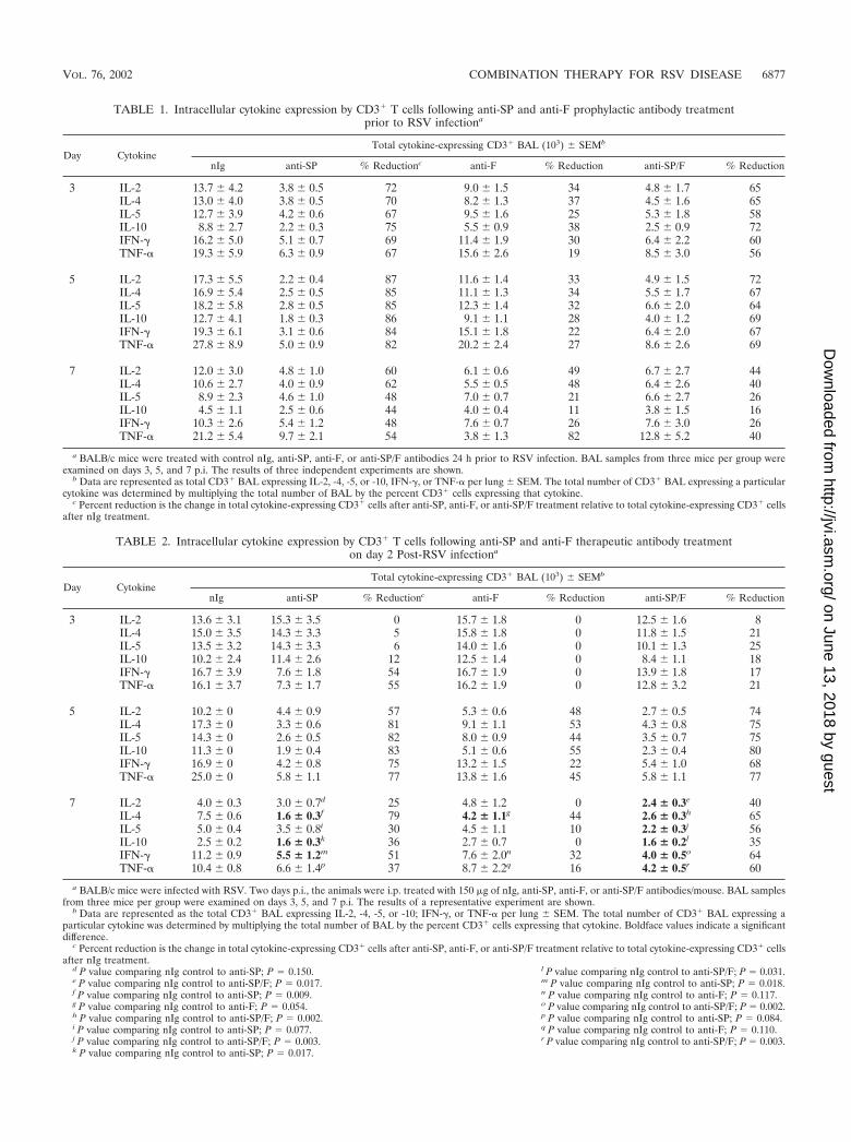
a BALB/c mice were treated with control nIg, anti-SP, anti-F, or anti-SP/F antibodies 24 h prior to RSV infection. BAL samples from three mice per group wereexamined on days 3, 5, and 7 p.i. The results of three independent experiments are shown.
b Data are represented as total CD3� BAL expressing IL-2, -4, -5, or -10, IFN-�, or TNF-� per lung � SEM. The total number of CD3� BAL expressing a particularcytokine was determined by multiplying the total number of BAL by the percent CD3� cells expressing that cytokine.
c Percent reduction is the change in total cytokine-expressing CD3� cells after anti-SP, anti-F, or anti-SP/F treatment relative to total cytokine-expressing CD3� cellsafter nIg treatment.
TABLE 2. Intracellular cytokine expression by CD3� T cells following anti-SP and anti-F therapeutic antibody treatmenton day 2 Post-RSV infectiona
Day CytokineTotal cytokine-expressing CD3� BAL (103) � SEMb
a BALB/c mice were infected with RSV. Two days p.i., the animals were i.p. treated with 150 �g of nIg, anti-SP, anti-F, or anti-SP/F antibodies/mouse. BAL samplesfrom three mice per group were examined on days 3, 5, and 7 p.i. The results of a representative experiment are shown.
b Data are represented as the total CD3� BAL expressing IL-2, -4, -5, or -10; IFN-�, or TNF-� per lung � SEM. The total number of CD3� BAL expressing aparticular cytokine was determined by multiplying the total number of BAL by the percent CD3� cells expressing that cytokine. Boldface values indicate a significantdifference.
c Percent reduction is the change in total cytokine-expressing CD3� cells after anti-SP, anti-F, or anti-SP/F treatment relative to total cytokine-expressing CD3� cellsafter nIg treatment.
d P value comparing nIg control to anti-SP; P 0.150.e P value comparing nIg control to anti-SP/F; P 0.017.f P value comparing nIg control to anti-SP; P 0.009.g P value comparing nIg control to anti-F; P 0.054.h P value comparing nIg control to anti-SP/F; P 0.002.i P value comparing nIg control to anti-SP; P 0.077.j P value comparing nIg control to anti-SP/F; P 0.003.k P value comparing nIg control to anti-SP; P 0.017.
l P value comparing nIg control to anti-SP/F; P 0.031.m P value comparing nIg control to anti-SP; P 0.018.n P value comparing nIg control to anti-F; P 0.117.o P value comparing nIg control to anti-SP/F; P 0.002.p P value comparing nIg control to anti-SP; P 0.084.q P value comparing nIg control to anti-F; P 0.110.r P value comparing nIg control to anti-SP/F; P 0.003.
VOL. 76, 2002 COMBINATION THERAPY FOR RSV DISEASE 6877
day 7 p.i., the numbers of cells were similar for all treatments.For the leukocyte subsets, the decrease was not always evidenton day 3 p.i. but was most evident on day 5 p.i. Anti-SP/Fantibody treatment reduced the total number of CD8� (P �0.05 [Fig. 1B]), CD4� (Fig. 1C), B220� (P � 0.05 [Fig. 1D]),and CD11b� (P � 0.05 [Fig. 1E]) cells and PMN (P � 0.05[Fig. 1F]) 6- to 10-fold by day 5 p.i. compared to nIg-treatedmice. In contrast, treatment with anti-F antibody alone wasassociated with an increase in CD8� and CD11b� cells on day3 p.i. and had minimal effect on the leukocyte subsets at latertime points (Fig. 1).
Therapeutic treatment of RSV infection. The results foranti-SP antibody prophylaxis suggested that similar treatmentmight be therapeutically effective during RSV infection. Earlyanti-SP or anti-SP/F antibody treatment on day 2 p.i., prior tothe period of maximal viral replication (day 5 p.i. [see Fig. 4]),led to a prompt and marked decrease of total pulmonary cellscompared to nIg antibody-treated mice (Fig. 2A). One dayafter antibody treatment, the numbers for all cell subsets weredecreased (P � 0.05) compared to anti-F or nIg antibody-treated mice. Treatment with anti-F antibody, relative to nIg,was associated with some reduction of CD8�, B220�, andCD11b� cells and PMN, but not CD4� cells, on day 3 p.i. (Fig.2B and D to F). By day 5 p.i., anti-F antibody treatment alonewas associated with a pronounced decrease in CD8� cells anda modest decrease in the other leukocyte subsets.
Late anti-SP antibody treatment in RSV infection was alsoeffective (Fig. 3). Administration of anti-SP or anti-SP/F anti-bodies on day 6 p.i. reduced the total number of cells infiltrat-ing the lung 1 day after treatment (Fig. 3A). Antibody treat-ment decreased the numbers of all leukocyte subsets examinedon day 7 p.i. compared to both nIg and anti-F antibody-treatedmice (Fig. 3B to F).
Cytokine expression after anti-SP and anti-F antibody treat-ments. Similar to cell numbers, the levels of intracellular cy-tokine expression were markedly decreased by anti-SP or anti-
SP/F antibody treatment (Tables 1, 2, and 3). This decrease ismost easily seen by examining the percent decrease of CD3�
BAL for the different treatments. In mice treated prophylac-tically with anti-SP or anti-SP/F antibody, cytokine expressionon days 3 and 5 p.i. was inhibited 56 to 87% (Table 1). By day7 p.i., this inhibition was less distinct and ranged from 16 to62%. Anti-F antibody treatment also reduced cytokine expres-sion, but the effect was less than that observed with anti-SP/Fantibody treatment (Table 1). For mice treated therapeuticallyon day 2 p.i. with anti-SP or anti-SP/F antibody, there was nota significant decrease in cytokine expression on day 3 p.i.,except for IFN-� and TNF-� in the anti-SP-treated mice; how-ever, all treatment groups showed a decrease compared to nIgtreatment on day 5 p.i. The decrease on day 5 p.i. was greaterwhen anti-SP antibodies were included in the treatment (57 to83%) than when they were not (anti-F treatment, 22 to 55%)(Table 2). On day 7 p.i., the greatest decrease in cytokineexpression was again most evident with treatments that in-cluded anti-SP antibodies, and the decrease following all treat-ments was greatest for IL-4 and IFN-� expression. Intracellularcytokine expression also decreased in mice treated with anti-SPor anti-SP/F antibody on day 6 p.i. (Table 3) and was greatestwhen anti-SP antibody was included in the treatment. Al-though treatment with anti-SP antibody reduced cytokine ex-pression on day 7 p.i. (26 to 72%), the greatest reduction incytokine expression was observed on day 9 p.i. (51 to 70%) formice treated with anti-SP antibodies (Table 3).
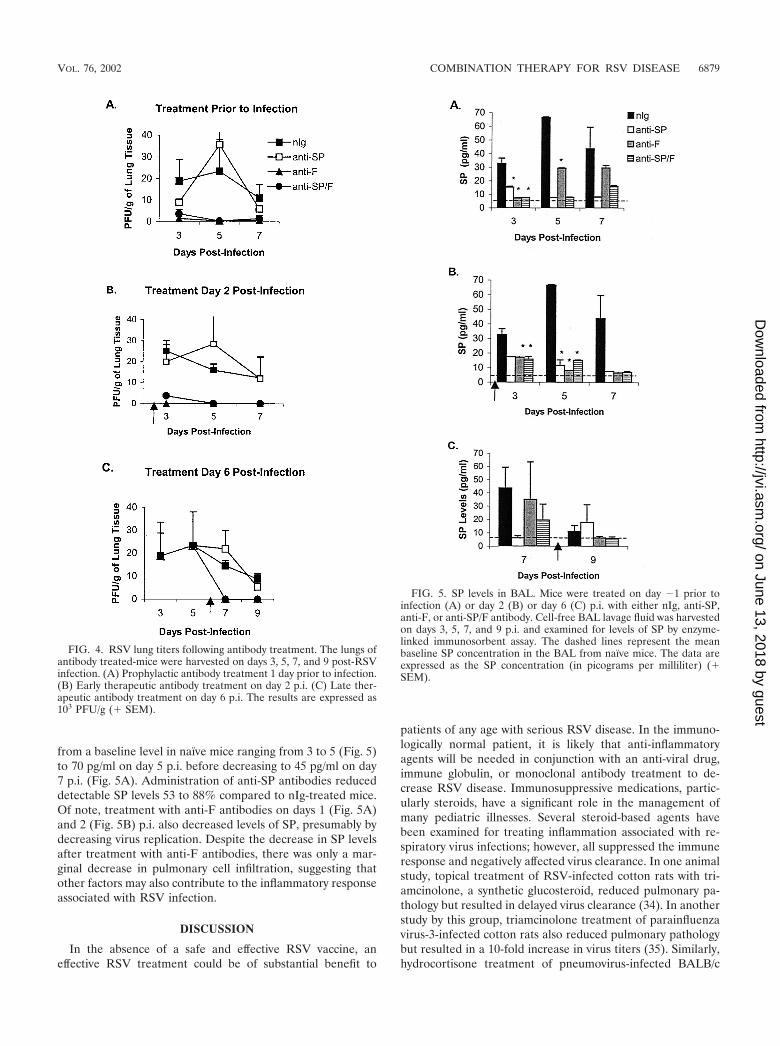
Virus replication and SP levels. As expected, administrationof anti-F antibodies alone markedly decreased the titer of virusin the lungs. Treatment with anti-SP antibodies did not alterthe titer of the virus recovered (Fig. 4), i.e., the titer of virus inmice treated with anti-SP antibodies was not significantly dif-ferent from the titer of nIg-treated mice, and the titer of virusin mice treated with anti-SP/F antibody was not significantlydifferent from that in mice treated with anti-F antibody alone.As previously shown, SP levels increased with RSV infection
TABLE 3. Intracellular cytokine expression by CD3� T cells following anti-SP and anti-F therapeutic antibody treatmenton day 6 post-RSV infectiona
Day CytokineTotal cytokine-expressing CD3� BAL (103) � SEMb
a BALB/c mice were infected with RSV. Six days p.i., the animals were i.p. treated with nIg, anti-SP, anti-F, or anti-SP/F antibodies. BAL samples from three miceper group were examined on days 3, 5, 7, and 9 p.i. The results of a representative experiment are shown.
b Data are represented as total CD3� BAL expressing IL-2, -4, -5, or -10, IFN-�, or TNF-� per lung � SEM. The total number of CD3� BAL expressing a particularcytokine was determined by multiplying the total number of BAL by the percent CD3� cells expressing that cytokine. Boldface values indicate a significant difference.
c Percent reduction is the change in total cytokine-expressing CD3� cells after anti-SP, anti-F, or anti-SP/F treatment relative to total cytokine-expressing CD3� cellsafter nIg treatment.
d P value comparing nIg control to anti-SP; P 0.042.e P value comparing nIg control to anti-SP; P 0.073.
from a baseline level in naïve mice ranging from 3 to 5 (Fig. 5)to 70 pg/ml on day 5 p.i. before decreasing to 45 pg/ml on day7 p.i. (Fig. 5A). Administration of anti-SP antibodies reduceddetectable SP levels 53 to 88% compared to nIg-treated mice.Of note, treatment with anti-F antibodies on days 1 (Fig. 5A)and 2 (Fig. 5B) p.i. also decreased levels of SP, presumably bydecreasing virus replication. Despite the decrease in SP levelsafter treatment with anti-F antibodies, there was only a mar-ginal decrease in pulmonary cell infiltration, suggesting thatother factors may also contribute to the inflammatory responseassociated with RSV infection.
DISCUSSION
In the absence of a safe and effective RSV vaccine, aneffective RSV treatment could be of substantial benefit to
patients of any age with serious RSV disease. In the immuno-logically normal patient, it is likely that anti-inflammatoryagents will be needed in conjunction with an anti-viral drug,immune globulin, or monoclonal antibody treatment to de-crease RSV disease. Immunosuppressive medications, partic-ularly steroids, have a significant role in the management ofmany pediatric illnesses. Several steroid-based agents havebeen examined for treating inflammation associated with re-spiratory virus infections; however, all suppressed the immuneresponse and negatively affected virus clearance. In one animalstudy, topical treatment of RSV-infected cotton rats with tri-amcinolone, a synthetic glucosteroid, reduced pulmonary pa-thology but resulted in delayed virus clearance (34). In anotherstudy by this group, triamcinolone treatment of parainfluenzavirus-3-infected cotton rats also reduced pulmonary pathologybut resulted in a 10-fold increase in virus titers (35). Similarly,hydrocortisone treatment of pneumovirus-infected BALB/c
FIG. 4. RSV lung titers following antibody treatment. The lungs ofantibody treated-mice were harvested on days 3, 5, 7, and 9 post-RSVinfection. (A) Prophylactic antibody treatment 1 day prior to infection.(B) Early therapeutic antibody treatment on day 2 p.i. (C) Late ther-apeutic antibody treatment on day 6 p.i. The results are expressed as103 PFU/g (� SEM).
FIG. 5. SP levels in BAL. Mice were treated on day �1 prior toinfection (A) or day 2 (B) or day 6 (C) p.i. with either nIg, anti-SP,anti-F, or anti-SP/F antibody. Cell-free BAL lavage fluid was harvestedon days 3, 5, 7, and 9 p.i. and examined for levels of SP by enzyme-linked immunosorbent assay. The dashed lines represent the meanbaseline SP concentration in the BAL from naïve mice. The data areexpressed as the SP concentration (in picograms per milliliter) (�SEM).
VOL. 76, 2002 COMBINATION THERAPY FOR RSV DISEASE 6879
mice had a deleterious effect of enhancing viral replicationand accelerating mortality (12). Prolonged virus sheddinghas also been shown in clinical studies of RSV-infectedchildren with compromised immune function due to steroidtherapy (16).
The concept of combining nonsteroidal anti-inflammatorytreatment with antiviral agents has been examined for nonres-piratory viruses, including rhinovirus-induced rhinitis (15) andherpes simplex virus keratitis (14). In one study, treatment ofrhinovirus infections with the anti-viral agent IFN-� and twoanti-inflammatory agents (ipratropium and naproxen) reducedthe overall symptoms and disease severity in treated individu-als (15). Likewise, combined treatment of herpetic stromalkeratitis with acyclovir and cyclosporin A reduced stromal in-filtration and the severity of stromal disease (14).
An alternative target for reducing inflammation associatedwith respiratory virus infection may be SP. SP has been shownto be an important contributor to airway inflammation anddisease. Recent data from animal studies show that RSV up-regulates the SP receptor (20, 33), and elevated levels of SPhave been detected in patients with asthma and obstructiveairway disease (38). Previously, we observed elevated pulmo-nary SP levels associated with RSV infection in BALB/c miceand showed that treatment with anti-SP antibodies was effec-tive in reducing the number of inflammatory cells and proin-flammatory cytokine expression (40). In this study, we demon-strate the effectiveness of combining anti-SP antibody with aneutralizing anti-F antibody in controlling both virus replica-tion and RSV-associated inflammation. The anti-F monoclonalIgG antibody used in this study (143-6C), recognizes the sameantigenic site (site A) as the anti-F monoclonal antibody usedfor prophylaxis in humans (palivizumab) (2, 18). As expected,this neutralizing monoclonal antibody inhibited virus replica-tion when given as prophylactic or therapeutic treatment, andas expected from previous studies, anti-F antibody treatmentwas inadequate at reducing pulmonary inflammation. In con-trast, both prophylactic and therapeutic treatment with anti-SPantibodies promptly reduced the number of inflammatorycells, and unlike steroid therapy, it did not prolong virus rep-lication. In addition, treatment with anti-SP antibodies de-creased the number of cells expressing proinflammatory cyto-kines.
In summary, this study shows that the anti-SP antibodies caneffectively reduce inflammation and suggests that anti-SP treat-ment may complement the antiviral effects of neutralizing an-ti-F antibody treatment or antiviral drugs to provide an effec-tive treatment for RSV disease.
ACKNOWLEDGMENTS
This research was supported in part by an appointment to the Re-search Participation Program at the Centers for Disease Control andPrevention, National Center for Infectious Diseases, Division of Viraland Rickettsial Diseases, administered by the Oak Ridge Institute forScience and Education through an interagency agreement between theU.S. Department of Energy and the Centers for Disease Control andPrevention.
We thank Les Jones (Centers for Disease Control and Prevention,Atlanta, Ga.) for technical assistance.
REFERENCES
1. Anderson, L. J., P. Bingham, and J. C. Hierholzer. 1988. Neutralization ofrespiratory syncytial virus by individual and mixtures of F and G proteinmonoclonal antibodies. J. Virol. 62:4232–4238.
2. Beeler, J. A., and K. van Wyke Coelingh. 1989. Neutralization epitopes of theF glycoprotein of respiratory syncytial virus: effect of mutation upon fusionfunction. J. Virol. 63:2941–2950.
3. Berczi, I., I. M. Chalmers, E. Nagy, and R. J. Warrington. 1996. The immuneeffects of neuropeptides. Baillieres Clin. Rheumatol. 10:227–257.
4. Berman, A. S., C. Chancellor-Freeland, G. Zhu, and P. H. Black. 1996.Substance P primes murine peritoneal macrophages for an augmentedproinflammatory cytokine response to lipopolysaccharide. Neuroimmuno-modulation 3:141–149.
5. Calvo, N., J. Reiriz, E. Perez-Navarro, and J. Alberch. 1996. Tachykininsprotect cholinergic neurons from quinolinic acid excitotoxicity in striatalcultures. Brain Res. 740:323–328.
6. Connors, M., P. L. Collins, C. Y. Firestone, and B. R. Murphy. 1991. Res-piratory syncytial virus (RSV) F, G, M2 (22K), and N proteins each induceresistance to RSV challenge, but resistance induced by M2 and N proteins isrelatively short-lived. J. Virol. 65:1634–1637.
7. Covas, M. J., L. A. Pinto, and R. M. Victorino. 1994. Disturbed immuno-regulatory properties of the neuropeptide substance P on lymphocyte pro-liferation in HIV infection. Clin. Exp. Immunol. 96:384–388.
8. Crowe, J. E., Jr., B. R. Murphy, R. M. Chanock, R. A. Williamson, C. F.Barbas III, and D. R. Burton. 1994. Recombinant human respiratory syncy-tial virus (RSV) monoclonal antibody Fab is effective therapeutically whenintroduced directly into the lungs of RSV-infected mice. Proc. Natl. Acad.Sci. USA 91:1386–1390.
9. De Giorgio, R., P. L. Tazzari, G. Barbara, V. Stanghellini, and R. Cori-naldesi. 1998. Detection of substance P immunoreactivity in human periph-eral leukocytes. J. Neuroimmunol. 82:175–181.
10. De Vincenzo, J. P., D. Leombruno, R. J. Soiffer, and G. R. Siber. 1996.Immunotherapy of respiratory syncytial virus pneumonia following bonemarrow transplantation. Bone Marrow Transplant. 17:1051–1056.
11. Dickerson, C., B. Undem, B. Bullock, and R. A. Winchurch. 1998. Neuropep-tide regulation of proinflammatory cytokine responses. J. Leukoc. Biol. 63:602–605.
12. Domachowske, J. B., C. A. Bonville, D. Ali-Ahmad, K. D. Dyer, A. J. Easton,and H. F. Rosenberg. 2001. Glucocorticoid administration accelerates mor-tality of pneumovirus-infected mice. J. Infect. Dis. 184:1518–1523.
13. Fisher, R. G., J. E. Crowe, Jr., T. R. Johnson, Y. W. Tang, and B. S. Graham.1999. Passive IgA monoclonal antibody is no more effective than IgG atprotecting mice from mucosal challenge with respiratory syncytial virus.J. Infect. Dis. 180:1324–1327.
14. Gunduz, K., and O. Ozdemir. 1997. Topical cyclosporin as an adjunct totopical acyclovir treatment in herpetic stromal keratitis. Ophthalmol. Res.29:405–408.
15. Gwaltney, J. M., Jr. 1992. Combined antiviral and antimediator treatment ofrhinovirus colds. J. Infect. Dis. 166:776–782.
16. Hall, C. B., K. R. Powell, N. E. MacDonald, C. L. Gala, M. E. Menegus, S. C.Suffin, and H. J. Cohen. 1986. Respiratory syncytial viral infection in childrenwith compromised immune function. N. Engl. J. Med. 315:77–81.
17. Ho, W. Z., J. P. Lai, X. H. Zhu, M. Uvaydova, and S. D. Douglas. 1997.Human monocytes and macrophages express substance P and neurokinin-1receptor. J. Immunol. 159:5654–5660.
18. Johnson, S., C. Oliver, G. A. Prince, V. G. Hemming, D. S. Pfarr, S. C. Wang,M. Dormitzer, J. O’Grady, S. Koenig, J. K. Tamura, R. Woods, G. Bansal, D.Couchenour, E. Tsao, W. C. Hall, and J. F. Young. 1997. Development of ahumanized monoclonal antibody (MEDI-493) with potent in vitro and invivo activity against respiratory syncytial virus. J. Infect. Dis. 176:1215–1224.
19. Khoshoo, V., and D. Edell. 2000. Ribavarin in ventilated respiratory syncytialvirus bronchiolitis: a randomized, placebo-controlled trial. Am. J. Respir.Crit. Care Med. 162:333–334.
20. King, K. A., C. Hu, M. M. Rodriguez, R. Romaguera, X. Jiang, and G.Piedimonte. 2001. Exaggerated neurogenic inflammation and substance Preceptor upregulation in RSV-infected weanling rats. Am. J. Respir. CellMol. Biol. 24:101–107.
21. Kurt-Jones, E. A., L. Popova, L. Kwinn, L. M. Haynes, L. P. Jones, R. A.Tripp, E. E. Walsh, M. W. Freeman, D. T. Golenbock, L. J. Anderson, andR. W. Finberg. 2000. Pattern recognition receptors TLR4 and CD14 mediateresponse to respiratory syncytial virus. Nat. Immunol. 1:398–401.
22. Lambert, N., P. L. Lescoulie, B. Yassine-Diab, G. Enault, B. Mazieres, C. DePreval, and A. Cantagrel. 1998. Substance P enhances cytokine-inducedvascular cell adhesion molecule-1 (VCAM-1) expression on cultured rheu-matoid fibroblast-like synoviocytes. Clin. Exp. Immunol. 113:269–275.
23. Lambrecht, B. N., P. R. Germonpre, E. G. Everaert, I. Carro-Muino, M. DeVeerman, C. de Felipe, S. P. Hunt, K. Thielemans, G. F. Joos, and R. A.Pauwels. 1999. Endogenously produced substance P contributes to lympho-cyte proliferation induced by dendritic cells and direct TCR ligation. Eur.J. Immunol. 29:3815–3825.
24. Lee, H. R., W. Z. Ho, and S. D. Douglas. 1994. Substance P augments tumor
necrosis factor release in human monocyte-derived macrophages. Clin. Di-agn. Lab. Immunol. 1:419–423.
25. Maggi, C. A. 1997. The effects of tachykinins on inflammatory and immunecells. Regul. Pept. 70:75–90.
26. Maggi, C. A. 1995. The mammalian tachykinin receptors. Gen. Pharmacol.26:911–944.
27. Malley, R., J. DeVincenzo, O. Ramilo, P. H. Dennehy, H. C. Meissner, W. C.Gruber, P. J. Sanchez, H. Jafri, J. Balsley, D. Carlin, S. Buckingham, L.Vernacchio, and D. M. Ambrosino. 1998. Reduction of respiratory syncytialvirus (RSV) in tracheal aspirates in intubated infants by use of humanizedmonoclonal antibody to RSV F protein. J. Infect. Dis. 178:1555–1561.
28. Manske, J. M., E. L. Sullivan, and S. M. Andersen. 1995. Substance Pmediated stimulation of cytokine levels in cultured murine bone marrowstromal cells. Adv. Exp. Med. Biol. 383:53–64.
29. Nakagawa, N., H. Sano, and I. Iwamoto. 1995. Substance P induces theexpression of intercellular adhesion molecule-1 on vascular endothelial cellsand enhances neutrophil transendothelial migration. Peptides 16:721–725.
30. Pascual, D. W., and K. L. Bost. 1990. Substance P production by P388D1macrophages: a possible autocrine function for this neuropeptide. Immunol-ogy 71:52–56.
31. Payan, D. G. 1989. Neuropeptides and inflammation: the role of substance P.Annu. Rev. Med. 40:341–352.
32. Pernow, B. 1985. Role of tachykinins in neurogenic inflammation. J. Immu-nol. 135:812S-815S.
33. Piedimonte, G., M. M. Rodriguez, K. A. King, S. McLean, and X. Jiang.1999. Respiratory syncytial virus upregulates expression of the substance Preceptor in rat lungs. Am. J. Physiol. 277:L831–L840.
34. Prince, G. A., A. Mathews, S. J. Curtis, and D. D. Porter. 2000. Treatmentof respiratory syncytial virus bronchiolitis and pneumonia in a cotton ratmodel with systemically administered monoclonal antibody (Palivizumab)and glucocorticosteroid. J. Infect. Dis. 182:1326–1330.
35. Prince, G. A., and D. D. Porter. 1996. Treatment of parainfluenza virus type
3 bronchiolitis and pneumonia in a cotton rat model using topical antibodyand glucocorticosteroid. J. Infect. Dis. 173:598–608.
36. Rameshwar, P., and P. Gascon. 1995. Substance P (SP) mediates productionof stem cell factor and interleukin-1 in bone marrow stroma: potentialautoregulatory role for these cytokines in SP receptor expression and induc-tion. Blood 86:482–490.
37. Stott, E. J., G. Taylor, L. A. Ball, K. Anderson, K. K. Young, A. M. King, andG. W. Wertz. 1987. Immune and histopathological responses in animalsvaccinated with recombinant vaccinia viruses that express individual genes ofhuman respiratory syncytial virus. J. Virol. 61:3855–3861.
38. Tomaki, M., M. Ichinose, M. Miura, Y. Hirayama, H. Yamauchi, N. Naka-jima, and K. Shirato. 1995. Elevated substance P content in induced sputumfrom patients with asthma and patients with chronic bronchitis. Am. J.Respir. Crit. Care Med. 151:613–617.
39. Tripp, R. A., D. Moore, L. Jones, W. Sullender, J. Winter, and L. J. Ander-son. 1999. Respiratory syncytial virus G and/or SH protein alters Th1 cyto-kines, natural killer cells, and neutrophils responding to pulmonary infectionin BALB/c mice. J. Virol. 73:7099–7107.
40. Tripp, R. A., D. Moore, J. Winter, and L. J. Anderson. 2000. Respiratorysyncytial virus infection and G and/or SH protein expression contribute tosubstance P, which mediates inflammation and enhanced pulmonary diseasein BALB/c mice. J. Virol. 74:1614–1622.
41. Walker, T. A., S. Khurana, and S. J. Tilden. 1994. Viral respiratory infec-tions. Pediatr. Clin. N. Am. 41:1365–1381.
42. Weltzin, R., S. A. Hsu, E. S. Mittler, K. Georgakopoulos, and T. P. Monath.1994. Intranasal monoclonal immunoglobulin A against respiratory syncytialvirus protects against upper and lower respiratory tract infections in mice.Antimicrob. Agents Chemother. 38:2785–2791.
43. Win, N., D. Mitchell, S. Pugh, and N. H. Russell. 1992. Successful therapywith ribavirin of late onset respiratory syncytial virus pneumonitis compli-cating allogeneic bone transplantation. Clin. Lab. Haematol. 14:29–32.
44. Yonehara, N., Y. Imai, T. Shibutani, and R. Inoki. 1989. Participation ofsubstance P in inflammatory responses. Adv. Exp. Med. Biol. 247B:529–534.
VOL. 76, 2002 COMBINATION THERAPY FOR RSV DISEASE 6881