62
Version 5.01 9/01 Catalog #E8000S ® BioLabs NEW ENGLAND Inc. pMAL ™ Protein Fusion and Purification System Expression and Purification of Proteins from Cloned Genes Instruction Manual

Version 5.019/01Catalog #E8000S
®BioLabsNEW ENGLAND
Inc.
pMAL™ Protein Fusionand Purification System
Expression and Purification of Proteins from Cloned Genes
I n s t r u c t i o n M a n u a l

Table of Contents
System Components . . . . . . . . . . . . . . . . . . . . . . . . . . . . . . . . . . . . . . . . . . . . . . . . . . . . . 2Introduction . . . . . . . . . . . . . . . . . . . . . . . . . . . . . . . . . . . . . . . . . . . . . . . . . . . . . . . . . . . . . . . . 5Construction of the Fusion Plasmid . . . . . . . . . . . . . . . . . . . . . . . . . . . . . . . . . . . . 7
Choice of Vector . . . . . . . . . . . . . . . . . . . . . . . . . . . . . . . . . . . . . . . . . . . . . . . . . . . . . . . 8Sequencing . . . . . . . . . . . . . . . . . . . . . . . . . . . . . . . . . . . . . . . . . . . . . . . . . . . . . . . . . . . . 9Creating a Blunt-ended Fragment . . . . . . . . . . . . . . . . . . . . . . . . . . . . . . . . . . . 9Cloning a PCR Fragment . . . . . . . . . . . . . . . . . . . . . . . . . . . . . . . . . . . . . . . . . . 14
Pilot Experiment . . . . . . . . . . . . . . . . . . . . . . . . . . . . . . . . . . . . . . . . . . . . . . . . . . . . . . . . . 19Affinity Chromatography . . . . . . . . . . . . . . . . . . . . . . . . . . . . . . . . . . . . . . . . . . . . . . . 23
Method I: Fusion Protein from Total Cell Extract . . . . . . . . . . . . . . . . 23Method II: Exported Fusion Protein . . . . . . . . . . . . . . . . . . . . . . . . . . . . . . . 26Regenerating the Amylose Resin Column . . . . . . . . . . . . . . . . . . . . . . . 27
Cleavage of the Fusion Protein . . . . . . . . . . . . . . . . . . . . . . . . . . . . . . . . . . . . . . . 28Denaturing the Fusion Protein . . . . . . . . . . . . . . . . . . . . . . . . . . . . . . . . . . . . . 29Separating the Protein of Interest from MBP . . . . . . . . . . . . . . . . . . . . 30
DEAE-Sepharose Chromatography . . . . . . . . . . . . . . . . . . . . . . . . . . . 30Removal of Maltose and Rebinding to Amylose Resin . . . . . 32
Media and Solutions . . . . . . . . . . . . . . . . . . . . . . . . . . . . . . . . . . . . . . . . . . . . . . . . . . . . 33Troubleshooting and Tips . . . . . . . . . . . . . . . . . . . . . . . . . . . . . . . . . . . . . . . . . . . . . . 34Protein Fusion and Purification Strain List . . . . . . . . . . . . . . . . . . . . . . . . . . . 51Molecular Weights of pMAL Proteins . . . . . . . . . . . . . . . . . . . . . . . . . . . . . . . . . 54References . . . . . . . . . . . . . . . . . . . . . . . . . . . . . . . . . . . . . . . . . . . . . . . . . . . . . . . . . . . . . . 55
Notice to Buyer/User: The buyer/user has a nonexclusive license to use the pMAL™ vectors forresearch purposes only. A license to use the pMAL™ vectors for commercial purposes is availablefrom New England Biolabs, Inc.US Patent 5,643,758.
© 1991-2001 New England Biolabs1

System Components
pMAL™-c2X 10 µgpMAL™-p2X 10 µg
Supplied in 10 mM Tris-HCl, 1 mM EDTA, pH 7.5. Store at –20°C
Amylose Resin 15 mlSupplied pre-swollen in 20% ethanol. Binding capacity > 3 mg/ml bedvolume. Store at 4°C
Factor Xa 50 µgPurified from bovine plasma; MW 42,400 Daltons (two disulfide-linkedchains, MW ~ 30 kDa and ~ 20 kDa). 1 µg of Factor Xa will cleave 50 µgof the test substrate MBP paramyosin∆Sal to 95% completion in 8 hoursor less at 23°C in 50 µl. Supplied in 20 mM HEPES, 500 mM NaCl, 2 mMCaCl2, 50% glycerol, pH 8.0. Store at –20°C
Anti-MBP Antiserum 25 µlRabbit serum prepared using purified maltose-binding protein. Suggesteddilution for Western blotting or ELISA 1:10,000. Store at –20°C.
2

MBP2* 10 µg (40 µg/ml)
Expressed from a derivative of pMAL-c2X, including amino acid residuescoded for by the polylinker up to the Xmn I site. MW 42,698 Daltons.Supplied in SDS-PAGE Sample Buffer. Store at –20°C.
MBP2*-paramyosin ∆Sal 100 µg 5 mg/ml
An MBP fusion protein as a positive control for Factor Xa cleavage. Suppliedin 20 mM Tris-HCl, 200 mM NaCl, 50% glycerol, pH 7.2. MW 70,216Daltons. Store at –20°C.
E. coli host TB1
F– ara ∆(lac-proAB) [φ80dlac ∆(lacZ)M15] rpsL(StrR) thi hsdR. Suppliedfrozen in 25% glycerol (not competent). Store at –70°C.
Information presented herein is accurate and reliable to the best of ourknowledge and belief, but is not guaranteed to be so. Nothing herein is to beconstrued as recommending any practice or any product in violation of anypatent or in violation of any law or regulation. It is the user’s responsibility todetermine for himself or herself the suitability of any material and/or proce-dure for a specific purpose and to adopt such safety precautions as may benecessary.
3
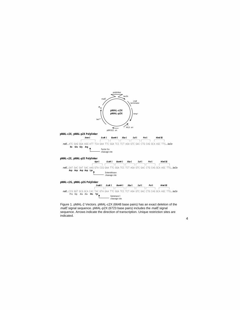
pMAL-c2XpMAL-p2X
pBR322 ori
lacl
P
malE
polylinker
lacZα
q
tac
rrnBterminator
Amp
M13 ori+
–
r
Kpn I EcoR I BamH I Xba I Sal I Pst I Hind III
malE...GAT GAC GAT GAC AAG GTA CCG GAA TTC GGA TCC TCT AGA GTC GAC CTG CAG GCA AGC TTG... lacZαAsp Asp Asp Asp Lys
pMAL-c2E, pMAL-p2E Polylinker
SnaB I EcoR I BamH I Xba I Sal I Pst I Hind III
malE...CCG GGT GCG GCA CAC TAC GTA GAA TTC GGA TCC TCT AGA GTC GAC CTG CAG GCA AGC TTG... lacZαPro Gly Ala Ala His Tyr
pMAL-c2G, pMAL-p2G Polylinker
Xmn I EcoR I BamH I Xba I Sal I Pst I Hind III
malE...ATC GAG GGA AGG ATT TCA GAA TTC GGA TCC TCT AGA GTC GAC CTG CAG GCA AGC TTG...lacZαIle Glu Gly Arg
pMAL-c2X, pMAL-p2X Polylinker
Figure 1. pMAL-2 Vectors. pMAL-c2X (6648 base pairs) has an exact deletion of themalE signal sequence. pMAL-p2X (6723 base pairs) includes the malE signalsequence. Arrows indicate the direction of transcription. Unique restriction sites areindicated.
Factor Xacleavage site
▲
Enterokinasecleavage site
▲
Genenase Icleavage site
▲
4

Introduction
The pMAL™-2 vectors (Figure 1) provide a method for expressing andpurifying a protein produced from a cloned gene or open reading frame.The cloned gene is inserted downstream from the malE gene of E. coli,which encodes maltose-binding protein (MBP), resulting in the expressionof an MBP fusion protein (1,2). The method uses the strong “tac” promoterand the malE translation initiation signals to give high-level expression ofthe cloned sequences (3,4), and a one-step purification of the fusionprotein using MBP’s affinity for maltose (5). The vectors express the malEgene (with or without its signal sequence) fused to the lacZα gene. Restric-tion sites between malE and lacZα are available for inserting the codingsequence of interest. Insertion inactivates the β-galactosidase α-fragmentactivity of the malE-lacZα fusion, which results in a blue to white colorchange on Xgal plates when the construction is transformed into an α-complementing host such as TB1 (6) or JM107 (7). When present, thesignal peptide on pre-MBP directs fusion proteins to the periplasm. Forfusion proteins that can be successfully exported, this allows folding anddisulfide bond formation to take place in the periplasm of E. coli, as well asallowing purification of the protein from the periplasm (8). The vectors carrythe lacIq gene, which codes for the Lac repressor. This keeps expressionfrom Ptac low in the absence of IPTG induction. The pMAL-2 vectors alsocontain the sequence coding for the recognition site of a specific protease,located just 5´ to the polylinker insertion sites. This allows MBP to becleaved from the protein of interest after purification. The pMAL-c2X andpMAL-p2X vectors that are included in the system encode the site forFactor Xa (9, 10). Factor Xa cleaves after its four amino acid recognitionsequence, so that few or no vector-derived residues are attached to the
5

protein of interest, depending on the site used for cloning. pMAL vectorscontaining sites for alternative proteases are also available (see Figure 1).The vectors pMAL-c2G (NEB #N8068) and pMAL-p2G (NEB #N8069)encode the site for Genenase™I (NEB #P8075), which cleaves following thesequence His-Tyr. The vectors pMAL-c2E (NEB #N8066) and pMAL-p2E(NEB #N8067) encode the site for Enterokinase (NEB #P8070), whichcleaves following the sequence Asp-Asp-Asp-Asp-Lys.
In the large majority of cases, fusion protein expressed from a pMAL-c2plasmid constitutes 20–40% of the total cellular protein, while fusion proteinexpressed from a pMAL-p2 plasmid constitutes 1–5% of the total cellularprotein. For pMAL-c2 vectors, a band corresponding to the fusion proteincan usually be seen by running a small sample of induced cells on an SDS-PAGE gel. The yield of fusion protein from the affinity purification ranges upto 200 mg/liter culture, with typical yields in the range of 10–40 mg/liter.The yield varies greatly depending upon the sequences fused to malE. Incases where the yield has been compared directly, the pMAL-c2 vectors (nosignal sequence) give approximately 10-fold more protein in the affinitypurification than pMAL-p2 vectors. The pMAL-p2 vectors are useful forcases where export to the periplasm is desirable, e.g. for purification ordisulfide bond formation. 75% of the fusions made so far have worked in theaffinity purification. In the cases that have not worked, the fusion binds to thecolumn poorly or not at all, is degraded by E. coli proteases, or is insoluble.The pMAL-2 vectors also contain the origin of DNA replication of the E. colibacteriophage M13, which allows the production of single-stranded DNA byinfection of cells bearing a pMAL-2 plasmid with a helper phage (11). Thesingle-stranded plasmid DNA can be used for sequencing using a primeravailable from NEB (see Sequencing, page 9), or for oligonucleotide-directed mutagenesis.
6

Construction of the Fusion Plasmid
To produce a fusion protein in the pMAL-2 vectors, the gene or open readingframe of interest must be inserted into the pMAL-2 vectors so that it is in thesame translational reading frame as the vector’s malE gene. The vectorshave a polylinker containing a restriction site for cloning fragments directlydownstream of the specific protease site. A number of other restriction sitesare also available for cloning fragments downstream of the primary site, orfor directional cloning of a blunt/sticky-ended fragment. Inserts cloned intothe primary site produce a protein of interest that, after protease cleavage,contains no vector-derived amino acids (9,10). Factor Xa, Genenase I andEnterokinase will not cleave fusion proteins that have a proline immediatelyfollowing the last residue of their respective sites (the P1´ position), so thefirst three bases of the insert should not encode Pro when cloning into theprimary site. In addition, Factor Xa fails to cleave sites where the P1´ residueis arginine, and Genenase I fails to cleave if the P1´ residue is isoleucine.Several strategies may be employed to create an appropriate fragment tosubclone. It is assumed that the sequence of interest includes a translationalstop codon at its 3´ end; if not, one should be engineered into the cloningstrategy. Alternatively, a linker containing a stop codon (e.g. NEB #S1061S)can be inserted into one of the downstream polylinker sites, or the stopcodon present in the Xba I site can be shifted into appropriate reading frame(e.g., by filling in the site with Klenow).
7

Choice of Vector
The pMAL vectors come in six versions, for expressing fusion proteins withone of three different specific protease sites in either the cytoplasm or theperiplasm. Choice among the three proteases is determined empirically,since the three dimensional structure of the fusion protein determinessusceptibility to cleavage. Choice between the cytoplasmic and periplasmicversions can be guided by the descriptions below.
pMAL-c2 series The malE gene on these vectors has a deletion of thesignal sequence, leading to cytoplasmic expression of thefusion protein. This vector generally produces more fusionprotein than the pMAL-p2 series. Fusion proteins thatcannot be exported are more stable in pMAL-c2 vectors.
pMAL-p2 series The signal sequence of the malE gene on this vector isintact, potentially allowing fusion proteins to be exportedto the periplasm. The pMAL-p2 vectors are the bestchoice if it is known that the protein of interest does notfold properly in the E. coli cytoplasm, or that it requiresdisulfide bonding to fold correctly (8). The pMAL-p2vectors may also be the best choice if it is known that theprotein of interest is a secreted protein, or is the extracel-lular domain of a transmembrane protein.
8
Sequencing
Inserts in the pMAL vectors can be sequenced using primers available fromNew England Biolabs. The malE Primer (NEB #S1237S) initiates sequencenear the 3´ end of malE, 78-81 bases upstream of the primary site in thepolylinker. This primer can be used for sequencing single stranded DNAproduced using the vector’s M13 origin, or for sequencing plasmid DNA. The3´ end of inserts can be sequenced from the lacZα side using the M13/pUCSequencing Primers (NEB #’s S1211S, S1212S, or S1224S). The se-quences of the pMAL vectors are available from New England Biolabs onour web page at www.neb.com or e-mail a request to [email protected].
Creating a Blunt-ended Fragment
Three strategies that can be used to create a blunt-ended fragment to insertinto the Xmn I site of the pMAL-2X vectors are presented. The samestrategies can be used for the pMAL-2G and pMAL-2E vectors by substitut-ing (respectively) SnaB I and Kpn I/T4 polymerase for Xmn I. Whererestriction enzymes are used to create an end, the enzyme must be chosenfrom among those that do not cut within the gene of interest. In all cases, it isdesirable to use a restriction enzyme that creates an overhang at the 3´ endof the gene (Hind III in the examples) to direct the orientation of the insertand increase ligation efficiency in the final step. The first codon following theFactor Xa site can be any codon except those coding for proline andarginine. Keep in mind that for strategies that employ oligonucleotides, thefragment (or the oligonucleotides used to make it) must be kinased if thevector is phosphatased after cutting.
9

↓
↓
↓
Strategy I: Depending on the exact sequence of the gene of interest, onecan sometimes create a restriction site at the 5´ end of the gene of interestby oligonucleotide-directed mutagenesis (11,12).
Example:
5´ GACC ATG ACC ATC...gene of interest
...AAGCTT 3´ CTGG TAC TGG TAG... ...TTCGAA
mutate to
5´ GACC ATG TCC ATC... 3´ CTGG TAC AGG TAG...
Creates a Tth111 I site GACN/NNGTCCTGNN/NCAG
Cut with Tth111 I
5´ ATG TCC ATC... 3´ AC AGG TAG...
Fill in with DNA Polymerase I, Large Fragment (NEB #M0210)and all four dNTP's, heat inactivate the DNA polymerase, thendigest with Hind III
ATG TCC ATC...gene of interest
...ATAC AGG TAG... ...TTCGAA
Cut the pMAL-2X vector with Xmn I and Hind III, mix with theprepared fragment, and ligate (see detailed ligation protocol inCloning a PCR fragment, page 14).
10

adapter
Figure 2. Creating ablunt-ended fragmentwith an adapter
gene of interest
malE
Strategy II: Synthesize an adaptor that recreates the start of the gene,up to a convenient restriction site early in the gene (in this example Bss H II)(Figure 2).
Example:Bss H II Hind III
5´ GACC GAA ACC ATC TTC AGG AGG CGC GCA... gene of ...AAGCTT 3´ CTGG CTT TGG TAG AAG TCC TCC GCG CGT... interest ...TTCGAA
Synthesize the oligonucleotides:
5´ GAA ACC ATC TTC AGG AGG 3´3´ CTT TGG TAG AAG TCC TCC GCG C 5´
Cut your fragment with Bss H II and Hind III, cut the pMAL-2X vector withXmn I and Hind III, mix with annealed oligonucleotides (20X molar excess)and ligate (see detailed ligation protocol in Cloning a PCR fragment,page 14).
1st codon of gene of interest
↓
11
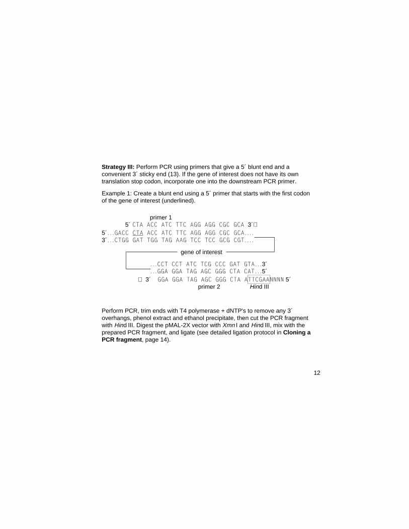
Strategy III: Perform PCR using primers that give a 5´ blunt end and aconvenient 3´ sticky end (13). If the gene of interest does not have its owntranslation stop codon, incorporate one into the downstream PCR primer.
Example 1: Create a blunt end using a 5´ primer that starts with the first codonof the gene of interest (underlined).
primer 1 5´ CTA ACC ATC TTC AGG AGG CGC GCA 3´⇒5´...GACC CTA ACC ATC TTC AGG AGG CGC GCA....3´...CTGG GAT TGG TAG AAG TCC TCC GCG CGT....
gene of interest
...CCT CCT ATC TCG CCC GAT GTA...3´
...GGA GGA TAG AGC GGG CTA CAT...5´ ⇐ 3´ GGA GGA TAG AGC GGG CTA ATTCGAANNNN 5´
primer 2 Hind III
Perform PCR, trim ends with T4 polymerase + dNTP's to remove any 3´overhangs, phenol extract and ethanol precipitate, then cut the PCR fragmentwith Hind III. Digest the pMAL-2X vector with Xmn I and Hind III, mix with theprepared PCR fragment, and ligate (see detailed ligation protocol in Cloning aPCR fragment, page 14).
12

Example 2: Design a primer that incorporates a restriction site for a Type IISenzyme, 5´ to the first codon of the gene of interest.
Bsa I primer 1 5´ NNNGGTCTCNATG ACC ATC TTC AGG AGG CGC GCA 3´⇒ 5´ ....GACC ATG ACC ATC TTC AGG AGG CGC GCA... 3´ ....CTGG TAC TGG TAG AAG TCC TCC GCG CGT...
gene of interest
same primer 2 as example 1
Perform PCR, cut with Bsa I [GGTCTC(1/5)], fill in with Klenow, heat inactivatethe Klenow, then cut with Hind III. Cut the pMAL-2X vector with Xmn I andHind III, mix with the prepared PCR fragment and ligate (see detailed ligationprotocol, page 14).
↓
13

Cloning a PCR Fragment
The procedure below is for cloning a fragment produced by PCR into a pMALvector. It is assumed that the PCR fragment is approximately 1 kb, beginswith a blunt end, and that a stop codon followed by a Hind III site has beenincorporated into the 3´ end (see Strategy III, page 12).
1. Streak the TB1 cells on an LB plate. Prepare competent cells (14).Any competent cells can be used for the cloning; the cells must containthe lacZ∆M15 allele for the α-complementation to occur.
2. Prepare a PCR fragment of the gene of interest as outlined in Strategy IIIon page 12.
3. Digest 0.5 µg of the pMAL vector DNA in 20 µl of 1X NEBuffer 2 (sup-plied as a 10X stock) with 10 units of Xmn I and 10 units of Hind III at37°C for 1 hour. Heat inactivate the enzymes by incubating at 65°C for10 minutes.
4. Check for complete digestion of the pMAL digest by running 4 µl on anagarose gel. At the same time, run a sample of the PCR fragment toestimate its concentration.
5. Digest 0.5 µg of the PCR fragment in 20 µl of 1X NEBuffer 2 with10 units of Hind III. Heat inactivate the Hind III by incubating at 65°C for10 minutes.
6. Add EDTA to both digests to a final concentration of 20 mM. Add anequal volume of a 1:1 phenol/chloroform mixture to the restrictiondigests, mix, and remove the aqueous (top) phase and place in a freshtube. Repeat with chloroform alone.
14

Alternatively, the DNA fragments can be isolated from a low melting pointagarose gel, or purified using one of the many commercially-availablefragment purification kits.
7. Add 10 µg glycogen or tRNA as carrier to both digests, then add 1/9thvolume 3 M sodium acetate, mix, and add an equal volume isopropanol.Incubate at room temperature for 10 minutes.
8. Microcentrifuge for 15 minutes. Pour off the supernatant, rinse the pelletwith 70% ethanol, and allow to dry.
9. Resuspend each sample in 25 µl of 10 mM Tris-HCl, 1 mM EDTA, pH 8.0.It can be helpful to run another sample of each preparation on an agarosegel to re-estimate the concentration at this point.
10. Mix: 2 µl vector digest (40 ng)1 µl insert digest (20 ng)Add 14 µl water, then heat the DNA mixture at 45°C for 5 minutes.Cool on ice, then add:2 µl 10X T4 DNA Ligase Buffer (supplied as a 10X stock)1 µl (~400 units) T4 DNA Ligase (NEB #M0202S)Incubate at 16°C for 2 hours to overnight.
11. Heat at 65°C for 5 minutes; cool on ice.
12. Mix the ligation reaction with 25 µl competent TB1 (or any lacZα-comple-menting strain) and incubate on ice for 5 minutes. Heat to 42°C for2 minutes.
15
13. Add 0.1 ml LB and incubate at 37°C for 20 minutes. Spread on an LBplate containing 100 µg/ml ampicillin (do not plate on IPTG; see below).Incubate overnight at 37°C. Pick colonies with a sterile toothpick andstab onto a master LB amp plate and an LB amp plate containing 80 µg/ml Xgal and 0.1 mM IPTG. Incubate at 37°C for 8 to 16 hours. Deter-mine the Lac phenotype on the Xgal plate and recover the “white”clones from the corresponding patch on the master plate. It can behelpful to perform a control transformation with about 1 ng of the uncutpMAL vector as well - while most inserts prevent expression of anylacZα fragment, it is possible to get some α-fragment activity in cloneswith inserts. In this case, a difference in the shade of blue can usuallybe seen by comparing to transformants containing the vector alone. Analternative way to use the blue-white screen is to replica plate on LBamp and LB amp Xgal IPTG. One can also divide the ligation mixtureinto two aliquots, and plate one of them on 80 µg/ml Xgal, 0.1 mM IPTGto observe the percentage of clones with inserts, and if it is acceptablyhigh, screen the transformants on the LB amp plate directly by prepar-ing plasmid DNA.
Note: Because of the strength of the Ptac promoter, transformants takenfrom a plate containing IPTG can contain mutant plasmids that haveeither 1) lost part or all of the fusion gene, or 2) no longer express it athigh levels.
14. Screen for the presence of inserts in one or both of the following ways:A. Prepare miniprep DNA (15). Digest with an appropriate restriction
endonuclease to determine the presence and orientation of theinsert (16).
B. i) Grow a 5 ml culture in LB amp broth to 2 x 10⁸ cells/ml(A600 of ~0.5).
16

ii) Withdraw a 1 ml sample. Microcentrifuge for 2 minutes, discardthe supernatant and resuspend the cells in 50 µl protein gelSDS-PAGE Sample Buffer (17).
iii) Add IPTG to the remaining culture to 0.3 mM, for example 15 µlof a 0.1 M stock solution. Incubate at 37°C with good aerationfor 2 hours.
iv) Withdraw a 0.5 ml sample. Microcentrifuge for 2 minutes,discard the supernatant and resuspend the cells in 100 µl SDS-PAGE sample buffer.
v) Place samples in a boiling water bath for 5 minutes. Electro-phorese 15 µl of each sample on a 10% SDS-PAGE gel alongwith a set of protein MW standards and 15 µl of the suppliedMBP2* in SDS-PAGE Sample Buffer. Stain the gel withCoomassie brilliant blue (17,18).
An induced band should be visible at a position corresponding to themolecular weight of the fusion protein. A band at or around the position ofMBP2* (MW 42.5 kDa) indicates either an out of frame fusion or a severeprotein degradation problem. These can usually be distinguished by per-forming a Western blot using the anti-MBP serum (19); even with severeprotein degradation, a full length fusion protein can be detected on theWestern. The molecular weight of the MBP-β-gal-α fusion is 50.8 kDa.
17

Figure 3. Flow chart for the pilot experiment.
Grow Cells
Add IPTG
Divide Culture and Harvest Cells
Sample 1: Uninduced Cells
Sample 2: Induced Cells
For pMAL-c2 and p2
Constructs
For pMAL-p2Constructs Only
Resuspend in Column BufferPrepare Total Cell Extract
Resuspend in Tris/sucrosePrepare Periplasmic Extract
Sample 3: Crude ExtractSample 4: Insoluble Matter
Sample 6: Periplasmic Extract(Osmotic-shock Fluid)
Test Amylose Resin Binding
SDS-PAGE
Sample 5: Protein Boundto Amylose
18

Pilot Experiment
A small scale experiment is described to determine the behavior of a particu-lar MBP fusion protein. This protocol results in five (pMAL-c2 vectors) or six(pMAL-p2 vectors) samples: uninduced and induced cells, a total cell crudeextract, a suspension of the insoluble material from the crude extract, afraction containing protein that binds to the amylose resin, and (for pMAL-p2constructions) a periplasmic fraction prepared by the cold osmotic shockprocedure (20) (Figure 3).
1. Inoculate 80 ml rich broth + glucose & amp (see Media and Solutions,page 33) with 0.8 ml of an overnight culture of cells containing the fusionplasmid.
2. Grow at 37°C with good aeration to 2 x 10⁸ cells/ml (A600 of ~0.5). Take asample of 1 ml and microcentrifuge for 2 minutes (sample 1: uninducedcells). Discard supernatant and resuspend the cells in 50 µl SDS-PAGESample Buffer. Vortex and freeze at –20°C.
3. Add IPTG (isopropylthiogalactoside) to the remaining culture to afinal concentration of 0.3 mM, e.g. 0.24 ml of a 0.1 M stock in H2O (seeMedia and Solutions). Continue incubation at 37°C for 2 hours. Withdrawa 0.5 ml sample and microcentrifuge for two minute (sample 2: inducedcells). Discard supernatant and resuspend the cells in 100 µl SDS-PAGESample Buffer. Vortex to resuspend cells and freeze at –20°C.Additional time points at 1 and 3 hours can be helpful in trying to decidewhen to harvest the cells for a large scale prep.
4. Divide the remaining culture into two aliquots. Harvest the cells bycentrifugation at 4000 x g for 10 minutes. Discard the supernatants andresuspend one pellet in 5 ml of Column Buffer (see Media and
19

Solutions), for protocol A. For pMAL-p2 constructions, resuspend theother pellet in 10 ml 30 mM Tris-HCl, 20% sucrose, pH 8.0, for protocol B(8 ml / 0.1 g cells wet weight).
Protocol A (all constructions)
5A. Freeze the cells in Column Buffer in a dry ice-ethanol bath (or overnightat –20°C; –20°C is more effective than –70°C, but takes longer). Thawin cold water.
6A. Place the cells in an ice-water bath and sonicate in short pulses of15 seconds or less. Monitor the release of protein using the Bradfordassay (21), adding 10 µl of the sonicate to 1.5 ml Bradford reagent andmixing. Continue sonication until the released protein reaches amaximum (usually about 2 minutes); a standard containing 10 µl of10 mg/ml BSA in 1.5 ml of Bradford reagent can be helpful as anapproximate endpoint.
7A. Centrifuge at 9,000 x g at 4°C for 20 minutes. Decant the supernatant(crude extract) and save on ice. Resuspend the pellet in 5 ml ColumnBuffer. This is a suspension of the insoluble matter. Add 5 µl 2x SDS-PAGE Sample Buffer to 5 µl of the crude extract and insoluble matterfractions (samples 3 and 4, respectively).
8A. Place ~200 µl of the amylose resin in a microfuge tube and spin brieflyin a microcentrifuge. Remove the supernatant by aspiration and discard.Resuspend the resin in 1.5 ml Column Buffer, then microcentrifugebriefly and discard the supernatant; repeat. Resuspend the resin in200 µl of Column Buffer. Mix 50 µl of crude extract with 50 µl of theamylose resin slurry. Incubate for 15 minutes on ice. Microcentrifuge1 minute, then remove the supernatant and discard. Wash the pelletwith 1 ml Column Buffer, microcentrifuge 1 minute, and resuspend theresin in 50 µl SDS-PAGE Sample Buffer (sample 5: protein bound toamylose). 20

Protocol B (pMAL-p2 constructions only)
5B. Add 20 µl 0.5 M EDTA (1 mM final conc.) to the cells in Tris/sucroseand incubate for 5–10 minutes at room temperature with shaking orstirring.
6B. Centrifuge at 8000 x g at 4°C for 10 minutes, remove all the superna-tant, and resuspend the pellet in 10 ml ice-cold 5 mM MgSO4.
7B. Shake or stir for 10 minutes in an ice-water bath.
8B. Centrifuge as above. The supernatant is the cold osmotic shock fluid.Add 10 µl 2X SDS-PAGE Sample Buffer to 10 µl of the cold osmoticshock fluid (sample 6).
SDS-PAGE (all constructions)
9. Place the samples in a boiling water bath for 5 minutes. Microcentrifugefor 1 minute. Load 20 µl of the of uninduced cells, induced cells andamylose resin samples (avoid disturbing the pellets), and all of theremaining samples, on a 10% SDS-PAGE gel (17).
10. (Optional) Run an identical SDS-PAGE gel(s) after diluting the samples1:10 in SDS-sample buffer. Prepare a Western blot(s) and develop withanti-MBP serum and, if available, serum directed against the protein ofinterest (19).
If the fusion is in the periplasmic fraction, consider using Method IIin Affinity Chromatography. In this case, another pilot to optimizeexpression and export may be desirable. If the protein is insoluble,modify the conditions of cell growth to attempt to produce solublefusion. Two changes that have helped in previous cases are i)changing to a different strain background, and ii) growing the cells ata lower temperature (8).
21
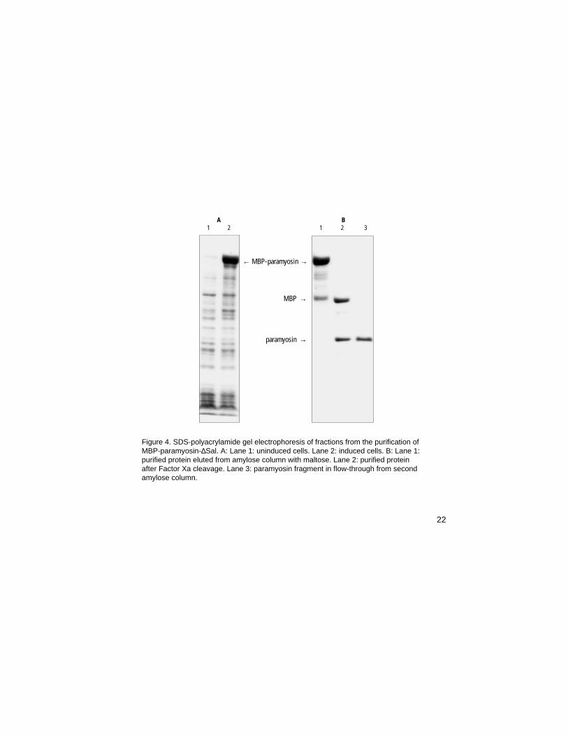
Figure 4. SDS-polyacrylamide gel electrophoresis of fractions from the purification ofMBP-paramyosin-∆Sal. A: Lane 1: uninduced cells. Lane 2: induced cells. B: Lane 1:purified protein eluted from amylose column with maltose. Lane 2: purified proteinafter Factor Xa cleavage. Lane 3: paramyosin fragment in flow-through from secondamylose column.
A B1 2 1 2 3
← MBP-paramyosin →
MBP →
paramyosin →
22

Affinity Chromatography
Two protocols for purification of a fusion protein from a 1 liter culture aredetailed below. Method I is designed for purification of a soluble fusionprotein expressed in the cytoplasm from a pMAL-c2 vector. However, thecrude extract prepared by this method also contains periplasmic proteins, soit can be used to prepare secreted fusion proteins expressed from a pMAL-p2 vector as well. Method II is designed for purification of a secreted fusionprotein expressed from a pMAL-p2 vector. It results in a periplasmic fractionthat contains many fewer E. coli proteins than the total cell crude extractprepared by method I, but the procedure is more difficult to scale up.
Method I
1. Inoculate 1 liter rich broth + glucose & ampicillin (see Media and Solu-tions, page 33) with 10 ml of an overnight culture of cells containing thefusion plasmid.Glucose is necessary in the growth medium to repress the maltose geneson the chromosome of the E. coli host, one of which is an amylase whichcan degrade the amylose on the affinity resin.
2. Grow to 2 x 10⁸ cells/ml (A600 ~0.5). Add IPTG to a final concentration of0.3 mM, e.g. 72 mg or 3 ml of a 0.1 M stock in H2O (see Media andSolutions). Incubate the cells at 37°C for 2 hours.The period of time and the temperature to use during expression de-pends on several factors (stability of the protein, host strain, etc.), andvariations can be tried to find optimum conditions for expression.
23

3. Harvest the cells by centrifugation at 4000 x g for 20 minutes and discardthe supernatant. Resuspend the cells in 50 ml Column Buffer (see Mediaand Solutions).For many unstable proteins, most of the degradation happens duringharvest and cell breakage. Therefore, it is best to harvest the cells quicklyand keep them chilled. 50 ml of Column Buffer is based on the expecta-tion of about 5 grams cells/liter, i.e. 10 ml for every gram of cells (wetweight).The EDTA in the lysis buffer is to help inhibit proteases that have a Ca++
cofactor. Addition of PMSF (phenyl methylsulfonyl fluoride) and/or otherprotease inhibitors may help in some cases.DTT or β–mercaptoethanol can be included to prevent interchain disulfidebond formation upon lysis (disulfide bonds usually do not formintracellularly in E. coli). For more about variations and additions to theColumn Buffer, see Media & Solutions, page 33.
4. Freeze sample in a dry ice-ethanol bath (or overnight at –20°C; –20°C ismore effective than –70°C, but takes longer). Thaw in cold water.
5. Place sample in an ice-water bath and sonicate in short pulses of15 seconds or less. Monitor the release of protein using the Bradfordassay (21), adding 10 µl of the sonicate to 1.5 ml Bradford reagent andmixing. Continue sonication until the released protein reaches a maxi-mum (usually about 2 minutes sonication time); a standard containing10 µl of 10 mg/ml BSA can be helpful as an approximate endpoint.
6. Centrifuge at 9,000 x g for 30 minutes. Save the supernatant (crudeextract). Dilute the crude extract 1:5 with Column Buffer.
7. Pour the amylose resin in a 2.5 x 10 cm column. Wash the column with8 column volumes of Column Buffer.The amount of resin depends on the amount of fusion protein produced.
24
The resin binds about 3 mg/ml bed volume, so a column of about 15 mlshould be sufficient for a yield of up to 45 mg fusion protein/liter culture.A 50 ml syringe plugged with silanized glass wool can be substituted forthe 2.5 cm column, but the glass wool should cover the bottom of thesyringe (not just in the tip) so the column will have an acceptable flowrate.
8. Load the diluted crude extract at a flow rate of [10 x (diameter of columnin cm)2 ]ml/hour. This is about 1 ml/minute for a 2.5 cm column.The dilution of the crude extract is intended to reduce the proteinconcentration to about 2.5 mg/ml. If your crude extract is less concen-trated, don’t dilute it as much. In general, 1 g wet weight of cells givesabout 50–120 mg protein.
9. Wash with 12 column volumes of Column Buffer.
The column can be washed overnight, if it has a safety loop to prevent itfrom running dry (see Figure 5, page 31). In this case, it is better torestart the column with elution buffer (step 10), rather than continuing thewash. Avoid loading the column overnight.
10. Elute the fusion protein with Column Buffer + 10 mM maltose. Collect10 to 20 fractions of 3 ml each (fraction size = 1/5th column volume).The fusion protein usually starts to elute within the first 5 fractions, andshould be easily detected by UV absorbance at 280 nm or the Bradfordprotein assay.
11. Pool the protein-containing fractions. If necessary, concentrate to about1 mg/ml in an Amicon Centricon or Centriprep concentrator, an Amiconstirred-cell concentrator, or the equivalent.
25

Method II
1. Inoculate 1 liter rich broth + glucose & ampicillin with 10 ml of an over-night culture of cells containing the fusion plasmid.
2. Grow to 2 to 4 x 10⁸ cells/ml (A600 ~0.5). Add IPTG to a final concentrationof 0.3 mM, e.g. 72 mg/l or 3 ml of a 0.1 M stock in H2O. Incubate the cellsat 37°C for 2 hours.The period of time and the temperature to use during expression de-pends on several factors (stability of the protein, host strain, etc.), andvariations can be tried to find optimum conditions (see Pilot Experi-ment). In addition, partial induction of exported proteins may lead tohigher yields, since protein export in E. coli may not be able to keep upwith full level Ptac expression.
3. Harvest the cells by centrifugation at 4000 x g for 20 minutes and discardthe supernatant. Resuspend the cells in 400 ml 30 mM Tris-HCl, 20%sucrose, pH 8.0 (80 ml for each gram of cells wet weight). Add EDTA to1 mM and incubate for 5–10 minutes at room temperature with shakingor stirring.
4. Centrifuge at 8000 x g for 20 minutes at 4°C, remove all the supernatant,and resuspend the pellet in 400 ml of ice-cold 5 mM MgSO4. Shake orstir for 10 minutes in an ice bath.
5. Centrifuge at 8000 x g for 20 minutes at 4°C. The supernatant is the coldosmotic shock fluid.
6. Add 8 ml of 1 M Tris-HCl, pH 7.4 to the osmotic shock fluid.
7. Continue from Method I, step 7 (page 24).
26

Regenerating the Amylose Resin Column
The resin may be reused three to five times when regenerated with thefollowing sequence of washes:
Water: 3 column volume0.1% SDS: 3 column volumesWater: 1 column volumesColumn Buffer: 3 column volumes
Please note that although the column can be washed at 4°C, 0.1% SDS willeventually precipitate at that temperature. It is therefore recommended thatthe SDS solution be stored at room temperature until needed, and rinsed outof the column promptly. The resin may be reused three to five times. Uponrepeated use, trace amounts of amylase in the E. coli extract decrease thebinding capacity of the column. It is recommended that the column bewashed promptly after each use.
27

Cleavage of the Fusion Protein
For Factor Xa and Genenase I, fusion protein cleavage is carried out at aw/w ratio of 1% the amount of fusion protein (e.g., 1 mg Factor Xa for areaction containing 100 mg fusion protein). The reaction mixture can beincubated for 3 hours to several days, at room temperature or 4°C. Depend-ing on the particular fusion protein, the amount of protease can be adjustedwithin the range of 0.1–5.0%, to get an acceptable rate of cleavage. Forenterokinase cleavage, 1 unit is used for every 50 µg fusion protein. Again,conditions can be adjusted according to the characteristics of a particularfusion. All the proteases will cleave at non-canonical sites in some proteins;for some fusions, there is a correlation between instability of the protein ofinterest in E. coli and cleavage at additional sites (unpublished observa-tions). Presumably this cleavage activity depends on the three dimensionalconformation of the fusion protein. For fusions that are resistant to cleavage,two strategies can sometimes help. Inclusion of small amounts of SDS(0.005–0.05%) in the reaction appears to relax the fusion enough to allow forcleavage in some cases (22). The window of SDS concentrations that workcan be small, so a pilot titration with different SDS concentrations is neces-sary. Another strategy that sometimes helps is to denature the fusion torender the protease site accessible to cleavage (see below and reference 9).
1. If necessary, concentrate the fusion protein to at least 1 mg/ml.All three proteases will work in the Column Buffer + maltose used to elutethe fusion protein. In addition, they will work in a variety of other buffers,with NaCl concentrations from 0 to 500 mM and pH values around 8.
2. Do a pilot experiment with a small portion of your protein.Example: Mix 20 µl fusion protein at 1 mg/ml, with either: 1 µl Factor Xaor Genenase I, diluted to 200 µg/ml, or 0.4 U Enterokinase
28

In a separate tube, place 5 µl fusion protein with no protease (mockdigestion). Incubate the tubes at room temperature. At 2, 4, 8, and24 hours, take 5 µl of the reaction, add 5 µl 2x SDS-PAGE SampleBuffer, and save at 4°C. Prepare a sample of 5 µl fusion protein + 5 µl 2Xsample buffer (uncut fusion).
3. Boil the 6 samples for 5 minutes and run on an SDS-PAGE gel (17).
4. Scale the pilot experiment up for the portion of the fusion protein to becleaved. Save at least a small sample of the uncut fusion as a reference.
5. Check for complete cleavage by SDS-PAGE.
Denaturing the fusion protein
1. Either dialyze the fusion against at least 10 volumes 20 mM Tris-HCl, 6 Mguanidine hydrochloride, pH 7.4 for 4 hours, or add guanidine hydrochlo-ride directly to the sample to give a final concentration of 6 M.
2. Dialyze against 100 volumes Column Buffer, 2 times at 4 hours each.During refolding, one has to balance between two objectives. For theprotease to cleave it must be present before the protein has completelyrefolded, so removing the denaturant quickly is desirable. However, whenthe denaturant is removed quickly some proteins will fail to refoldproperly and precipitate. Stepwise dialysis against buffer containingdecreasing amounts of guanidine hydrochloride can prevent precipitationof the fusion protein; halving the guanidine concentration at each step isconvenient, but cases where 0.1 M steps are necessary have beenreported. However, if the fusion protein is able to refold into a protease-resistant conformation, it may be better to dialyze away the denaturant inone step and take the loss from precipitation in order to maximize theamount of cleavable fusion protein recovered.Go to step 2 or 4 above, as appropriate.
29

Separating the Protein of Interest from MBP After Protease Cleavage
Method I: DEAE-Sepharose™ ion exchange chromatography
This method potentially purifies the target protein away from MBP and theprotease, but also provides an additional purification step for removing tracecontaminants. A disadvantage is that occasionally the peak containing theprotein of interest overlaps with MBP or the protease, resulting in poorseparation. The procedure is written for quantities <25 mg, and can bescaled up for larger amounts.
1. Dialyze the fusion protein cleavage mixture vs. 20 mM Tris-HCl,25 mM NaCl, pH 8.0 (2 or 3 changes of 100 volumes, at least2 hours each).
2. Wash about 6 ml of DEAE-Sepharose in 20 ml of 10 mM Tris-HCl, 25 mMNaCl, pH 8.0 a couple of times, letting the resin settle and pouring off thesupernatant between washes.
3. Pour the resin into a 1 x 10 cm column to give a bed volume of 5 ml(6–7 cm bed height).
4. Wash the column with 15 ml of the same buffer.
5. Load the fusion protein cleavage mixture onto the column. Collect 2.5 mlfractions of the column flow-through.
6. Wash the column with 3–5 column volumes of the same buffer. Continuecollecting 2.5 ml fractions.
7. Start a gradient of 25 mM NaCl to 500 mM NaCl (25 ml each) in 20 mMTris-HCl, pH 8.0 (see Figure 5). Collect 1 ml fractions.
30

8. Determine which fractions contain protein by measuring A280, or by theBradford or Lowry method. The MBP elutes as a sharp peak at 100–150 mM NaCl. Factor Xa elutes at about 400 mM NaCl, Genenase I andEnterokinase flow through the column. The target protein may flowthrough the column, or it may elute during the gradient. Electrophoresethe relevant fractions on an SDS-PAGE gel. Pool the fractions containingthe target protein free of MBP and concentrate as desired.
If your protein is not separated from MBP using DEAE-Sepharosechromatography, other chromatography resins can be tried. SP-Sepharose (phosphate buffer at pH 7.0; MBP flows through) and gelfiltration have been used successfully.
magneticstirrer
25 ml of 25 mM NaCl buffer
25 ml of500 mM NaCl buffer
Bridge
fraction collector
safetyloop
Figure 5. Diagram ofsetup for DEAE-Sepharosecolumn chromatography
31

Method II: Removal of maltose by hydroxyapatite chromatography anddomain separation by rebinding MBP to amylose
This method requires 2 steps, but since no dialysis is needed and bothcolumns are step-eluted, the procedure is fairly simple. It removes MBP fromthe cleavage mixture, but not the protease or any other trace contaminants.In addition, any MBP that has been denatured or otherwise damaged will notbind to the amylose column. The procedure must be carried out at roomtemperature to avoid precipitation of the phosphate buffer. This procedure iswritten for quantities <25 mg, but can be scaled up for larger amounts.
1. Swell 1 g hydroxyapatite in 20 mM sodium phosphate, 200 mM NaCl, pH7.2. Allow the resin to settle and pour off excess buffer (removes fineparticles). Add fresh buffer and repeat twice.
2. Pour the hydroxyapatite into a 1 x 10 cm column (a disposable polypropy-lene column such as a BioRad Econo-Pac™ can be used).
3. Load the fusion protein cleavage mixture onto the column.
4. Wash with 80 ml of the same buffer (washes away the maltose).
5. Elute with 0.5 M Na phosphate, pH 7.2 (stock solution in Media andSolutions). Collect 2 ml fractions. Assay for protein by A280, Bradfordassay or Lowry (21). Most of the protein usually elutes in the first 8 ml.
6. Pour a 15 ml amylose column as described in Affinity Chromatography.
7. Load the hydroxyapatite-eluted protein onto the amylose column. Collectthe flow-through as 5 ml fractions. Protein in the flow-through should befree of MBP and consist primarily of the target protein. Assay for proteinby A280, Bradford assay or Lowry. The protein of interest should flowthrough the column by the seventh fraction.
32

Media & SolutionsRich medium + glucose & ampicillin:per liter: 10 g tryptone, 5 g yeast extract, 5 g NaCl, 2 g glucoseautoclave; add sterile ampicillin to 100 µg/ml
0.1 M IPTG stock1.41 g IPTG (isopropyl-β-D-thiogalactoside), hemidioxane adduct, or1.19 g IPTG; add H2O to 50 ml; filter sterilize, store at 4°C;hemidioxane adduct is stable for ~ 6 months, dioxane-free is light sensitive
Column BufferPER LITER FINAL CONCENTRATION
20 ml 1.0 M Tris-HCl, pH 7.4 20 mM Tris-HCl11.7 g NaCl 200 mM NaCl2.0 ml 0.5 M EDTA 1 mM EDTA
optional:1.0 ml 1M sodium azide 1 mM azide0.7 ml β-mercaptoethanol 10 mM β-ME
or or154 mg DTT 1 mM DTT
Notes on additions or changes to the Column BufferThe conditions under which MBP fusions will bind to the column are flexible, and theColumn Buffer can be modified without adversely effecting the affinity purification. Buffers otherthan Tris-HCl that are compatible include MOPS, HEPES, and phosphate, at pH values around 7.MBP binds to amylose primarily by hydrogen bonding, so higher ionic strength does not decreaseits affinity. Nonionic detergents such as Triton X-100 and Tween 20 have been seen to interferewith the affinity of some fusions, while other fusions are unaffected.
0.5 M sodium phosphate buffer, pH 7.2 (stock)A) 69.0 g NaH2PO4•H2O, to 1 liter with H2OB) 134.0 g Na2HPO4•7H2O, to 1 liter with H2OMix 117 ml (A) with 383 ml (B). Store at room temperature.
33

Troubleshooting and Tips
1. Cloning and Transformation1.1 Differences among the pMAL vectors . . . . . . . . . . . . . . . . . . . . . . . . . . . . . . . . . . 361.2 Strain recommendations . . . . . . . . . . . . . . . . . . . . . . . . . . . . . . . . . . . . . . . . . . . . . . . . . . 371.3 Using the blue/white selection . . . . . . . . . . . . . . . . . . . . . . . . . . . . . . . . . . . . . . . . . . . 371.4 Inefficient transformation . . . . . . . . . . . . . . . . . . . . . . . . . . . . . . . . . . . . . . . . . . . . . . . . . 371.5 Sequencing primers . . . . . . . . . . . . . . . . . . . . . . . . . . . . . . . . . . . . . . . . . . . . . . . . . . . . . . . 381.6 Inability to clone . . . . . . . . . . . . . . . . . . . . . . . . . . . . . . . . . . . . . . . . . . . . . . . . . . . . . . . . . . . . 381.7 Obtaining pMAL sequences . . . . . . . . . . . . . . . . . . . . . . . . . . . . . . . . . . . . . . . . . . . . . . 381.8 Strand packaged by M13 helper phage . . . . . . . . . . . . . . . . . . . . . . . . . . . . . . . . 38
2. Expression2.1 Fusion protein is degraded . . . . . . . . . . . . . . . . . . . . . . . . . . . . . . . . . . . . . . . . . . . . . . . 392.2 Fusion protein is insoluble . . . . . . . . . . . . . . . . . . . . . . . . . . . . . . . . . . . . . . . . . . . . . . . . 392.3 Fusion protein not expressed . . . . . . . . . . . . . . . . . . . . . . . . . . . . . . . . . . . . . . . . . . . . 412.4 Only MBP-sized protein expressed . . . . . . . . . . . . . . . . . . . . . . . . . . . . . . . . . . . . . 422.5 Effect of export (secretion, using pMAL-p2 vectors)
on solubility and stability of the fusion . . . . . . . . . . . . . . . . . . . . . . . . . . . . . . . . . . 422.6 Expressing small peptides . . . . . . . . . . . . . . . . . . . . . . . . . . . . . . . . . . . . . . . . . . . . . . . . 43
3. Affinity Purification3.1 Fusion protein does not bind to amylose column . . . . . . . . . . . . . . . . . . . . . . 433.2 Amylose resin regeneration . . . . . . . . . . . . . . . . . . . . . . . . . . . . . . . . . . . . . . . . . . . . . . 443.3 Nonionic detergents and binding . . . . . . . . . . . . . . . . . . . . . . . . . . . . . . . . . . . . . . . . 443.4 Different buffers and salt concentrations . . . . . . . . . . . . . . . . . . . . . . . . . . . . . . . 44
34

3.5 Degradation of the fusion upon cell lysis . . . . . . . . . . . . . . . . . . . . . . . . . . . . . . . 443.6 Batch purification . . . . . . . . . . . . . . . . . . . . . . . . . . . . . . . . . . . . . . . . . . . . . . . . . . . . . . . . . . 453.7 Purification in the presence of denaturants . . . . . . . . . . . . . . . . . . . . . . . . . . . . 453.8 Storage of resin at –20°C . . . . . . . . . . . . . . . . . . . . . . . . . . . . . . . . . . . . . . . . . . . . . . . . . 45
4. Factor Xa Cleavage4.1 Fusion protein is cleaved internally . . . . . . . . . . . . . . . . . . . . . . . . . . . . . . . . . . . . . 464.2 Control substrates . . . . . . . . . . . . . . . . . . . . . . . . . . . . . . . . . . . . . . . . . . . . . . . . . . . . . . . . . 464.3 Inactivation of Factor Xa . . . . . . . . . . . . . . . . . . . . . . . . . . . . . . . . . . . . . . . . . . . . . . . . . . 464.4 Removal of Factor Xa . . . . . . . . . . . . . . . . . . . . . . . . . . . . . . . . . . . . . . . . . . . . . . . . . . . . . 474.5 Fusion will not cleave . . . . . . . . . . . . . . . . . . . . . . . . . . . . . . . . . . . . . . . . . . . . . . . . . . . . . 474.6 Molecular weight and calculated pI . . . . . . . . . . . . . . . . . . . . . . . . . . . . . . . . . . . . . 474.7 Cleavage in glycerol . . . . . . . . . . . . . . . . . . . . . . . . . . . . . . . . . . . . . . . . . . . . . . . . . . . . . . . 474.8 Cleavage in urea, guanidine hydrochloride and SDS . . . . . . . . . . . . . . . . . 484.9 Cleavage with fusion bound to the column . . . . . . . . . . . . . . . . . . . . . . . . . . . . . 48
5. Separation of Fusion Protein Domains and Storage5.1 Difficulties with removal of maltose by dialysis . . . . . . . . . . . . . . . . . . . . . . . . 485.2 How to store proteins . . . . . . . . . . . . . . . . . . . . . . . . . . . . . . . . . . . . . . . . . . . . . . . . . . . . . 49
6. MBP2*6.1 What is MBP2*? . . . . . . . . . . . . . . . . . . . . . . . . . . . . . . . . . . . . . . . . . . . . . . . . . . . . . . . . . . . 496.2 Crystal structure of MBP . . . . . . . . . . . . . . . . . . . . . . . . . . . . . . . . . . . . . . . . . . . . . . . . . . 506.3 How much of MBP is dispensable for binding? . . . . . . . . . . . . . . . . . . . . . . . . 506.4 Kd, pI and extinction coefficient for MBP2* . . . . . . . . . . . . . . . . . . . . . . . . . . . . . 506.5 Origin of the MBP region of the pMAL vectors . . . . . . . . . . . . . . . . . . . . . . . . . 506.6 Is MBP a monomer? . . . . . . . . . . . . . . . . . . . . . . . . . . . . . . . . . . . . . . . . . . . . . . . . . . . . . . 50
35

1. Cloning and Transformation
1.1 What are the differences among the various pMAL vectors?
The pMAL-c, -cRI and -p are the earliest versions of the pMAL vectors.pMAL-c and pMAL-p have a Stu I site in the polylinker for cloning blunt-ended fragments. Because the second half of the Stu I site codes forproline, if you clone an EcoR I fragment into pMAL-c or pMAL-p, theFactor Xa site reads IEGRP, and RP won't cut with Factor Xa. pMAL-cRI was designed as a short-term solution to fix this problem, bychanging the polylinker to code for IEGRI upstream of the EcoR I site.The pMAL-c2 and pMAL-p2 vectors are the next generation of pMALvectors. These vectors avoid the problem with Factor Xa cleavage byusing an Xmn I site instead of Stu I. They also have a spacer betweenmalE and the Factor Xa site which allows some fusions to bind moretightly to the amylose resin, and an M13 origin for making singlestranded DNA. The third generation of pMAL vectors is distinguished bythe addition of vectors that substitute an Enterokinase or Genenase Isite for the Factor Xa site. These vectors are called pMAL-c2E andpMAL-p2E (Enterokinase), and pMAL-c2G and pMAL-p2G (Genenase).The Factor Xa versions are now called pMAL-c2X and pMAL-p2X forconsistency. This third generation of vectors have a few minor modifica-tions outside the polylinker as well. The Nde I site in the pBR322 originwas destroyed by filling in, and an Nde I site at the ATG of malE wasadded by site directed mutagenesis. This allows a malE fusion to becut out in order to subclone it, for example into a eukaryotic vector. TheNco I site in malE was destroyed, as was the Ava I site in the M13origin, making the Ava I site upstream of the Factor Xa site unique. In allvectors, the “-c’’ designation refers to cytoplasmic expression, i.e. thesignal sequence that directs MBP to the periplasmic space has been
36

deleted. Vectors that are designated “-p’’ refer to periplasmic expres-sion, and these contain the wild-type malE signal sequence.
1.2 What strain(s) do you recommend as hosts for the pMAL vectors?
The strain we have used most frequently is TB1, NEB #E4122S, whichis JM83 hsdR. There is nothing special about it with respect to thepMAL system, but it has given the best results when consideringplasmid stability, expression and purification. We have used a numberof other strains successfully for certain proteins. We also use otherstrains in response to a particular problem (for example, see 2.1). Onecan start with TB1, or one can use whatever competent cells arereadily available and then try TB1 or another strain if a problem withexpression or purification develops.
1.3 Can I clone into pMAL vectors just as I would into pUC vectors, i.e.plating my transformants on Xgal/IPTG plates and picking whitecolonies?
No. Even though the β-galactosidase α fragment is the same on thepUC and pMAL vectors, the tac promoter on the pMAL vectors is muchstronger than the lac promoter on the pUC vectors. If cells bearing apMAL vector are induced with IPTG, the cells eventually die. The blue-to-white screen is done by replica plating (or picking and stabbing) ontoa master amp plate and an amp Xgal plate containing 0.1 mM IPTG.
1.4 What is the transformation efficiency of the pMAL vectors?
The pMAL vectors transform about 1/10th as well as pUC and pBR322.High amp concentration (>100 ug/ml) can cause lower efficiencies aswell.
37
1.5 What primers should I use to sequence the ends of my insert after I cloneit into a pMAL vector?
Use the malE primer (NEB #S1237S) on the 5´ side of the insert. If youwant to sequence the 3´ junction use pUC/M13 primers that bind to thelacZα region (e.g. NEB #S1224S).
1.6 What are some of the possible explanations for an inability to clone aninsert into a pMAL vector?
The most common explanation for this is technical difficulties with thesubcloning. Another explanation is that expression of the fusion is toxicto E. coli. The tac promoter induction ratio on the pMAL plasmids isabout 1:50, so if the induced level of the fusion is 40% of the total cellularprotein, the uninduced level works out to 0.8%. This amount of a proteincan be toxic, either because of its function (e.g. a protease) or becauseof its general properties (e.g. very hydrophobic).
1.7 How can I obtain the sequences of the pMAL vectors?
The pMAL sequences are available on the New England Biolabs website at www.neb.com, by e-mail from [email protected], and by FAX (callNEB Technical Support, 1-800-632-7799).
1.8 Which strand of DNA is packaged when cells carrying the pMALvectors are superinfected with an M13 helper phage such as M13KO7(NEB #N0315S)?
The strand corresponding to the antisense strand of the malE gene ispackaged. This is the strand that is complementary to the polylinkersequence shown in the 2000/01 NEB Catalog on page 276. The malEprimer (NEB #S1237S) hybridizes to the strand that is packaged by thehelper phage.
38
2. Expression
2.1 When we analyze our fusion protein expression by Western blot usingthe anti-MBP serum, only a small fraction of the protein is full-length,while most of it migrates close to the MBP2* marker.
It is likely that the fusion protein is degraded, leaving a stable MBP-sizedbreakdown product. In this case, try using a protease deficient host. A listof strains, which are free with an order or for the price of shipping, can befound on page 51. For cytoplasmic expression, the most proteasedeficient strain is CAG629 (#E4125S) – it is also, however, the mostdifficult to work with. ER2508 (#E4127S) and CAG597 (#E4123S) aregood alternatives. For periplasmic expression, the most proteasedeficient strain is CAG597 (#E4123S); KS1000 (#E4128S) and UT5600(#E4129S) might be worth trying as well. The CAG strains are difficult totransform, and often require electroporation to introduce the fusionplasmid.
2.2 My fusion protein is insoluble; is there anything I can do to get itexpressed as soluble protein?
Expressing at a lower temperature is the first thing to try. One can go aslow as 15°C by moving an incubator into the cold room. Of course, thecells grow very slowly at these temperatures, so grow the culture at 37°Cand shift to the low temperature when adding IPTG. One also has toincrease the time of induction to compensate for the slower growth – arule of thumb is 2X for every 7°C. Other references for solubility problemsinclude:
One of the original papers describing how expression at lower tempera-tures can produce soluble protein:Bishai, W.R., Rappuoli, R. and Murphy, J.R. (1987) High-level expression
39
of a proteolytically sensitive diphtheria toxin fragment in E. coli. J. Bact.169, 5140–5151.
Same for an exported protein:Takagi, H. et al. (1988) Control of folding of proteins secreted by a highexpression secretion vector, pIN-III-ompA: 16-fold increase in produc-tion of active subtilisin E in E. coli. Biotechnology 6, 948–950.
A method for growing the cells under osmotic stress, which can alsohelp produce soluble protein:Blackwell, J.R. and Horgan, R. (1991) A novel strategy for productionof a highly expressed recombinant protein in an active form. FEBSLetters 295, 10–12.
Review on methods to make correctly-folded protein in E. coli :Georgiou, G. and Valax, P. (1996) Expression of correctly foldedproteins in E. coli. Current Opinion in Biotechnology 7, 190–197.
Reviews on insolubility:Schein, C.H. (1989) Production of soluble recombinant proteins inbacteria. Biotechnology 7, 1141–1149.Schein, C.H. (1990) Solubility as a function of protein structure andsolvent components. Biotechnology 8, 308–317.Wilkinson, D.L. and Harrison, R.G. (1991) Predicting the solubility ofrecombinant proteins in E. coli. Biotechnology 9, 443–448.Kiefhaber, T. et al. (1991) Protein aggregation in vitro and in vivo: Aquantitative model of the kinetic competition between folding andaggregation. Biotechnology 9, 825–829.
Reviews on refolding:Zardeneta, G. and Horowitz, P.M. (1994) Detergent, liposome, andmicelle-assisted protein refolding. Anal Biochem 223, 1–6.Chaudhuri, J.B. (1994) Refolding recombinant proteins: Process
40

strategies and novel approaches. Ann NY Acad Sci 721, 374–385.Gilbert, H.F. (1994) Protein chaperones and protein folding. Curr OpinBiotechnol 5, 534–539.Schein, C.H. (1991) Optimizing protein folding to the native state inbacteria. Curr Opin Biotechnol 2, 746–750.Kelley, R.F. and Winkler, M.E. (1990) Folding of eukaryotic proteinsproduced in E. coli. Genet Eng (NY) 12, 1–19.Marston, F.A. and Hartley, D.L. (1990) Solubilization of protein aggre-gates. Methods in Enzymology 182, 264–282.
Extracting from membrane with lauroylsarcosine:Frankel, S., Sohn, R. and Leinwand, L. (1991) The use of Sarkosyl ingenerating soluble protein after bacterial expression. Proc Natl AcadSci USA, 88, 1192–1196.
2.3 When I run my uninduced and induced crude extracts on SDS-PAGEside by side, I don’t see an induced band.
There are a couple of possible explanations. Inserts cloned in a pMAL-p2X vector have about a 8- to 16-fold reduced level of expression whencompared to the same insert in a pMAL-c2X vector. This often reducesthe amount of expression to the point where there is no visible inducedband. In addition, some foreign genes are poorly expressed in E. coli,even when fused to a highly expressed carrier gene. Possible explana-tions are message instability or problems with translation – sometimesit is due to the presence of multiple rare codons in the gene of interest,and in these cases overexpression of the corresponding tRNA can help(23). Even in cases where a band is not visible, one can get yields upto 5 or 6 mg/liter of culture.
41

2.4 I’ve cloned my insert, but after SDS-PAGE the only induced bandpresent is the size of MBP2*.
There are two likely explanations for this result. If the protein of interest isin the wrong translational reading frame, an MBP2*-sized band will beproduced by translational termination at the first in-frame stop codon. Ifthe protein of interest is very unstable, an MBP2*-sized breakdown pro-duct is usually produced (MBP is a very stable protein). The best way todistinguish between these possibilities is to run a Western blot using anti-MBP antiserum (NEB #E8030S). If proteolysis is occurring, at least asmall amount of full-length fusion can almost always be detected. DNAsequencing of the fusion junction using the malE primer (NEB #S1237S)will confirm a reading frame problem. If the problem is proteolysis, youmight want to try one of the protease deficient strains from the list onpage 51.
2.5 What are the possible effects of export (secretion, using a pMAL-p2vector) on solubility/stability of the fusion?
Initiating export through the cytoplasmic membrane puts a fusion proteinon a different folding pathway – a difference in the solubility or stability ofthe protein is determined by whether this folding pathway leads to adifferent 3-dimensional structure for the protein. Some proteins, like MBPitself, can fold properly either in the cytoplasm or when exported to theperiplasm. However, the normal folding pathway for some proteins isincompatible with passage through the membrane, the fusion proteingets stuck in the membrane and cannot fold properly; this can lead todegradation (24). Other proteins, especially ones that have multipledisulfide bonds, only fold properly when exported (the E. coli cytoplasmis a reducing environment, and the proteins that catalyze disulfide bondformation are present in the periplasm; 25). When this class of protein is
42

expressed in the cytoplasm, it may fold improperly and become degradedor insoluble.
2.6 What is the minimum size of a fragment that can be cloned into pMAL andexpressed fused to MBP? Can short peptide sequences (about 10 aminoacids) be added onto MBP?
You can use the MBP system to express short peptides. However, forevery 40 mg of MBP (42.5 kDa) one gets about 1 mg of a 10 amino acidpeptide (1.1 kDa).
3. Affinity Purification
3.1 Much of my fusion protein flows through the amylose column. Is thereanything I can do to improve my fusion’s affinity for the amylose column?
A MBP fusion protein might not stick to the amylose column because ofthe presence of some factor in the extract that interferes with binding, orbecause of a low intrinsic affinity. Factors in the crude extract that caninterfere with binding include nonionic detergents (see 3.3) and cellularcomponents that are released during alternative methods of lysis(lysozyme/sonication or multiple passes through a French press). Inaddition, cells grown in LB and similar media have substantial amounts ofan amylase that interferes with binding, presumably by either cutting thefusion off the column or by releasing maltose that elutes the fusion fromthe column. By including glucose in the media, expression of this amylaseis repressed and the problem is alleviated. A low intrinsic affinity could becaused by an interaction between the protein of interest and MBP thateither blocks or distorts the maltose-binding site. Although this may beinherent in the protein of interest, sometimes the problem can be allevi-ated by shortening or lengthening the polypeptide that is fused to MBP.
43

3.2 How many times can I use the amylose column?
The most important variable in determining the useful life of theamylose resin is the amount of time it is in contact with trace amountsof amylase present in the crude extract (see 3.1). Under normalconditions (crude extract from 1 liter of cells grown in LB + 0.2%glucose, 15 ml column), the column loses 1–3% of its initial bindingcapacity each time it is used. If the yield of fusion protein under theseconditions is 40 mg, this means that after 3 to 5 runs there would be adecrease in the yield. In practice, we often use a column 8 or 10 timesbefore we notice a significant drop in the yield.
3.3 What is known about binding in the presence of nonionic detergents?
Some fusion proteins do not bind efficiently (<5% binding) in thepresence of 0.2% Triton X–100 or 0.25% Tween 20, while other fusionsare unaffected. For one fusion that does not bind in 0.25% Tween 20,diluting the Tween to 0.05% restores about 80% of the binding.
3.4 Can I substitute a different buffer and/or salt concentration in theColumn Buffer?
Yes, we have tried HEPES, MOPS, and phosphate buffers (at pH'sfrom 7.0 to 8.5) instead of Tris-HCl in the Column Buffer with similarresults. NaCl or KCl concentrations of 25 mM to 1 M are also compat-ible with the affinity purification.
3.5 I see my intact fusion protein by SDS-PAGE when I run cells boiled inSample Buffer, but when I check the crude extract the fusion isdegraded.
For fusions expressed in the cytoplasm, in many cases most of thedegradation happens during harvest and lysis. Harvesting promptly and
44

lysing the cells quickly may help. In other cases, degradation occurswhen the fusion protein is exposed to periplasmic or outer membraneproteases (26–28). The best strategy in either case is to use a hostwhich is deficient in the offending protease(s) (see 2.1, and the strainlist starting on page 51).
3.6 Can I perform a batch purification using the amylose resin?
Yes, batch purification works well, although it is difficult to wash all thenonspecific proteins away as effectively as in a column due to theincluded volume in the resin. The resin can withstand centrifugation atup to 6000 x g. A good compromise is to load the resin in a batchmode, by incubating with shaking for 2 hours to overnight, then pour itin a column to wash and elute. Dilution of the crude extract is not ascritical for loading the column by the batch method.
3.7 Can MBP fusions be purified in the presence of denaturants like ureaor guanidine-HCl?
No, MBP’s affinity to amylose and maltose depends on hydrogenbonds, that in turn are positioned by the three-dimensional structure ofthe protein. Agents that interfere with hydrogen bonds or the structureof the protein interfere with binding as well.
3.8 Is the amylose resin damaged by storage at –20°C? When our kitarrived, it was placed at –20°C, but I see that the recommendedstorage temperature for the amylose resin is 4°C.
The resin will freeze at –20°C but the performance of the resin is notdegraded by one freeze/thaw cycle. After the ethanol is removed, theresin should be stored at 4°C to prevent damage from freezing.
45

4. Factor Xa Cleavage
4.1 Factor Xa seems to be cleaving my protein at several sites, even thoughthe protein does not contain any IEGR sequences.
The specificity of Factor Xa reported in our catalog is as referenced inNagai and Thøgersen (1987) (10). The basis for this specificity is that thenatural Factor Xa sites in prothrombin are IEGR (or sometimes IDGR),and many examples of fusions with IEGR are cut specifically. However,proteins can be cleaved at other basic residues, depending on the context(29–32). A number of the secondary sites (but not all) that have beensequenced show cleavage following gly-arg. We have also seen acorrelation between proteins that are unstable in E. coli and cleavage atsecondary sites with Factor Xa, suggesting that these proteins are in apartially unfolded state. We’ve tried altering the reaction conditions toincrease the specificity, but with no success. Other site-specific proteases,such as Enterokinase and Genenase I, are now available as alternativesto Factor Xa.
4.2 Are there any control substrates for Factor Xa?
The Protein Fusion and Purification System comes with an MBP2*-paramyosin-∆Sal fusion as a positive control for Factor Xa cleavage(NEB #E8051S). Sigma also sells a colorimetric substrate, N-benzoyl-ile-glu-gly-arg-p-nitroanilide (Sigma, #B 7020).
4.3 How can Factor Xa be inactivated?
The best way is to add dansyl-glu-gly-arg-chloromethyl ketone(Calbiochem, #251700) to a final concentration of 2 µM, and incubate for1 minute at room temperature. This compound irreversibly inactivates theFactor Xa. It reacts with the active site histidine, so it could conceivablyreact with other sites on the protein of interest, but this is unlikely at thelow concentration used. 46
4.4 How can Factor Xa be removed from the reaction mix after cleavage?
Factor Xa can be removed by passing the reaction mix over a smallbenzamidine-agarose column (Pharmacia, #17-0568-01 or Sigma,#B 2768). When 50 µg of Factor Xa is passed over a 0.5 ml column,less than 0.2% of the activity flows through.
4.5 My protein cleaves very poorly with Factor Xa. Is there anything I cando to improve cleavage?
We presume that, in these cases, the fusion protein folds so that theFactor Xa site is inaccessible. In theory, anything that perturbs thestructure might uncover the site. We’ve tried increasing the tempera-ture, changing buffers and salt conditions, and adding detergents. Theonly thing that worked was low concentrations of SDS (0.01 to0.05%)(33). Another researcher found that calcium worked – hisprotein of interest was a calcium binding protein, supporting the ideathat anything that might change the conformation, such as a cofactor orsubstrate analog, could have a dramatic effect.
4.6 What is the molecular weight and pI of Factor Xa?
The molecular weight of Factor Xa is 42,400 Da. It consists of twodisulfide-linked chains, 26,700 and 15,700. On our SDS-PAGE gelsthey run as 30 kDa and 20 kDa. The pI of Factor Xa is around 5.0 (34),and the calculated pI of Factor Xa is 5.09.
4.7 What is maximum concentration of glycerol that Factor Xa can tolerateduring cleavage?
We have tested Factor Xa cleavage in up to 20% glycerol, where it stillcleaves at about half the normal rate.
47

4.8 How is the rate of Factor Xa cleavage affected by urea, guanidinehydrochloride and SDS?
The activity of Factor Xa on the chromogenic substrate Bz-IEGR-pNA inthe presence of these denaturants is as follows:Urea: In 0.25 M urea, Factor Xa cleaves at about 33% its normal rate; at0.5 M, 25% its normal rate, in 1 M urea, about 10% its normal rate, whilein 2 M urea no cleavage is detected.Guanidine: In 0.25 M guanidine hydrochloride it cleaves at about 15% itsnormal rate, and in 0.5 M it cleaves at about 5% the normal rate.SDS: Factor Xa is unaffected by concentrations of SDS below 0.005%.At 0.01% it cleaves at about half its normal rate, and at 0.03% at aboutone-third normal. At 0.1% and above no cleavage is detected.
4.9 Can MBP fusions be digested with Factor Xa while bound to the amyloseresin?
Cutting a bound fusion with Factor Xa has been done, (35, unpublishedresults). It has two problems that make it less than ideal. First, it requiresa lot of Factor Xa. With the fusion immobilized, it takes 5% for 24–48hours to get cleavage roughly equivalent to 1% for 24 hour in solution.The second problem is that during the incubation, some of the MBP fallsoff the column. This may be because there are trace amounts of amylasebound to the column too, and the amylase liberates enough maltose overtime to elute some of the MBP.
5. Separation of Fusion Protein Domains and Storage
5.1 In order to rebind MBP to the column, the maltose must be removed.Can this be done by dialysis?
Dialysis does not work very well to remove maltose from maltose-binding
48

protein. This is a general phenomenon of binding protein/ligand interac-tions; after the free ligand is gone, ligand that is released from the bindingsite usually finds another binding site before it encounters the dialysismembrane (36). We have determined empirically that binding the fusionto a chromatography resin and then washing away the maltose is muchmore effective. We prefer standard chromatography (e.g. DEAE) as theseparation step, since it can separate the Factor Xa and MBP from theprotein of interest. In case MBP co-elutes with the protein of interest, weinclude a large volume washing step to remove the maltose beforestarting the salt gradient. This way, the mixture can be run over anamylose column afterward if necessary.
5.2 How should I store my protein after it is purified?
Most proteins can be stored for at least a few days at 4°C withoutdenaturing. For long term storage, one can either freeze at –70°C ordialyze into 50% glycerol and store at –20°C. When storing at –70°C,aliquot the protein so only the portion to be used must be thawed –repeated freeze/thaw cycles denature many proteins.
6. MBP2*
6.1 What is MBP2*? Is it different from wild-type MBP produced from E.coli?MBP2* is the protein produced from pMAL-c2X that has a stop codonlinker (NEB #S1061S) cloned into the Xmn I site. It differs from wild-typeMBP by the addition of a methionine at the amino terminus (as do allfusions made in pMAL-c2 vectors), the deletion of the last four residuesof wild-type MBP, and the addition of the residues encoded by thepolylinker.
49

6.2 Has the crystal structure of MBP been determined?
The references for the crystal structure of MBP are Spurlino, J.C. et al.(1991) J. Biol. Chem. 266, 5202–5219 (37), and Sharff, A.J. et al. (1992)Biochem. 31, 10657–10663 (38).
6.3 How much of MBP is dispensable for binding?
The exact region of MBP necessary for binding has not been deter-mined, but the structure indicates that most of the protein is necessary.From the structure, it appears that very few, if any, residues could bedeleted at the C-terminus (other than the polylinker residues, of course).It is possible that some of the N-terminus could be deleted, but so farthis has not been tested.
6.4 What is the Kd, pI and extinction coefficient for MBP2*?
The Kd of MBP for maltose is 3.5 µM; for maltotriose, 0.16 µM (39).MBP2*'s extinction coefficient is 1.5 (1 mg/ml, 1 cm path length) and itscalculated pI is 4.9.
6.5 What is the origin of the MBP region of the pMAL vectors?
The malE gene in the pMAL vectors was derived from the Hin f Ifragment of the E. coli malB region. The Hin f I fragment lacks the lastfour amino acids of wild-type malE, and, of course, additional aminoacids are added as encoded by the polylinker.
6.6 Is MBP2* a monomer or a higher oligomer?
MBP2* is a monomer. There is one published report that MBP candimerize in 10 mM Tris-HCl (40) but we have not been able to reproducethis result with MBP2*. Gel filtration chromatography in both ColumnBuffer and 10 mM Tris-HCl gives a single peak of about 42 kDa.
50

Protein Fusion and Purification Strain List
TB1 (#E4122S) F– ara ∆(lac-proAB) [φ80dlac ∆(lacZ)M15] rpsL(StrR) thi hsdR
= JM83 hsdR
ER2507 (#E4121S) F– ara-14 leuB6 fhuA2 ∆(argF-lac)U169 lacY1 glnV44 galK2rpsL20 xyl-5 mtl-5 ∆(malB) zjc::Tn5(KanR) ∆(mcrC-mrr)HB101
= PR700 fhuA = RR1 ∆(malB) ∆(lac)U169 pro+ fhuA
The malE gene is included in the malB deletion, so thisstrain does not make any MBP from the chromosome. Itdoes not have the lacZ∆M15 allele, so it cannot be usedfor α-complementation (no blue-to-white screen on Xgal).This strain can be transformed with high efficiency, similarto RR1 and HB101.
Protease deficient strains:
ER2508 (#E4127S) F– ara-14 leuB6 fhuA2 ∆(argF-lac)U169 lacY1lon::miniTn10(TetR) glnV44 galK2 rpsL20 xyl-5 mtl-5 ∆(malB)zjc::Tn5(KanR) ∆(mcrC-mrr)HB101
= PR745 fhuA = RR1 lon ∆(malB) ∆(lac)U169 pro+ fhuA
The malE gene is included in the malB deletion, so thisstrain does not make any MBP from the chromosome.This strain can be transformed with fairly high efficiency,about 10X down from RR1 and HB101.
CAG626 (#E4124S) F– lacZ(am) pho(am) lon trp(am) tyrT[supC(ts)] rpsL mal(am)
CAG626 is Eco K r+m+, so your plasmid has to bemodified (i.e., come from an m+ strain such as TB1, JM83,
51

JM107, etc.) in order to get transformants; the transfor-mation frequency is about 100X down from othercommon strains used for recombinant DNA work, so ithelps to use electroporation (41).
CAG597 (#E4123S) F– lacZ(am) pho(am) tyrT[supC(ts)] trp(am) rpsL rpoH(am)165zhg::Tn10 mal(am)
rpoHam = htpRam, codes for the heat-shock sigmafactor; this strain has a temperature-sensitive ambersuppressor (tyrTts), and should be maintained at 30°C.When you induce your expression system (e.g. whenyou add IPTG), shift the cells to 37° or 42°C. CAG597 isEco K r+m+, so your plasmid has to be modified (i.e.,come from an m+ strain such as TB1, JM83, JM107, etc.)in order to get transformants; the transformation fre-quency is about 100X down from other common strainsused for recombinant DNA work, so it helps to useelectroporation (41).
CAG629 (#E4125S) F– lacZ(am) pho(am) lon supC(ts) trp(am) rpsL rpoH(am)165zhg::Tn10 mal(am)
rpoHam = htpRam, codes for the heat-shock sigmafactor; this strain has a temperature-sensitive ambersuppressor (tyrTts), and should be maintained at 30°C.When you induce your expression system (e.g. whenyou add IPTG), shift the cells to 37° or 42°C. CAG597 isEco K r+m+, so your plasmid has to be modified (i.e.,come from an m+ strain such as TB1, JM83, JM107, etc.)in order to get transformants; the transformation fre-
52

quency is about 100X down from other common strainsused for recombinant DNA work, so it helps to useelectroporation (41).
CAG748 (#E4126S) F– thr::Tn10(TetR) dnaJ259 leu fhuA2 lacY ∆(lac)X90 glnV44 thi
This strain has a mutation in the dnaJ gene, which codesfor a chaperonin. This defect has been shown to stabilizecertain mutant proteins expressed in E. coli, e.g. mutantsof lambda repressor. (dnaJ mutant with linked Tn10);(42, 43).
KS1000 (#E4128S) F´lacIq lac+ pro+/ ara ∆(lac-pro) ∆(tsp)≡ ∆(prc)::KanR
eda51::Tn10(TetR) gyrA rpoB thi-1 argI(am)
This strain is defective in Prc, a periplasmic protease,which can cleave proteins that are overexpressed in thecytoplasm when the cells are lysed to make a crudeextract. The original name of this protease is Tsp (tailspecific protease) (26).
UT5600 (#E4129S) F– ara-14 leuB6 secA6 lacY1 proC14 tsx-67 ∆(ompT-fepC)266entA403 trpE38 rfbD1 rpsL109 xyl-5 mtl-1 thi-1
This strain is deficient in an outer-membrane proteasethat cleaves between sequential basic amino acids (e.g.arg-arg). It can cleave proteins that are overexpressed inthe cytoplasm when the cells are lysed to make a crudeextract. (CGSC7092); (27, 28, 44).
53

Molecular Weights of pMAL Proteins
MBP2* 42,482 Dal
MBP2*-β-gal α fragment 50,843 Dalmade from the pMAL-c2X vector with no insert
MBP2*-paramyosin∆Sal 70,204 Dal
Paramyosin∆Sal domain 27,753 Dalproduced by factor Xa cleavage of MBP2*-paramyosin
Factor Xaconsists of two disulfide linked chainscalculated molecular weights are derived from the sequence,and don’t include post-translational modifications
entire, calculated 42,324 Dallight chain 15,676 Dalheavy chain 26,666 DalOn reducing SDS-PAGE, the heavy chain runs at 30 kDaand the light chain runs at 20 kDa
Enterokinase 26,262 Dal
Genenase I 27,405 Dal
54

References1. Guan, C., Li, P., Riggs, P.D. and Inouye, H. (1987) Vectors that facilitate the
expression and purification of foreign peptides in Escherichia coli by fusion to maltose-binding protein. Gene 67, 21–30.
2. Maina, C.V., Riggs, P.D., Grandea, A.G. III, Slatko, B.E., Moran, L.S., Tagliamonte,J.A., McReynolds, L.A. and Guan, C. (1988) A vector to express and purify foreignproteins in Escherichia coli by fusion to, and separation from, maltose binding protein.Gene 74, 365–373.
3. Amann, E. and Brosius, J. (1985) ‘ATG vectors’ for regulated high-level expression ofcloned genes in Escherichia coli. Gene 40, 183–190.
4. Duplay, P., Bedouelle, H., Fowler, A., Zabin, I., Saurin, W. and Hofnung, M. (1984)Sequences of the malE gene and of its product, the maltose-binding protein ofEscherichia coli. K12. J. Biol. Chem. 259, 10606–10613.
5. Kellerman, O.K. and Ferenci, T. (1982) Maltose binding protein from E. coli. Methods inEnzymology 90, 459–463.
6. Johnston, T.C., Thompson, R.B. and Baldwin, T.O. (1986) Nucleotide sequence of theluxB gene of Vibrio harveyi and the complete amino acid sequence of the beta subunitof bacterial luciferase. J. Biol. Chem. 261, 4805–4811.
7. Yanisch-Perron, C., Vieira, J. and Messing, J. (1985) Improved M13 phage cloningvectors and host strains: nucleotide sequences of the M13mp18 and pUC19 vectors.Gene 33, 103–119.
8. Lauritzen, C., Tüchsen, E., Hansin, P.E and Skovgaard, O. (1991) BPTI and N-terminalextended analogs generated by Factor Xa cleavage and cathepsin C trimming ofa fusion protein expressed in Escherichia coli. Protein Expression and Purification 2,372–378; Takagi, H., Morinaga, Y., Tsuchiya, M., Ikemura, G. and Inouye, M.(1988) Control of folding of proteins secreted by a high expression secretion vector,pIN-III-ompA: 16-fold increase in production of active subtilisin E in Escherichia coli.Bio/technology 6, 948–950.
55
9. Nagai, K. and Thøgersen, H. C. (1984) Generation of β–globin by sequence-specificproteolysis of a hybrid protein produced in Escherichia coli. Nature 309, 810–812.
10. Nagai, K. and Thøgersen, H. C. (1987) Synthesis and sequence-specific proteolysis ofhybrid proteins produced in Escherichia coli. Methods in Enzymol. 153, 461–481.
11. Sambrook, J., Fritsch, E.F., and Maniatis, T. (1989) Phagemids: plasmids containingan origin of replication derived from a filamentous bacteriophage, in MolecularCloning:A Laboratory Manual. Cold Spring Harbor Laboratory Press, Cold SpringHarbor, pp.4.17–4.19; Ibid, Large scale preparation of single stranded bacteriophageM13 DNA, p. 4.32.
12. Kunkel, T.A., Roberts, J.D. and Zakour, R.A. (1987) Rapid and efficient site-specificmutagenesis without phenotypic selection. Methods Enzymol. 154, 367–382;Sambrook, J., Fritsch, E.F., and Maniatis, T. (1989) Oligonucleotide-mediatedmutagenesis, in Molecular Cloning: A Laboratory Manual. Cold Spring HarborLaboratory Press, Cold Spring Harbor, pp.15.51–15.80.
13. Coen, D.M. (1991). Enzymatic amplification of DNA by PCR, in Current Protocols inMolecular Biology, Ausebel, F.M. et al. (eds). Greene Publishing & Wiley-Interscience,New York, pp15.1.1–15.1.7; Sambrook, J., Fritsch, E.F., and Maniatis, T. (1989) In vitroamplification of DNA by the polymerase chain reaction, in Molecular Cloning: ALaboratory Manual. Cold Spring Harbor Laboratory Press, Cold Spring Harbor,pp.14.1–14.35.
14. Sambrook, J., Fritsch, E.F., and Maniatis, T. (1989) Preparation and transformation ofcompetent E. coli, in Molecular Cloning: A Laboratory Manual. Cold Spring HarborLaboratory Press, Cold Spring Harbor, pp. 1.74–1.84.
15. Sambrook, J., Fritsch, E.F., and Maniatis, T. (1989) Small-scale preparations of plasmidDNA, in Molecular Cloning: A Laboratory Manual. Cold Spring Harbor LaboratoryPress, Cold Spring Harbor, pp. 1.25–1.28.
16. Bloch, K.D. (1989) Digestion of DNA with restriction endonucleases, in CurrentProtocols in Molecular Biology, Ausebel, F.M. et al. (eds) Greene Publishing & Wiley-Interscience, New York, pp 3.1.1–3.2.5.
56
17. Sambrook, J., Fritsch, E.F., and Maniatis, T. (1989) SDS-polyacrylamide gelelectrophoresis of proteins, in Molecular Cloning: A Laboratory Manual. ColdSpring Harbor Laboratory Press, Cold Spring Harbor, pp. 18.47–18.55; Smith, J.A.(1989). One-dimensional gel electrophoresis of proteins, in Current Protocols inMolecular Biology, Ausebel, F.M. et al. (eds) Greene Publishing & Wiley-Interscience,New York, pp. 10.2.1–10.2.7.
18. Sasse, J. (1989) Detection of proteins in gels, in Current Protocols in MolecularBiology, Ausebel, F.M. et al. (eds) Greene Publishing & Wiley-Interscience, New York,pp.10.6.1–10.6.3.
19. Sambrook, J., Fritsch, E.F., and Maniatis, T. (1989) Transfer of proteins from SDS-polyacrylamide gels to solid supports: immunological detection of immobilizedproteins (Western blotting), in Molecular Cloning: A Laboratory Manual. Cold SpringHarbor Laboratory Press, Cold Spring Harbor, pp. 18.60–18.75; Winston, S.E., Fuller,S.A. and Hurrell, J.G.R. (1989) Western Blotting, in Current Protocols in MolecularBiology, Ausebel, F.M. et al. (eds) Greene Publishing & Wiley-Interscience, New York,pp.10.8.1–10.8.6.
20. Neu, H.C. and Heppel, L.A. (1965) The release of enzymes from Escherichia coli byosmotic shock and during the formation of spheroplasts. J. Biol. Chem. 240, 3685–3692.
21. Smith, J.A. (1989) Quantitation of proteins, in Current Protocols in Molecular Biology,Ausebel, F.M. et al. (eds) Greene Publishing & Wiley-Interscience, New York,pp. 10.1.1–10.1.3.
22. Ellinger, S., Mach, M., Korn, K and Jahn, G. (1991) Cleavage and purification ofprokaryotically expressed HIV gag and env fusion proteins for detection of HIVantibodies in the ELISA. Virology 180, 811–813.
23. Schenk, P.M., Baumann, S., Mattes, R. and Steinbiß (1995) Improved high-levelexpression system for eukaryotic genes in Escherichia coli using T7 RNA polymeraseand rare argtRNAs. BioTechniques 19, 196–200.
24. Gentz, R., Kuys, Y., Zwieb, C. Taatjes, D.,Taatjes, H., Bannwarth, W., Stueber, D. andIbrahimi, I. (1988) Association of degradation and secretion of three chimericpolypeptides in Escherichia coli. J. Bact. 170, 2212–20.
57
25. Bardwell J.C.A., McGovern, K. and Beckwith, J. (1991) Identification of a proteinrequired for disulfide bond formation in vivo. Cell 67, 581–589.
26. Silber, K.R., Keiler, K.C. and Sauer, R.T. (1992) Tsp: a tail-specific protease thatselectively degrades proteins with nonpolar C termini. Proc. Natl. Acad. Sci. USA 89,295–299.
27. Grodberg, J. and Dunn, J.J. (1988) ompT encodes the Escherichia coli outer membraneprotease that cleaves T7 RNA polymerase during purification. J. Bacteriol. 170, 1245–1253.
28. Sugimura, K and Higashi, N. (1988) A novel outer-membrane-associated protease inEscherichia coli. J. Bacteriol. 170, 3650–3654.
29. Nagai, K., Perutz, M.F. and Poyart, C. (1985) Oxygen binding properties of humanmutant hemoglobins synthesized in Escherichia coli. Proc Natl. Acad. Sci. USA 82,7252–7255.
30. Quinlan, R.A., Moir, R.D. and Stewart, M. (1989) Expression in Escherichia coli offragments of glial fibrillary acidic protein: characterization, assembly properties andparacrystal formation. J. Cell Sci. 93, 71–83.
31. Eaton, D., Rodriguez, H. and Vehar, G.A. (1986) Proteolytic processing of human factorVIII. Correlation of specific cleavages by thrombin, Factor Xa, and activated protein Cwith activation and inactivation of factor VIII coagulant activity. Biochem. 25, 505–512.
32. Wearne, S.J. (1990) Factor Xa cleavage of fusion proteins: elimination of non-specificcleavage by reversible acylation. FEBS Lett. 263, 23–26.
33. Ellinger, S., Mach, M., Korm, K. and Jahn, G. (1991) Cleavage and purification ofprokaryotically expressed HIV gag and env fusion proteins for detection of HIVantibodies in the ELISA. Virol. 180, 811–8113.
34. DiScipio, R.G. and Davie, E.W. (1979) Characterization of protein S, a gamma-carboxyglutamic acid containing protein from bovine and human plasma. Biochemistry18, 899.
35. Rawlings, D.J. and Kaslow, D.C. (1992) A novel 40-kDa membrane-associated EF handcalcium-binding protein in Plasmodium falciparum. J. Biol. Chem. 267, 3976–3982.
58

36. Silhavy, T.J., Szmelcman, S., Boos, W. and Schwartz, M. (1975) On the significance ofthe retention of ligand by protein. Proc. Natl. Acad. Sci. USA 72, 2120–2124.
37. Spurlino, J.C., Lu, G. and Qiocho, F.A. (1991) The 2.3-Å resolution structure of themaltose- or maltodextrin-binding protein, a primary receptor of bacterial active transportand chemotaxis. J. Biol. Chem. 266, 5202–5219.
38. Sharff, A.J., Rodseth, L.E., Spurlino, J.C. and Quiocho, F.A. (1992) Crystallographicevidence of a large ligand-induced hinge-twist motion between the two domains ofthe maltodextrin binding protein involved in active transport and chemotaxis. Biochem.31, 10657–10663.
39. Miller, D.M., Olson, J.S., Pflugrath, J.W. and Quiocho, F.A. (1983) Rates of ligandbinding to periplasmic proteins involved in bacterial transport and chemotaxis. J. Biol.Chem. 258, 13665–13672.
40. Richarme, G. (1982) Associative properties of the Escherichia coli galactose bindingprotein and maltose binding protein. Biochem. Biophys. Res. Comm. 105, 476–481.
41. Baker, T.A., Grossman, A.D. and Gross, C.A. (1984) A gene regulating the heatshock response in Escherichia coli also affects proteolysis. Proc. Natl. Acad. Sci. USA81, 6779–6783.
42. Straus, D.B., Walter, W.A. and Gross, C.A. (1988) Escherichia coli heat shock genemutants are defective in proteolysis. Genes Dev. 2, 897–904.
43. Reidhaar-Olson, J.F., Parsell, D.A. and Sauer, R.T. (1990) An essential proline in λrepressor is required for resistance to intracellular proteolysis. Biochem. 29, 7563–7571.
44. Elish, M.E., Pierce, J.R. and Earhart, C.F. (1988) Biochemical analysis of spontaneousfepA mutants of Escherichia coli. J. Gen. Microbiol. 134, 1355–1364.
59

Genenase™ I#P8075S 50 µg#P8075L 250 µg
Anti-MBP Antiserum#E8030S 0.2 ml
Anti-MBP Antibody#E8031S 0.08 ml
MBP2* Protein#E8044S 1.0 mg#E8044L 5.0 mg
malE Primer#S1237S 0.5 A260 units
M13/pUC Sequencing Primer#S1224S 0.5 A260 units
M13K07 Helper Phage#N0315S 1.8 ml
MBP2-paramyosin ∆Sal#E8051S 100 µg
E.coli host TB1#E4122S
Components Sold Separately
pMAL-c2X#N8076S 10 µg
pMAL-p2X#N8077S 10 µg
pMAL-c2E#N8066S 10 µg
pMAL-p2E#N8067S 10 µg
pMAL-c2G#N8068S 10 µg
pMAL-p2G#N8069S 10 µg
Amylose Resin#E8021S 15 ml#E8021L 100 ml
Factor Xa#P8010S 50 µg#P8010L 250 µg
Enterokinase#P8070S 5 units#P8070L 25 units 60

®BioLabsNEW ENGLAND
Inc.
New England Biolabs GmbHBrüningstrasse 50, Geb.G 81065926 Frankfurt am MainGermanyTelephone (0)69/305 23140Free Call 0800/246 5227 (0800 BIOLABS)Fax (0)69/305 23149Free Fax 0800/246 5229 (0800 BIOLABX)e-mail: [email protected]
New England Biolabs (UK) Ltd.73 Knowl PieceWilbury WayHitchin, HertfordshireSG4 0TYEngland, UKTelephone (01462) 420616Call Free 0800 318486Fax (01462) 421057Fax Free 0800 435682e-mail: [email protected]
New England Biolabs, Inc.32 Tozer RoadBeverly, MA 01915-5599USATelephone (978) 927-5054Toll Free (USA orders) 1-800-632-5227Toll Free (USA Tech) 1-800-632-7799Fax (978) 921-1350e-mail: [email protected]
New England Biolabs, Ltd.3397 American Drive, Unit 12Mississauga, Ontario, L4V 1T8CanadaTelephone (905) 672-3370Toll Free 1-800-387-1095Fax (905) 672-3414Fax Toll Free 1-800-563-3789e-mail: [email protected]


