www.aging-us.com 16099 AGING INTRODUCTION IBD is a chronic nonspecific inflammatory disease of the intestinal tract, with diarrhea, abdominal pain, and bloody stool as its primary clinical manifestations, which is a common autoimmune disease in China and many European countries and America in recent years [1, 2]. It is mainly caused by intestinal barrier dysfunction. At present, the pathology and etiological mechanism of IBD still need further exploration. Interleukin-21 (IL-21) is mainly derived from one of the living CD4T cells shared with other members (IL-2, IL- 4, IL-13, and IL-15) and the γc receptor subunit of helper T cells (Th). IL-21 receptor (IL-21R) contains IL-21Rα and γc subunit. IL-21 is an essential member of the IL-2 family. IL-21 is expressed in thymus, spleen, and other lymphoid tissues. After binding to IL-21R, IL-21 promotes the activation of transcription factors such as STAT1 and STAT3 through the JAK/STAT signal pathway. IL-21 could regulate the proliferation of B cells, promote the production of immunoglobulin (Ig) G, and inhibit the production of IgE [3]. Previous studies have confirmed that in the process of inflammatory bowel disease, parasite infection, and other diseases, IL-21 stimulated T cell proliferation, www.aging-us.com AGING 2020, Vol. 12, No. 16 Research Paper IL-21 mediates microRNA-423-5p /claudin-5 signal pathway and intestinal barrier function in inflammatory bowel disease Mu Wang 1,2,* , Jian Guo 3,4,* , Yi-Qing Zhao 5 , Jun-Ping Wang 5 1 Department of Neurology, Shanxi Provincial People's Hospital, The Affiliated People's Hospital of Shanxi Medical University, Taiyuan 030012, Shanxi Province, China 2 Department of Metabolism, Digestion and Reproduction, Faculty of Medicine, Imperial College London, London W12 0NN, United Kingdom 3 The Institutes of Biomedical Sciences, Shanxi University, Taiyuan 030006, Shanxi Province, China 4 Department of General Surgery, Shanxi Provincial People's Hospital, The Affiliated People's Hospital of Shanxi Medical University, Taiyuan 030012, Shanxi Province, China 5 Department of Gastroenterology, Shanxi Provincial People's Hospital, The Affiliated People's Hospital of Shanxi Medical University, Taiyuan 030001, Shanxi Province, China *Equal contribution Correspondence to: Jun-Ping Wang; email: [email protected]Keywords: IBD, inflammation, microRNA-423-5p, IL-21, claudin-5 Received: April 10, 2020 Accepted: June 05, 2020 Published: August 28, 2020 Copyright: Wang et al. This is an open-access article distributed under the terms of the Creative Commons Attribution License (CC BY 3.0), which permits unrestricted use, distribution, and reproduction in any medium, provided the original author and source are credited. ABSTRACT Inflammatory bowel disease (IBD) is a group of chronic and recurrent nonspecific inflammatory disorders, including Crohn's disease (CD) and ulcerative colitis (UC). Due to the persistent inflammation of intestinal mucosa caused by immune disorders, barrier dysfunction may be an essential cause of the pathogenesis of IBD. Therefore, exploring the mechanism is very important to clarify the pathogenesis of IBD. In our research, we provided evidence of IL-21 function in IBD. The junction complex protein claudin-5 may be a downstream gene of the IL-21. Anti-IL-21 administrated prevented DSS-simulative colitis via recovering claudin-5 expression in the human colonic epithelial cells. Meanwhile, we described that miR-423-5p could be involved in IL-21/ claudin-5 pathway by regulating NF-κB/MAPKs/JNK signaling pathway, which may provide a new therapeutic target for IBD.
Transcript
www.aging-us.com 16099 AGING
INTRODUCTION
IBD is a chronic nonspecific inflammatory disease of
the intestinal tract, with diarrhea, abdominal pain, and
bloody stool as its primary clinical manifestations,
which is a common autoimmune disease in China and
many European countries and America in recent years
[1, 2]. It is mainly caused by intestinal barrier
dysfunction. At present, the pathology and etiological
mechanism of IBD still need further exploration.
Interleukin-21 (IL-21) is mainly derived from one of the
living CD4T cells shared with other members (IL-2, IL-
4, IL-13, and IL-15) and the γc receptor subunit of
helper T cells (Th). IL-21 receptor (IL-21R) contains
IL-21Rα and γc subunit. IL-21 is an essential member
of the IL-2 family. IL-21 is expressed in thymus, spleen,
and other lymphoid tissues. After binding to IL-21R,
IL-21 promotes the activation of transcription factors
such as STAT1 and STAT3 through the JAK/STAT
signal pathway. IL-21 could regulate the proliferation of
B cells, promote the production of immunoglobulin (Ig)
G, and inhibit the production of IgE [3]. Previous
studies have confirmed that in the process of
inflammatory bowel disease, parasite infection, and
other diseases, IL-21 stimulated T cell proliferation,
www.aging-us.com AGING 2020, Vol. 12, No. 16
Research Paper
IL-21 mediates microRNA-423-5p /claudin-5 signal pathway and intestinal barrier function in inflammatory bowel disease
Mu Wang1,2,*, Jian Guo3,4,*, Yi-Qing Zhao5, Jun-Ping Wang5 1Department of Neurology, Shanxi Provincial People's Hospital, The Affiliated People's Hospital of Shanxi Medical University, Taiyuan 030012, Shanxi Province, China 2Department of Metabolism, Digestion and Reproduction, Faculty of Medicine, Imperial College London, London W12 0NN, United Kingdom 3The Institutes of Biomedical Sciences, Shanxi University, Taiyuan 030006, Shanxi Province, China 4Department of General Surgery, Shanxi Provincial People's Hospital, The Affiliated People's Hospital of Shanxi Medical University, Taiyuan 030012, Shanxi Province, China 5Department of Gastroenterology, Shanxi Provincial People's Hospital, The Affiliated People's Hospital of Shanxi Medical University, Taiyuan 030001, Shanxi Province, China *Equal contribution
Correspondence to: Jun-Ping Wang; email: [email protected] Keywords: IBD, inflammation, microRNA-423-5p, IL-21, claudin-5 Received: April 10, 2020 Accepted: June 05, 2020 Published: August 28, 2020
Copyright: Wang et al. This is an open-access article distributed under the terms of the Creative Commons Attribution License (CC BY 3.0), which permits unrestricted use, distribution, and reproduction in any medium, provided the original author and source are credited.
ABSTRACT
Inflammatory bowel disease (IBD) is a group of chronic and recurrent nonspecific inflammatory disorders, including Crohn's disease (CD) and ulcerative colitis (UC). Due to the persistent inflammation of intestinal mucosa caused by immune disorders, barrier dysfunction may be an essential cause of the pathogenesis of IBD. Therefore, exploring the mechanism is very important to clarify the pathogenesis of IBD. In our research, we provided evidence of IL-21 function in IBD. The junction complex protein claudin-5 may be a downstream gene of the IL-21. Anti-IL-21 administrated prevented DSS-simulative colitis via recovering claudin-5 expression in the human colonic epithelial cells. Meanwhile, we described that miR-423-5p could be involved in IL-21/ claudin-5 pathway by regulating NF-κB/MAPKs/JNK signaling pathway, which may provide a new therapeutic target for IBD.
and other diseases [6]. The claudin protein family is a
cytoskeleton protein with tight junctions between cells.
It was first extracted from the chicken liver by Furuse et
al. in 1998 [7], and the claudin-1 gene was cloned for
the first time in 1999 [8]. Since then, other members of
the claudin family have been gradually discovered. So
far, 27 members of the claudin family have been cloned
[9]. Claudin-5, a member of the Claudin family, also
has the function of fence and barrier, that is, it can
control the communication of ions between cells,
prevent the mixing of different molecules in the apical
membrane and basolateral membrane of epithelial cells,
and play an essential role in maintaining cell polarity
[10].
In our study, we revealed the potential mechanism of
the IL-21 pathway in IBD. We confirmed that claudin-5
may be downstream of IL-21 and found that miR-423-
5p played a crucial mediating function in regulating IL-
21/claudin-5 pathway and intestinal barrier function by
NF-κB/MAPKs/JNK signaling pathway. This newly
discovered IL-21-miR-423-5p-claudin-5 pathway may
be a target for IBD therapy.
RESULTS
Abnormally expression of IL-21 in ulcerative colitis
patients
IL-21 stimulates fibroblasts to secrete extracellular
matrix-degrading enzymes and epithelial cells to secrete
T cell chemical inducer MIP-3α, which is an
inflammatory factor. To explore the role of IL-21 in
IBD, we examined the expression of IL-21 in colonic
mucosa from UC patients and healthy volunteers. RT-
PCR results performed the significantly increased
expression level of IL-21 in UC patients, while there
was almost no expression of IL-21 in the normal colon
tissues (Figure 1A). Meanwhile, the protein level of IL-
21 was also upregulated in colonic mucosa from UC
patients (Figure 1B). Similar results were also
performed in the IHC staining of colonic samples
(Figure 1C).
For further research, we established a C57BL/6 mouse
model of acute colitis with DSS and treated it with IL-
21 neutralizing antibody. Compared with the DSS
group, the bodyweight of mice treated with anti-IL-21
recovered (Figure 1D). After assessing the level of IL-
21, we found that DSS induced the expression of IL-21,
which was recovered by anti-IL-21 (Figure 1E).
Further, we found that DSS induced the expression of
inflammatory cytokines (IL1β, IL6, and TNFα).
Compared with the DSS group, the expression of IL1β,
IL6, and TNFα were decreased in anti-IL-21 treated
mice (Figure 1F). We found that mice treated with IL-
21 neutralizing antibody had a protective effect on
experimental colitis compared with the DSS group
(Figure 1G). As a biochemical test of acute enteritis,
colonic MPO activity also showed that IL-21
neutralizing antibody could significantly reduce
inflammation (Figure 1H). Further, we detected the
intestinal barrier function of mice, and the results
showed that IL-21 neutralizing antibody could decrease
the level of FD40 in the serum of DSS mice (Figure 1I).
Meanwhile, we detected the components of the NF-
κB/MAPKs/JNK signaling pathway. DSS-induced the
phosphorylation of NF-κB, ERK1/2, JNK, and P38,
which were inhibited by anti-IL-21 (Figure 1J). These
results suggested that fighting against IL-21 can relieve
the symptoms of UC.
IL-21 regulates the intestinal epithelial barrier
function by targeting claudin-5
The value of trans-epithelial resistance is an index to
detect the tightness of colonic intercellular connection.
The effect of IL-21 on intestinal epithelial barrier
function was detected via TEER experiments. We found
that IL-21 decreased the TEER, while IL-21
neutralizing antibodies markedly recovery the tight
junction (Figure 2A).
The damage of the colonic mucosal barrier function is
one of the pathogenesis of UC. Occludin, claudin, and
zonulae occludente (ZO) in colonic epithelial cells play
an essential role in intestinal tight junction function. We
detected the level of claudin-1, claudin-4, claudin-5,
claudin-8, claudin-11, occludin, and ZO-1 by
employing the RT-PCR assay. We observed that
claudin-1, claudin-5, claudin-8, occludin, and ZO-1
were decreased after IL-21 treatment in cells (Figure
2B). Except for claudin-1 and claudin-8, claudin-5 is the
most significantly abnormal expression in Caco-2 cells
after IL-21 treatment. We speculated that claudin-5
might be downstream of IL-21 in UC. Then we
www.aging-us.com 16101 AGING
constructed the plasmid for upregulating the level of
claudin-5 (claudin-5), vector plasmid (vector) was
described as a negative control, we transfected claudin-
5/vector into Caco-2 cells after treated with or without
IL-21, we observed that overexpression of claudin-5
restored the injured-intestinal epithelial barrier function
after IL-21 treatment (Figure 2C). We also evaluated
the permeability of Caco-2 monolayer with fluorescein
isothiocyanate-dexamethasone (FD4). Similar to the
results of TEER, the overexpression of claudin-5
restored the change of FD 4 level caused by IL-21
(Figure 2D). Meanwhile, we assessed the protein level
of claudin-5 in Caco-2 cells after IL-21 and IL-21
neutralizing antibodies treatment. Western blot assay
results showed that IL-21 inhibited the level of claudin-
5, and IL-21 neutralizing antibodies recovered the level
of claudin-5 (Figure 2E, 2F). Further, we detected the
expression of claudin-5 after IL-21 administrated by
immunofluorescence experiment, similar to western blot
assay, IL-21 blocked the expression of claudin-5
(Figure 2G). In summary, IL-21 regulated the function
of the intestinal barrier by controlling the level of
claudin-5.
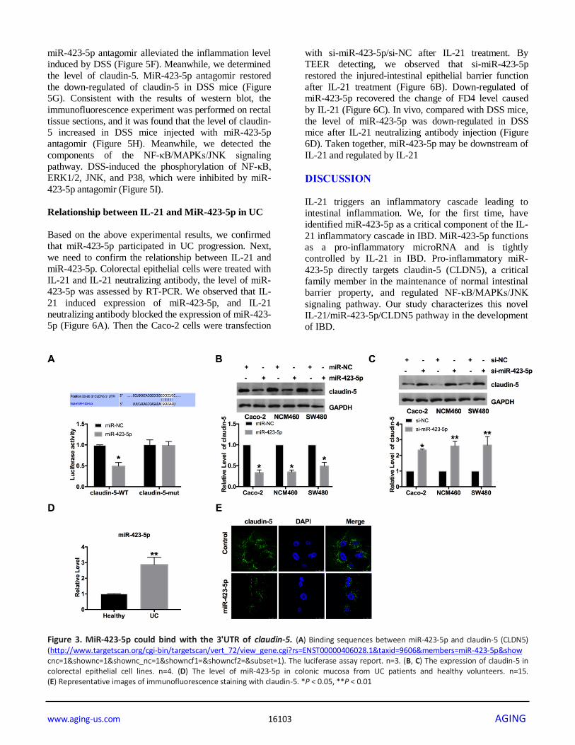
MiR-423-5p binds with 3’UTR of claudin-5.
Through the prediction of the bioinformatics website,
we found that there are binding sequences between
miR-423-5p and claudin-5. By performing luciferase
assay, we ensured that miR-423-5p could interact and
bind with 3'UTR of claudin-5 (Figure 3A). Then we
transfected the miR-423-5p mimic/miR-NC into three
colorectal epithelial cells (Caco-2, NCM460, and
SW480 cells). Then the protein level of claudin-5 was
assessed in different cell lines. Figure 3B showed that
forced expression of miR-423-5p could inhibit the level
Figure 1. Increased expression of IL-21 in UC patients. (A) The expression of IL-21 in colonic mucosa from UC patients and healthy volunteers. n=15. (B) The protein level of IL-21in human samples. n=5. (C) Representative images of immunohistochemistry staining with IL-21. (D) Changes in body weight in different groups of mice. n=10. (E) The protein level of IL-21 in different groups. n=10. (F) The mRNA level of IL1β, IL6, and TNFα in plasma. n=5. (G) The length of the colon in different groups of mice. (H) MPO activity detection in different groups of mice.n=5. (I) Serum FITC- dextran was used as a monitoring index of intestinal permeability. n=6. (J) The protein level of NF-κB/MAPKs/JNK signaling pathway components in different groups. n=5 *P < 0.05, **P < 0.01.
www.aging-us.com 16102 AGING
of claudin-5. Further, si-miR-423-5p/si-NC were
transfected into cells for detecting the level of claudin-
5. The assays performed, blocking the level of miR-
423-5p would restore the level of claudin-5 (Figure 3C).
Meanwhile, we found that the level of miR-423-5p was
abnormally elevated in UC patients (Figure 3D).
Further, we detected the level of claudin-5 in Caco-2
cells by immunofluorescence experiment, similar to
western blot assay. MiR-423-5p blocked the expression
of claudin-5 (Figure 3E).
MiR-423-5p regulates the intestinal epithelial
barrier function by targeting claudin-5
We found that miR-423-5p mimic transfection reduced
the TEER of Caco-2 cells. However, si-miR-423-5p
significantly enhanced the tight junction of Caco-2 cells
(Figure 4A). Then we detected the level of claudin-1,
claudin-5 was also decreased after miR-423-5p mimic
transfection in cells (Figure 4B). We transfected
claudin-5 into Caco-2 cells after transfecting with miR-
423-5p mimic/miR-NC, we observed that
overexpression of claudin-5 restored the injured-
intestinal epithelial barrier function after forced
expression of miR-423-5p (Figure 4C). Similar to the
results of TEER, the overexpression of claudin-5
restored the change of FD4 level caused by
overexpression miR-423-5p (Figure 4D). Taken
together, miR-423-5p would control the intestinal
epithelial barrier function by targeting claudin-5.
MiR-423-5p antagomir administrated performs
protection in DSS mice.
Next, we established a C57BL/6 mouse model of acute
colitis with DSS and treated it with miR-423-5p
antagomir. Compared with the DSS group, the
bodyweight of mice treated with miR-423-5p antagomir
recovered (Figure 5A). Further, we found that DSS
induced the expression of IL1β, IL6, and TNFα.
Compared with the DSS group, the expression of IL1β,
IL6 and TNFα were decreased in miR-423-5p
antagomir injected mice (Figure 5B)
We also found that mice treated with miR-423-5p
antagomir had a protective effect on experimental
colitis compared with the DSS group (Figure 5C).
Colonic MPO activity also showed that miR-423-5p
antagomir could significantly reduce inflammation
(Figure 5D). Further, miR-423-5p antagomir down-
regulated the level of FD40 in the serum of DSS
mice (Figure 5E). H&E staining also performed that
Figure 2. IL-21 regulates the intestinal epithelial TJ barrier function by targeting claudin-5. (A) The effect of IL-21 on intestinal epithelial barrier function. n=3. (B) RT-PCR was employed to detect the mRNA level of claudin-1, claudin-4, claudin-5, claudin-8, claudin-11, occludin, and ZO-1. n=7. (C) The effect of claudin-5 on TEER in Caco-2 cells. n=3. (D) The FD4 level was detected after claudin-5/vector transfection in IL-21 treated cells. n=5. (E, F) The protein level of claudin-5 in different groups. n=4. (G) Representative images of immunofluorescence staining with claudin-5. *P < 0.05, **P < 0.01.
www.aging-us.com 16103 AGING
miR-423-5p antagomir alleviated the inflammation level
induced by DSS (Figure 5F). Meanwhile, we determined
the level of claudin-5. MiR-423-5p antagomir restored
the down-regulated of claudin-5 in DSS mice (Figure
5G). Consistent with the results of western blot, the
immunofluorescence experiment was performed on rectal
tissue sections, and it was found that the level of claudin-
5 increased in DSS mice injected with miR-423-5p
antagomir (Figure 5H). Meanwhile, we detected the
components of the NF-κB/MAPKs/JNK signaling
pathway. DSS-induced the phosphorylation of NF-κB,
ERK1/2, JNK, and P38, which were inhibited by miR-
423-5p antagomir (Figure 5I).
Relationship between IL-21 and MiR-423-5p in UC
Based on the above experimental results, we confirmed
that miR-423-5p participated in UC progression. Next,
we need to confirm the relationship between IL-21 and
miR-423-5p. Colorectal epithelial cells were treated with
IL-21 and IL-21 neutralizing antibody, the level of miR-
423-5p was assessed by RT-PCR. We observed that IL-
21 induced expression of miR-423-5p, and IL-21
neutralizing antibody blocked the expression of miR-423-
5p (Figure 6A). Then the Caco-2 cells were transfection
with si-miR-423-5p/si-NC after IL-21 treatment. By
TEER detecting, we observed that si-miR-423-5p
restored the injured-intestinal epithelial barrier function
after IL-21 treatment (Figure 6B). Down-regulated of
miR-423-5p recovered the change of FD4 level caused
by IL-21 (Figure 6C). In vivo, compared with DSS mice,
the level of miR-423-5p was down-regulated in DSS
mice after IL-21 neutralizing antibody injection (Figure
6D). Taken together, miR-423-5p may be downstream of
IL-21 and regulated by IL-21
DISCUSSION
IL-21 triggers an inflammatory cascade leading to
intestinal inflammation. We, for the first time, have
identified miR-423-5p as a critical component of the IL-
21 inflammatory cascade in IBD. MiR-423-5p functions
as a pro-inflammatory microRNA and is tightly
controlled by IL-21 in IBD. Pro-inflammatory miR-
423-5p directly targets claudin-5 (CLDN5), a critical
family member in the maintenance of normal intestinal
barrier property, and regulated NF-κB/MAPKs/JNK
signaling pathway. Our study characterizes this novel
IL-21/miR-423-5p/CLDN5 pathway in the development
of IBD.
Figure 3. MiR-423-5p could bind with the 3'UTR of claudin-5. (A) Binding sequences between miR-423-5p and claudin-5 (CLDN5) (http://www.targetscan.org/cgi-bin/targetscan/vert_72/view_gene.cgi?rs=ENST00000406028.1&taxid=9606&members=miR-423-5p&show cnc=1&shownc=1&shownc_nc=1&showncf1=&showncf2=&subset=1). The luciferase assay report. n=3. (B, C) The expression of claudin-5 in colorectal epithelial cell lines. n=4. (D) The level of miR-423-5p in colonic mucosa from UC patients and healthy volunteers. n=15. (E) Representative images of immunofluorescence staining with claudin-5. *P < 0.05, **P < 0.01
in CD [14, 15]. It has been proved that the active
interaction between mucosal immune cells and non-
immune cells promotes tissue destruction, and cytokines
must be regulated. IL-21 is one of them, is
overexpressed in CD tissues, and supports the response
of Th1 cells to progressive inflammation. This suggests
that IL-21 can regulate other inflammatory pathways in
the digestive tract except to enhance the immune
response of Th1 cells [16–18]. Similarly, with the
previous study, we also found that IL-21 was
upregulation in IBD mice. After treating with IL-21
neutralizing antibody, the mice showed a decreased
level of inflammatory and in IBD mice.
Tight junction (TJ) exists between epithelial cells and
between epithelial cells and vascular endothelial cells,
which is responsible for regulating the transmembrane
Figure 4. MiR-423-5p regulates the intestinal epithelial barrier function by targeting claudin-5. (A) The effect of miR-423-5p on intestinal epithelial barrier function. n=3. (B) RT-PCR was employed to detect the mRNA level of claudin-1, claudin-4, claudin-5, claudin-8, claudin-11, occludin, and ZO-1. n=6. (C) The effect of miR-423-5p and claudin-5 on TEER in Caco-2 cells. n=4. (D) The FD4 level was detected after claudin-5/vector transfection with miR-423-5p mimic/miR-NC. n=5. *P < 0.05, **P < 0.01.
www.aging-us.com 16105 AGING
transport of ions and solute molecules and maintaining
transmembrane protein families: occludin and claudin
and peri-membrane proteins (zonulae occludente, ZO)
[19]. Studies have shown that the expression of claudin-
2 in the colon of patients with active ulcerative colitis is
significantly increased. Compared with UC, CD showed
the main Th1 immune response. In this context, the
central pro-inflammatory cytokines TNF α and
interferon-γ have been shown to increase the expression
of claudin-2 in HT-29/B6 and Caco-2 cells [20].
Decreased expression and localization of claudin-3 in
the inflammatory colon of patients with CD. In
summary, the claudin family plays a crucial role in IBD.
MicroRNA plays a vital role in many biological
processes. Intestinal inflammation is an important
aspect. Cellular stress, inflammation, and other factors
can induce the expression of MicroRNA, thus affecting
a variety of biological processes and play an
inflammatory or anti-inflammatory role. The gene
mutation and inactivation of MicroRNA will lead to
down-regulation or overexpression of MicroRNA,
which is closely related to intestinal immunity and
inflammation. Sanctuary et al. [21] found that down-
regulation of miR-106a expression can attenuate
intestinal inflammation mediated by TNF- α, and miR-
106a knockout can reduce chronic ileitis in mice, and
Figure 5. The effect of miR-423-5p in DSS mice. (A) Changes in body weight in different groups of mice. n=10. (B) The mRNA level of IL1β, IL6, and TNFα in plasma. n=5. (C) The length of the colon in different groups of mice. (D) MPO activity detection in different groups of mice. n=4. (E) Serum FITC- dextran was used as a monitoring index of intestinal permeability. n=5. (F) Representative images of H&E staining. (G) The expression of claudin-5 in different groups. n=4. (H) Representative images of immunofluorescence staining with claudin-5. (I) The protein level of NF-κB/MAPKs/JNK signaling pathway components in different groups. n=5. *P < 0.05, **P < 0.01.
www.aging-us.com 16106 AGING
out that it is related to the inhibition of post-transcriptional
regulation of IL-10 release through the binding of NF- κB
promoter, which proves that miR-106a has the potential to
treat chronic inflammation including IBD. In addition,
miR-16 is overexpressed in the colonic mucosa of patients
with UC by inhibiting colonic mucosa. The expression of
the A2a receptor inhibits the activation of the NF- κB
signal pathway and regulates immune and inflammatory
response [22]. In our model, however, we found that miR-
423-5p is a pro-inflammatory miRNA. A similar pro-
inflammatory role of miR-423-5p has been reported in
other models [23]. Thus, future studies are needed to
explore the miRNA/target RNA interactome network in
the IL-21 pathway.
The intestinal tract is a relatively complex system in the
human body, and it is the internal ecosystem of the
human body, in which there is an ecological
balance composed of countless kinds of bacteria. The
barrier system of harmful substances in the intestinal
tract of the human body is to protect human health.
Important tools. In hemorrhagic shock. Sepsis, etc. [6].
It will cause damage to the intestinal tract of
patients, especially for some elderly patients, because of
their high age, low immunity, decreased body
function, accompanied by the simultaneous existence
of a variety of diseases, and the degradation of intestinal
function, the intestinal barrier function is even more
weakened. Therefore, it is of considerable significance
for patients to judge early whether the patient has an
intestinal mucosal injury and carry out effective
intervention [24]. With the continuous in-depth
study of intestinal barrier function in the clinic, unique
and comprehensive testing can also be carried out. With
Figure 6. IL-21 regulates miR-423-5p in UC. (A) The expression of miR-423-5p in colorectal epithelial cell lines. n=8. (B) TEER was determined in Caco-2 cells. n=4. (C) The FD4 level was detected after si-miR-423-5p/si-NC transfection in IL-21 treated cells. n=5. (D) The expression of miR-423-5p in different groups. n=7. *P < 0.05, **P < 0.01.
www.aging-us.com 16107 AGING
the constant development of medicine, it provides a
significant reference value for the clinical.
MATERIALS AND METHODS
Human colon samples
Human IBD colonic tissue was taken from IBD patients
and healthy volunteers in Shanxi Provincial People's
Hospital. All participants signed the informed consent
form. The research scheme was approved by the Ethics
Committee of Shanxi Provincial People's Hospital, and
the experimental method was in line with the Helsinki
Declaration.
Cell cultures
Caco-2 cells were seeded in DMEM (Biological
Industries, Israel) supplemented with 10% FBS
(Biological Industries). SW480 cells were cultured in
1640 (Biological Industries, Israel) medium
supplemented with 10% FBS. NCM460 cells were
cultured in M3: BaseF medium (Biological Industries,
Israel) supplemented with 10% FBS and 1% penicillin
and streptomycin sulfate (Gibco, USA).
Cell transfection
For the siRNA/mimic/plasmid transfection, 2 × 10⁵
cells per well were plated in a 6-well plate. After
adhering for 24 hours, si-miR-423-5p/si-NC, miR-
NC/miR-423-5p, or claudin-5/vector (RiboBio, China)
were added to the transfection medium with
Lipofectamine 2000 (Thermo Fisher Scientific, USA)
for 6 hours at 37°C in a CO2 incubator. After
transfection, the cells were supplemented with a normal
culture medium and cultured at 37°C/5% CO2 for up to
48 hours before harvest.
Western blot
Total proteins were extracted from cells and tissues.
The cells and tissues were lysed with 20 μL of RIPA
Mice were purchased from Beijing Charles river and fed
in the experimental Animal Center of Shanxi Provincial
People's Hospital with the standard condition. Day 1 to
Day 7: C57BL/6 in the model group was fed 3% Dextran
Sodium Sulfate (DSS) aqueous solution, while the mice
in the untreated group drank normal water. Day 8 to Day
14: replace fresh DSS-free drinking water. On day 14,
mice were intraperitoneally injected with 3%
pentobarbital sodium and were killed by excessive
anesthesia with a dose of 90 mL/kg, and the organ and
tissue were removed for follow-up study. The research
protocol of this study was approved by the Animal Care
and Use Committee of Shanxi Provincial People's
Hospital.
Mice were treated with IL-21-neutralizing antibodies
(0.5 mg/kg) by intraperitoneal injection at day 8-10
after DSS treated, at day 14, mice were intraperitoneally
injected with 3% pentobarbital sodium and were killed
by excessive anesthesia with a dose of 90 mL/kg, and
the organ and tissue were removed for follow-up study.
For inhibition of miR-423-5p, mice in the antagomir-
423-5p (50 nmol, RiBoBio, China) group were dosed by
tail vein injection on the 7th day.
Measurement of myeloperoxidase activity
Myeloperoxidase (MPO) activity was measured in the
distal colonic tissue obtained from control and mice with
colitis. The inflamed distal colon (5 cm) was removed.
MPO is an enzyme found primarily in neutrophils;
measurement of MPO has been widely used as a marker
for intestinal inflammation [25]. MPO activity was
measured according to the protocol described by
Krawisz et al. [26]. Briefly, after the samples were
weighed, tissue samples were homogenized in a buffer
(0.5% hexadecyltrimethylammonium bromide in 50 mM
potassium phosphate buffer, pH 6.0) for 1 min. The
samples were frozen in liquid nitrogen, thawed three
times, and centrifuged at 20,000 × g for 20 min at 4°C
using a microcentrifuge. Aliquots of supernatants (20 ml)
were mixed with 980 ml of O-dianisidine. Absorbance
was recorded at 450 nm every 1 min over a period of 10
min by ELISA. MPO activity was expressed as units/g of
tissue. An enzyme unit was defined as the conversion of
1 mol of H2O2 per min at 25°C.
Statistical analysis
Data were calculated as means ± SEM and analyzed by
GraphPad 7.0. T-test and two-way ANOVA were used
to analyze the data P < 0.05 was considered as a
statistically significant difference.
CONFLICTS OF INTEREST
The authors declared no conflicts of interest.
FUNDING
This study was supported by the Science Foundation of
Health and the family planning commission of Shanxi
Province (No.2017020); The International Science
and Technology Cooperation Project of Shanxi
(No.2013081066).
REFERENCES
1. Laube R, Yau Y, Selinger CP, Seow CH, Thomas A, Wei
Chuah S, Hilmi I, Mao R, Ong D, Ng SC, Wei SC, Banerjee R, Ahuja V, et al. Knowledge and attitudes towards pregnancy in females with inflammatory bowel disease - an international, multi-centre study. J Crohns Colitis. 2020. [Epub ahead of print].
2. Ng SC, Shi HY, Hamidi N, Underwood FE, Tang W, Benchimol EI, Panaccione R, Ghosh S, Wu JC, Chan FK, Sung JJ, Kaplan GG. Worldwide incidence and prevalence of inflammatory bowel disease in the 21st century: a systematic review of population-based studies. Lancet. 2018; 390:2769–78.
3. Qiu L, Yu Q, Zhou Y, Zheng S, Tao J, Jiang Q, Yuan G. Functionally impaired follicular helper T cells induce regulatory B cells and CD14+ human leukocyte antigen-DR- cell differentiation in non-small cell lung cancer. Cancer Sci. 2018; 109:3751–61.
https://doi.org/10.1111/cas.13836 PMID:30325558
4. Faghih M, Rostami-Nejad M, Amani D, Sadeghi A, Pourhoseingholi MA, Masotti A, Zali MR. Analysis of IL17A and IL21 expression in the small intestine of celiac disease patients and correlation with circulating thioredoxin level. Genet Test Mol Biomarkers. 2018; 22:518–25.
5. Caielli S, Veiga DT, Balasubramanian P, Athale S, Domic B, Murat E, Banchereau R, Xu Z, Chandra M, Chung CH, Walters L, Baisch J, Wright T, et al. A CD4+ T cell population expanded in lupus blood provides B cell help through interleukin-10 and succinate. Nat Med. 2019; 25:75–81.
6. Holmberg FE, Seidelin JB, Yin X, Mead BE, Tong Z, Li Y, Karp JM, Nielsen OH. Culturing human intestinal stem cells for regenerative applications in the treatment of inflammatory bowel disease. EMBO Mol Med. 2017; 9:558–70.
7. Furuse M, Fujita K, Hiiragi T, Fujimoto K, Tsukita S. Claudin-1 and -2: novel integral membrane proteins localizing at tight junctions with no sequence similarity to occludin. J Cell Biol. 1998; 141:1539–50.
8. Swisshelm K, Machl A, Planitzer S, Robertson R, Kubbies M, Hosier S. SEMP1, a senescence-associated cDNA isolated from human mammary epithelial cells, is a member of an epithelial membrane protein superfamily. Gene. 1999; 226:285–95.
9. Mineta K, Yamamoto Y, Yamazaki Y, Tanaka H, Tada Y, Saito K, Tamura A, Igarashi M, Endo T, Takeuchi K, Tsukita S. Predicted expansion of the claudin multigene family. FEBS Lett. 2011; 585:606–12.
11. Parrish-Novak J, Dillon SR, Nelson A, Hammond A, Sprecher C, Gross JA, Johnston J, Madden K, Xu W, West J, Schrader S, Burkhead S, Heipel M, et al. Interleukin 21 and its receptor are involved in NK cell expansion and regulation of lymphocyte function. Nature. 2000; 408:57–63.
https://doi.org/10.1038/35040504 PMID:11081504
12. Tang L, Chen C, Gao X, Zhang W, Yan X, Zhou Y, Guo L, Zheng X, Wang W, Yang F, Liu G, Sun J, Hou J, Li Y. Interleukin 21 reinvigorates the antiviral activity of hepatitis B virus (HBV)-specific CD8+ T cells in chronic HBV infection. J Infect Dis. 2019; 219:750–59.
13. Xing R, Sun L, Wu D, Jin Y, Li C, Liu X, Zhao J. Autoantibodies against interleukin-21 correlate with disease activity in patients with rheumatoid arthritis. Clin Rheumatol. 2018; 37:75–80.
14. Wang Y, Jiang X, Zhu J, Yue D, Zhang X, Wang X, You Y, Wang B, Xu Y, Lu C, Sun X, Yoshikai Y. IL-21/IL-21R signaling suppresses intestinal inflammation induced by DSS through regulation of th responses in lamina propria in mice. Sci Rep. 2016; 6:31881.
https://doi.org/10.1038/srep31881 PMID:27545302
15. Araki A, Nara H, Rahman M, Onoda T, Li J, Juliana FM, Jin L, Murata K, Takeda Y, Asao H. Role of interleukin-21 isoform in dextran sulfate sodium (DSS)-induced colitis. Cytokine. 2013; 62:262–71.
16. Liu Z, Yang L, Cui Y, Wang X, Guo C, Huang Z, Kan Q, Liu Z, Liu Y. Il-21 enhances NK cell activation and cytolytic activity and induces Th17 cell differentiation in inflammatory bowel disease. Inflamm Bowel Dis. 2009; 15:1133–44.
17. Neil JA, Matsuzawa-Ishimoto Y, Kernbauer-Hölzl E, Schuster SL, Sota S, Venzon M, Dallari S, Galvao Neto A, Hine A, Hudesman D, Loke P, Nice TJ, Cadwell K. IFN-I and IL-22 mediate protective effects of intestinal viral infection. Nat Microbiol. 2019; 4:1737–49.
19. Kuo WT, Shen L, Zuo L, Shashikanth N, Ong ML, Wu L, Zha J, Edelblum KL, Wang Y, Wang Y, Nilsen SP, Turner JR. Inflammation-induced Occludin Downregulation Limits Epithelial Apoptosis by Suppressing Caspase-3 Expression. Gastroenterology. 2019; 157:1323–1337.
20. Rosenthal R, Luettig J, Hering NA, Krug SM, Albrecht U, Fromm M, Schulzke JD. Myrrh exerts barrier-stabilising and -protective effects in HT-29/B6 and caco-2 intestinal epithelial cells. Int J Colorectal Dis. 2017; 32:623–34.
22. Tian T, Zhou Y, Feng X, Ye S, Wang H, Wu W, Tan W, Yu C, Hu J, Zheng R, Chen Z, Pei X, Luo H. MicroRNA-16 is putatively involved in the NF-κB pathway regulation in ulcerative colitis through adenosine A2a receptor (A2aAR) mRNA targeting. Sci Rep. 2016; 6:30824.
https://doi.org/10.1038/srep30824 PMID:27476546
23. Herbert C, Sebesfi M, Zeng QX, Oliver BG, Foster PS, Kumar RK. Using multiple online databases to help identify microRNAs regulating the airway epithelial cell response to a virus-like stimulus. Respirology. 2015; 20:1206–12.
https://doi.org/10.1111/resp.12606 PMID:26289417
24. Camara-Lemarroy CR, Metz L, Meddings JB, Sharkey KA, Wee Yong V. The intestinal barrier in multiple sclerosis: implications for pathophysiology and therapeutics. Brain. 2018; 141:1900–16.
26. Krawisz JE, Sharon P, Stenson WF. Quantitative assay for acute intestinal inflammation based on myeloperoxidase activity. Assessment of inflammation in rat and hamster models. Gastroenterology. 1984; 87:1344–50.