49
21.Which of the following conditions is frequently associated with these cells? Hemolytic
| Date post: | 26-Oct-2015 |
| Category: |
Documents |
| Upload: | sophia-davis |
| View: | 1 times |
| Download: | 0 times |

21.Which of the following conditions is frequently associated with these cells?Hemolytic anemiaChronic blood

lossMegaloblastic anemiaSickle cell anemiaFeedback Hypersegmented neutrophils are associated with megaloblastic anemias.22.The cell indicated by

the arrow in this illustration is called:Hair cellSmudge cellMegakaryocyteBlastFeedback Smudge Cells (bare nuclei) are commonly

seen in blood smears from patients withchronic lymphocytic leukemia.23.Identify the urine sediment elements shown by the arrow:Cholesterol

crystalsUric acid crystalsAmorphous u ra tecrystalsCystine crystalsFeedback Cystine crystals are characteristically seen as colorless

hexagonal plates in acidurine. They may be confused with hexagonal uric acid crystals. They can bedifferentiated from uric acid by their solubility in dilute

hydrochloric acid versuscrystalline uric acid, which is not soluble in dilute hydrochloric acid. The cyanide-nitroprusside test can be used to confirm the

presence of cystine in urine. Cystinecrystals are not present in normal urine.24.Identify the urine sediment elements shown by the arrow:


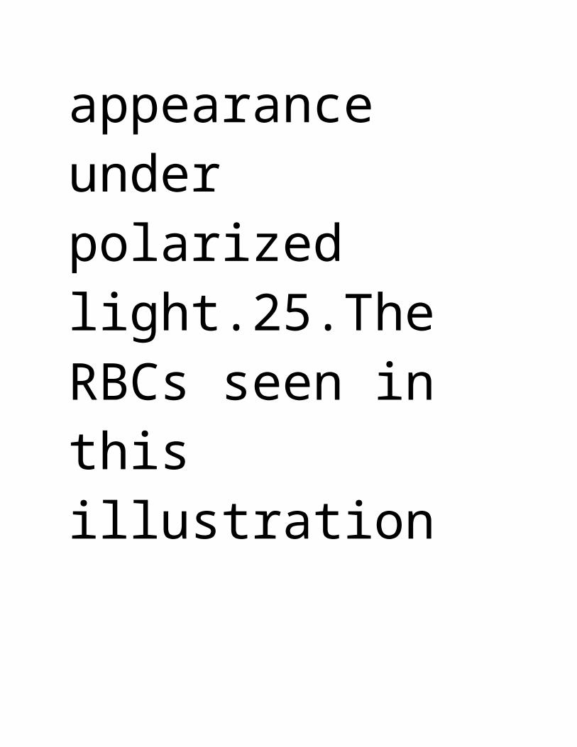
YeastWBCsFatStarchcrystalsFeedback Starch crystals often contaminate normal urine. They appear round to oval, arehighly

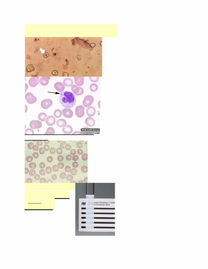
refractile, with an irregular indentation in the center. They may exhibit the"Maltese cross" appearance under polarized light.25.The RBCs seen in this

illustration are indicative of:Normal cellsMicrocytic cellsMacrocytic cellsHypochromiccellsFeedback

Normal red cells are uniformly shaped and sized with 2-3mm of central pallor.26.The McFarland Comparison Card shown in the

illustration is used to:Perform sensitivity testingStandardize concentrations of organismsQ.C. broth

mediaDetermine MICs27.The WBC indicated by the arrow in this illustration is exhibiting:Dohle bodiesHyperseg
mentationDegranulationPelger-Huet


anomalyFeedback Pelger-Huet anomoly is an autosomal dominant condition characterized byhyposegmentation of neutrophils. Decreased nuclear

segmentation may also be seenas an acquired disorder in cases of granulocytic leukemia, myeloproliferativedisorders, infections,
and after exposure to certain drugs.Image courtesy and copyright of the Clinical Chemistry and Hematology Laboratory, WadsworthCenter, NY State Department of

Health (http://www.wadsworth.org)28.The abnormal RBC shape seen in this illustration is:Sickle cell Thorn cellFragmented

cellCrenated cellFeedback Crenated red cells usually occur as an artifact during the preparation of bloodsmears. Acknowledgement

I m thank full to my God he give meAnd also I thank full my friend Mr. Sami Khan he help me in down load, Thanks,Muhammad Younis

Lab. Technician NNP, Rabigh, [email protected]


