Page 1
ORIGINAL PAPER
New Penicillium and Talaromyces species from honey, pollenand nests of stingless bees
Renan N. Barbosa . Jadson D. P. Bezerra . Cristina M. Souza-Motta .
Jens C. Frisvad . Robert A. Samson . Neiva T. Oliveira . Jos Houbraken
Received: 15 January 2018 / Accepted: 31 March 2018 / Published online: 13 April 2018
� The Author(s) 2018
Abstract Penicillium and Talaromyces species have
a worldwide distribution and are isolated from various
materials and hosts, including insects and their
substrates. The aim of this study was to characterize
the Penicillium and Talaromyces species obtained
during a survey of honey, pollen and the inside of nests
of Melipona scutellaris. A total of 100 isolates were
obtained during the survey and 82% of those strains
belonged to Penicillium and 18% to Talaromyces.
Identification of these isolates was performed based on
phenotypic characters and b-tubulin and ITS sequenc-
ing. Twenty-one species were identified in Penicillium
and six in Talaromyces, including seven new species.
These new species were studied in detail using a
polyphasic approach combining phenotypic, molecu-
lar and extrolite data. The four new Penicillium
species belong to sections Sclerotiora (Penicillium
fernandesiae sp. nov., Penicillium mellis sp. nov.,
Penicillium meliponae sp. nov.) and Gracilenta
(Penicillium apimei sp. nov.) and the three new
Talaromyces species to sections Helici (Talaromyces
pigmentosus sp. nov.), Talaromyces (Talaromyces
mycothecae sp. nov.) and Trachyspermi (Talaromyces
brasiliensis sp. nov.). The invalidly described species
Penicillium echinulonalgiovense sp. nov. was also
isolated during the survey and this species is validated
here.
Keywords 8 new taxa � Aspergillaceae � Fungalecology � Polyphasic approach � Taxonomy �Trichocomaceae
Introduction
Stingless bees comprise a diverse group of highly
eusocial insects occurring throughout the tropical
regions in the world. They are important honey
producers and pollinators of several plants (Ramırez
et al. 2010; Brown and Oliveira 2014). An example of
a stingless bee species is Melipona scutellaris
Electronic supplementary material The online version ofthis article (https://doi.org/10.1007/s10482-018-1081-1) con-tains supplementary material, which is available to authorizedusers.
R. N. Barbosa � R. A. Samson � J. Houbraken (&)
Westerdijk Fungal Biodiversity Institute, Uppsalalaan 8,
3584 CT Utrecht, The Netherlands
e-mail: [email protected]
R. N. Barbosa � J. D. P. Bezerra � C. M. Souza-Motta �N. T. Oliveira
Departamento de Micologia Prof. Chaves Batista,
Universidade Federal de Pernambuco, Av. Prof. Moraes
Rego, s/n, Centro de Biociencias, Cidade Universitaria,
CEP: 50670-901 Recife, PE, Brazil
J. C. Frisvad
Department of Biotechnology and Biomedicine,
Technical University of Denmark, 2800 Kongens Lyngby,
Denmark
123
Antonie van Leeuwenhoek (2018) 111:1883–1912
https://doi.org/10.1007/s10482-018-1081-1(0123456789().,-volV)(0123456789().,-volV)
Page 2
(Hymenoptera: Apidae: Meliponini), an indigenous
species occurring in the North-eastern part of Brazil
and considered to be one of the first species to be
domesticated in the Americas (Kerr 1996; Silva et al.
2013). In this part of Brazil, M. scutellaris is the main
bee species in meliponiculture (stingless beekeeping).
Meliponiculture in the rural areas is a sustainable
activity and the honey from these bees is widely
appreciated as a food source. The composition of the
honey of the stingless bees differs from that of bees of
the genus Apis (honey bees) (Vit et al. 2004). The
honey of stingless bees contains, in comparison to
honey of honey bees, a more complex mixture of
carbohydrates and contains other types of organic
acids, proteins, minerals, vitamins, pollen grains and
enzymes (Almeida-Muradian et al. 2013). Recently,
the interest in honey produced by stingless bees
increased. Besides being a food source, also several
other functionalities are linked to this type of honey,
such as antiseptic, antimicrobial, anti-inflammatory
and wound-healing properties (Silva et al. 2013; Rao
et al. 2016).
Penicillium and Talaromyces are fungal genera
classified in the order Eurotiales. In the dual nomen-
clature era (pre 2012), Talaromyces was known as a
sexual genus related to Penicillium and other genera.
In the last decade, the genera Talaromyces and
Penicillium were re-defined due to new taxonomic
insights and the introduction of single name nomen-
clature (Houbraken and Samson 2011; Samson et al.
2011; McNeill et al. 2012; Yilmaz et al. 2014).
Currently, Penicillium and Talaromyces are separate
genera that contain both sexual and asexual species.
Visagie et al. (2014) accepted 354 Penicillium species
and Yilmaz et al. (2014) 88 Talaromyces species, and
these numbers are rapidly increasing (Houbraken et al.
2016a). Several of the new species that are being
discovered are found during ecology and biodiversity
studies of specific substrates or habitats (Houbraken
et al. 2016a). Describing new species from poorly
explored substrates and habitats, like those related to
meliponiculture, will add to our knowledge on biodi-
versity. With this information, future studies will also
be able to better understand the ecology of fungi in
these type of environments.
Fungi, such as Penicillium and Talaromyces, can
have a strong association to a specific substrate
(Peterson et al. 2003; Kobayashi et al. 2008; Visagie
2012, Li et al. 2012; Rivera et al. 2012; Yilmaz et al.
2014). The genera Aspergillus, Penicillium, Mon-
ascus andMucor are commonly associated with bees
or their products (Egorova 1971; Gilliam et al. 1989;
Eltz et al. 2002; Ferraz et al. 2008; Barbosa et al.
2017). Most fungi associated with bees and nests
have a saprophytic lifestyle, but fungi can also have a
mutualistic relationship with bees (Menezes et al.
2015). On the other hand, fungi are also reported to be
pathogenic to many bee species and cause serious
problems in honey bee (Apis mellifera) brood.
Aspergillus flavus is the primary species responsible
for stonebrood, a disease where dead and mummified
larvae are present in the brood cells, but also other
Aspergilli such as Aspergillus fumigatus and Asper-
gillus niger are reported as aetiological agents of this
disease (Gilliam and Vandenberg 1988; Foley et al.
2014; Lopes et al. 2015; Sarwar 2016). Though it is
generally accepted that infection only occurs in
weakened colonies, the specific conditions predis-
posing the onset of disease are not fully understood
(Shoreit and Bagy 1995).
Fungi play an important role in many ecosystems;
however, only a limited number of studies dealt with
the association between stingless bees in Brazil and
filamentous fungi (e.g. Oliveira and Morato 2000;
Ferraz et al. 2008; Gois et al. 2010) and yeasts (e.g.
Teixeira et al. 2003; Rosa et al. 2003; Daniel et al.
2013; Barbosa et al. 2016). In the present study, we
analysed three different substrates associated withM.
scutellaris bees: bee pollen, nests and honey. In
nature, theM. scutellaris bee nests are mainly located
in tree hollows, and they are kept by beekeepers in
artificial wooden hives. The bees use cerumen (a
mixture of wax and floral resins) for the construction
of their nests and this material is also used inside
nests in storage pots, brood cells and entrance
openings (Cortopassi-Laurino et al. 2006; Pianaro
et al. 2007). The floral pollen is collected, packed into
pollen pellets, and subsequently stored inside the nest
by worker bees. This stored pollen is referred to as
‘bee bread’. The pollen spectrum has been studied in
the past to get insight in the bee colony’s food
requirements, pollinating functions and the plant
species visited by the bees (Cortopassi-Laurino et al.
2007).
In this paper, we focus on the identification of
Penicillium and Talaromyces species isolated from
three different substrates (bee pollen, nests and honey)
associated to M. scutellaris in the Atlantic Rainforest
1884 Antonie van Leeuwenhoek (2018) 111:1883–1912
123
Page 3
in Brazil. Phenotypic characters, combined with ITS
and partial b-tubulin (BenA) sequences were applied
to identify the isolates. Four Penicillium and three
Talaromyces species could not be assigned to any
known species and are described here as new. Those
species are described using a polyphasic approach
including morphology, ITS, BenA, calmodulin (CaM)
and/or RNA polymerase II second largest subunit
(RPB2) sequences and extrolites profiles.
Materials and methods
Strains
Six collections were performed between January and
June 2014 in the tropical forest in Pernambuco, Brazil
(8�703000S, 34�5203000W and 8�403600S, 34�5703400W).
During each collection, four hives were sampled.
Stingless bees process honey and pollen in cerumen
pots. Per hive, four samples of the honey pots and four
of the pollen pots were collected and combined,
resulting in one mixed sample of each substrate. In the
same hives, also the surface of brood cells and the
pollen and honey pots were sampled using sterile
cotton swabs (in total 48 swabs). Analysis of the
samples was performed using dichloran 18% glycerol
agar (DG18) and malt extract agar supplemented with
chloramphenicol as described in Barbosa et al. (2017).
The isolates were subsequently deposited in the
Micoteca URM culture collection (Federal University
of Pernambuco, Recife, Brazil) and ex-type strains in
the CBS culture collection, housed at the Westerdijk
Fungal Biodiversity Institute, Utrecht, The Nether-
lands (underMaterial Transfer Agreement—MTANo.
01/2016/Micoteca URM) (Tables 1, 2). Holotype
material (slide preparation) is deposited at Herbario
Pe. Camille Torrend (Federal University of Pernam-
buco, Recife, Brazil). New species names and asso-
ciated information were deposited in MycoBank.
Morphological analyses
For morphological analysis, the strains were three-
point inoculated onto creatine agar (CREA), Czapek
yeast extract agar (CYA), CYA supplemented with
5% NaCl (CYAS), dichloran 18% glycerol agar
(DG18), malt extract agar (MEA, Oxoid), oatmeal
agar (OA) and yeast extract sucrose agar (YES). All
Petri dishes were incubated at 25 �C for 7 days and
additional CYA andMEA plates were incubated at 15,
30 and 37 �C. Media preparation, inoculation and
incubation were performed as described in Samson
et al. (2010). Colony diameters were measured after
7 days of incubation and colony characteristics
recorded (e.g. presence of soluble pigments, exudates,
obverse and reverse colony colours, colour of
mycelium). Microscopic observations of the asexual
stage were made from colonies grown on MEA. The
presence of a sexual stage was determined from
cultures incubated on CYA, MEA and OA for at least
40 days at 25 �C. Lactic acid (60%) was used as
mounting fluid and 96% ethanol was used to remove
excess conidia. A Zeiss Stereo Discovery V20
dissecting microscope and a Zeiss AX10 Imager A2
light microscope, both equipped with Nikon DS-Ri2
cameras, were used to capture digital images using the
software NIS-Elements D v4.50. The size, shape and
pigmentation of microscopic features were recorded.
DNA isolation, PCR and sequencing
Genomic DNA extractions were made from 7 days old
colonies grown on MEA using the UltraClean Micro-
bial DNA kit (MoBio Laboratories, Solana Beach,
CA, USA). Polymerase chain reaction (PCR) ampli-
fication of the ITS barcode (ITS1, 5.8S rDNA and
ITS2), BenA, CaM and RPB2 gene regions were
performed using methods described by Samson et al.
(2010) and Houbraken et al. (2012). The PCR products
were sequenced in both directions with the same
primers using the BigDye� Terminator v. 3.1 Cycle
Sequencing Kit (Applied Biosystems Life Technolo-
gies, Carlsbad, CA, USA) and purified with Sephadex,
according to the manufacturers’ recommendations.
Contigs were assembled in the SeqMan (v.10.0.1;
Madison, WI, USA) program using the forward and
reverse sequence. Newly generated sequences were
deposited in the NCBI nucleotide database (GenBank)
and the European Nucleotide Archive (Table 1).
Phylogenetic analysis
Sequence datasets were generated by combining the
newly generated sequences with reference (preferably
ex-type) sequences from NCBI (Visagie et al. 2014;
Yilmaz et al. 2014; Taniwaki et al. 2015; Visagie et al.
2015; Chen et al. 2016; Laich and Andrade 2016; Luo
Antonie van Leeuwenhoek (2018) 111:1883–1912 1885
123
Page 4
Table 1 Details of strains isolated in this study and used in the phylogenetic analyses
Species Strain numbers Substrate; location Sequence accession numbers
ITS BenA CaM RPB2
Penicillium apimei URM 7591 T =
CBS 142502
Honey of Melipona scutellaris;
Recife, Pernambuco, Brazil
MF278310 LT854641 LT882717 LT854650
Penicillium
echinulonalgiovense
URM 7599 Bee pollen of Melipona scutellaris;
Recife, Pernambuco, Brazil
MF278311 LT882667 LT882670 LT882673
Penicillium
echinulonalgiovense
RB 217 Inside nest of Melipona scutellaris;
Recife, Pernambuco, Brazil
MF278312 LT882668 LT882671 LT882674
Penicillium
echinulonalgiovense
RB 218 Inside nest of Melipona scutellaris;
Recife, Pernambuco, Brazil
MF278313 LT882669 LT882672 LT882675
Penicillium
fernandesiae
URM 7600 T =
CBS 142500
Inside nest of Melipona scutellaris;
Recife, Pernambuco, Brazil
MF278314 LT854645 LT854649 LT854654
Penicillium
meliponae
URM 7602 T =
CBS 142495
Honey of Melipona scutellaris;
Recife, Pernambuco, Brazil
MF278315 LT854644 LT854648 LT854653
Penicillium mellis URM 7605 T =
CBS 142499
Honey of Melipona scutellaris;
Recife, Pernambuco, Brazil
MF278316 LT854643 LT854647 LT854652
Penicillium mellis URM 7611 Inside nest of Melipona scutellaris;
Recife, Pernambuco, Brazil
MF278317 LT882629 LT882634 LT882634
Penicillium mellis RB 9 Inside nest of Melipona scutellaris;
Recife, Pernambuco, Brazil
MF278318 LT882625 LT882630 LT882635
Penicillium mellis RB 69 Honey of Melipona scutellaris;
Recife, Pernambuco, Brazil
MF278319 LT882626 LT882631 LT882636
Penicillium mellis RB 85 Inside nest of Melipona scutellaris;
Recife, Pernambuco, Brazil
MF278320 LT882627 LT882632 LT882637
Penicillium mellis RB 110 Inside nest of Melipona scutellaris;
Recife, Pernambuco, Brazil
MF278321 LT882628 LT882633 LT882638
Penicillium sp. URM 7610 =
CBS 142497
Bee pollen of Melipona scutellaris;
Recife, Pernambuco, Brazil
MF278322 LT882642 LT882646 LT882651
Talaromyces
brasiliensis
URM 7618 T =
CBS 142493
Honey of Melipona scutellaris;
Recife, Pernambuco, Brazil
MF278323 LT855560 LT855563 LT855566
Talaromyces
brasiliensis
URM 7619 Inside nest of Melipona scutellaris;
Recife, Pernambuco, Brazil
MF278324 LT882640 LT882642 LT882644
Talaromyces
brasiliensis
URM 7620 Inside nest of Melipona scutellaris;
Recife, Pernambuco, Brazil
MF278325 LT882641 LT882643 LT882645
Talaromyces
mycothecae
URM 7622 T =
CBS 142494
Inside nest of Melipona scutellaris;
Recife, Pernambuco, Brazil
MF278326 LT855561 LT855564 LT855567
Talaromyces
mycothecae
URM 7623 Inside nest of Melipona scutellaris;
Recife, Pernambuco, Brazil
MF278327 LT882646 LT882649 LT882652
Talaromyces
mycothecae
RB 95 Inside nest of Melipona scutellaris;
Recife, Pernambuco, Brazil
MF278328 LT882647 LT882650 LT882653
Talaromyces
mycothecae
RB 171 Inside nest of Melipona scutellaris;
Recife, Pernambuco, Brazil
MF278329 LT882648 LT882651 LT882654
Talaromyces
pigmentosus
URM 7624 T =
CBS 142805
Inside nest of Melipona scutellaris;
Recife, Pernambuco, Brazil
MF278330 LT855562 LT855565 LT855568
Talaromyces
pigmentosus
URM 7625 Bee pollen of Melipona scutellaris;
Recife, Pernambuco, Brazil
MF278331 LT882655 LT882659 LT882663
Talaromyces
pigmentosus
RB 30 Inside nest of Melipona scutellaris;
Recife, Pernambuco, Brazil
MF278332 LT882656 LT882660 LT882664
1886 Antonie van Leeuwenhoek (2018) 111:1883–1912
123
Page 5
et al. 2016; Romero et al. 2016; Rong et al. 2016;
Visagie et al. 2016; Yilmaz et al. 2016; Guevara-Suarez
et al. 2017; Wang et al. 2017a, b). The sequences were
aligned using MAFFT v.7 (Katoh and Standley 2013)
and manually optimized using MEGA v. 6.06 (Tamura
et al. 2013). Individual alignments were concatenated
by using Mesquite v. 3.04 (Maddison and Maddison
2016). The most suitable substitution model was
determined using jModelTest v. 2.1.7 (Posada 2008).
Phylogenetic trees were constructed using Maximum
likelihood analyses (ML) using RAxML-HPC v. 8.2.8
(Stamatakis 2014) BlackBox with 1 000 rapid boot-
strap inferences via the CIPRES science gateway
(http://www.phylo.org/) (Miller et al 2010), while
Bayesian inference (BI) analysis was performed in
MrBayes 3.2.2 (Ronquist et al. 2012). In the Bayesian
analyses, every 1 000 generations was sampled and the
first 25% of the samples were discarded. Trees were
visualized in FigTree v. 1.1.2 (Rambaut 2009) and
edited in Adobe Illustrator v. 5.1. Bayesian inference
(BI) posterior probabilities (pp) values and bootstrap
(bs) values are labelled at the nodes. Values less than
0.95 pp and 70% bootstrap support are not shown.
Branches with full support in Bayesian and RAxML
analyses are thickened. Values below 0.95 pp and 70%
are not shown and indicated with a hyphen. Aligned
datasets and trees were uploaded to TreeBase (www.
treebase.org) under submission number 21965.
Extrolite analysis
Extrolites were extracted from the Penicillium strains
after growing them on CYA, YES and MEA at 25 �Cfor 7 days. The Talaromyces strains were inoculated
on CYA, YES, MEA and OA, and incubated at 25 �Cfor 14 days. Three agar plugs of each medium were
extracted as previously described (Smedsgaard 1997;
Houbraken et al. 2012). After extraction, the liquid
was transferred to a clean screw-cap vial and evapo-
rated to dryness. The dried extracts were re-dissolved
in methanol by ultrasonication and filtered through a
0.45 lm filter. The extracts were analysed by ultra-
high performance liquid chromatography with diode-
array detection (UHPLC-DAD) (Houbraken et al.
2012). The detected eluted compounds were identified
by comparison of the retention time, retention index
and the UV spectrummeasured at 200–600 nm against
UV spectra from made from standards and data from
literature (Nielsen et al. 2011; Klitgaard et al. 2014).
Results
Isolation and identification
During this study on the fungal diversity of substrates
related to stingless bees, isolates belonging to various
genera [e.g. Aspergillus, Fusarium, Monascus (Bar-
bosa et al. 2017), Penicillium, Talaromyces] were
isolated. This study focusses on the identification of
the detected Penicillium and Talaromyces diversity.
The number of Talaromyces species (and isolates)
detected during this study is low compared to Peni-
cillium. Eighty-two Penicillium and 18 Talaromyces
isolates were obtained during the survey on fungi
present in honey, bee pollen and inside the nests of
Melipona scutellaris bees. Phenotypic characters,
combined with ITS and partial BenA sequences were
used to identify isolates. In total, 21 Penicillium and
six Talaromyces species were present among the
investigated isolates. Among those, five Penicillium
and three Talaromyces species displayed unique
characters deviating from known species. Seven of
those eight species are described here as new (see
Taxonomy section), and one isolate (RB115), belong-
ing to section Lanata-Divaricata, will be described
Table 1 continued
Species Strain numbers Substrate; location Sequence accession numbers
ITS BenA CaM RPB2
Talaromyces
pigmentosus
RB 96 Bee pollen of Melipona scutellaris;
Recife, Pernambuco, Brazil
MF278333 LT882657 LT882661 LT882665
Talaromyces
pigmentosus
RB 100 Inside nest of Melipona scutellaris;
Recife, Pernambuco, Brazil
MF278334 LT882658 LT882662 LT882666
T ex-type strain, URM URM Culture Collection (www.ufpe.br/micoteca), Brazil, RB personal working collection of Renan Barbosa,
CBS culture collection of the Westerdijk Fungal Biodiversity Institute, The Netherlands
Antonie van Leeuwenhoek (2018) 111:1883–1912 1887
123
Page 6
Table 2 Overview of isolated species from honey, bee pollen and nests
Species Section Isolate numbers Honey Pollen Nests Total
Penicillium apimei sp.
nov.
Gracilenta URM 7591 T= CBS 142502 1 1
Penicillium brocae Sclerotiora RB 001; RB 036; RB 035; RB 036; RB 046; RB 064; RB
075; RB 079; RB 082; RB 090; RB 093; RB 101; RB
116; RB 123; RB 124; RB 125; RB 181; RB 182; RB
184; RB 186; RB 193; RB 225
9 13 22
Penicillium
chermesinum
Charlesia RB 114 1 1
Penicillium
citreosulfuratum
Exilicaulis RB 094 1 1
Penicillium citrinum Citrina RB 006; RB 028; RB 032; RB 047; RB 068; RB 250; RB
086; RB 104; RB 109; RB 119; RB 134; RB 185; RB
187; RB 192; RB 198; RB 206
4 2 10 16
Penicillium
echinulonalgiovense
sp. nov.
Lanata-
Divaricata
RB 201; RB 217; RB 218 1 2 3
Penicillium fellutanum Charlesia RB 112; RB 113 2 2
Penicillium
fernandesiae sp. nov.
Sclerotiora URM 7600 T = CBS 142500 1 1
Penicillium mallochii Sclerotiora RB 138; RB 151; RB 152 3 3
Penicillium meliponae
sp. nov.
Sclerotiora URM 7602 T = CBS 142495 1 1
Penicillium mellis sp.
nov.
Sclerotiora URM 7605 T= CBS 142499; URM 7611; RB 09; RB 69;
RB 85; RB 110
2 4 6
Penicillium paxilli Citrina RB 127; RB 128 2 2
Penicillium rubens Chrysogena RB 014; RB 153; RB 161; RB 192
RB 210; RB 235
6 6
Penicillium
sanshaense
Sclerotiora URM 7617 T = CBS 142496 1 1
Penicillium
sclerotiorum
Sclerotiora RB 056; RB 121; RB 129; RB 237 1 1 2 4
Penicillium shearii Citrina RB 034; RB 073; RB 248 3 3
Penicillium singorense Lanata-
Divaricata
RB 202 1 1
Penicillium sp. Lanata-
Divaricata
URM 7610 = CBS 142497 1 1
Penicillium steckii Citrina RB 065; RB 088; RB 089; RB 137 4 4
Penicillium
sumatraense
Citrina RB 149 1 1
Penicillium wotroi Lanata-
Divaricata
RB 010; RB 158 1 1 2
Talaromyces
brasiliensis sp. nov.
Trachyspermi URM 7618 T = CBS 142493; URM 7619; URM 7620 1 2 3
Talaromyces
calidicanius
Talaromyces RB 183 1 1
Talaromyces
mycothecae sp. nov.
Talaromyces URM 7622 T = CBS 142494; URM 7623; RB 95; RB
171
4 4
Talaromyces
pigmentosus sp. nov.
Helici URM 7624 T= CBS 142805; URM 7625; RB 30; RB 96;
RB 100
2 3 5
Talaromyces scorteus Islandici RB 072; RB 114; RB 148; RB 167 3 1 4
1888 Antonie van Leeuwenhoek (2018) 111:1883–1912
123
Page 7
elsewhere. Three new Penicillium species belong to
section Sclerotiora and one to section Gracilenta; the
three new Talaromyces are classified in sections
Helici, Talaromyces and Trachyspermi. An overview
of the species isolated during this study is given in
Table 2. The highest Penicillium and Talaromyces
occurrence frequency was observed in the samples
collected from the inside of nests (66%). The majority
of the isolated Penicillium species belonged to
sections Sclerotiora (46%) and Citrina (30%). Peni-
cillium brocae was most frequently isolated (22%),
followed by Penicillium citrinum (16%), Penicillium
rubens (6%) and Penicillium mellis sp. nov. (6%).
Phylogeny
The phylogenetic relationship of the new Penicillium
and Talaromyces species with accepted species was
determined by analysis of single and concatenated
sequence datasets of three or four loci (ITS, BenA,
CaM and/or RPB2). An overview of the length of each
dataset and the most optimal substitution model is
given Table 3. The multigene phylograms are show in
the manuscript and the single gene trees in Supple-
mentary data.
Penicillium section Gracilenta
Section Gracilenta contains four species, P. angusti-
porcatum, P. estinogenum, P. gracilentum and P.
macrosclerotiorum. Penicillium apimei sp. nov. is in
all phylogenies, with high statistical support
([ 0.95 pp,[ 70% bs), related to P. macrosclerotio-
rum (Fig. 1). ITS, BenA and CaM sequences can
distinguish all species in this section.
Penicillium section Lanata-Divaricata
Isolates URM 7599, RB 217 and RB 218 cluster
together in all phylograms, and always close to P.
echinulonalgiovense CBS 328.59. The BenA phy-
logeny shows that these three isolates and P. echin-
ulonalgiovense CBS 328.59 are related with full
support to P. cataractum DAOMC 250534. The
CaM, ITS, RPB2 and combined phylogenies could
not resolve the phylogenetic relationship of these
isolates (Fig. 2, Suppl. Figures 2, 3).
Penicillium section Sclerotiora
Isolate URM 7602T (Penicillium meliponae sp. nov.)
resides in a well-supported clade with P. maximae
NRRL 2060T and P. austrosinicum HMAS 248734T
(ITS: 0.99 pp, 99% bs; BenA: 1.00 pp, 100% bs;CaM:
1.00 pp, 93% bs). Penicillium fernandesiae sp. nov.
(URM 7600T) clusters with P. hirayamae CBS
229.60T in our ITS (\ 0.95 pp, 91% bs) phylogram.
Analysis of the BenA, ITS and combined dataset
shows that this species belongs to a large clade
containing e.g. P. sclerotiorum, P. maximae and P.
hirayamae, the so-called P. sclerotiorum-clade. Iso-
lates URM 7605T, URM 7611, RB 9, RB 69, RB 85
and RB 110 resolved in all analyses in a single,
distinct, well-supported branch and are described here
as P. mellis sp. nov. Analysis of BenA and ITS
sequences could not resolve the phylogenetic position
of P. mellis sp. nov. in section Sclerotiora. This
species has, in the CaM phylogram, a basal position to
a clade containing e.g. P. bilaiae, P. brocae and P.
adametzioides. Penicillium mellis sp. nov. takes a
basal position to P. bilaiae and related species in the
phylogeny based on a combined dataset of ITS, BenA
and CaM sequences (Fig. 3). This relationship is
Table 2 continued
Species Section Isolate numbers Honey Pollen Nests Total
Talaromyces
wortmanii
Islandici RB 130 1 1
Total 23 11 66 100
T ex-type strain, URM URM Culture Collection (www.ufpe.br/micoteca), Brazil, RB personal working collection of Renan Barbosa,
CBS culture collection of the Westerdijk Fungal Biodiversity Institute, The Netherlands
Antonie van Leeuwenhoek (2018) 111:1883–1912 1889
123
Page 8
supported with a high posterior probability value
(0.99), but a low bootstrap percentage (\ 70%). A
limited number of RPB2 sequences are available for
section Sclerotiora and therefore no phylogenetic
analysis was performed for this locus.
Talaromyces section Helici
Nine species are currently accepted in section Helici.
The combined phylogenetic analysis (Fig. 4) revealed
the presence of two well supported clades. One clade
contained the species T. reverso-olivaceus, T. helicus, T.
georgiensis, T. boninensis and T. varians (clade 1) and
the other T. aerugineus, T. diversiformis, T. bohemicus
and T. cinnabarinus (clade 2). Five strains isolated
during this study clustered together in all (single gene)
phylogenies and are here described as a new species
named Talaromyces pigmentosus. Talaromyces pigmen-
tosus sp. nov. clusters in clade 1 with T. reverso-
olivaceus, T. helicus, T. boninensis and T. varians. The
combined analysis showed, with high statistical support,
that the T. pigmentosus sp. nov. isolates have a basal
position to these clade 1 members (Fig. 4).
Talaromyces section Talaromyces
The phylogenetic relationship of T. mycothecae sp.
nov. is difficult to determine based on the single gene
phylogenies (Suppl. Figures 7, 8). In the BenA anal-
ysis, the species is close to T. neofusisporus, T.
amestolkiae, T. ruber, T. stollii (0.99 pp,\ 70% bs)
and the species is in the CaM and RPB2 phylogenies
close to T. ruber, T. amestolkiae and T. stolii, though
with poor or no support (CaM\ 0.95 pp,\ 70% bs;
RPB2 1.00 pp,\ 70% bs). The phylogenetic relation-
ship based on the BI analysis of the combined dataset
indicated a relationship with T. neofusisporus, T.
amestolkiae, T. ruber and T. stollii (1.00 pp); how-
ever, no statistical support in the ML analysis was
found (\ 70% bs) (Fig. 5).
Talaromyces section Trachyspermi
Isolates URM 7618T, URM 7619 and URM 7620
formed a clade together in all analyses. This set of
isolates is described here as a new species named
P. angustiporcatum CBS 202.84 T
P. gracilentum CBS 599.73 T
P. estinogenum CBS 329.59 T
P. apimei URM 7591 T
P. macrosclerotiorum CBS 116871 T
P. abidjanum CBS 246.67 T0.03
1/-
0.99/91
Fig. 1 Phylogeny based on the combined ITS, BenA, CaM and
RPB2 data set for species classified in Penicillium section
Gracilenta. Penicillium abidjanum CBS 246.67 was chosen as
outgroup
Table 3 Sequence data sets and models used in the phylogenetic analyses
Section ITS
(bp)
Substitution
model
BenA
(bp)
Substitution
model
CaM
(bp)
Substitution
model
RPB2
(bp)
Substitution
model
Penicillium
sect. Gracilenta
493 TrN?G 444 GTR?G 570 K80?G 895 TrN?G
Penicillium sect. Lanata-
Divaricata
500 GTR?G 443 GTR?G 499 GTR?G 755 GTR?G
Penicillium
sect. Sclerotiora
536 GTR?G 406 GTR?G 456 TrN?G n/a n/a
Talaromyces sect. Helici 464 HKY?G 432 HKY?G 564 TrN?G 852 TrN?G
Talaromyces
sect. Talaromyces
459 TrN?G 397 HKY?G 515 TrN?G 706 HKY?G
Talaromyces
sect. Trachyspermi
472 GTR?G 394 TrN?G 515 K80?G 517 GTR?G
n/a not available
1890 Antonie van Leeuwenhoek (2018) 111:1883–1912
123
Page 9
T. brasiliensis. The phylogenetic relationship of this
species with other members of this section is
unknown. The analysis of the combined dataset
indicates that this species is basal to T. assistuensis,
T. atroroseus, T. minioluteus, T. systylus, T. tra-
chyspermus, T. ucrainicus and T. udagawae, but
statistical support is lacking (\ 0.95 pp,\ 70% bs)
(Fig. 6).
Extrolites
The majority of investigated Penicillium and Talar-
omyces species were producers of different kinds of
extrolites. An overview of results is given in Table 4.
Penicillium apimei sp. nov. produced spinulosin, four
members of the geodin biosynthetic family (asterric
acid, erdin, geodin, sulochrins) and an uncharacterized
compound belonging to ‘‘biosynthetic family G’’. The
new species in section Sclerotiora produced sclero-
tiorins, patulodin (or similar) and kojic acid. Our fresh
isolate of P. echinulonalgiovense (sect. Lanata-Di-
varicata) produced xanthoepocin and andrastin A.
Talaromyces mycothecae produced duclauxin, a com-
pound with a rubropunctatin chromophore and various
extrolites also produced by other members of section
Talaromyces. The new species T. pigmentosus
(sect. Helici) and T. brasiliensis (sect. Trachyspermi)
produced several uncharacterized extrolites that
appear to be unique for the species.
Taxonomy
Penicillium apimei R.N. Barbosa, Souza-Motta, N.T.
Oliveira & Houbraken sp. nov. (Figure 7)
MycoBank: MB 822208
Etymology: apimei refers to APIME, the stingless
beekeeping association in Pernambuco, Brazil, which
gave support for collecting samples used for this study.
Diagnosis: Penicillium apimei sp. nov. belongs to
section Gracilenta and is phylogenetically unique.
The species is strictly monoverticillate, grows well on
MEA and CYA at 25 �C and is able to grow 37 �C.Type: BRAZIL: Pernambuco: Recife, from honey of
Melipona scutellaris, April 2014, R.N. Barbosa.
Holotype (slide preparation) is deposited in the
URM Mycology Herbarium (Recife, Brazil): URM
90489; ex-type strains URM 7591 = CBS 142502.
P. ochrochloron CBS 357.48 T
P. curticaule CBS 135127 T
P. cremeogriseum CBS 223.66 T
P. excelsum ITAL 7572 T
P. lineolatum CBS 188.77 T
P. levitum CBS 345.48 T
P. caperatum CBS 443.75 T
P. coeruleum CBS 141.45 T
P. oxalicum CBS 219.30 TP. glabrum CBS 125543 T
P. annulatum CBS 135126 T
P. araracuaraense CBS 113149 T
P. vanderhammenii CBS 126216 T
P. piscarium CBS 362.48 TP. terrarumae CBS 131811 T
P. janthinellum CBS 340.48 T
P. reticulisporum CBS 122.68 T
P. pulvillorum CBS 280.39 T
P. vasconiae CBS 339.79 T
P. ludwigii CBS 417.68 T
P. skrjabinii CBS 439.75 T
P. daleae CBS 211.28 T
P. brasilianum CBS 253.55 T
P. ehrlichii CBS 324.48 T
P. cluniae CBS 326.89 T
P. bissettii DAOMC 167011 T
P. mariae-crucis CBS 271.83 T
P. brefeldianum CBS 235.81 T
P. echinulonalgiovense RB 218
P. penarojense CBS 113178 T
P. abidjanum CBS 246.67 T
P. meloforme CBS 445.74 T
P. paraherquei CBS 338.59 T
P. subrubescens DTO 188-D6
P. elleniae CBS 118135 T
P. rolfsii CBS 368.48 T
P. svalbardense EXF 1307
P. echinulonalgiovense URM 7599
P. griseopurpureum CBS 406.65 T
P. raperi CBS 281.58 T
P. glaucoroseum NRRL 908 T
P. cataractum DAOMC 250534 T
P. panissanguineum DAOMC 250562 T
P. limosum CBS 339.97 T
P. ortum CBS 135669 T
P. camponotum DAOMC 250557 T
P. koreense KACC 47721 T
P. javanicum CBS 341.48 T
P. singorense DTO 133-C6
P. malacosphaerulum CBS 135120 T
P. tanzanicum DAOMC 250514 T
P. infrabuccalum DAOMC 250537 T
P. amphipolaria DAOMC 250551 T
P. echinulonalgiovense CBS 328 59 T
P. wotroi CBS 118171 T
P. echinulonalgiovense RB 217
P. onobense CBS 174.81 T
Penicillium sp. RB115
P. pedernalense F01-11 T
P. simplicissimum CBS 372.48 T
P. zonatum CBS 992.72 T
1/98
1/98
1/97
1/97
0.05
Fig. 2 Phylogeny based on the combined ITS, BenA and CaM
data set for species classified in Penicillium section Lanata-
Divaricata. Penicillium glabrum CBS 125543 was chosen as
outgroup
Antonie van Leeuwenhoek (2018) 111:1883–1912 1891
123
Page 10
ITS barcode: MF278310. Alternative markers:
BenA = LT854641; CaM = LT882717; RPB2 =
LT854650.
Colony diam, 7 days (in mm): CYA 29–31; CYA
15 �C 10–12; CYA 30 �C 40–41; CYA 37 �C 7–9;
MEA 25–27; MEA 15 �C 14–15; MEA 30 �C 39–40;
MEA 37 �C 6–8; DG18 22–23; CYAS 20–23; OA
29–30; YES 38–40; CREA 22–23.
Colony characters: CYA, 25 �C, 7 days: Colonies
moderately deep, radially sulcate; margins entire, low,
narrow; mycelium white; colony texture velvety;
sporulation moderate; conidial colour en masse grey-
ish green; exudate clear to yellowish; soluble pigment
yellow amber to brownish; reverse brown. MEA, 25
�C, 7 days: Colonies plane, slightly raised at centre,
radially sulcate; margins entire, low, narrow; myce-
lium white sometimes inconspicuously grey; colony
texture velvety to floccose; sporulation moderate to
strong; conidial colour en masse greyish green;
exudate present as small clear droplets; soluble
pigment absent, reverse brownish. YES, 25 �C,7 days: Colonies moderately deep, radially and con-
centrically sulcate; margins low, narrow, entire;
0.06
P. verrucisporum HMAS 248819 T
P. sclerotiorum NRRL 2074 T
P. herquei CBS 336.48 T
P. mellis RB 69
P. austrosinicum HMAS 248734 T
P. brocae NRRL 31479 T
P. alexiae CBS 134558 T
P. viticola FKI 4410 T
P. daejeonium CNU 100097 T
P. johnkrugii DAOM 239943 T
P. exsudans HMAS 248735 T
P. cf. herquei HMAS 248816
P. cainii DAOM 239914 T
P. meliponae URM 7602 T
P. mellis URM 7611
P. restingae 43M6
P. cf. herquei HMAS 248817
P. jacksonii DAOM 239937 T
P. angulare NRRL 28157 T
P. bilaiae NRRL 3391 T
P. arianeae CBS 134559 T
P. mellis RB 9
P. mellis URM 7605 T
P. choerospondiatis HMAS 248813 T
P. mellis RB 85
P. mallochii DAOM 239917 T
P. guanacastense DAOM 239912 T
P. lilacinoechinulatum CBS 454.93 T
P. sanshaense HMAS 248820 T
P. hirayamae CBS 229.60 T
P. amaliae CBS 134209 T
P. adametzii CBS 209.28 T
P. fernandesiae URM 7600 T
P. jugoslavicum CBS 192.87 T
P. maximae NRRL 2060 T
P. mellis RB 110
P. roqueforti CBS 221.30 T
P. malachiteum CBS 647.95 T
P. adametzioides CBS 313.59 T
P. vanoranjei CBS 134406 T
1/970.96/95
-/91
1/99
0.99/- 0.99/-
0.95/91
0.99/-
0.99/-1/95
0.99/-
1/97
1/-
1/99
Fig. 3 Phylogeny based on the combined ITS, BenA, and CaM
data set for species classified in Penicillium section Sclerotiora.
Penicillium glabrum CBS 125543 was chosen as outgroup
0.05
T. pigmentosus RB 96
T. pigmentosus URM 7625
T. cinnabarinus CBS 267.72 T
T. pigmentosus URM 7624 T
T. pigmentosus RB 30
T. aerugineus CBS 350.66 T
T. bohemicus CBS 545.86 T
T. diversiformis CBS 141931 T
T. reverso-olivaceus CBS 140672 T
T. ucrainicus CBS 162.67 T
T. pigmentosus RB 100
T. helicus CBS 335.48 T
T. boninensis CBS 650.95 T
T. varians CBS 386.48 T
0.96/-
1/99
1/-
Fig. 4 Phylogeny based on the combined ITS, BenA, CaM and
RPB2 data set for species classified in Talaromyces section
Helici. Talaromyces ucrainicus CBS 162.67 was chosen as
outgroup
1892 Antonie van Leeuwenhoek (2018) 111:1883–1912
123
Page 11
mycelium white to grey; colony texture velvety;
sporulation moderate to strong, conidia en masse
greyish green; exudate absent; soluble pigment yel-
low; reverse yellow to brownish elsewhere. DG18, 25
�C, 7 days: Colonies plane, raised at centre; margins
low, entire; mycelium white; colony texture velvety;
sporulation moderate; conidial colour en masse grey-
ish green; exudate absent; soluble pigment absent;
reverse yellow, sometimes inconspicuously greenish.
OA, 25 �C, 7 days: Colonies flat, entire; margins
regular; mycelium white to inconspicuously yellow;
colony texture velvety; sporulation sparse; conidial
colour en masse greyish green; exudate absent; soluble
pigment yellow; reverse yellowish to cream. CYAS 25
�C, 7 days: Colonies plane, raised at centre, radially
and concentrically sulcate; margins low, narrow,
entire; mycelium white; colony texture velvety;
sporulation sparse, conidial colour en masse greyish;
exudate absent; soluble pigment brownish; reverse
T. ucrainicus CBS 162.67 T
T. trachyspermus CBS 373.48 T
T. solicola CBS 133445 T
T. brasiliensis URM 7618 T
T. brasiliensis URM 7620
T. assiutensis CBS 147.78 T
T. systylus BAFCcult 3419 T
T. atroroseus CBS 133442 T
T. purpurogenus CBS 286.36 T
T. heiheensis HMAS 248789 T
T. convolutus CBS 100537 T
T. aerius CBS 140611 TT. albobiverticillius CBS 133440 T
T. erythromellis CBS 644.80 TT. rubrifaciens GCMCC 3.17658
T. brasiliensis URM 7619
T. austrocalifornicus CBS 644.95 TT. diversus CBS 320.48 T
1/-
1/90
1/99
1/97
1/99
1/-
0.97/-
0.04
T. minnesotensis CBS 142381 T T. udagawae CBS 579.72 T
T. minioluteus CBS 642.68 T
Fig. 6 Phylogeny based on the combined ITS, BenA, CaM and
RPB2 data set for species classified in Talaromyces section
Trachyspermi. Talaromyces purpurogenus CBS 286.36 was
chosen as outgroup
0.05
T. australis CBS 137102 T
T. neofusisporus AS 3.15415 T
T. angelicus KACC 46611 T
T. mycothecae URM 7623
T. mycothecae RB 95
T. rubicundus CBS 342.59 T
T. viridis CBS 114.72 T
T. oumae-annae CBS 138208 T
T. qii AS 3.15414 T
T. funiculosus CBS 272.86 T
T. duclauxii CBS 322.48 T
T. thailandensis CBS 133147 T
T. fuscoviridis CBS 193.69 T
T. aculeatus CBS 289.48 T
T. primulinus CBS 321.48 T
T. siamensis CBS 475.88 T
T. stipitatus CBS 375.48 T
T. liani CBS 225.66 T
T. amestolkiae CBS 132696 T
T. dendriticus CBS 660.80
T. mycothecae RB 171
T. adpressus CBS 140620 T
T. fusiformis CBS 140637 T
T. derxii CBS 412.89 T
T. calidicanius CBS 112002 T
T. galapagensis CBS 751.74 TT. macrosporus CBS 317.63 T
T. mycothecae URM 7622 T
T. ruber CBS 132704 T
T. flavus CBS 310.38 T
T. purpurogenus CBS 286.36 T
T. cnidii KACC 46617 T
T. apiculatus CBS 312.59 T
T. flavovirens CBS 102801 T
T. beijingensis CBS 140617 T
T. verruculosus CBS 388.48 T
T. aurantiacus CBS 314.59 T
T. indigoticus CBS 100534 T
T. mangshanicus HMAS 248733 TT. intermedius CBS 152.65 T
T. pinophilus CBS 631.66 T
T. panamensis CBS 128.89 T
T. euchlorocarpius DTO 176-I3 T
T. muroii CBS 756.96 T
T. stollii CBS 408.93 T
T. sayulitensis CBS 138204 T
T. viridulus CBS 252.87 T
T. stellenboschiensis CBS 135665 T
T. marneffei CBS 388.87 T
T. veerkampii CBS 500.78 T
T. kendrickii CBS 136666 T
1/96
1/99
0.98/-
1/-
1/97
0.96/-0.97/-
1/-
0.97/-
1/-
0.98/-
1/-
0.97/-
1/98
0.91/-
1/-
0.94/-
1/98
Fig. 5 Phylogeny based on the combined ITS, BenA, CaM and
RPB2 data set for species classified in Talaromyces section
Talaromyces. Talaromyces dendriticus CBS 660.80 was chosen
as outgroup
Antonie van Leeuwenhoek (2018) 111:1883–1912 1893
123
Page 12
Table 4 Extrolites detected in the investigated Penicillium and Talaromyces species
Species Strain examined Extrolites
Penicillium apimei URM 7591 T = CBS
142502
Asterric acid, (-)-bisdechlorogeodin, erdin, geodin, spinulosin X, sulochrin
Penicillium brocae RB 075; RB 125 Brocaenol, pyranonigrin F, spinulosin X
Penicillium
chermesinum
RB 144 Extrolites with end-absorbtion
Penicillium
citreosulfuratum
RB 094 Citroviridin; pyrenocins
Penicillium citrinum RB 028; RB 059 Citrinin; quinolactacin; citrinadin
Penicillium
echinulonalgiovense
RB 201 Andrastin A, xanthoepocin
Penicillium
fernandesiae
URM 7600 T = CBS
142500
Rotiorin, sclerotiorin and other members of the sclerotiorin biosynthetic family
Penicillium mallochii RB 151; RB 152 Atlantinone A
Penicillium
meliponae
URM 7602 T = CBS
142495
Rotiorin, sclerotiorin and other members of the sclerotiorin biosynthetic family
Penicillium mellis URM 7605 T = CBS
142499; URM 7611
Kojic acid; Kojic acid and sclerotiorin
Penicillium
fellutanum
RB 112; RB 113 Many extrolites with end absorbtion
Penicillium paxilli RB 127; RB 128 Pyrenocine; paxillin; paspaline; paspalinine
Penicillium sp. RB 115 Atlantinone A, fumitremorgin A, B & C, verruculogen
Penicillium rubens RB 014; RB 153 Andrastin A; glandicolins; roquefortine C; meleagrin; chrysogine, meleagrin,
roquefortine C, sorbicillins
Penicillium
sanshaense
URM 7617 T = CBS
142496
Atrovenetin, emodin, an emodin bisanthron, naphthalic anhydride, members of
the herqueinone biosynthetic family
Penicillium
sclerotiorum
RB 056; RB 237 Extrolite with orthosporin chromophore, rotiorin, sclerotiorin and other related
extrolites
Penicillium shearii RB 034; RB 073 Indole alkaloids; paspaline; paxillin; shearinins with an extended chromophore
Penicillium
singorense
RB 202 Special shearinins, paspaline or paspaline-like
Penicillium steckii RB 065; RB 088 Isochromantoxin; quinolactacin
Penicillium
sumatrense
RB 149 Curvularin; daldinins
Penicillium wotroi RB 010 Xanthoepocin
Talaromyces
brasiliensis
URM 7618 T = CBS
142493; URM 7619
Many extrolites detected, none of them could be identified, and none of them
have been observed in other Talaromyces or Penicillium species before.
Talaromyces
calidicanius
RB 183 duclauxin and other members of the duclauxin biosynthetic family
Talaromyces
mycothecae
URM 7622 T = CBS
142494; URM 7623
Duclauxin and other duclauxins, compound with a rubropunctatin chromophore,
many further extrolites detected, none of them could be identified, and none of
them have observed in other Talaromyces or Penicillium species before.
Talaromyces
pigmentosus
URM 7624 T= CBS
142805; URM 7625Many extrolites detected, none of them could be identified, and none of them
have been observed in other Talaromyces or Penicillium species before.
Talaromyces scorteus RB 072; RB 114 Rugulosin and skyrin detected in, several unknown extrolites
Talaromyces
wortmannii
RB 130 Rugulovasine A; rugulosin; skyrin; ukulactones
T ex-type strain, URM URM Culture Collection (www.ufpe.br/micoteca), Brazil, RB personal working collection of Renan Barbosa,
CBS culture collection of the Westerdijk Fungal Biodiversity Institute, The Netherlands
1894 Antonie van Leeuwenhoek (2018) 111:1883–1912
123
Page 13
brown. CREA, 25 �C, 7 days: good growth, acid
production absent.
Micromorphology: Conidiophores strictly
monoverticillate. Stipes smooth walled, 25–90 9
1–4.5 lm, vesiculate, up to 4 lm in diam. Phialides
4–10 per stipe, ampulliform, 6.5–9.5 9 2.0–3.0 lm.
Conidia smooth walled, globose, 2.0–3.0 9
2.0–3.0 lm. Sclerotia or ascomata not observed.
Notes: Houbraken and Samson (2011) did not
report any significant similarities shared between
species belonging to section Gracilenta, except that
all species weren’t able to grow at 37 �C and had
brown reverses on Czapek agar or CYA. The reverse
colony colour of P. apimei on CYA and YES is also in
shades of brown, but the species is unique in for its
ability to grow at 37 �C. This species is phylogenet-ically most closely related to P. macrosclerotiorum.
Besides its ability to grow at 37 �C, it can further be
differentiated from this species by the absence of
sclerotia and slower growth on YES (38–40 vs
54–56 mm).
Penicillium echinulonalgiovense S. Abe ex Houbra-
ken & R.N. Barbosa sp. nov.
MycoBank: MB822213
= Penicillium echinulonalgiovense S. Abe, Journal
of General and Applied Microbiology 2: 80. 1956.
[MB536546]. (nom. inval., Art. 39.1.).
Diagnosis: Penicillium echinulonalgiovense sp.
nov. is phylogenetically unique. Colonies on CYA
incubated at 25 �C for 7 days attain a diameter of
33–37 mm and on CYA 37 �C 8–12 mm. The growth
on CREA is weak, the colony diameter 23–27 mm,
and no acid compounds are produced. The conidio-
phore stipes are rough walled, and conidia are globose
to subglobose and echinulate.
Type: JAPAN: unrecorded source, S. Abe. Holotype:
CBS H–23172; ex-type strains CBS 328.59 = ATCC
18314 = FAT 907 = FRR 638 = IFO 6229 = IMI
068213 = QM 7301.
ITS barcode: GU981587. Alternative markers:
BenA = GU981631; CaM = KX961269; RPB2 =
KX961301.
Additional material examined. Australia, Atherton
Tableland, Queensland, soil, R. van Leeuwen & J.
Houbraken, DTO 030-D8; China, Hong Kong, soil,
isol. by W. Gams & A. Aptroot, CBS 102417;
Indonesia, Yogyakarta, storage room, DTO 232-C6;
Netherlands, industrial installation, J. Houbraken,
CBS 115322; Madagascar, Ifaty, forest soil, coll. F.
Hagen, isol. J. Houbraken, DTO 088-A2; Malaysia,
Langkawi, soil of rainforest, coll. R.A. Samson, isol. J.
Houbraken, DTO 054-A1; USA, Florida, soil from
citrus grove, R.A. Samson, DTO 010-A5. Brazil, Bee
pollen of Melipona scutellaris URM 7599; inside of
nests of Melipona scutellaris RB 217; RB 218 coll.
R.N.Barbosa.
Notes: Penicillium echinulonalgiovense was
described without a Latin diagnosis. To validate the
species, an English diagnosis is given above, with the
name of the original author maintained. The ITS and
partial BenA and CaM sequence data had sufficient
discriminatory power to differentiate P. echinulonal-
giovense (CBS 328.59T) from P. simplicissimum and
other described species in section Lanata-Divaricata.
In the BenA analysis (Suppl. Figure 2), P. echinu-
lonalgiovense is related to P. cataractum DAOMC
250534T and P. mariae-crucis (CBS 271.83T). Peni-
cillium echinulonalgiovense can be differentiated
from those species by its ability to grow on CYA
incubated at 37 �C (8–12 mm). Furthermore, P.
cataractum grows moderately well on CREA and
produces high levels of acid compounds on this
medium. Both P. echinulonalgiovense and P. mariae-
crucis grow poorly on CREA and do not produce acid
compounds. In addition, the reverse colours on CYA
differ. The reverse colour of P. echinulonalgiovense
on CYA is dark brown in the centre and beige
towards the margins, the reverse colour of P. mariae-
crucis is blackish brown and those of P. cataractum
greyish yellow to greyish orange (Visagie et al.
2016).
Penicillium fernandesiae R.N. Barbosa, Souza-Motta,
N.T. Oliveira & Houbraken sp. nov. (Figure 8)
MycoBank: MB822209
Etymology: Named in honour of prof. Maria Jose
Fernandes, mycologist working with Aspergillus and
Penicillium in the former Institute of Mycology of the
University of Recife (IMUR), Pernambuco, Brazil.
Diagnosis: Red soluble pigments produced on
CYA, no growth on MEA and CYA at 37 �C,restricted growth on CYA, MEA, YES, CYAS and
no acid production on CREA.
Type: BRAZIL: Pernambuco: Recife, inside nests of
Melipona scutellaris, May 2014, R.N. Barbosa.
Holotype (slide preparation) is deposited in the
Antonie van Leeuwenhoek (2018) 111:1883–1912 1895
123
Page 14
Fig. 7 Morphological characters of Penicillium apimeiCBS 142502. a Colonies from left to right (top row)MEA, CYA, YES and OA;
(bottom row) CYA reverse, MEA reverse, YES reverse and CREA. b Texture on CYA. c Texture on MEA. d Conidia. e–hConidiophores. Scale bars 10 lm
1896 Antonie van Leeuwenhoek (2018) 111:1883–1912
123
Page 15
URM Mycology Herbarium (Recife, Brazil): URM
90490; ex-type strains URM 7600 = CBS 142500.
ITS barcode: MF278314. Alternative markers:
BenA = LT854645; CaM = LT854649; RPB2 =
LT854654.
Colony diam, 7 days (in mm): CYA 15–18;
CYA15 �C 5–6; CYA30 �C 20–22; CYA37 �C no
growth; MEA 15–17; MEA 15 �C 4–5; MEA 30 �C20–22; MEA 37 �C no growth; DG18 17–18; CYAS
15–17; OA 6–8; YES 21–22; CREA 3–4.
Colony characters: CYA, 25 �C, 7 days: Colonies
moderately deep, gently radially sulcate; margins low,
undulate, entire; mycelium yellow; colony texture
velvety to floccose; sporulation absent; conidial colour
en masse indeterminable; exudate orange; soluble
pigment in shades of red; reverse orange to brownish
at centre. MEA, 25 �C, 7 days: Colonies convex;
margins low, narrow, entire; mycelium white, some-
times inconspicuously yellow; colony texture floc-
cose; sporulation absent; conidial colour en masse
indeterminable; exudate clear at centre and sometimes
orange close the margins; soluble pigment absent;
reverse orange. YES, 25 �C, 7 days: Colonies mod-
erately deep, radially and concentrically sulcate;
margins low, narrow, entire; mycelium white to
slightly inconspicuously yellow; colony texture floc-
cose; sporulation absent; conidial colour en masse
indeterminable; exudate orange; soluble pigment
absent; reverse brownish to orange. DG18, 25 �C,7 days: Colonies moderately deep, gently radially
sulcate; margins low, narrow, entire; mycelium yel-
low, texture velvety to floccose; sporulation absent;
conidial colour en masse indeterminable; exudate
orange; soluble pigment absent; reverse orange. OA,
25 �C, 7 days: Colonies flat, margins irregular;
mycelium yellow; sporulation absent, conidial colour
en masse indeterminable; exudate clear orange;
soluble pigment absent; reverse orange. CYAS 25
�C, 7 days: Colonies moderately deep, radially and
concentrically sulcate; margins low, narrow, entire;
mycelium white; colony texture floccose; sporulation
absent; conidial colour en masse indeterminable;
exudate orange; soluble pigment absent; reverse
brownish orange. CREA, 25 �C, 7 days: Very weak
growth, acid production absent.
Micromorphology: Conidiophores strictly
monoverticillate. Stipes smooth walled, 7.5–20 9
1.5–2.0 lm, non-vesiculate. Phialides 4–7 per stipe,
ampulliform, 6–11 9 2.0–3.0 lm. Conidia smooth
walled, globose, 2–3 lm. Sclerotia or ascomata not
observed.
Notes: Penicillium fernandesiae sp. nov. belongs to
the P. sclerotiorum-clade. The species produces
sclerotiorins and these compounds are shared with P.
hirayamae, P. meliponae and P. sclerotiorum and
other species in the P. sclerotiorum-clade, which is in
line with its phylogenetic placement. Penicillium
fernandesiae produces red soluble pigments on CYA
and these are not produced by the closely related
species P. hirayamae. Red soluble pigment production
is shared with P. adametzioides, a phylogenetically
distant species (Visagie et al. 2013).
Penicillium meliponae R.N. Barbosa, Souza-Motta,
N.T. Oliveira & Houbraken sp. nov. (Figure 9)
MycoBank: MB822210
Etymology: meliponae, refers to Melipona scutel-
laris, the stingless bee species investigated in this
study.
Diagnosis: Penicillium meliponae sp. nov. have
colony diameter on CYA, MEA, DG18, CYAS and
CREA generally below to 32 mm. The species grows
moderately well on CREA and has a strong acid
production.
Type: BRAZIL: Pernambuco: Recife, honey of
Melipona scutellaris, June 2014, R.N. Barbosa.
Holotype (slide preparation) is deposited in the
URM Mycology Herbarium (Recife, Brazil): URM
90491; ex-type strains URM 7602 = CBS 142495.
ITS barcode: MF278315. Alternative markers:
BenA = LT854644; CaM = LT854648; RPB2 =
LT854653.
Colony diam, 7 days (in mm): CYA 30–32;
CYA15 �C 15–16; CYA30 �C 25–28; CYA37 �C no
growth; MEA 30–31; MEA 15 �C 9–10; MEA 30 �C24–25; MEA 37 �C no growth; DG18 25–26; CYAS
23–25; OA 26–28; YES 40–43; CREA 17–18.
Colony characters: CYA, 25 �C, 7 days: Colonies
radially sulcate, slightly raised at centre; margins low,
narrow, entire; mycelium white; colony texture floc-
cose; sporulation absent at margin, strong in centre;
conidial colour en masse greyish green; exudate
orange; soluble pigment orange; reverse brown at
centre, orange at the margins and yellow at the
borders. MEA, 25 �C, 7 days: Colonies plane, mod-
erately deep, lightly radially sulcate; margins entire,
low, narrow, entire; mycelium white and slightly
orange; colony texture floccose at centre somewhat
Antonie van Leeuwenhoek (2018) 111:1883–1912 1897
123
Page 16
Fig. 8 Morphological characters of Penicillium fernandesiae CBS 142500. a Colonies from left to right (top row) MEA, CYA, YES
and OA; (bottom row) CYA reverse, MEA reverse, YES reverse and CREA. b Texture on CYA. c Texture on MEA. d Conidia e–iConidiophores. Scale bars 10 lm
1898 Antonie van Leeuwenhoek (2018) 111:1883–1912
123
Page 17
Fig. 9 Morphological characters of Penicillium meliponae CBS 142495. a Colonies from left to right (top row) MEA, CYA, YES and
OA; (bottom row) CYA reverse, MEA reverse, YES reverse and CREA. b Texture on CYA. c Texture on MEA. d Conidia. e–iConidiophores. Scale bars 10 lm
Antonie van Leeuwenhoek (2018) 111:1883–1912 1899
123
Page 18
velvety in some areas close the margins; sporulation
strong in centre, weak at margins; conidial colour en
masse greyish; exudate hyaline to pale orange; soluble
pigment absent; reverse dull orange. YES, 25 �C,7 days: Colonies moderately deep, raised at centre,
randomly sulcate; margins low, narrow, entire;
mycelium white, sometimes inconspicuously orange;
colony texture floccose; sporulation sparse; conidial
colour en masse greyish in some areas; exudate
orange; soluble pigment absent; reverse reddish brown
at centre fading to orange close to margin and yellow
in the borders. DG18, 25 �C, 7 days: Colonies
moderately deep, lightly sulcate; margins entire;
mycelium white; colony texture floccose to velvety;
sporulation sparse; conidial colour en masse indeter-
minable; exudate orange; soluble pigment absent;
reverse orange at centre to yellow in the margins. OA,
25 �C, 7 days: Colonies plane, not sulcate; margins
entire; mycelium yellow, sometimes white; colony
texture velvety, sporulation sparse, conidial colour
somewhat greyish, exudate orange, in small droplets;
soluble pigment absent; reverse orange at centre to
yellow at the margins. CYAS, 25 �C, 7 days: Colonies
slightly raised, radially and concentrically sulcate;
margins low, narrow, entire; mycelium white; colony
texture floccose; sporulation absent to moderate at
centre; conidial colour en masse greyish; exudate
orange, soluble pigment absent; reverse reddish brown
at centre fading to orange close to margin and yellow
at the borders. CREA, 25 �C, 7 days: Moderate
growth; acid produced.
Micromorphology: Conidiophores strictly
monoverticillate. Stipes smooth walled 22.0–45 9
2.5–3.5 lm, vesiculate 4.0–6.5 lm. Phialides 4–12
per stipe, ampulliform, 6.0–9.0 9 2.5–4.0 lm. Coni-
dia smooth walled, subglobose, 2.0–3.0 lm. Sclerotia
not observed produced.
Notes: Penicillium meliponae sp. nov. is phyloge-
netically most closely related to P. maximae and P.
austrosinicum. Penicillium meliponae sp. nov. pro-
duces smaller colonies on CYA, MEA, CYAS and
CREA after 7 days incubation at 25 �C than P.
austrosinicum and P. maximae. Furthermore, P.
meliponae sp. nov. has a strong acid production on
CREA, while P. maximae lacks acid production on
CREA (Visagie et al. 2013). Penicillium aus-
trosinicum produces subglobose, rough walled coni-
dia, the conidia of P. meliponae sp. nov. are
subglobose and smooth and those of P. maximae are
ellipsoidal and smooth. Additionally, P. meliponae sp.
nov. and P. maximae do not produce sclerotia, while P.
austrosinicum does (Wang et al. 2017a).
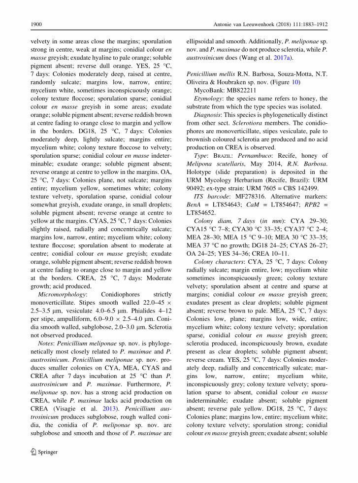
Penicillium mellis R.N. Barbosa, Souza-Motta, N.T.
Oliveira & Houbraken sp. nov. (Figure 10)
MycoBank: MB822211
Etymology: the species name refers to honey, the
substrate from which the type species was isolated.
Diagnosis: This species is phylogenetically distinct
from other sect. Sclerotiora members. The conidio-
phores are monoverticillate, stipes vesiculate, pale to
brownish coloured sclerotia are produced and no acid
production on CREA is observed.
Type: BRAZIL: Pernambuco: Recife, honey of
Melipona scutellaris, May 2014, R.N. Barbosa.
Holotype (slide preparation) is deposited in the
URM Mycology Herbarium (Recife, Brazil): URM
90492; ex-type strain: URM 7605 = CBS 142499.
ITS barcode: MF278316. Alternative markers:
BenA = LT854643; CaM = LT854647; RPB2 =
LT854652.
Colony diam, 7 days (in mm): CYA 29–30;
CYA15 �C 7–8; CYA30 �C 33–35; CYA37 �C 2–4;
MEA 28–30; MEA 15 �C 9–10; MEA 30 �C 33–35;
MEA 37 �C no growth; DG18 24–25; CYAS 26–27;
OA 24–25; YES 34–36; CREA 10–11.
Colony characters: CYA, 25 �C, 7 days: Colony
radially sulcate; margin entire, low; mycelium white
sometimes inconspicuously green; colony texture
velvety; sporulation absent at centre and sparse at
margins; conidial colour en masse greyish green;
exudates present as clear droplets; soluble pigment
absent; reverse brown to pale. MEA, 25 �C, 7 days:
Colonies low, plane; margins low, wide, entire;
mycelium white; colony texture velvety; sporulation
sparse, conidial colour en masse greyish green;
sclerotia produced, inconspicuously brown, exudate
present as clear droplets; soluble pigment absent;
reverse cream. YES, 25 �C, 7 days: Colonies moder-
ately deep, radially and concentrically sulcate; mar-
gins low, narrow, entire; mycelium white,
inconspicuously grey; colony texture velvety; sporu-
lation sparse to absent, conidial colour en masse
indeterminable; exudate absent; soluble pigment
absent; reverse pale yellow. DG18, 25 �C, 7 days:
Colonies plane; margins low, entire; mycelium white;
colony texture velvety; sporulation strong; conidial
colour en masse greyish green; exudate absent; soluble
1900 Antonie van Leeuwenhoek (2018) 111:1883–1912
123
Page 19
Fig. 10 Morphological characters ofPenicilliummellisCBS 142499. aColonies from left to right (top row)MEA, CYA, YES andOA;
(bottom row) CYA reverse, MEA reverse, YES reverse and CREA. b Texture on CYA. c Texture on MEA. d Conidia. e Sclerotia. f–jConidiophores. Scale bars 10 lm
Antonie van Leeuwenhoek (2018) 111:1883–1912 1901
123
Page 20
pigment absent; reverse pale. OA, 25 �C, 7 days:
Colonies flat, margins regular; mycelium white;
colony texture velvety, sporulation dense, conidial
colour en masse greyish; exudate present as clear
droplets; soluble pigment absent; reverse white to
pale. CYAS 25 �C, 7 days: Colonies radially and
concentrically sulcate; margins low, entire; mycelium
white; colony texture velvety; sporulation moderate to
strong; conidial colour en masse greyish green;
exudate absent; soluble pigment absent; reverse
brownish. CREA, 25 �C, 7 days: moderate growth,
no acid production.
Micromorphology: Conidiophores strictly
monoverticillate. Stipes smooth walled, 25–40 9
2.0–3.5 lm, vesicilate 4.0–5.0 lm. Phialides 5–12
per stipe, ampulliform, 6.5–9.0 9 2.0–3.0 lm. Coni-
dia smooth walled, globose to subglobose,
2.0–3.0 lm. Sclerotia present, 150–250 lm.
Additional material examined. Brazil, Pernambuco,
Recife, Inside nest of Melipona scutellaris, R.N.
Barbosa, URM 7611; RB 9; RB 85; RB 110; honey
of Melipona scutellaris, R.N. Barbosa, RB 69.
Notes: Penicillium mellis sp. nov. is phylogeneti-
cally unique. It can be distinguished from other
members in section Sclerotiora by its ability to
produce pale to brownish coloured sclerotia on
MEA, CYA and OA.
Talaromyces brasiliensis R.N. Barbosa, Souza-Motta,
N.T. Oliveira & Houbraken sp. nov. (Figure 11)
MycoBank: MB822214
Etymology: Named after Brazil, the country of
origin of the type strain.
Diagnosis: Talaromyces brasiliensis sp. nov. is
phylogenetically unique. This species grows restricted
on CYA and MEA at 25 �C and growth is absent to
poor at 37 �C. The phialides of T. brasiliensis are
ampulliform and the conidia globose and finely
roughened.
Type: BRAZIL: Pernambuco: Recife, honey of
Melipona scutellaris, June 2014, R.N. Barbosa.
Holotype (slide preparation) is deposited in the
URM Mycology Herbarium (Recife, Brazil): URM
90494; ex-type strains URM 7618 = CBS 142493.
ITS barcode: MF278323. Alternative markers:
BenA = LT855560; CaM = LT855563; RPB2 =
LT855566.
Colony diam, 7 days (in mm): CYA 5–6;
CYA15 �C 3–4; CYA30 �C 5–6; CYA37 �C no
growth; MEA 14–15; MEA 15 �C 6–7; MEA 30 �C14–15; MEA 37 �C 4–5; DG18 10–11; CYAS no
growth; OA 12–13; YES 6–8; CREA no growth.
Colony characters: CYA, 25 �C, 7 days: Colonies
plane; margins entire; mycelium white; colony texture
loosely floccose; sporulation poor; conidia en masse
greyish green; exudates absent; soluble pigments
absent; reverse cream to brownish. MEA, 25 �C,7 days: Colonies plane; margins entire; mycelium
white; colony texture loosely funiculose to floccose;
sporulation strong; conidia en masse greyish; exudates
absent; soluble pigments absent; reverse cream to
yellow. YES, 25 �C, 7 days: Colonies loosely deep;
margins entire; mycelium white; colony texture floc-
cose; sporulation absent; conidia en masse indeter-
minable; exudates absent; soluble pigments absent;
reverse cream to yellow. DG18, 25 �C, 7 days:
Colonies raised at centre; margins entire, deep;
mycelium white, occasionally inconspicuously grey;
colony texture floccose; sporulation poor at centre,
conidia en masse greyish; exudates absent; soluble
pigments absent; reverse brown to pale. OA, 25 �C,7 days: Colonies plane; margins entire; mycelium
white, occasionally light yellow; colony texture vel-
vety; sporulation strong at centre, week at margin;
conidia en masse dull green; exudates present as small
hyaline droplets; soluble pigments absent; reverse
white to inconspicuously black. CREA 25 �C, 7 days:
no growth.
Micromorphology: Conidiophores biverticillate,
stipes smooth walled, 20–50 9 2.5–4 lm. Metulae
5–6, 8–11 9 2.5–3.5 lm. Phialides 3–4 per stipe,
ampulliform tapering to very fine necks, 7–11
(–14) 9 2.0–3 lm; conidia globose, finely rough-
ened, 2–3 lm. Ascomata not observed.
Additional material examined. Brazil, Pernambuco,
Recife, Inside nest of Melipona scutellaris, R.N.
Barbosa, URM 7619; URM 7620.
Notes: Section Trachyspermi comprise species that
normally grow slowly on CYA and slightly faster on
MEA. Talaromyces brasiliensis sp. nov. also grows
restricted on CYA (5–6 mm) and better on MEA
(14–15 mm), confirming the phylogenetic results.
Talaromyces brasiliensis sp. nov. is phylogenetically
distinct (Fig. 6).
Talaromyces mycothecae R.N. Barbosa, Souza-Motta,
N.T. Oliveira & Houbraken sp. nov. (Figure 12)
MycoBank: MB822215
1902 Antonie van Leeuwenhoek (2018) 111:1883–1912
123
Page 21
Fig. 11 Morphological characters of Talaromyces brasiliensis CBS 142493. a Colonies from left to right (top row) MEA, CYA, YES
and OA; (bottom row) CYA reverse, MEA reverse, YES reverse and CREA. b Texture on CYA. c Texture on MEA. d Conidia. e–hConidiophores. Scale bars 10 lm
Antonie van Leeuwenhoek (2018) 111:1883–1912 1903
123
Page 22
Fig. 12 Morphological characters of Talaromyces mycothecae CBS 142494. a Colonies from left to right (top row) MEA, CYA, YES
and OA; (bottom row) CYA reverse, MEA reverse, YES reverse and CREA. b Texture on CYA. c Texture on MEA. d Conidia. e–hConidiophores. Scale bars 10 lm
1904 Antonie van Leeuwenhoek (2018) 111:1883–1912
123
Page 23
Etymology: In honour of Micoteca URM (URM,
University Recife Mycology), an important Latin-
American Fungal Culture Collection founded by
mycologist Augusto Chaves Batista.
Diagnosis: The reverse colour on MEA and OA is
wine red. The species produces red coloured exudate
droplets on YES and no acid compounds are produced
on CREA. Furthermore, T. mycothecae sp. nov. grows
well on CYA 37 �C and produces smooth walled,
fusiform to ellipsoidal shaped conidia.
Type: BRAZIL: Pernambuco: Recife, inside nests of
Melipona scutellaris, Feb 2014, R.N. Barbosa. Holo-
type (slide preparation) is deposited in the URM
Mycology Herbarium (Recife, Brazil): URM 90495;
ex-type strains URM 7622 = CBS 142494.
ITS barcode: MF278326. Alternative markers:
BenA = LT855561; CaM = LT855564; RPB2 =
LT855567.
Colony diam, 7 days (in mm): CYA 20–23;
CYA15 �C 2–5; CYA 30 �C 28–30; CYA 37 �C18–20; MEA 29–30; MEA 15 �C 3–6; MEA 30 �C38–40; MEA 37 �C 20–22; DG18 10–12; CYAS no
growth; OA 24–25; YES 25–26; CREA 4–5.
Colony characters: CYA, 25 �C, 7 days: Colonies
plane, margins entire; mycelium white occasionally
inconspicuously yellow; colony texture velvety to
floccose; sporulation strong, conidia en masse greyish
to dull green; exudates present as small clear droplets;
soluble pigments absent; reverse yellow amber to dark
brown at centre. MEA, 25 �C, 7 days: Colonies plane;
margin entire, mycelium white; colony texture vel-
vety; sporulation strong; conidia en masse greyish;
exudates absent; soluble pigments absent; reverse
yellow amber to wine-reddish. YES, 25 �C, 7 days:
Colonies crateriform; margins entire; mycelium
white; colony texture floccose; sporulation strong;
conidia en masse greyish to dull green; exudates
present as small red droplets; soluble pigments absent;
reverse red near margins to wine-reddish in centre.
DG18, 25 �C, 7 days: Colonies plane; margins entire;
mycelium white; colony texture floccose; sporulation
sparse; conidia en masse green; exudates present as
small red droplets; soluble pigments absent; reverse
cream at margins to reddish at centre. OA, 25 �C,7 days: Colonies plane; margins low; mycelium white
occasionally inconspicuously greenish; colony texture
velvety to granular; sporulation abundant, conidia en
masse dull green; exudates present as small clear
droplets; soluble pigments absent; reverse reddish.
CREA 25 �C, 7 days: Very weak growth, acid
production absent.
Micromorphology: Conidiophores biverticillate;
stipes smooth, 55–105 9 2–3 lm; metulae 3–4,
11.5–15.5 9 2.5–4 lm. Phialides 3–5 per stipe,
acerose, 9.5–12.5 9 2.5–3.5 lm. Conidia smooth,
fusiform to ellipsoidal, 2.5–4 9 3–3.5 lm. Ascomata
not observed.
Additional material examined. Brazil, Pernambuco,
Recife, isolated from inside nest of Melipona scutel-
laris, R.N. Barbosa, URM 7623; RB 95; RB 171.
Notes: Altough the relationship if Talaromyces
mycothecae sp. nov. is difficult to determine, the
species seems to be phylogenetically most closely
related to T. neofusisporus, T. stollii, T. amestolkiae
and T. ruber. Talaromyces neofusisporus produces
synnemata on CYA and YES, and grows poorly at 37
�C (2–3 mm, CYA, 7 days) (Wang et al. 2016). In
contrast, no synnemata and good growth at 37 �C(18–20 mm, CYA, 7 days) is observed for T. mycothe-
cae. Yilmaz et al. (2012) used various characters, such
as the ability to grow at 37 �C, the colony texture on
MEA and CYA and the production of acid compounds
on CREA to differentiate T. amestolkiae, T. ruber and
T. stollii. No acid is produced on CREA by T.
mycothecae and this is shared with T. ruber (T.
amestolkiae and T. stollii are poor acid producers).
Talaromyces mycothecae sp. nov. attains a diameter of
18–20 mm after 7 days on CYA at 37 �C and this is
faster than T. amestolkiae (8–15 mm) and T. ruber
(14–18 mm), but slower than T. stollii (25–35 mm)
(Yilmaz et al. 2014). Based on the data above, T.
mycothecae phenotypically resembles T. ruber. The
characteristic yellow and red pigmented mycelium on
YES of T. ruber is not observed in the T. mycothecae
sp. nov. cultures.
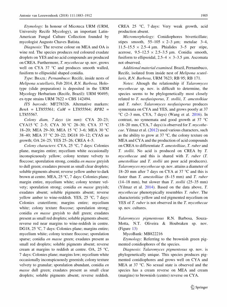
Talaromyces pigmentosus R.N. Barbosa, Souza-
Motta, N.T. Oliveira & Houbraken sp. nov.
(Figure 13)
MycoBank: MB822216
Etymology: Referring to the brownish green pig-
mented conidiophores of the species.
Diagnosis: Talaromyces pigmentosus sp. nov. is
phylogenetically unique. This species produces pig-
mented conidiophores and grows well on CYA and
MEA at 37 �C. No sexual state is observed and the
species has a cream reverse on MEA and cream
(margins) to brownish (centre) reverse on CYA.
Antonie van Leeuwenhoek (2018) 111:1883–1912 1905
123
Page 24
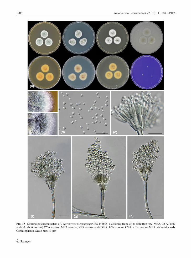
Fig. 13 Morphological characters of Talaromyces pigmentosus CBS 142805. a Colonies from left to right (top row) MEA, CYA, YES
and OA; (bottom row) CYA reverse, MEA reverse, YES reverse and CREA. b Texture on CYA. c Texture on MEA. d Conidia. e–hConidiophores. Scale bars 10 lm
1906 Antonie van Leeuwenhoek (2018) 111:1883–1912
123
Page 25
Type: BRAZIL: Pernambuco: Recife, inside nests of
Melipona scutellaris, June 2014, R.N. Barbosa.
Holotype (slide preparation) is deposited in the
URM Mycology Herbarium (Recife, Brazil): URM
90496; ex-type strains URM 7624 = CBS 142805.
ITS barcode: MF278330. Alternative markers:
BenA = LT855562; CaM = LT855565; RPB2 =
LT855568.
Colony diam, 7 days (in mm): CYA 23–24; CYA
15 �C 5–7; CYA 30 �C 34–35; CYA 37 �C 35–36;
MEA 23–24; MEA 15 �C 6–8; MEA 30 �C 34–35;
MEA 37 �C 33–34; DG18 7–9; CYAS 2–3; OA
24–25; YES 23–24; CREA 2–4.
Colony characters: CYA, 25 �C, 7 days: Colonies
moderately deep; margins entire; mycelium white
sometimes inconspicuously green; colony texture
velvety; sporulation absent; conidial colour en masse
cannot be determinate; exudates absent; soluble pig-
ments absent; reverse white to cream at margins to
brownish in centre. MEA, 25 �C, 7 days: Colonies
moderately deep, sunken at centre; margin entire;
mycelium white; colony texture velvety; sporulation
sparse; conidia en masse greyish; exudates present as
small hyaline droplets; soluble pigments absent;
reverse cream. YES, 25 �C, 7 days: Colonies moder-
ately deep, sunken, raised at centre; margins entire;
mycelium white; colony texture floccose; sporulation
sparse to absent, conidia en masse greyish; soluble
pigments absent; exudates absent; reverse cream to
yellow. DG18, 25 �C, 7 days: Colonies raised at
centre; margins low, plane; mycelium white; colony
texture floccose; sporulation absent, conidia en masse
indeterminable; exudates absent; soluble pigments
absent; reverse brown at centre, light cream to white at
margin. OA, 25 �C, 7 days: Colonies low, plane;
margins low, plane; mycelium white; colony texture
velvety; sporulation absent; conidia en masse indeter-
minable; exudates absent; soluble pigments absent;
reverse light cream. CYAS, 25 �C, 7 days: Colonies
low, plane; margins low, plane; mycelium white;
colony texture velvety; sporulation absent; conidia en
masse indeterminable; exudates absent; soluble pig-
ments absent; reverse white. CREA 25 �C, 7 days:
Very weak growth, acid production absent.
Micromorphology: Conidiophores biverticillate,
brownish green pigmented. Stipes smooth walled,
17–65 9 2–4 lm. Metulae 3–4, divergent,
7–11 9 2–2.5 lm. Phialides, 3–6 per stipe, acerose,
7–11 9 2–3 lm; conidia smooth walled, globose to
subglobose, 2.0 9 3.0 lm. Ascomata not observed.
Additional material examined. Brazil, Pernambuco,
Recife, from bee pollen of Melipona scutellaris, R.N.
Barbosa, URM 7625; RB 96, RB 171; from inside nest
of Melipona scutellaris RB 30; RB 100.
Notes: Talaromyces pigmentosus sp. nov. is phylo-
genetically closely related to T. helicus, T. boninensis
and T. reverso-olivaceus and shares the ability to grow
well on CYA incubated at 37 �C (10–34 mm, 7 days).
Talaromyces boninensis and T. helicus produce a
sexual state and this is not observed T. reverso-
olivaceus and T. pigmentosus sp. nov.. The new
species can also be differentiated from T. reverso-
olivaceus by its ability to produce brownish green
pigmented stipes. The production of pigmented stipes
is also shared with the phylogenetically more distant
species T. varians (also in sect. Helici) and T.
ptychoconidium (sect. Purpurei) (Yilmaz et al. 2014).
Discussion
Penicillium and Talaromyces species are well-known
cosmopolitan filamentous fungi that play various roles
in natural ecosystems, agriculture and biotechnology.
Both genera have a sectional infrageneric classifica-
tion system. Currently, 26 sections are accepted in
Penicillium (Houbraken and Samson 2011; Houbra-
ken et al. 2016b) and seven in Talaromyces (Yilmaz
et al. 2014). A current monograph on Penicillium is
lacking, but many Penicillium sections are studied
using a polyphasic approach or multigene phylogenies
(e.g. Houbraken et al. 2014; Peterson et al. 2015;
Visagie et al. 2015; Houbraken et al. 2016b) and all
Talaromyces sections were treated in detail in the
monograph of Yilmaz et al. (2014). ITS and BenA
sequences are proposed identification markers for
Penicillium and Talaromyces and often are generated
in taxonomic studies. In many of those studies it is
shown that ITS sequencing is insufficient for Penicil-
lium identifications as closely related species often
share similar or identical sequences. On the other
hand, this locus works relatively well to assign species
to sections. Besides ITS sequences, we also generated
BenA sequences of all isolated Penicillium and
Talaromyces species. The BenA sequences are used
for identification of Penicillium and Talaromyces
species, as this is the recommended identification
Antonie van Leeuwenhoek (2018) 111:1883–1912 1907
123
Page 26
marker (Visagie et al. 2014; Yilmaz et al. 2014). With
exception of the new species, all other isolates
obtained during our survey could be reliably identified
using BenA sequences.
Among the Penicillia, section Sclerotiora (46%)
isolates were most frequently detected during this
study. Penicillium brocae (27%) was predominantly
present among the isolates belonging to section
Sclerotiora, followed by P. mellis sp. nov. (7%), P.
sclerotiorum (5%), P. mallochii (4%), P. sanshaense
(1%), P. fernandesiae sp. nov. (1%) and P. meliponae
sp. nov. (1%). Section Sclerotoria species generally
produce monoverticillate conidiophores and excep-
tions are the biverticillate conidiophores observed in
P. choerospondiatis, P. herquei, P. malachiteum, P.
sanshaense and P. verrucisporum. They also have
bright yellow or orange pigments, which may occur in
the mycelium, sclerotia, ascocarps, soluble pigments
and/or colony reverse pigmentation (Houbraken and
Samson 2011; Rivera and Seifert 2011; Visagie et al.
2013). The species isolated during our survey also
produced monoverticillate conidiophores, bright
coloured colonies, sclerotia and/or mycelium, and
none of the strains produced a sexual state. Interest-
ingly, P. brocae was the most predominant Penicil-
lium present in our study. This species was originally
described from coffee berry borers (galleries, cuticle,
feces and guts) in Mexico (Peterson et al. 2003), and
more recently detected in faeces of another, unrelated
beetle, Eufallia sp. (Wang and Chan 2015). Along
with the description of P. brocae, Peterson et al.
(2003) suggested that this species produces exogenous
sterols necessary for the coffee berry borer’s devel-
opment and thus is mutualistically associated with the
insect. Similar to other insects, bees are unable to
synthesize sterols and, thus, exogenous sterol is
required (Ferreira-Caliman et al. 2012). A recent
study showed that essential steroid precursors, needed
for the development of Scaptotrigona depilis bees, are
proved by a Zygosaccharomyces species (Paludo et al.
2018). Whether a symbiosis exists between steroids
produced by Penicillium and Talaromyces species and
Melipona scutellaris bees needs to be further inves-
tigated. Also P. mallochii and P. guanacastense, two
species related to P. brocae, are associated with guts
and faeces of leaf-eating caterpillars (Rivera et al.
2012) suggesting an association of other section
Sclerotiora members with insects as well.
Isolates belonging to section Citrina made up for
30% of all Penicillia. Penicillium citrinum was most
frequently detected (20%), followed by P. steckii
(5%), P. shaerii (4%), P. paxilli (2%), and P.
sumatrense (1%). These species are characterised by
symmetrically biverticillate conidiophores, flask
shaped phialides, small-sized conidia, and some
species like P. shaerii produce greyish brown cleis-
tothecia. These species are common in soils and have a
preference for (sub)tropical soils. It’s unknown
whether these species are saprotrophs or if they are
associated with stingless bees. Section Citrina species
produce various bioactive extrolites (mycotoxins,
antibiotics) such as citrinin, curvularin, paspaline,
paspalinine and paxillin (Houbraken et al. 2011a). The
presence of these extrolites in honey and pollen
samples wasn’t subject of this study; however, if
present, they can affect the quality of the honey and
pollen. How insects cope with mycotoxins has rarely
been investigated (Glinski and Jarosz 2000; Traniello
et al. 2001; Keller et al. 2014).
Another group of isolates obtained in this study
belong to Lanata-Divaricata, a section re-established
by Houbraken and Samson (2011). Most of the section
Lanata-Divaricata species grow rapidly in culture.
The conidiophores of these species are often strongly
divaricate and have metulae that are born terminally,
subterminally and in intercalary positions. Useful
characters for identification are the shape and orna-
mentation of the conidia, growth on CYA incubated at
37 �C, colony diameters and morphology (e.g. reverse
colours on CYA, YES), and growth rate on CREA.
Three Lanata-Divaricata species were isolated: P.
singorense, P. wotroi and P. echinulonalgiovense.
One isolate (RB 115) represents a novel species in
sect. Lanata-Divaricata, and this species will be
described elsewhere (Y-Z Diao et al., in progress).
Penicillium singorense and P. wotroi are known
species and the former was originally described from
house dust in Thailand. Recent collections show that
P. singorense has a worldwide distribution and this
species is isolated in USA (Florida), Korea and China
(J. Houbraken, pers. observations). The distribution of
P. wotroi seems to be restricted to South-America
(Brazil, Argentina) (Houbraken et al. 2011b). The
name P. echinulonalgiovense was invalidly published
because it was described without a Latin description or
diagnosis (Art. 39.1.; Melbourne Code). In subsequent
treatments using morphological characters, this
1908 Antonie van Leeuwenhoek (2018) 111:1883–1912
123
Page 27
species was placed in synonymy with P. janthinellum
(Smith 1963) and/or P. simplicissimum (Pitt 1979;
Stolk and Samson 1983). However, molecular data
shows that this species is distinct (Houbraken et al.
2011b) and we therefore reinstate this species as
distinct. A search in the DTO and CBS collection
shows that this species has a worldwide distribution (J.
Houbraken, unpubl. results). Section Lanata-Divari-
cata species are usually found in soil and (decaying)
leaves (Houbraken et al. 2011b), but various other
substrates are listed in literature. These species
probably have a broad ecological niche. For example
P. excelsum was isolated from bees and ants, but also
from other substrates such as flowers, leaves, Brazil
nut kernels and shells (Taniwaki et al. 2015).
Insects are adapted to different ecosystems and
have symbiotic and/or pathogenic associations with
fungi and other microorganisms (Bode 2011; Mello
et al. 2016). Studies investigating the fungi associated
with bees (sensu lato) revealed that some fungi are
common saprophytes in the environment of the
beehive. Bees collect plant pollen and nectar from
different kinds of plants and inevitably, also fungal
fragments will be introduced to the bee pollen (e.g.
Eltz et al. 2002; Barbosa et al. 2017; Paludo et al.
2018). The nutritional value of a fungal spore is lower
than that of pollen; however, it is speculated that
fungal spores can serve as a complement to the bee
diet since the availability is high and harvest is
relatively easy (Oliveira and Morato 2000; Eltz et al.
2002). Only a limited number of reports about fungi
associated with stingless bees are found in literature,
and the reports related to Penicillium and Talaromyces
are even rarer (Ferraz et al. 2008). According to
Pandey et al. (1983), pollen grains can secret sub-
stances that inhibit microbial spore germination. After
collection, the pollen grains are processed by bees into
bee bread and this product normally has low water
activity. Many different fungal species can be found in
honey, but these are probably latently present and will
not grow due to the low water activity of the product.
The natural introduction of fungi in the bee environ-
ment most likely occurs in the period between
collection of the plant pollen, formation of bee pollen
and drying and storage of the pollen in the nests.
An important characteristic of Penicillium and
Talaromyces species is the production of a diverse
range of bioactive extrolites (Nielsen et al. 2017). The
extrolite production of the species that were isolated
during this study was investigated. The (combination
of) detected extrolites can potentially play a role in the
interaction between different organisms (Frisvad
2008). Our results demonstrate that the detected
species are able to produce several extrolites, includ-
ing the mycotoxin citrinin. The occurrence of myco-
toxins and mycotoxigenic fungi has been recorded in
bee pollen (sensu lato) around the world (e.g.
Gonzalez et al. 2005; Kacaniova et al. 2011;
Rodrıguez-Carrasco et al. 2013). Logically, the pres-
ence of this mycotoxins (e.g. citrinin) is unwanted and
can negatively affect the quality of the bee pollen for
human consumption. Extrolite function depends on
their ecological interaction. Insects are well adapted to
feed on plants that contain a broad spectrum of
(chemical) compounds (Dowd 1992). They have a
long evolutionary history of interacting with fungi and
it is known that some insects can use fungi as feed
(Dowd 1992). Often, mycotoxins co-occur with other
fungal extrolites for which no function is known.
Following the analogy with the situation in higher
plants, it is possible that these co-occurring fungal
extrolites can synergize (or antagonize) the toxicity of
co-occurring mycotoxins (Dowd 1992). In nature,
fungal metabolites can provide various fitness advan-
tages ranging from protection to competition with
other microbes for niche securement (Rohlfs and
Churchill 2011). Penicillium apimei sp. nov. and
Monascus are both isolated from stingless bees and are
both producers of compounds belonging to the geodin
biosynthetic family (Barbosa et al. 2017). It is
tempting to speculate that this group of metabolites
might have particular function in the bee habitat.
Various extrolites could not be identified in our
experimental conditions and could represent novel
bioactive compounds. During this study, the quality of
the nests and the health of the bees was followed over a
long time and no disease was observed. If any of these
(novel) compounds were secreted in the bee pollen or
nests, then these compounds probably didn’t have an
(large) effect on the bees health.
Data on the functional relationship between fungi
and stingless bees are scarce. Further studies on fungi
from honey, inside nests, bee body, stored pollen and
native plants are needed to understand the relation-
ships between these organisms in tropical ecosystems,
and the benefits that such fungi can possibly confer on
their hosts. This study could serve as the first step for
more detailed studies on ecological interactions
Antonie van Leeuwenhoek (2018) 111:1883–1912 1909
123
Page 28
between stingless bees, fungi and their bioactive
extrolites.
Acknowledgements Wewould like to thank National Council
for Scientific and Technological Development (CNPq) (Process
201478/2015-3—SWE) and Coordination for the Improvement
of Higher Education Personnel (CAPES) for financial support
and scholarship for Renan N. Barbosa and Associacao
Pernambucana de Apicultores e Meliponicultores (APIME) is
thanked for their help in collecting the honey/pollen samples.
We would like to acknowledge the URM Culture Collection
staff; Martin Meijer and Bart Kraak from the Westerdijk Fungal
Biodiversity Institute for their support and Konstanze Bench for
nomenclatural assistance. Renan N. Barbosa received
scholarship and financial support from the National Council
for Scientific and Technological Development (CNPq) (Process
201478/2015-3—SWE) and the Coordination for the
Improvement of Higher Education Personnel (CAPES).
Conflict of interest All authors declare that they have no
conflict of interest.
Open Access This article is distributed under the terms of the
Creative Commons Attribution 4.0 International License (http://
creativecommons.org/licenses/by/4.0/), which permits unre-
stricted use, distribution, and reproduction in any medium,
provided you give appropriate credit to the original
author(s) and the source, provide a link to the Creative Com-
mons license, and indicate if changes were made.
References
Almeida-Muradian LB, Stramm KM, Horita A et al (2013)
Comparative study of the physicochemical and palyno-
logical characteristics of honey from Melipona subnitida
and Apis mellifera. IFST 48:1698–1706
Barbosa RN, Bezerra JDP, Souza-Motta CM, Gomes BS, Costa
CMC, Melo HF (2016) Prospection on yeasts from sting-
less bees honey in Brazilian Tropical Dry Forest (Caa-
tinga). Gaia Scientia 10:151–159
Barbosa RN, Leong S-L, Vinnere-Pettersson O et al (2017)
Phylogenetic analysis of Monascus and new species from
honey, pollen and nests of stingless bees. Stud Mycol
86:29–51
Bode HB (2011) Insect-associated microorganisms as a source
for novel secondary metabolites with therapeutic potential.
In: Vilcinskas A (ed) Insect Biotechnology, vol 2. Springer,
Netherlands, pp 77–93
Brown JC, Oliveira ML (2014) The impact of agricultural col-
onization and deforestation on stingless bee (Apidae:
Meliponini) composition and richness in Rondnia, Brazil.
Apidologie 42:172–188
Chen AJ, Sun BD, Houbraken J et al (2016) New Talaromyces
species from indoor environments in China. Stud Mycol
84:119–144
Cortopassi-Laurino M, Imperatriz-Fonseca VL, Roubik DW
et al (2006) Global meliponiculture: challenges and
opportunities. Apidologie 37:275–292
Cortopassi-Laurino M, Velthuis HHW, Nogueira-Neto P (2007)
Diversity of stingless bees from the Amazon forest in
Xapuri (Acre), Brazil. Exp Appl Entomol 18:105–114
Daniel HM, Sao Thiago-Calaca PS, Rosa CA et al (2013)
Starmerella neotropicalis f.a., sp. nov., a new yeast species
found in bees and pollen from Brazil and Cuba. Int J Syst
Evol Microbiol 63:3896–3903
Dowd PF (1992) Insect interactions with mycotoxin-producing
fungi and their hosts. In: Bhatnagar D, Lillehoj EB, Arora
DK (eds) Handbook of Applied Mycology, vol 5. Myco-
toxins in Ecological Systems. Marcel Dekker, New York,
pp 137–155
Egorova AI (1971) Preservative microflora in stored pollen.
Veterinariya 8:40–41
Eltz T, Bruhl CA, Gorke C (2002) Collection of mold (Rhizopus
sp.) spores in lieu of pollen by the stingless bee Trigona
collina. J Trop Ecol 12:441–445
Ferraz RE, Lima PM, Pereira DS, Freitas CCO, Feijo FMC
(2008) Microbiota fungica de Melipona subnitida Ducke
(Hymenoptera: Apidae). Neotrop Entomol 37:345–346
Ferreira-Caliman F, Silva CI, Mateus S, Zucchi R, Nascimento
FS (2012) Neutral sterols of cephalic glands of stingless
bees and their correlation with sterols from pollen. Psyche
2012:ID 982802. https://doi.org/10.1155/2012/982802
Foley K, Fazio G, Jensen AB, Hughes WO (2014) Nutritional
limitation and resistance to opportunistic Aspergillus par-
asites in honey bee larvae. J Invertebr Pathol 111:68–73
Frisvad JC (2008) Fungi in cold ecosystems. In: Margesin R,
Schinner F, Marx JC, Gerday C (eds) Psychrophiles: from
biodiversity to biotechnology. Springer-Verlag, Berlin
Heidelberg
Gilliam M, Vandenberg JD (1988) Fungi. In: Morse RA,
Nowogrodzki R (eds) Honey Bee Pests, Predators, and
Diseases. Comell University Press, Ithaca, NY
Gilliam M, Prest DB, Lorenz BJ (1989) Microbiology of pollen
and bee bread: taxonomy and enzymology of molds. Api-
dologie 20:53–68
Glinski Z, Jarosz J (2000) The honeybee defense in mycotic
deseases. Honeybee Sci 21:69–70
Gois GC, Carneiro GG, Rodrigues AE et al (2010) Qualidade
microbiologica do mel de abelhas Melipona scutellaris.
PUBVET 4: 769
Gonzalez G, Hinojo MJ, Mateo R, Medina A, JimenezM (2005)
Occurrence of mycotoxin producing fungi in bee pollen. Int
J Food Microbiol 105:1–9
Guevara-Suarez M, Sutton DA, Gene J (2017) Four new species
of Talaromyces from clinical sources. Mycoses. https://doi.
org/10.1111/myc.12640
Houbraken J, Samson RA (2011) Phylogeny of Penicillium and
the segregation of Trichocomaceae into three families.
Stud Mycol 70:1–51
Houbraken J, Frisvad JC, Samson RA (2011a) Taxonomy of
Penicillium section Citrina. Stud Mycol 70:53–138
Houbraken J, Lopez-Quintero CA, Frisvad JC et al (2011b)
Penicillium araracuarense sp. nov., Penicillium elleniae
sp. nov., Penicillium penarojense sp. nov., Penicillium
vanderhammenii sp. nov. and Penicillium wotroi sp. nov.,
1910 Antonie van Leeuwenhoek (2018) 111:1883–1912
123
Page 29
isolated from leaf litter. Int J Syst Evol Microbiol
61:1462–1475
Houbraken J, Spierenburg H, Frisvad JC (2012) Rasamsonia, a
new genus comprising thermotolerant and thermophilic
Talaromyces and Geosmithia species. Antonie Van
Leeuwenhoek 101:403–421
Houbraken J, Visagie CM, Meijer M et al (2014) A taxonomic
and phylogenetic revision of Penicillium section Asper-
gilloides. Stud Mycol 78:373–451
Houbraken J, Samson RA, Yilmaz N (2016a) Taxonomy of
Aspergillus, Penicillium and Talaromyces and its signifi-
cance for biotechnology. In: de Vries RP, Gelber IB,
Andersen MR (eds) Aspergillus and Penicillium in the
Post-Genomic Era. Caister Academic Press, Norfolk,
pp 1–15
Houbraken J, Wang L, Lee HB, Frisvad JC (2016b) New sec-
tions in Penicillium containing novel species producing
patulin, pyripyropens or other bioactive compounds. Per-
soonia 36:299–314
Kacaniova M, Juracek M, Chlebo R et al (2011) Mycobiota and
mycotoxins in bee pollen collected from different areas of
Slovakia. J Environ Sci Health B 46:623–629
Katoh K, Standley DM (2013) MAFFT multiple sequence
alignment software version 7: improvements in perfor-
mance and usability. Mol Biol Evol 30:772–780
Keller KM, Deveza MV, Koshiyama AS et al (2014) Fungi
infection in honeybee hives in regions affected by Brazilian
sac brood. Arq Bras Med Vet Zootec 66:1471–1478
Kerr WE (1996) Tiuba: a Abelha do Maranhao. EDUFMA, Sao
Luıs
Klitgaard A, Iversen A, Andersen MR (2014) Aggressive
dereplication using UHPLC-DAD-QTOF—screening
extracts for up to 3000 fungal secondary metabolites. Anal
Bioanal Chem 406:1933–1943
Kobayashi C, Fukasawa Y, Hirose D et al (2008) Contribution of
symbiotic mycangial fungi to larval nutrition of a leaf-
rolling weevil. Evol Ecol 22:711–722
Laich F, Andrade J (2016) Penicillium pedernalense sp. nov.,
isolated from whiteleg shrimp heads waste compost. Int J
Syst Evol Microbiol 66:4382–4388
Li X, Guo W, Ding J (2012) Mycangial fungus benefits the
development of a leaf-rolling weevil, Euops chinesis. J In-
sect Physiol 58:867–873
Lopes LQS, Quatrin PM, De SouzaME, DeAlmeida Vaucher R,
Vianna Santos RC (2015) Fungal infections in honey bees.
Fungal Genomics Biol 4:118. https://doi.org/10.4172/
2165-8056.1000118
Luo Y, Lu X, Bi W, Liu F, Gao W (2016) Talaromyces rubri-
faciens, a new species discovered from heating, ventilation
and air conditioning systems in China. Mycologia
108:773–779
Maddison WP, Maddison DR (2016) Mesquite: a modular
system for evolutionary analysis, version 3.11. http://
mesquiteproject.org
McNeill J, Barrie FF, Buck WR (eds) (2012) International Code
of Nomenclature for algae, fungi, and plants (Melbourne
Code) Koeltz Scientific Books, Konigstein (Regnum veg-
etabile no. 154)
Mello AP, Correa EB, Barbosa-Silva AM, Bezerra-GusmaoMA
(2016) Fungi associated with nests of Nasutitermes
corniger (Motschulsky) (Isoptera: Nasutitermitinae) in a
semiarid region of Brazil. Entomotropica 31:302–310
Menezes C, Vollet-Neto A, Marsaioli AJ et al (2015) A
Brazilian social bee must cultivate fungus to survive. Curr
Biol 25:2851–2855
Miller MA, Pfeiffer W, Schwartz T (2010) Creating the CIPRES
Science Gateway for inference of large phylogenetic trees.
In: IEEE Gateway Computing Environments workshop
(GCE), pp 1–8
Nielsen KF, Mansson M, Rank C (2011) Dereplication of
microbial natural products by LC-DAD-TOFMS. J Nat
Prod 74:2338–2348
Nielsen JC, Grijseels S, Prigent S et al (2017) Global analysis of
biosynthetic gene clusters reveals vast potential of sec-
ondary metabolite production in Penicillium species. Nat
Microbiol 2:17044. https://doi.org/10.1038/nmicrobiol.
2017.44
Oliveira ML, Morato EF (2000) Stingless bees (Hymenoptera,
Meliponini) feeding on stinkhorn spores (Fungi, Phallales):
robbery or dispersal? Rev Bras Zool 17:881–884
Paludo CR, Menezes C, Silva-Junior EA et al (2018) Stingless
bee larvae require fungal steroid to pupate. Sci Rep 8:1122.
https://doi.org/10.1038/s41598-018-19583-9
Pandey DK, Tripathi PM, Tripathi RD, Dixit SN (1983) Fun-
gitoxicity in pollen grains. Grana 22:31–32
Peterson SW, Perez J, Vega F et al (2003) Penicillium brocae, a
new species associated with the coffee berry borer in
Chiapas, Mexico. Mycologia 95:141–147
Peterson SW, Jurjevic Z, Frisvad JC (2015) Expanding the
species and chemical diversity of Penicillium section
Cinnamopurpurea. PLoS ONE 10:e0121987. https://doi.
org/10.1371/journal.pone.0121987
Pianaro A, Flach A, Patricio EFLRA, Nogueira-Neto P, Mar-
saioli AJ (2007) Chemical Changes Associated with the
invasion of a Melipona scutellaris colony by Melipona
rufiventris workers. J Chem Ecol 33:971–984
Pitt JI (1979) The Genus Penicillium and its teleomorphic states
Eupenicillium and Talaromyces. Academic Press, London
Posada D (2008) jModelTest: phylogenetic model averaging.
Mol Biol Evol 25:1253–1256
Rambaut A (2009) FigTree v.1.3.1. Computer program and
documentation distributed by the author. http://tree.bio.ed.
ac.uk/software/
Ramırez SR, Nieh JC, Quental TB et al (2010) A molecular
phylogeny of the stingless bee genus Melipona (Hy-
menoptera: Apidae). Mol Phylogenet Evol 56:519–525
Rao PV, Krishnan KT, Salleh N, Gan SH (2016) Biological and
therapeutic effects of honey produced by honey bees and
stingless bees: a comparative review. Rev Bras Farmacogn
26:657–664
Rivera KG, Seifert KA (2011) A taxonomic and phylogenetic
revision of the Penicillium sclerotiorum complex. Stud
Mycol 70:139–158
Rivera KG, Dıaz J, Chavarrıa-Dıaz F et al (2012) Penicillium
mallochii and P. guanacastense, two new species isolated
from Costa Rican caterpillars. Mycotaxon 119:315–328
Rodrıguez-Carrasco Y, Font G, Manes J, Berrada H (2013)
Determination of mycotoxins in bee pollen by gas chro-
matography-tandem mass spectrometry. J Agric Food
Chem 61:1999–2005
Antonie van Leeuwenhoek (2018) 111:1883–1912 1911
123
Page 30
RohlfsM, Churchill AC (2011) Fungal secondary metabolites as
modulators of interactions with insects and other arthro-
pods. Fungal Genet Biol 48:23–34
Romero SM, Romero AI, Barrera V, Comerio R (2016) Talar-
omyces systylus, a new synnematous species from Argen-
tinean semi-arid soil. Nova Hedwigia 102:24–256
Rong C, Yuanwei M, Shouxian W et al (2016) Penicillium
chroogomphum, a new species in Penicillium section
Ramosa isolated from fruiting bodies of Chroogomphus
rutilus in China. Mycoscience 57:79–84
Ronquist F, Teslenko M, van der Mark P et al (2012) MrBayes
3.2: efficient bayesian phylogenetic inference and model
choice across a large model space. Syst Biol 61:539–542
Rosa CA, Lachance M-A, Silva JOC et al (2003) Yeast com-
munities associated with stingless bees. FEMS Yeast Res
4:271–275
Samson RA, Houbraken J, Thrane U et al (2010) Food and
indoor fungi. CBS Laboratory Manual Series 2. Centraal-
bureau voor Schimmelcultures, Utrecht, The Netherlands
Samson RA, Yilmaz N, Houbraken J et al (2011) Phylogeny and
nomenclature of the genus Talaromyces and taxa accom-
modated in Penicillium subgenus Biverticillium. Stud
Mycol 70:159–183
Sarwar M (2016) Challenges due to bacterial infections of the
honey bees and contributions to manage pest problems.
IJER 1:4–10
Shoreit MN, Bagy HM (1995) Mycoflora associated with
stonebrood disease in honey bee colonies in Egypt.
Microbiol Res 150:207–211
Silva TMS, Santos FP, Evangelista-Rodrigues A et al (2013)
Phenolic compounds, melissopalynological, physico-
chemical analysis and antioxidant activity of jandaıra
(Melipona subnitida) honey. J Food Compos Anal
29:10–18
Smedsgaard J (1997) Micro-scale extraction procedure for
standardized screening of fungal metabolite production in
cultures. J Chromatogr A 760:264–270
Stamatakis A (2014) RAxML Version 8: a tool for Phylogenetic
Analysis and Post-Analysis of Large Phylogenies. Bioin-
formatics 30:1312–1313
Stolk AC, Samson RA (1983) The ascomycete genus Eupeni-
cillium and related Penicillium anamorphs. Stud Mycol
23:1–149
Tamura K, Stecher G, Peterson D et al (2013) MEGA6:
Molecular Evolutionary Genetics Analysis version 6.0.
Mol Biol Evol 30:2725–2729
Taniwaki MH, Pitt JI, Iamanaka BT et al (2015) Penicillium
excelsum sp. nov. from the Brazil nut tree ecosystem in the
Amazon basin. PLoS ONE 10:e0143189. https://doi.org/
10.1371/journal.pone.0143189
Teixeira ACP, Marini MM, Nicoli JR et al (2003) Starmerella
meliponinorum sp. nov., a novel ascomycetous yeast
species associated with stingless bees. Int J Syst Evol
Microbiol 53:339–343
Traniello JFA, Rosengaus RB, Savoie K (2001) The develop-
ment of immunity in a social insect: evidence for the group
facilitation of disease resistance. Proc Natl Acad Sci USA
99:6838–6842
Visagie CM (2012) The polyphasic taxonomy of Penicillium
and Talaromyces spp. isolated from the diverse fynbos
biome. PhD Thesis, Stellenbosch University
Visagie CM, Houbraken J, Rodriques C et al (2013) Five new
Penicillium species in section Sclerotiora: a tribute to the
Dutch Royal family. Persoonia 31:42–62
Visagie CM, Houbraken J, Frisvad JC et al (2014) Identification
and nomenclature of the genus Penicillium. Stud Mycol
78:343–372
Visagie CM, Houbraken J, Seifert KA, Samson RA, Jacobs K
(2015) Four new Penicillium species isolated from the
fynbos biome in South Africa, including a multigene
phylogeny of section Lanata-Divaricata. Mycol Prog
14:96. https://doi.org/10.1007/s11557-015-1118-z
Visagie CM, Renaud JB, Burgess KMN, Malloch DW, Clark D,
Ketch L, Urb M, Louis-Seize G, Assabgui R, Sumarah
MW, Seifert KA (2016) Fifteen new species of Penicil-
lium. Persoonia 36:247–280
Vit P, Medina M, Enriquez ME (2004) Quality standards for
medicinal uses of Meliponinae honey in Guatemala,
Mexico and Venezuela. Bee World 85:2–5
Wang Y-Z, Chan M-L (2015) A Penicillium and a Talaromyces
new to Taiwan. Coll Res 28:1–4
Wang Q-M, Zhang Y-H, Wang B, Wang L (2016) Talaromyces
neofusisporus and T. qii, two new species of section
Talaromyces isolated from plant leaves in Tibet, China. Sci
Rep 6:18622. https://doi.org/10.1038/srep18622
Wang X-C, Chen K, Zeng Z-Q, Zhuang W-Y (2017a) Phy-
logeny and morphological analyses of Penicillium section
Sclerotiora (Fungi) lead to the discovery of five new spe-
cies. Sci Rep 7:8233. https://doi.org/10.1038/s41598-017-
08697-1
Wang X-C, Chen X, Qin W-T, Zhuang W-Y (2017b) Talar-
omyces heiheensis and T. mangshanicus, two new species
from China. Mycol Prog 16:73–81
Yilmaz N, Houbraken J, Hoekstra ES et al (2012) Delimitation
and characterisation of Talaromyces purpurogenus and
related species. Persoonia 29:39–54
Yilmaz N, Visagie CM, Houbraken J (2014) Polyphasic tax-
onomy of the genus Talaromyces. Stud Mycol 78:175–342
Yilmaz N, Visagie CM, Frisvad JC et al (2016) Taxonomic re-
evaluation of species in Talaromyces section Islandici,
using a polyphasic approach. Persoonia 36:37–56
1912 Antonie van Leeuwenhoek (2018) 111:1883–1912
123