New saurichthyid actinopterygian fishes from the Anisian (Middle Triassic) of southwestern China WU FEIXIANG, SUN YUANLIN, XU GUANGHUI, HAO WEICHENG, JIANG DAYONG, and SUN ZUOYU Wu, F.X., Sun, Y.L., Xu, G.H., Hao, W.C., Jiang, D.Y., and Sun, Z.Y. 2011. New saurichthyid actinopterygian fishes from the Anisian (Middle Triassic) of southwestern China. Acta Palaeontologica Polonica 56 (3): 581–614. A new genus Sinosaurichthys of the Saurichthyidae with three new species, S. longipectoralis, S. longimedialis, and S. minuta, are described and compared with Saurichthys. The new genus is represented by more than a hundred almost com− plete skeletons, collected from the strata corresponding to the Upper Member of the Guanling Formation (Pelsonian, Anisian, Middle Triassic) of two localities: Yangjuan of Panxian County, Guizhou Province, and Dawazi of Luoping, Yunnan Province, China. Sinosaurichthys differs from Saurichthys in having an unusual dermal pectoral girdle, high in− sertion of pectoral fin, relatively dorsally positioned axial skeleton in the abdominal region, and in the absence of branchiostegal rays. These differences are supposed to reflect the different life styles between the two genera. Sino− saurichthys, like the extant needlefish, probably has a better ability for cruising in surface water than Saurichthys. In addi− tion, these species of Sinosaurichthys are compared, and their morphological differences also probably reflect individual adaptations for different habitats at the two localities. Key words: Actinopterygii, Saurichthyidae, Sinosaurichthys, Anisian, Triassic, Guizhou, Yunnan, China. Wu Feixiang [[email protected]] and Xu Guanghui [[email protected]], Key Laboratory of Orogenic Belts and Crustal Evolution, School of Earth and Space Sciences, Peking University, Beijing 100871, China; Key Laboratory of Evolutionary Systematics of Vertebrates, Institute of Vertebrate Paleontology and Paleoanthropology, Chinese Acad− emy of Sciences, Beijing 100044, China; Sun Yuanlin, [[email protected]] (corresponding author), Hao Weicheng [whao@ pku.edu.cn], Jiang Dayong [[email protected]], and Sun Zuoyu [sunzuoyu@ pku.edu.cn], Key Laboratory of Orogenic Belts and Crustal Evolu− tion, School of Earth and Space Sciences, Peking University, Beijing 100871, China. Received 13 January 2010, accepted 29 September 2010, available online 5 October 2010. Introduction The Saurichthyidae (Saurichthyiformes) is a group of spe− cialized Mesozoic actinopterygian fishes, characterized by a long and slender body and rostrum, posteriorly located dorsal and anal fins, abbreviated diphycercal caudal fin and gener− ally reduced squamation with several longitudinal rows of scales. They were originally interpreted to be ambush preda− tors with a life style similar to that of the pike Esox or garpike Lepisosteus (Tintori 1990; Rieppel 1992). The fossil record of this family showed great morphological variations and a world−wide distribution (Beltan and Tintori 1980; Rieppel 1985; Thies 1985; Gozzi 2004; Mutter et al. 2008; Kogan et al. 2009; Wu et al. 2009). A handful of genera (Saurichthys Agassiz, 1834; Ichthyorhynchus Bellotti, 1857; Belonorhyn− chus Bronn, 1858; Giffonus Costa, 1862; Acidorhynchus Stensiö, 1925; Brevisaurichthys Beltan, 1972; Systolichthys Beltan, 1972; and Eosaurichthys Liu and Wei, 1988) were proposed previously, but some of them (e.g., Ichthyorhyn− chus, Belonorhynchus, Giffonus, Brevisaurichthys, Systo− lichthys) were considered to be synonyms of Saurichthys (Stensiö 1925; Cartanyà 1999). Except the Late Permian Eosaurichthys from southeastern China (Liu and Wei 1988) and the Early Jurassic Acidorhynchus from Europe and North America (Stensiö 1925; Gardiner 1960; Neuman and Wilson 1985; Thies 1985), all other taxa of this group are re− stricted to the Triassic and so far ascribed only to the genus Saurichthys (sensu Stensiö 1925). More than 30 species were named under Saurichthys based not only on isolated teeth or fragmentary bones (see Rieppel 1985 and references therein; Mutter et al. 2008; Kogan et al. 2009; Wu et al. 2009; Zhang et al. 2010). However, the lack of distinguishable generic features among these species, except those shared by all saurichthyids (such as the elongate, slender jaws, conical teeth, and the so−called abbreviated diphycercal caudal fin), leaves great challenges to a taxonomic revision of this group. In recent years, abundant fossil fishes have been recov− ered from the Middle Triassic in western Guizhou and east− ern Yunnan (Fig. 1), including diverse actinopterygians and a few sarcopterygians (Tintori et al. 2008; Sun et al. 2009). Among these actinopterygians, the saurichthyids are abun− dant both in quantity and diversity, but only two species of http://dx.doi.org/10.4202/app.2010.0007 Acta Palaeontol. Pol. 56 (3): 581–614, 2011
Transcript
New saurichthyid actinopterygian fishes from the Anisian(Middle Triassic) of southwestern China
WU FEIXIANG, SUN YUANLIN, XU GUANGHUI, HAO WEICHENG, JIANG DAYONG,
and SUN ZUOYU
Wu, F.X., Sun, Y.L., Xu, G.H., Hao, W.C., Jiang, D.Y., and Sun, Z.Y. 2011. New saurichthyid actinopterygian fishesfrom the Anisian (Middle Triassic) of southwestern China. Acta Palaeontologica Polonica 56 (3): 581–614.
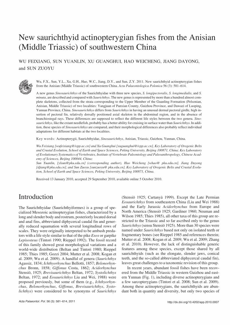
A new genus Sinosaurichthys of the Saurichthyidae with three new species, S. longipectoralis, S. longimedialis, and S.minuta, are described and compared with Saurichthys. The new genus is represented by more than a hundred almost com−plete skeletons, collected from the strata corresponding to the Upper Member of the Guanling Formation (Pelsonian,Anisian, Middle Triassic) of two localities: Yangjuan of Panxian County, Guizhou Province, and Dawazi of Luoping,Yunnan Province, China. Sinosaurichthys differs from Saurichthys in having an unusual dermal pectoral girdle, high in−sertion of pectoral fin, relatively dorsally positioned axial skeleton in the abdominal region, and in the absence ofbranchiostegal rays. These differences are supposed to reflect the different life styles between the two genera. Sino−saurichthys, like the extant needlefish, probably has a better ability for cruising in surface water than Saurichthys. In addi−tion, these species of Sinosaurichthys are compared, and their morphological differences also probably reflect individualadaptations for different habitats at the two localities.
Wu Feixiang [[email protected]] and Xu Guanghui [[email protected]], Key Laboratory of Orogenic Beltsand Crustal Evolution, School of Earth and Space Sciences, Peking University, Beijing 100871, China; Key Laboratoryof Evolutionary Systematics of Vertebrates, Institute of Vertebrate Paleontology and Paleoanthropology, Chinese Acad−emy of Sciences, Beijing 100044, China;Sun Yuanlin, [[email protected]] (corresponding author), Hao Weicheng [whao@ pku.edu.cn], Jiang Dayong[[email protected]], and Sun Zuoyu [sunzuoyu@ pku.edu.cn], Key Laboratory of Orogenic Belts and Crustal Evolu−tion, School of Earth and Space Sciences, Peking University, Beijing 100871, China.
Received 13 January 2010, accepted 29 September 2010, available online 5 October 2010.
Introduction
The Saurichthyidae (Saurichthyiformes) is a group of spe−cialized Mesozoic actinopterygian fishes, characterized by along and slender body and rostrum, posteriorly located dorsaland anal fins, abbreviated diphycercal caudal fin and gener−ally reduced squamation with several longitudinal rows ofscales. They were originally interpreted to be ambush preda−tors with a life style similar to that of the pike Esox or garpikeLepisosteus (Tintori 1990; Rieppel 1992). The fossil recordof this family showed great morphological variations and aworld−wide distribution (Beltan and Tintori 1980; Rieppel1985; Thies 1985; Gozzi 2004; Mutter et al. 2008; Kogan etal. 2009; Wu et al. 2009). A handful of genera (SaurichthysAgassiz, 1834; Ichthyorhynchus Bellotti, 1857; Belonorhyn−chus Bronn, 1858; Giffonus Costa, 1862; AcidorhynchusStensiö, 1925; Brevisaurichthys Beltan, 1972; SystolichthysBeltan, 1972; and Eosaurichthys Liu and Wei, 1988) wereproposed previously, but some of them (e.g., Ichthyorhyn−chus, Belonorhynchus, Giffonus, Brevisaurichthys, Systo−lichthys) were considered to be synonyms of Saurichthys
(Stensiö 1925; Cartanyà 1999). Except the Late PermianEosaurichthys from southeastern China (Liu and Wei 1988)and the Early Jurassic Acidorhynchus from Europe andNorth America (Stensiö 1925; Gardiner 1960; Neuman andWilson 1985; Thies 1985), all other taxa of this group are re−stricted to the Triassic and so far ascribed only to the genusSaurichthys (sensu Stensiö 1925). More than 30 species werenamed under Saurichthys based not only on isolated teeth orfragmentary bones (see Rieppel 1985 and references therein;Mutter et al. 2008; Kogan et al. 2009; Wu et al. 2009; Zhanget al. 2010). However, the lack of distinguishable genericfeatures among these species, except those shared by allsaurichthyids (such as the elongate, slender jaws, conicalteeth, and the so−called abbreviated diphycercal caudal fin),leaves great challenges to a taxonomic revision of this group.
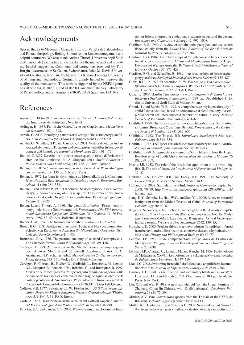
In recent years, abundant fossil fishes have been recov−ered from the Middle Triassic in western Guizhou and east−ern Yunnan (Fig. 1), including diverse actinopterygians anda few sarcopterygians (Tintori et al. 2008; Sun et al. 2009).Among these actinopterygians, the saurichthyids are abun−dant both in quantity and diversity, but only two species of
Saurichthys have been described recently (Wu et al. 2009;Zhang et al. 2010). Here, we name and describe three othernew species of the Saurichthyidae assigned to a new genus,Sinosaurichthys, from the two localities mentioned above.The morphological differences between the new genus andSaurichthys and their life styles are specially discussed.
Geological settingThe three new saurichthyid species described here comefrom two localities: one from Yangjuan of Panxian County,Guizhou Province and the other two from Dawazi of Luo−ping County, Yunnan Province (Fig. 1).
In the Yangjuan area of Panxian, Sinosaurichthys longi−pectoralis gen. et sp. nov. was recovered from a 3−meterthick vertebrate−rich layer in the upper part of the GuanlingFormation, associated with abundant, well preserved marinereptiles and other fossil fishes (Sun et al. 2008). This speciesis one of the quantitatively dominant vertebrates in that local−ity. Conodont analysis revealed that the fossiliferous layer iswithin the Nicoraella kockeli Zone, suggesting an early mid−dle Anisian age (Sun et al. 2006).
In the Dawazi area of Luoping, the studied materials werecollected from a more than 10−meter thick vertebrate−richlayer near the top of the Third Member of the Gejiu Forma−tion (a local lithostratigraphic unit applied in Yunnan Prov−ince, spanning from the upper Lower Triassic to upper Mid−dle Triassic, and its Third Member corresponding to theGuanling Formation used in adjacent Guizhou Province), as−sociated with abundant other fossil fishes. Sinosaurichthys
582 ACTA PALAEONTOLOGICA POLONICA 56 (3), 2011
CHINA
QingshanYangjuan
Xinmin
Dawazi
Pu'an
Fuyuan
Luoping
Xingyi
E 105°
N 25°
20 km
Panxian
Yangjuan Section
ko
cke
lico
nstr
icta
Ya
ng
liu
jin
gF
m.
Gu
an
lin
gF
m.(p
art
)
An
isia
n
Illy
ria
nP
els
on
ian
10
m
Sin
osa
urich
thys
lon
gip
ecto
ralis
ge
n.e
tsp
.n
ov.
Y12
Y11
Y10
Y13
0.5
m
Dawazi Section
Gu
an
lin
gF
orm
ati
on
(pa
rt)
10
m
0.5
m
D50
D37
D54
Sin
osa
urich
thys
min
uta
ge
n.e
tsp
.n
ov.
Sin
osa
urich
thys
lon
gim
ed
ialis
ge
n.e
tsp
.n
ov.
Sa
urich
thys
da
wa
zie
nsis
bifu
rca
ta
limestonemarly
limestonenodular
limestonechert
limestonebanded
limestone dolostonedolomiticlimestone
bentonitelayer
vertebratefossils
areniticdolostone
fossilsite
Xingren
Fig. 1. Locality map and lithological columns of two sections in Guizhou and Yunnan Provinces, China.
minuta was recovered from the lower part while S. longi−medialis occured in the upper part of the fossiliferous layer,about 3 to 5 meters above the former. The conodont Nicora−ella kockeli was also recovered in samples collected from thefossiliferous layer, suggesting the same age for this fauna asthat from Yangjuan of Panxian.
Material and methodsThis study is based on more than 150 specimens. Most ofthem were well−preserved and mechanically prepared withsharp needles under a stereomicroscope. The line drawingswere done based on photos, assisted with observation of thespecimens under a Nikon SMZ1500 binocular microscope.All specimens are deposited in the GMPKU.
The terminology of bones used in this paper followsStensiö (1925), Rieppel (1985), and Gardiner and Schaeffer(1989). The method of measurement is shown in Fig. 2.
Systematic paleontologyActinopterygii Woodward, 1891Saurichthyiformes Aldinger, 1937Saurichthyidae Stensiö, 1925Genus Sinosaurichthys nov.Type species: Sinosaurichthys longipectoralis sp. nov.; see below.Etymology: From Greek sino, China; saurichthys, the only genus ofsaurichthyids from the Triassic heretofore.
Diagnosis.—Pectoral fin inserting high on flank, at or abovemidline of body; cleithrum boot−shaped with high rectangularposterior blade, slender anterodorsal stem and short horizontalanteroventral arm; presence of large posttemporal−supraclei−thrum; parasphenoid with highly elevated posterior stem; axialskeleton arranged high in abdominal region of body; absenceof branchiostegals; anal fin situated closer to pelvic fin than tocaudal fin; cordate mid−dorsal scales much wider than mid−ventral ones; and extremely elongated first paired scale in analloop, expanding anteriorly and tapering posteriorly.
Species included.—Sinosaurichthys longipectoralis sp. nov.,S. longimedialis sp. nov., and S. minuta sp. nov.
Stratigraphic and geographic range.—Anisian, Middle Tri−assic, Guizhou and Yunnan, China.
Etymology: From Latin longus and pectoralis, referring to its exception−ally elongated pectoral fins.
Type material: Holotype GMPKU−P1233, a laterally compressed skullwith part of the postcranial skeleton. Paratypes: GMPKU−P1214, apostcranial skeleton with complete caudal region; and GMPKU−P1215,a laterally compressed skull.
Type locality: Yangjuan Village, Xinmin, Panxian County, GuizhouProvince, China.
Type horizon: The vertebrate fossiliferous horizon in the Upper Memberof the Guanling Formation (Pelsonian of Anisian, Middle Triassic) (Sunet al. 2006).
Diagnosis.—Type species of Sinosaurichthys, pectoral finsickle−shaped and extremely long, reaching about or morethan mandible length; cleithrum bearing anteriorly inclineddorsal stem and rather deep posterior blade with depth/lengthratio about 1.8; posttemporal−supracleithra meeting at mid−line; pelvic fin relatively long; axial skeleton consisting of noless than 210 neural arches between opercle and caudal fin(compared with 157–172 in S. longimedialis and 154–156 inS. minuta described below), including ca. 180 anterior oneswith neural spines; at least 90 mid−dorsal scales anterior todorsal fin. Fin formula: P 24–25, V 23–24, D/A ~55/~50, C36–38/36–38.
Description
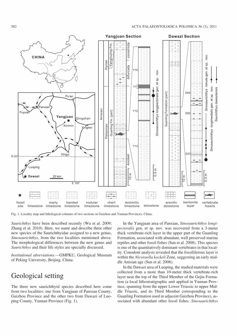
General appearance.—As a shared feature of the Saurich−thyidae, Sinosaurichthys longipectoralis has a long rostrumand an elongated, slender body. The standard length of thelargest specimen is over 560 mm. The skull length is 27% ofthe standard body length. The rostrum makes up 73–76% ofthe mandible length (Figs. 3A–C, 4; Table 1). It is similar toother members of the same genus described below in havingthe pectoral fins located near to the posterodorsal corner ofthe opercle, in contrast to Saurichthys that have pectoral finsmuch lower located as commonly in other actinopterygians.The pelvic fins are closer to the caudal fin than to the opercle.The dorsal and anal fins are opposite to each other and aremuch closer to the pelvic fins than to the caudal fin.
Endocranium.—The endocranium is poorly ossified and onlythe posterodorsal part of the orbitotemporal region was pre−served in some of laterally compressed specimens to carry twoforamina (Fig. 4A), probably related to the oculomotor nerve(III).
Snout.—The snout is composed of the paired rostro−pre−maxillae and nasalo−antorbitals, with a length up to 61–65%of the skull length (Fig. 4). The rostro−premaxilla is elon−
doi:10.4202/app.2010.0007
WU ET AL.—MIDDLE TRIASSIC SAURICHTHYID FISHES FROM CHINA 583
standard body length
length betweenopercle and pelvic fins
skull length
rostrumlength
mandiblelength
length fromanal to caudal fin
wid
th
length
de
pth
width
length
length frompelvic to anal fin
Fig. 2. Illustration showing measurement conventions. A. Body in lateralview. B. Median fin in lateral view. C. Mid−dorsal scales in dorsal view.
584 ACTA PALAEONTOLOGICA POLONICA 56 (3), 2011
gated and triangular, tapering anteriorly and carrying theethmoid commissure at its anterior tip. The rostro−premaxillameets its opposite pair medially and contacts with thefrontals and nasalo−antorbitals posterodorsally, and themaxilla posteriorly, with a series of conical teeth along itsoral margin (Figs. 4, 5). The rostro−premaxilla is ornamentedwith parallel striations, dipping posteroventrally, and a fewtubercles near its dorsal edge. The nasalo−antiorbital is trian−gular, in contact with the frontal dorsally and the rostro−premaxilla ventrally, and forms the anterior rim of the orbit.Two subovate external nares are present on this bone, ar−ranged in the same way as in other saurichthyids, and the an−terior one is distinctly larger than the posterior. The supra−orbital sensory canal enters the nasalo−antorbital from thefrontal and passes between the two nares to join the infra−orbital sensory canal at the ventral portion of this bone. The
nasalo−antorbital is ornamented with posteroventrally in−clined striations and patch of tubercles near the dorsal edge.
Dermal skull roof.—The skull roof consists of paired frontals,parietals, dermopterotics and extrascapulars (Figs. 4, 5A, C).The frontals are elongated and triangular, tapering anteriorlyand forming the main part of the anterior portion of the skullroof above the orbit. Posteriorly, the frontals are in contactwith the parietals and dermopterotics. The parietals are sub−circular and relatively large in proportion to the skull width,with a width about 60% of the skull width (Fig. 5A, C). Thedermopterotics compose the major part of the posterior por−tion of the skull roof. The posterior end of the dermopterotic islocated at the level of the anterior margin of the opercle. As acharacteristic feature of this family, the dermopterotics meet atthe mid−line of the skull roof posterior to the parietals with adistinct notch at the anterolateral margin of the bone on eachside (Figs. 5A, C, 6A) which was interpreted to accommodatethe opening of the spiracle canal in Saurichthys (Stensiö1925). The dermopterotic has a posterolaterally protrudingprocess as the articular facet for the extrascapular and post−temporal−supracleithrum. The part of the dermopterotic ante−rior to the notch is laterally bent downward to form a triangular
doi:10.4202/app.2010.0007
WU ET AL.—MIDDLE TRIASSIC SAURICHTHYID FISHES FROM CHINA 585
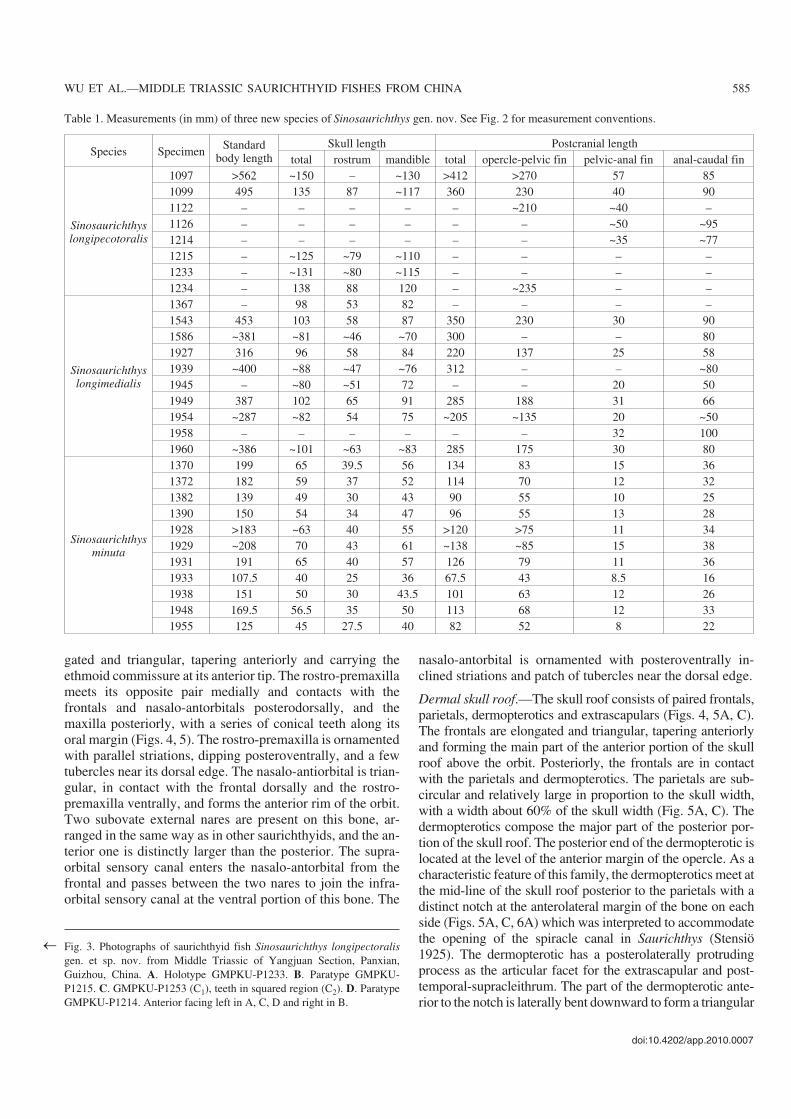
Table 1. Measurements (in mm) of three new species of Sinosaurichthys gen. nov. See Fig. 2 for measurement conventions.
Species Specimen Standardbody length
Skull length Postcranial lengthtotal rostrum mandible total opercle−pelvic fin pelvic−anal fin anal−caudal fin
Fig. 3. Photographs of saurichthyid fish Sinosaurichthys longipectoralisgen. et sp. nov. from Middle Triassic of Yangjuan Section, Panxian,Guizhou, China. A. Holotype GMPKU−P1233. B. Paratype GMPKU−P1215. C. GMPKU−P1253 (C1), teeth in squared region (C2). D. ParatypeGMPKU−P1214. Anterior facing left in A, C, D and right in B.
�
586 ACTA PALAEONTOLOGICA POLONICA 56 (3), 2011
lateral lobe to cover part of the cheek region between the orbitand the preopercle. The extrascapulars are small and subovate(Figs. 3A, 4A, 5C). The external surface of the skull roof is or−namented with dense tubercles.
Cheek and opercular series.—The orbit is subcircular to el−lipse shaped. Similar to Saurichthys curionii (Rieppel 1985),no supraorbital bones are developed and the frontal directlyforms the dorsal margin of the orbit (Figs. 4, 5A, C). Thedermosphenotic is a small slender bone, forming the postero−dorsal margin of the orbit. The infraorbital sensory canal ex−tends upward in the posterior half of this bone to enter thedermopterotic. The infraorbitals are poorly preserved and onlya few fragments can be identified along the posterior and ven−tral margins of the orbit in the holotype (Fig. 4A). The scle−rotic ring is partly exposed along the edge of the orbit in sev−eral specimens. The ring probably has four ossification centersbecause three elements are exposed in GMPKU−P1183 whichoccupies almost 3/4 of the orbital circumference (Fig. 6A).
The postorbital region is rather short, with a length evi−dently less than the skull depth, differing from that in mostknown species of Saurichthys (Stensiö 1925; Rieppel 1985)in which the length of the postorbital region is generallygreater than or equal to the skull depth. The maxilla andpreopercle are the main dermal elements of the cheek region.The maxilla is composed of a long and slender anterior orbitalportion that contacts the rostro−premaxilla anteriorly and ahighly expanded postorbital portion that contacts with thepreopercle. The oral margin of the maxilla is slightly concavebehind the orbit. The preopercle is deep and almost vertical,with a distinct concave anterior margin. The dorsal portion isexpanded and ventral portion is narrowed, showing a primi−tive condition as commonly in other lower actinopterygians(Fig. 4A, C). The quadratojugal is relatively small, in contactwith the maxilla anteriorly and the preopercle dorsally(Fig. 4A), showing more or less a plesiomorphic condition asin other lower actinopterygians (e.g. Mimia, Moythomasia,Pteronisculus, and Birgeria) (Nielsen 1942, 1949; Lehman1952; Gardiner 1984). This bone is greatly deduced or lost inneopterygians such as Amia (Grande and Bemis 1998).
As a shared feature of the Saurichthyidae, Sinosaurich−thys longipectoralis has a single large, semicircular opercleand lacks an independent subopercle (Figs. 3A–C1, 4A, B1,C). The opercle reaches 85–88% of the skull depth in height,with a depth/width ratio varying from 1.55 to 1.97 (average1.76; Table 2). A small process is developed in its straight an−terior margin at the level slightly higher than the joint of theupper and lower jaws. Externally the opercle shows radiatingand concentric striations with some tubercles near its dorsaland ventral edges. The medial side of the opercle is smoothexcept for a distinct circular recess posterior to the process
mentioned above. Neither gular plates nor branchiostegalrays are developed. In Saurichthys, the gular plates are ab−sent but there is generally one pair of branchiostegal rays.
Mandible.—The elongated lower jaw is as long as the upperjaw. Its maximum depth is less than half of the skull depth.The symphyseal region occupies about two fifth of the mandi−ble length. There is little difference in the arrangement of thedermal elements in lateral side of the mandible from that ofmost species of Saurichthys (Fig. 4A, B1, C). The dentary isthe largest ossification and covers almost the whole lateral sideof the mandible. It is ornamented with anteroventrally inclinedfine striations and a few tubercles along the ventral edge. Theangular is triangular, occupying the posteroventral portion ofthe mandible and sutures with the dentary anterodorsally (Fig.4A, B1, C). In the lateral side, the angular extends to the levelanterior to the posterior rim of the orbit, whereas ventrally thebone bends up dorsomedially to wrap the mandible along itsventral edge and continues forward beyond the anterior rim ofthe orbit. The angular is ornamented with coarse ridges that ra−diate from its posteroventral corner, and some tubercles alongits posterior and ventral edges. The supraangular is a smallslender element, located at the posterodorsal corner of themandible anterior to the articular.
In the lingual side of the mandible a long bone with manysmall teeth can be seen in specimen GMPKU−P1141, coveringalmost the whole length of the mandible (Fig. 5B). It should bethe fused prearticular and coronoids (= mixcoronoid of Stensiö1925). It is roughly acute triangular, high in the posterior partand tapering anteriorly, with a straight dorsal edge. The poste−rior part of this bone contacts the angular posteroventrally (Fig.5B). In the joint region with the upper jaw, there are two trans−verse depressions related to the articulation with the quadrate;therefore this region should be the ossification of the articular,similar with the situation in some other known saurichthyids(Stensiö 1925; Beltan 1968; Rieppel 1985). The adductor fossais elongated and deep, enclosed laterally by the dentary andsupraangular, medially by the prearticular−coronoid, and poste−riorly by the articular (Fig. 5B, E).
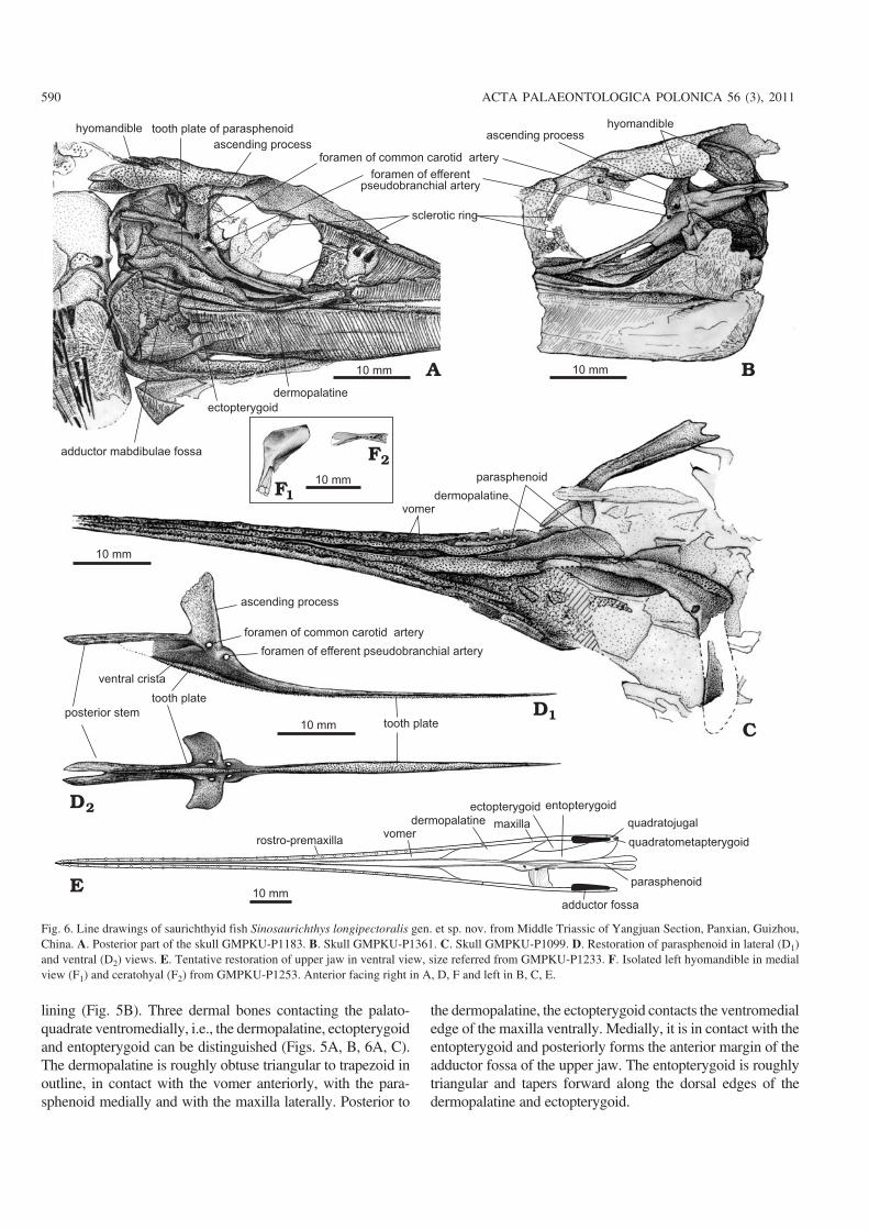
Palate.—The palate includes a pair of long and slender vo−mers (Fig. 6C, E), and a median parasphenoid. Although theparasphenoid is incompletely preserved in all specimens, itsgeneral morphology and structure can still be restored (Fig.6D). As in the Lower Triassic Saurichthys from Spitsbergenand Madagascar (Stensiö 1925; Beltan 1968), the parasphe−noid consists of a long anterior stem that has extended be−tween the paired vomers, a pair of large ascending processesthat have dorsolaterally extended to cover considerable por−tion of the otic and orbitotemporal regions of the neuro−cranium, and a posterior stem that has extended posteriorlyover the occipital region. It shows the following features thatdiffer from that of Saurichthys described by Stensiö (1925):(1) the posterior stem is highly elevated above the mid−pointof the skull depth with a high plate−like ventral crista (Fig.6A, B, D) while in Saurichthys it is only slightly elevatedwith a low ridge−like ventral crista. The elevation of the pos−
doi:10.4202/app.2010.0007
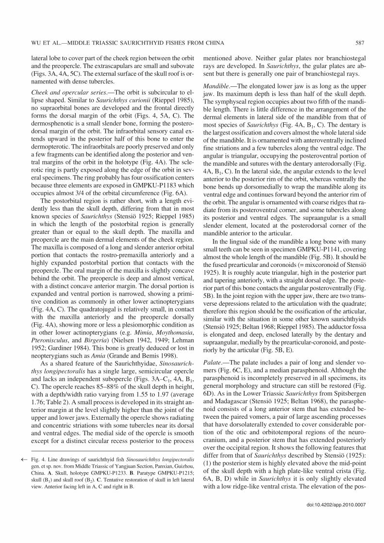
WU ET AL.—MIDDLE TRIASSIC SAURICHTHYID FISHES FROM CHINA 587
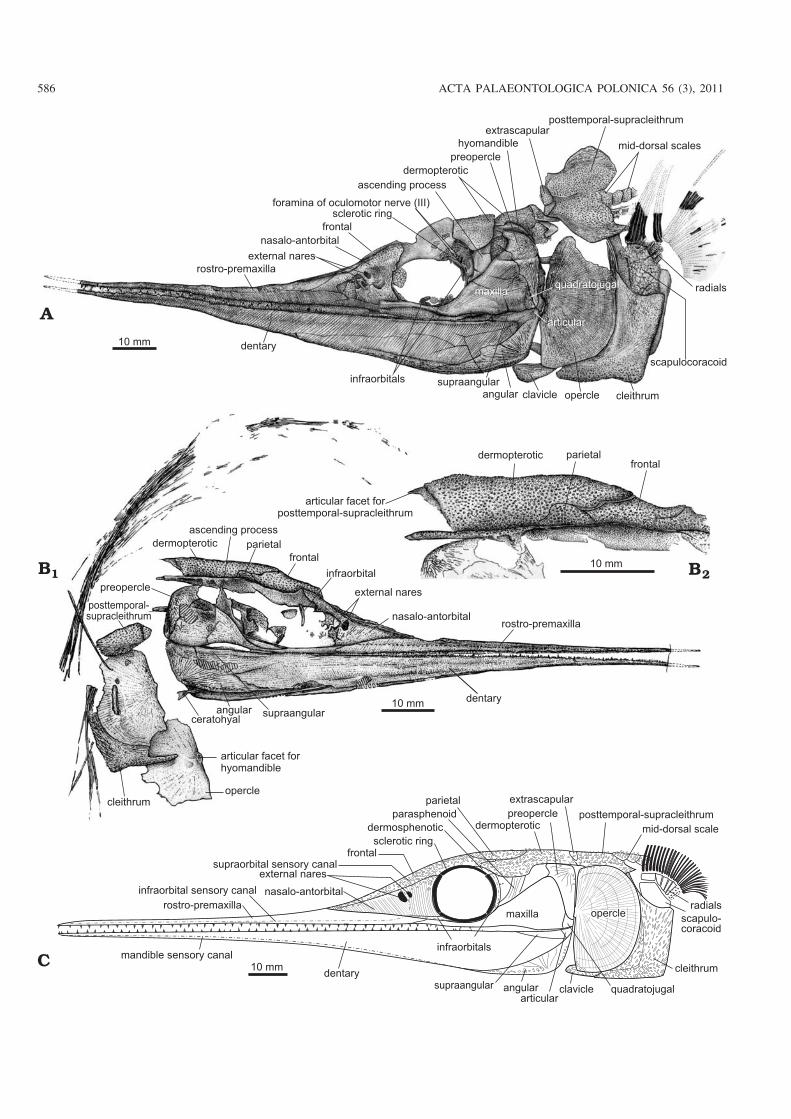
Fig. 4. Line drawings of saurichthyid fish Sinosaurichthys longipectoralisgen. et sp. nov. from Middle Triassic of Yangjuan Section, Panxian, Guizhou,China. A. Skull, holotype GMPKU−P1233. B. Paratype GMPKU−P1215;skull (B1) and skull roof (B2). C. Tentative restoration of skull in left lateralview. Anterior facing left in A, C and right in B.
�
terior stem of the parasphenoid in Sinosaurichthys isapparrently related to the elevation of the axial skeleton inthe abdominal region and is unique; (2) the tooth plate orpatch in the ventral (oral) face of the parasphenoid extendsposteriorly on the ventral crista posterior to the ascendingprocess while that in Saurichthys is relatively short, and onlyrestricted anterior to the ascending process; (3) the anteriorstem is very long, about three times of the length of the poste−rior one (Fig. 6C–E) while in the Lower Triassic Saurichthysthe anterior stem is almost as long as the posterior one. But insome later representatives of Saurichthys the anterior stemmay be longer than the posterior one because this feature isevidently related to the shortening of the postorbital region inthe saurichthyids; (4) the efferent pseudobranchial arteriespenetrate the parasphenoid through a pair of small ear−likeprocesses anterior to the ascending process (Fig. 6A, B, D).This paired ear−like process, more or less, resembles to thedermal basipterygoid process in some low actinopterygiansin morphology and position but it leaves no trace for articu−lating with the palatoquadrate. Maybe it represents an incipi−ent or highly reduced basipterygoid process. A similar condi−tion is present or probably present in some of the Middle Tri−assic Saurichthys, such as Saurichthys dawaziensis (Wu etal. 2009) and S. curionii (Rieppel 1985) but probablly absentin the Early Triassic Saurichthys (Stensiö 1925); (5) the fo−
ramina of the common carotid arteries are situated in the lat−eral wall of the ascending process with openings directedmore or less lateroventrally (Fig. 6A, B, D). In Saurichthysornatus the same foramina (originally interpreted as for theexternal carotid arteries by Stensiö (1925), and later assumedto have transmitted the common carotid arteries by Patterson(1975) are in the underside of the parasphenoid beneath theposterior margin of the ascending process (Stensiö 1925) andin S. curionii they are even anterior to the ascending process(Rieppel 1985).
Hyoid arches.—The hyomandible is almost vertical, havinga slightly broad, blade−like dorsal portion and a narrow, slen−der posteroventral portion (Figs. 5B, 6A, B, F1), slightly dif−ferent from the hockey stick−like one in Saurichthys hamil−toni (Stensiö 1925) and S. costasquamosus (Rieppel 1985).The blade−like dorsal portion has a distinct posteroventrallydirected ridge running through its medial surface and theposteroventral portion seems to contact with the palato−quadrate at a point slightly high above the mandibular articu−lation. No opercular process is present in the hyomandible.The symplectic is absent as in Saurichthys and other loweractinoptergyians. The ceratohyal can be seen in GMPKU−P1215 and 1253, being of a typical hour glass shape, narrowrod−like in the middle and expanded plate−like at the twoends (Figs. 4B1, 6F2).
588 ACTA PALAEONTOLOGICA POLONICA 56 (3), 2011
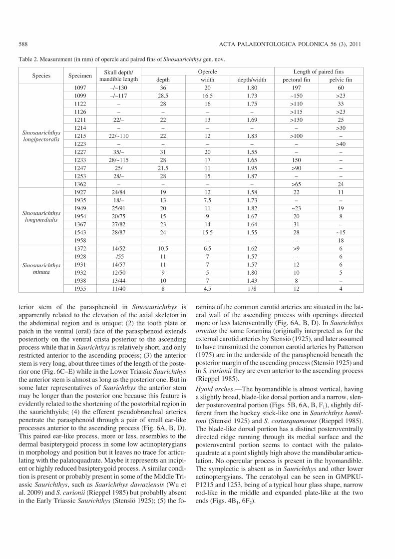
Table 2. Measurement (in mm) of opercle and paired fins of Sinosaurichthys gen. nov.
Species Specimen Skull depth/mandible length
Opercle Length of paired finsdepth width depth/width pectoral fin pelvic fin
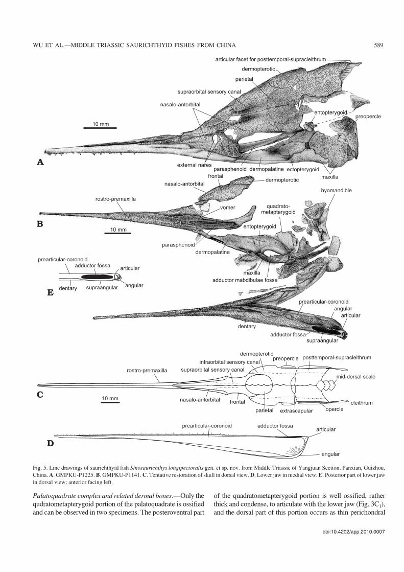
Palatoquadrate complex and related dermal bones.—Only thequdratometapterygoid portion of the palatoquadrate is ossifiedand can be observed in two specimens. The posteroventral part
of the quadratometapterygoid portion is well ossified, ratherthick and condense, to articulate with the lower jaw (Fig. 3C1),and the dorsal part of this portion occurs as thin perichondral
doi:10.4202/app.2010.0007
WU ET AL.—MIDDLE TRIASSIC SAURICHTHYID FISHES FROM CHINA 589
Fig. 5. Line drawings of saurichthyid fish Sinosaurichthys longipectoralis gen. et sp. nov. from Middle Triassic of Yangjuan Section, Panxian, Guizhou,China. A. GMPKU−P1225. B. GMPKU−P1141. C. Tentative restoration of skull in dorsal view. D. Lower jaw in medial view. E. Posterior part of lower jawin dorsal view; anterior facing left.
lining (Fig. 5B). Three dermal bones contacting the palato−quadrate ventromedially, i.e., the dermopalatine, ectopterygoidand entopterygoid can be distinguished (Figs. 5A, B, 6A, C).The dermopalatine is roughly obtuse triangular to trapezoid inoutline, in contact with the vomer anteriorly, with the para−sphenoid medially and with the maxilla laterally. Posterior to
the dermopalatine, the ectopterygoid contacts the ventromedialedge of the maxilla ventrally. Medially, it is in contact with theentopterygoid and posteriorly forms the anterior margin of theadductor fossa of the upper jaw. The entopterygoid is roughlytriangular and tapers forward along the dorsal edges of thedermopalatine and ectopterygoid.
590 ACTA PALAEONTOLOGICA POLONICA 56 (3), 2011
Fig. 6. Line drawings of saurichthyid fish Sinosaurichthys longipectoralis gen. et sp. nov. from Middle Triassic of Yangjuan Section, Panxian, Guizhou,China. A. Posterior part of the skull GMPKU−P1183. B. Skull GMPKU−P1361. C. Skull GMPKU−P1099. D. Restoration of parasphenoid in lateral (D1)and ventral (D2) views. E. Tentative restoration of upper jaw in ventral view, size referred from GMPKU−P1233. F. Isolated left hyomandible in medialview (F1) and ceratohyal (F2) from GMPKU−P1253. Anterior facing right in A, D, F and left in B, C, E.
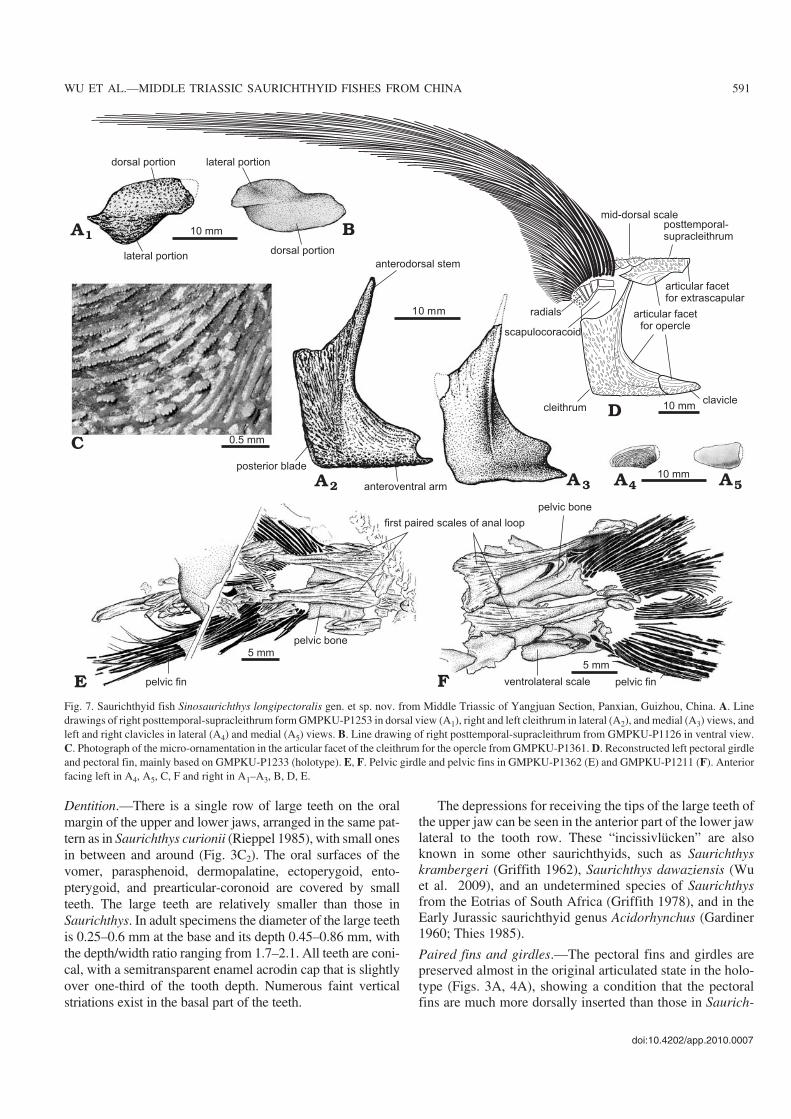
Dentition.—There is a single row of large teeth on the oralmargin of the upper and lower jaws, arranged in the same pat−tern as in Saurichthys curionii (Rieppel 1985), with small onesin between and around (Fig. 3C2). The oral surfaces of thevomer, parasphenoid, dermopalatine, ectoperygoid, ento−pterygoid, and prearticular−coronoid are covered by smallteeth. The large teeth are relatively smaller than those inSaurichthys. In adult specimens the diameter of the large teethis 0.25–0.6 mm at the base and its depth 0.45–0.86 mm, withthe depth/width ratio ranging from 1.7–2.1. All teeth are coni−cal, with a semitransparent enamel acrodin cap that is slightlyover one−third of the tooth depth. Numerous faint verticalstriations exist in the basal part of the teeth.
The depressions for receiving the tips of the large teeth ofthe upper jaw can be seen in the anterior part of the lower jawlateral to the tooth row. These “incissivlücken” are alsoknown in some other saurichthyids, such as Saurichthyskrambergeri (Griffith 1962), Saurichthys dawaziensis (Wuet al. 2009), and an undetermined species of Saurichthysfrom the Eotrias of South Africa (Griffith 1978), and in theEarly Jurassic saurichthyid genus Acidorhynchus (Gardiner1960; Thies 1985).
Paired fins and girdles.—The pectoral fins and girdles arepreserved almost in the original articulated state in the holo−type (Figs. 3A, 4A), showing a condition that the pectoralfins are much more dorsally inserted than those in Saurich−
doi:10.4202/app.2010.0007
WU ET AL.—MIDDLE TRIASSIC SAURICHTHYID FISHES FROM CHINA 591
Fig. 7. Saurichthyid fish Sinosaurichthys longipectoralis gen. et sp. nov. from Middle Triassic of Yangjuan Section, Panxian, Guizhou, China. A. Linedrawings of right posttemporal−supracleithrum form GMPKU−P1253 in dorsal view (A1), right and left cleithrum in lateral (A2), and medial (A3) views, andleft and right clavicles in lateral (A4) and medial (A5) views. B. Line drawing of right posttemporal−supracleithrum from GMPKU−P1126 in ventral view.C. Photograph of the micro−ornamentation in the articular facet of the cleithrum for the opercle from GMPKU−P1361. D. Reconstructed left pectoral girdleand pectoral fin, mainly based on GMPKU−P1233 (holotype). E, F. Pelvic girdle and pelvic fins in GMPKU−P1362 (E) and GMPKU−P1211 (F). Anteriorfacing left in A4, A5, C, F and right in A1–A3, B, D, E.
thys. Although most of the fin rays were weathered away,their impressions remain on the matrix. The bases of the finrays are arranged in an arc that slightly curves down back−ward dorsal to the radials. The fins are sickle−shaped, as longas or longer than the mandible length. The longest pectoralfins are recorded in GMPKU−P1097, reaching 257 mm long,about 1.84 times of the mandible length. In Saurichthys thepectoral fins are generally fan−shaped, with a length gener−ally no more than 1/6 of the mandible length. No segmenta−tion is observed in the fin rays. The anterior four to five finrays are unbranched and the remainder branch distally fivetimes maximally and twice minimally. The 7th fin ray is thelongest.
The dermal elements of the pectoral girdle are well pre−served (Figs. 3A–C1, 4, 7A, B). The posttemporal (= supra−scapular sensu Stensiö 1925; Lehman 1952; Rieppel 1980)and supracleithrum are fused into a large bone (here tenta−tively named as posttemporal−supracleithrum) consisting of arectangular, horizontal dorsal portion (= posttemporal) anda triangular, vertical ventrolateral portion (= supracleithrum)posterior to the dermopterotic and extrascapular and dorsal tothe opercle. The dorsal portion of the bone meets its fellow ofthe opposite side at the midline and the ventrolateral portionof this bone is partially overlapped by the opercle ventro−anteriorly and contacts the cleithrum posteriorly. The pores ofthe infraorbital sensory canal can be seen distributed along theborder of the two portions of this bone to extend anteriorly intothe dermopterotic and runs posteriorly into the body. The ex−posed surface of this composite bone is ornamented with tu−bercles and the area overlapped by the opercle has longitudi−nal ridges with serrations pointing upwards. The postclei−thrum is absent. The cleithrum is boot−shaped and consists of adeep rectangular posterior blade, a long, slender and curveddorsal stem, and a short horizontal anteroventral arm (Figs.3A, 4A, C, 7A2, A3, D), different from the typical triradiateone in other saurichthyids. The bone is concave anteriorly andborders the posterior margin of the opercle. The anterodorsaltip of the dorsal stem articulates with the posttemporal−supra−cleithrum. The depth of the posterior blade of the bone is morethan half of the skull depth with a depth/length ratio of about1.8. The horizontal anteroventral arm is low, with its anterioredge concave to fit the posterior margin of the clavicle. Aprominent keel in the medial surface runs from the radiationcenter of the bone upward to the tip of dorsal stem. The clavi−cle is suboval to subtriangular (Figs. 3A, 4A, 7A4, A5), with itsexpanded posterior end articulating with the cleithrum, ventralto the opercle (Fig. 7D). The exposed surfaces of the cleithrumand clavicle are decorated with spiny tubercles while the areasoverlapped by the opercle are ornamented by similar ridges tothose on posttemporal−supracleithrum with serrations pointingaway from the opercle (Fig. 7C).
The endoskeletal pectoral girdle is best preserved in theholotype, including the scapulocoracoid and the radials. Thescapulocoracoid is a deep plate−like structure emerging be−hind the dorsal stem of the cleithrum. It bears at least sevenradials along its dorsal margin. Except the anteriormost and
largest one, the rest decrease gradually in size posteriorly.These radials support approximately 25 fin rays distally(Figs. 3A, 4A, 7D).
The pelvic fins are situated closer to the caudal fin than tothe pectoral ones, with its distance to the caudal fin slightlymore than one−third of the distance between the opercle andthe caudal fin. Compared to Saurichthys and other species ofSinosaurichthys, the pelvic fins are quite long, more than theskull depth (Table 2). In Saurichthys and other two species ofthe new genus, the pelvic fins are generally less than or aslong as half of the skull depth. Each pelvic fin consists of23–24 unsegmented, distally branched fin rays that articulatewith the posterior edge of the pelvic bone (Fig. 7E, F). Thepelvic bone is subrectangular. No radials are preserved.
Unpaired fins.—The unpaired fins are relatively well pre−served in three specimens. The dorsal and anal fins are trian−gle−shaped and situated opposite to each other, slightly poste−rior to the pelvic fins (Fig. 3D). The depth/width ratio of thedorsal and anal fin is over 1.7, showing a higher aspect ratio(defined as depth2/area) than that in Saurichthys and the othertwo species of Sinosaurichthys. The estimated fin rays of thedorsal and anal fin in GMPKU−P1214 are no less than 55 and50, respectively. The anterior 12 fin rays are stout and un−branched, whereas the following ones are distally branchedonce or twice. The 12th fin ray is the longest. Generally, a max−imum of three to four segments are counted in the longest finray of both the dorsal and anal fins, but six segments are re−corded in the anal fin in GMPKU−P1223. Both the dorsal andanal fins are supported by the radials consisting of slenderproximal axonosts and small distal baseosts. There are 16axonosts on each fin. The axonosts are posterodorsally andanterodorsally directed in the dorsal and anal fin, respectively,and tend to decrease in depth posteriorly. The small and rect−angular baseosts are poorly ossified, lying distal to the axo−nosts.
The caudal fin is deeply forked and symmetrical, with36–38 fin rays in each lobe directly supported by the axialendoskeleton and with an angle between the leading edges ofthe two lobes varying from 84� to 100�. There are at least sixsegments in the epichordal lobe and five segments in thehypochordal lobe of the caudal fin with maximal three timesof distal bifurcation in GMPKU−P1089. The depth of thecaudal fin is about 2.8 times of the maximal body depth inGMPKU−P1214.
The basal and fringing fulcra are present in all medianfins. Six to seven basal fulcra can be seen in the anal fin inGMPKUM−P1214 and P−1122. Two to three basal fulcra ex−ist on each lobe of the caudal fin. The fringing fulcra occur assmall spine−like elements lying on the surface of marginleading rays distally, distinctly shorter than the basal fulcraand overlapping one by one. This condition probably can beassigned to the pattern B as proposed by Arratia (2008: 229).
Axial skeleton.—The axial skeleton consists of the neuraland haemal arches applied to the persisting notochord.In GMPKU−P1099, approximately 140 and 70–72 neural
592 ACTA PALAEONTOLOGICA POLONICA 56 (3), 2011
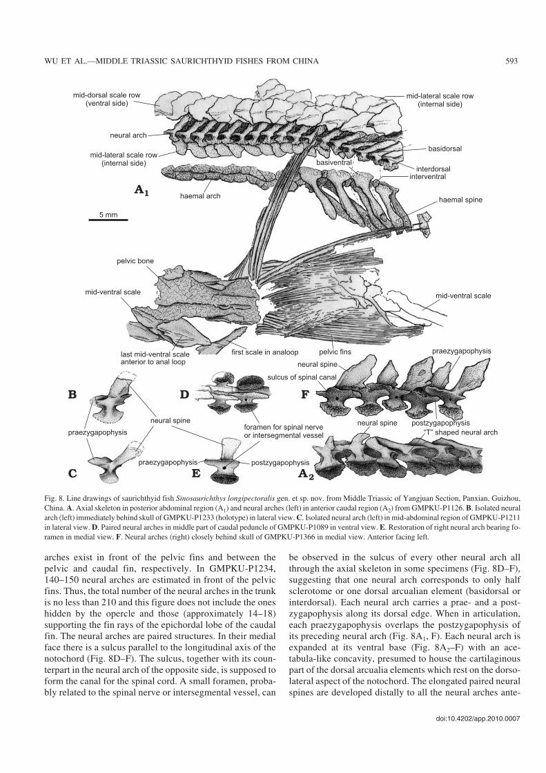
arches exist in front of the pelvic fins and between thepelvic and caudal fin, respectively. In GMPKU−P1234,140–150 neural arches are estimated in front of the pelvicfins. Thus, the total number of the neural arches in the trunkis no less than 210 and this figure does not include the oneshidden by the opercle and those (approximately 14–18)supporting the fin rays of the epichordal lobe of the caudalfin. The neural arches are paired structures. In their medialface there is a sulcus parallel to the longitudinal axis of thenotochord (Fig. 8D–F). The sulcus, together with its coun−terpart in the neural arch of the opposite side, is supposed toform the canal for the spinal cord. A small foramen, proba−bly related to the spinal nerve or intersegmental vessel, can
be observed in the sulcus of every other neural arch allthrough the axial skeleton in some specimens (Fig. 8D–F),suggesting that one neural arch corresponds to only halfsclerotome or one dorsal arcualian element (basidorsal orinterdorsal). Each neural arch carries a prae− and a post−zygapophysis along its dorsal edge. When in articulation,each praezygapophysis overlaps the postzygapophysis ofits preceding neural arch (Fig. 8A1, F). Each neural arch isexpanded at its ventral base (Fig. 8A2–F) with an ace−tabula−like concavity, presumed to house the cartilaginouspart of the dorsal arcualia elements which rest on the dorso−lateral aspect of the notochord. The elongated paired neuralspines are developed distally to all the neural arches ante−
doi:10.4202/app.2010.0007
WU ET AL.—MIDDLE TRIASSIC SAURICHTHYID FISHES FROM CHINA 593
Fig. 8. Line drawings of saurichthyid fish Sinosaurichthys longipectoralis gen. et sp. nov. from Middle Triassic of Yangjuan Section, Panxian, Guizhou,China. A. Axial skeleton in posterior abdominal region (A1) and neural arches (left) in anterior caudal region (A2) from GMPKU−P1126. B. Isolated neuralarch (left) immediately behind skull of GMPKU−P1233 (holotype) in lateral view. C. Isolated neural arch (left) in mid−abdominal region of GMPKU−P1211in lateral view. D. Paired neural arches in middle part of caudal peduncle of GMPKU−P1089 in ventral view. E. Restoration of right neural arch bearing fo−ramen in medial view. F. Neural arches (right) closely behind skull of GMPKU−P1366 in medial view. Anterior facing left.
rior to the caudal peduncle and the following 28 neuralarches anterior to the caudal fin do not carry neural spines,and thus look “T” shaped in lateral view (Fig. 8A2). Whenapproaching the caudal fin, the neural arches support the finrays of the epichordal lobe of the caudal fin.
The haemal arches are paired and lie along the ventro−lateral aspects of the notochord, opposite to the neural arches(Fig. 8A1). The haemal arches occur as poorly ossified small
suboval plates in the abdominal region and consist of twokinds of alternatively arranged bony plates in the caudal re−gion. One of them is well ossified and considerably large, al−most corresponding to two neural arches in length and bear−ing a distinct haemal spine, and the other is poorly ossifiedand very small, without a haemal spine. These two kinds ofhaemal arches should represent the separate basi− and inter−ventral arcualia elements (Fig. 8A1). Nearly twenty haemal
594 ACTA PALAEONTOLOGICA POLONICA 56 (3), 2011
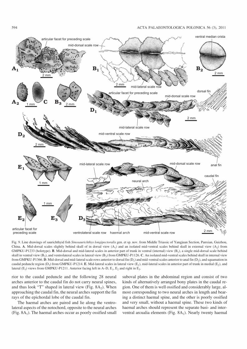
Fig. 9. Line drawings of saurichthyid fish Sinosaurichthys longipectoralis gen. et sp. nov. from Middle Triassic of Yangjuan Section, Panxian, Guizhou,China. A. Mid−dorsal scales slightly behind skull of in dorsal view (A1) and an isolated mid−ventral scales behind skull in external view (A2) fromGMPKU−P1233 (holotype). B. Mid−dorsal and mid−lateral scales in anterior part of trunk in ventral (internal) view (B1), a single mid−dorsal scale behindskull in ventral view (B2), and ventrolateral scales in lateral view (B3) from GMPKU−P1126. C. An isolated mid−ventral scales behind skull in internal viewfrom GMPKU−P1366. D. Mid−dorsal and mid−lateral scale rows anterior to dorsal fin (D1) and mid−ventral scales anterior to anal fin (D2), and squamation incaudal peduncle region (D3) from GMPKU−P1214. E. Mid−lateral scales in lateral view (E1), mid−lateral scales in anterior part of trunk in medial (E2), andlateral (E3) views from GMPKU−P1211. Anterior facing left in A–D, E1, E2 and right in E3.
arches with distinct haemal spines can be counted betweenthe pelvic and anal fin in GMPKU−P1214.
Based on the size relationship of haemal and neural archesin Saurichthys ornatus, Stensiö (1925) supposed that the basi−dorsal and interdorsal elements are equally developed andpossess the same shape in Saurichthys, but this viewpoint wasnot widely accepted. The presence of the foramen of theintersegmental vessels in the haemal arches of Saurichthysmadagascariensis (Lehman 1952), and one haemal arch cor−responding to one neural arch in Saurichthys curionii (Rieppel1985) had led to the idea that the neural arches of the saurich−thyids represent only the basidorsals (Arratia et al. 2001).However, the alternative distribution of the foramen in theneural arch in Sinosaurichthys longipectoralis and in Saurich−thys dawaziensis (Wu et al. 2009) provides evidence to sup−port Stensiö’s (1925) hypothesis and strongly suggests that theneural arches in saurichthyids, at least in some species ofSaurichthys and Sinosaurichthys are alternate basidorsal andinterdorsal. In other lower actinopterygians, the ossification ofbasidorsal is always larger than the interdorsal and neuralspines are usually only developed in the basidorsals. Conse−quently, the basidorsal and interdorsal equally−developedshould be considered as a possible synapomorphy of thesaurichthyids.
Squamation.—Similar to the Middle Triassic Saurichthysfrom Monte San Giorgio (Rieppel 1985, 1992) but differentfrom the species of Saurichthys of other areas of the world,Sinosaurichthys bears six longitudinal rows of scales, includ−ing one mid−dorsal, one mid−ventral, two mid−lateral and twoventrolateral rows. In the GMPKU−P1126, in addition to thesix rows, numerous small bony plates are scattered betweenthe scale rows.
The mid−dorsal scale row runs through the body lengthand only interrupted by the dorsal fin. Because of the incom−plete preservation, in specimen GMPKU−P1099, 81 mid−dorsal scales can be counted only between the skull and thelevel slightly posterior to the pelvic fins. In the other twospecimens, 16–17 mid−dorsal scales can be counted in thearea between the pelvic and dorsal fins. Thus, the total num−ber of the mid−dorsal scales anterior to the dorsal fin is no lessthan 90 and this number does not include those of the basalfulcra of the dorsal fin. The exposed part of the mid−dorsalscales are cordate, generally wider than long but tend to benarrower toward the dorsal fin with the width/length ratiovarying from about 2–2.1 to 0.7–0.75 from the skull to thedorsal fin (Fig. 9A1, B1, B2, D1). The posterior exposed partof each scale overlaps about the anterior one−third of the suc−ceeding scale and bears a plate−like ventral median crista(Fig. 9B). The surface of each scale is ornamented with 12 to20 longitudinal rows of posteriorly directed spine−like tuber−cles. The exposed parts of the scales just behind the dorsal finare relatively thick, longer than wide, with blunt spearheadshaped end. These scales tend to increase in size posteriorly.In the anterior part of each scale there is a concaved facetto accept the posterior part of the preceding scale (Fig. 9A1,D1, D3).
Different from the Middle Triassic Saurichthys fromMonte San Giorgio (Rieppel 1985) the mid−ventral scalerow of Sinosaurichthys longipectoralis begins to developjust behind the skull. This scale row consists of loosely ar−ticulated small suboval plates (Fig. 9A2, C) in anterior one−third part between the skull and pelvic fins. The followingscales increase in length posteriorly and become lanceolatetoward the pelvic fins. Slightly anterior to the pelvic fins,the mid−ventral scale row is branched to form the anal loop.Five pairs of the scales are, at least, involved in the analloop but the accurate number is not clear due to the preser−vation. The last scale anterior to the anal loop is elongated,with its posterior part expanded into a rhombic plate tooverlap on the first paired scale in the anal loop, which isalso very large, as long as four to five times of each of thesubsequent anal loop scales, expanding anteriorly and ta−pering posteriorly with some longitudinal fine grooves onits surface and a hook−like spine in the posterior end (Fig.7E, F). This paired large scale was incorrectly consideredby Wu et al. (2008) as a “clasper−like” gonopodium. Themid−ventral scales in the caudal region are lanceolate andsimilar to the mid−dorsal ones of the same region (Fig. 9D3).
The mid−lateral scale row runs through to the end of thebody. The scales are roughly triangular to rhombic in outline,differentiated into a narrow dorsal half that is ornamentedwith some posteriorly directed spine−like tubercles and awider, smooth ventral half (Fig. 9E). The dorsal half is evi−dently higher and more acute than the ventral half in thescales anterior to the dorsal fin and tends to decrease inheight posteriorly and becomes as deep as the ventral halfposterior to the dorsal fin. Generally, the length of twomid−lateral scales equals that of one mid−dorsal scale. No lat−eral line openings are observed in the scales.
The ventrolateral scale row commences a short distanceanterior to the pelvic fins and are interrupted by the pelvic fins.The scales are suboval to rhombic (Fig. 9B3) and smaller thanthe mid−lateral scales.
Etymology: From Latin longus and medialis, referring to its exception−ally elongated median fins.Type material: Holotype GMPKU−P1927, a laterally compressed, al−most complete skeleton. Paratypes: GMPKU−P1543, P1936, 1949,1954.Type locality: Dawazi, Luoping, Yunnan Province, China.Type horizon: Upper part of the fossiliferous strata near the top of theThird Member of the Gejiu Formation (Pelsonian of Anisian, MiddleTriassic).
Referred specimens.—GMPKU−P1367, 1380, 1388, 1586,1769, 1935, 1936, 1939, and 1958. Most of them are com−plete skeletons.
Diagnosis.—Medium−sized Sinosaurichthys (standard bodylength ranging from 270–470 mm) with unusually elongatedmedian fins with relatively few segments of fin rays; less num−ber of neural arches between opercle and caudal fin (approxi−
doi:10.4202/app.2010.0007
WU ET AL.—MIDDLE TRIASSIC SAURICHTHYID FISHES FROM CHINA 595
mately 157–172), less number of anterior ones with neuralspines (130–146); and less number of mid−dorsal scales infront of dorsal fin (69–86) than in type species; 14–15 distinct
haemal spines in caudal region; pectoral fin triangular shapedwith length about 1/3 of mandible length (shared with S.minuta described below); posttemporal−supracleithrum from
596 ACTA PALAEONTOLOGICA POLONICA 56 (3), 2011
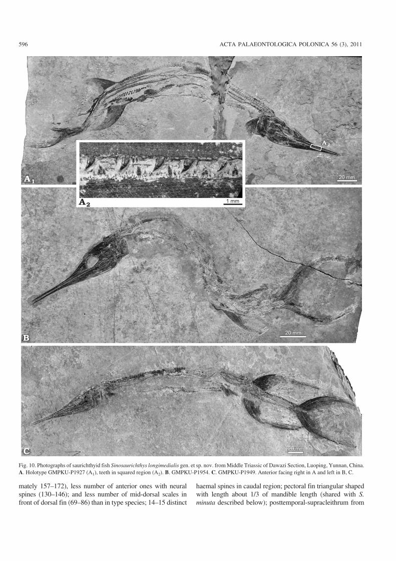
Fig. 10. Photographs of saurichthyid fish Sinosaurichthys longimedialis gen. et sp. nov. from Middle Triassic of Dawazi Section, Luoping, Yunnan, China.A. Holotype GMPKU−P1927 (A1), teeth in squared region (A2). B. GMPKU−P1954. C. GMPKU−P1949. Anterior facing right in A and left in B, C.
either side separated from each other by anterior mid−dorsalscales (shared with S. minuta described below); cleithrumplate (depth/length ratio ca. 1.2–1.25) much lower than in typespecies (approximately 1.8), but close to that in S. minuta (ap−proximately 1). Fin formula: P 18–19, V 18–20, D/A 44–49/>40–48, C 37–39/37–39.
Description
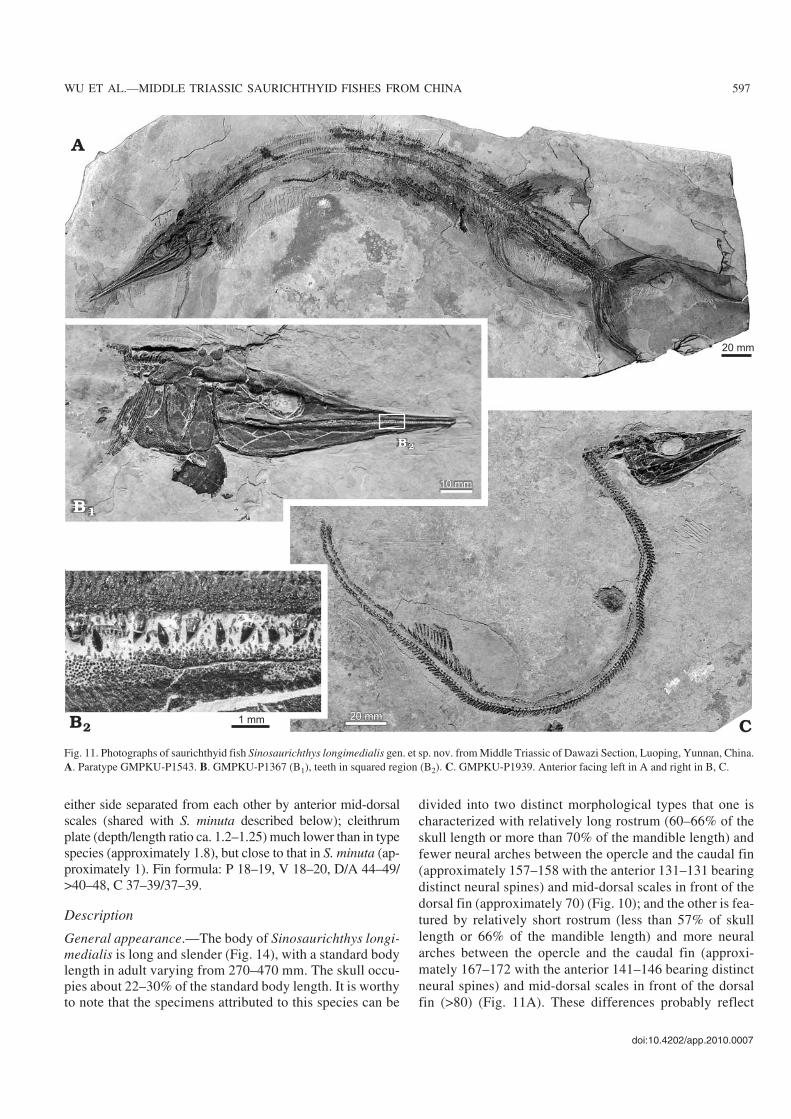
General appearance.—The body of Sinosaurichthys longi−medialis is long and slender (Fig. 14), with a standard bodylength in adult varying from 270–470 mm. The skull occu−pies about 22–30% of the standard body length. It is worthyto note that the specimens attributed to this species can be
divided into two distinct morphological types that one ischaracterized with relatively long rostrum (60–66% of theskull length or more than 70% of the mandible length) andfewer neural arches between the opercle and the caudal fin(approximately 157–158 with the anterior 131–131 bearingdistinct neural spines) and mid−dorsal scales in front of thedorsal fin (approximately 70) (Fig. 10); and the other is fea−tured by relatively short rostrum (less than 57% of skulllength or 66% of the mandible length) and more neuralarches between the opercle and the caudal fin (approxi−mately 167–172 with the anterior 141–146 bearing distinctneural spines) and mid−dorsal scales in front of the dorsalfin (>80) (Fig. 11A). These differences probably reflect
doi:10.4202/app.2010.0007
WU ET AL.—MIDDLE TRIASSIC SAURICHTHYID FISHES FROM CHINA 597
Fig. 11. Photographs of saurichthyid fish Sinosaurichthys longimedialis gen. et sp. nov. from Middle Triassic of Dawazi Section, Luoping, Yunnan, China.A. Paratype GMPKU−P1543. B. GMPKU−P1367 (B1), teeth in squared region (B2). C. GMPKU−P1939. Anterior facing left in A and right in B, C.
598 ACTA PALAEONTOLOGICA POLONICA 56 (3), 2011
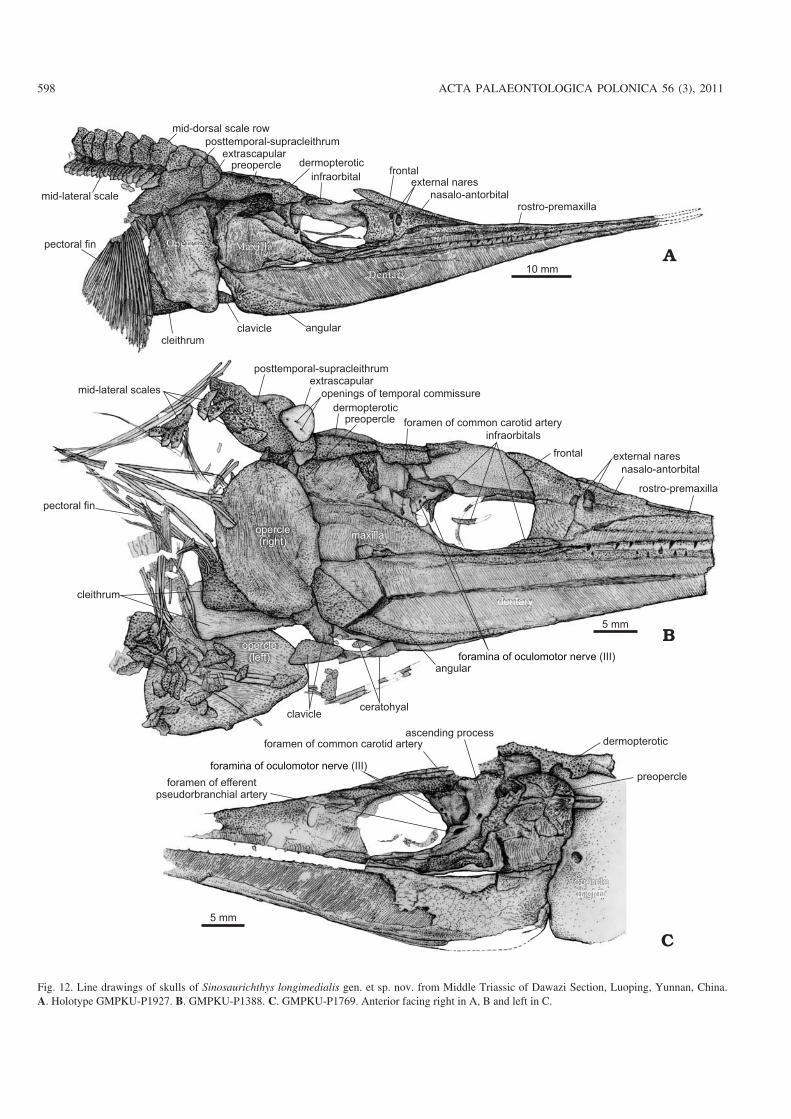
Fig. 12. Line drawings of skulls of Sinosaurichthys longimedialis gen. et sp. nov. from Middle Triassic of Dawazi Section, Luoping, Yunnan, China.A. Holotype GMPKU−P1927. B. GMPKU−P1388. C. GMPKU−P1769. Anterior facing right in A, B and left in C.
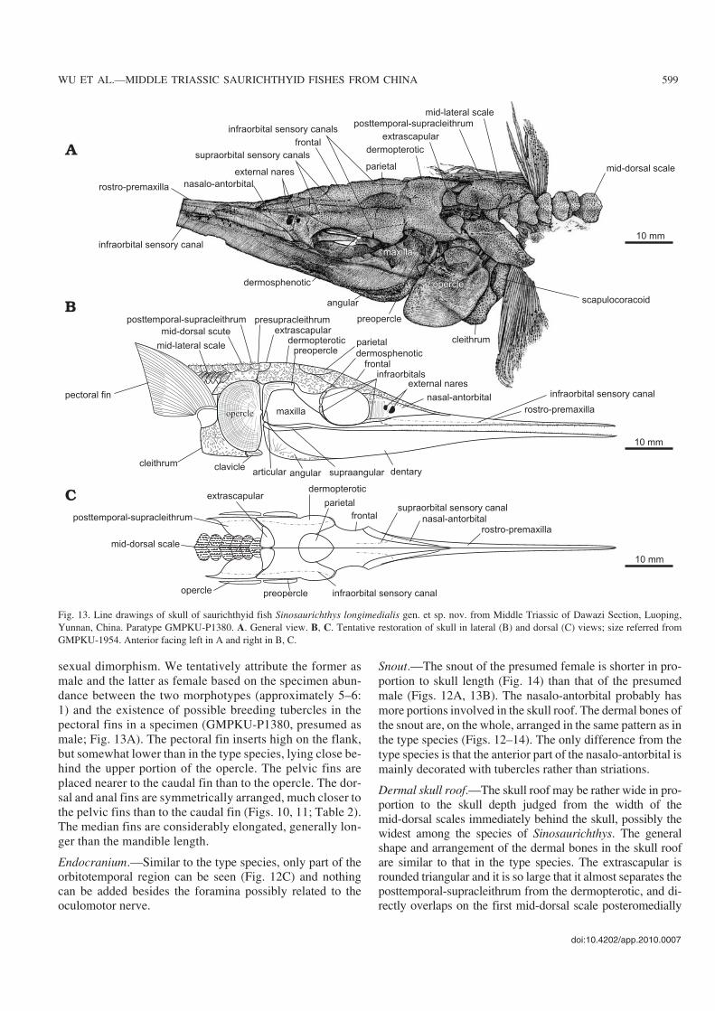
sexual dimorphism. We tentatively attribute the former asmale and the latter as female based on the specimen abun−dance between the two morphotypes (approximately 5–6:1) and the existence of possible breeding tubercles in thepectoral fins in a specimen (GMPKU−P1380, presumed asmale; Fig. 13A). The pectoral fin inserts high on the flank,but somewhat lower than in the type species, lying close be−hind the upper portion of the opercle. The pelvic fins areplaced nearer to the caudal fin than to the opercle. The dor−sal and anal fins are symmetrically arranged, much closer tothe pelvic fins than to the caudal fin (Figs. 10, 11; Table 2).The median fins are considerably elongated, generally lon−ger than the mandible length.
Endocranium.—Similar to the type species, only part of theorbitotemporal region can be seen (Fig. 12C) and nothingcan be added besides the foramina possibly related to theoculomotor nerve.
Snout.—The snout of the presumed female is shorter in pro−portion to skull length (Fig. 14) than that of the presumedmale (Figs. 12A, 13B). The nasalo−antorbital probably hasmore portions involved in the skull roof. The dermal bones ofthe snout are, on the whole, arranged in the same pattern as inthe type species (Figs. 12–14). The only difference from thetype species is that the anterior part of the nasalo−antorbital ismainly decorated with tubercles rather than striations.
Dermal skull roof.—The skull roof may be rather wide in pro−portion to the skull depth judged from the width of themid−dorsal scales immediately behind the skull, possibly thewidest among the species of Sinosaurichthys. The generalshape and arrangement of the dermal bones in the skull roofare similar to that in the type species. The extrascapular isrounded triangular and it is so large that it almost separates theposttemporal−supracleithrum from the dermopterotic, and di−rectly overlaps on the first mid−dorsal scale posteromedially
doi:10.4202/app.2010.0007
WU ET AL.—MIDDLE TRIASSIC SAURICHTHYID FISHES FROM CHINA 599
Fig. 13. Line drawings of skull of saurichthyid fish Sinosaurichthys longimedialis gen. et sp. nov. from Middle Triassic of Dawazi Section, Luoping,Yunnan, China. Paratype GMPKU−P1380. A. General view. B, C. Tentative restoration of skull in lateral (B) and dorsal (C) views; size referred fromGMPKU−1954. Anterior facing left in A and right in B, C.
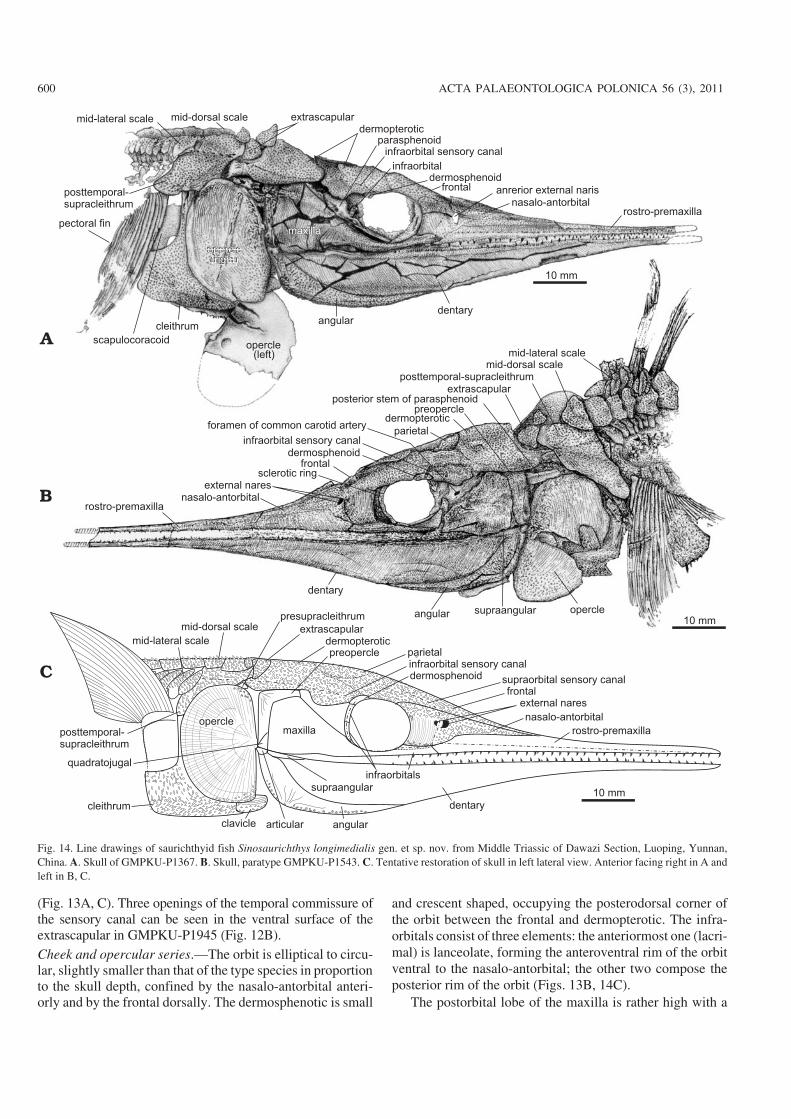
(Fig. 13A, C). Three openings of the temporal commissure ofthe sensory canal can be seen in the ventral surface of theextrascapular in GMPKU−P1945 (Fig. 12B).Cheek and opercular series.—The orbit is elliptical to circu−lar, slightly smaller than that of the type species in proportionto the skull depth, confined by the nasalo−antorbital anteri−orly and by the frontal dorsally. The dermosphenotic is small
and crescent shaped, occupying the posterodorsal corner ofthe orbit between the frontal and dermopterotic. The infra−orbitals consist of three elements: the anteriormost one (lacri−mal) is lanceolate, forming the anteroventral rim of the orbitventral to the nasalo−antorbital; the other two compose theposterior rim of the orbit (Figs. 13B, 14C).
The postorbital lobe of the maxilla is rather high with a
600 ACTA PALAEONTOLOGICA POLONICA 56 (3), 2011
Fig. 14. Line drawings of saurichthyid fish Sinosaurichthys longimedialis gen. et sp. nov. from Middle Triassic of Dawazi Section, Luoping, Yunnan,China. A. Skull of GMPKU−P1367. B. Skull, paratype GMPKU−P1543. C. Tentative restoration of skull in left lateral view. Anterior facing right in A andleft in B, C.
truncated rather than a convex dorsal margin that is almostfully covered by the dorsal portion of the preopercle (Figs.12–14), different from that in the type species, but similar tothat in S. minuta described below.
Similar to the type species, the opercular series consists ofa single large semicircular opercle (Figs. 12–14), with thedepth/width ratio varying from 1.55 to 1.82 (average 1.67)(Table 2). The gular and branchiostegal rays are absent.
doi:10.4202/app.2010.0007
WU ET AL.—MIDDLE TRIASSIC SAURICHTHYID FISHES FROM CHINA 601
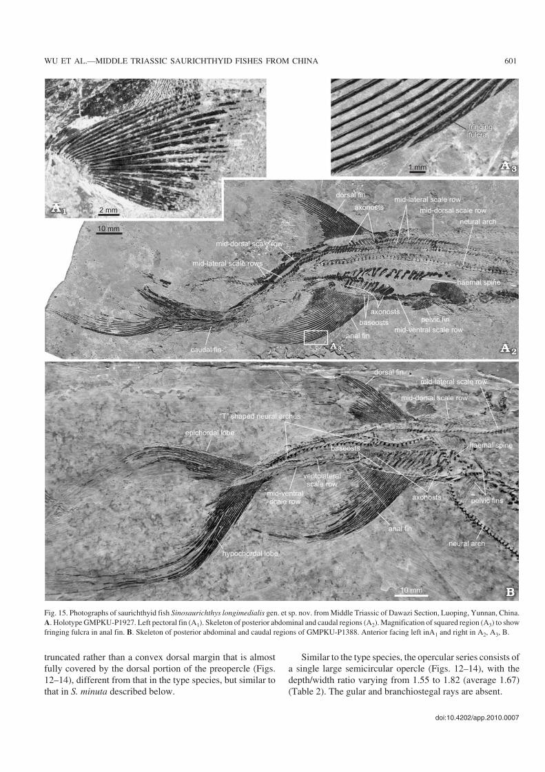
Fig. 15. Photographs of saurichthyid fish Sinosaurichthys longimedialis gen. et sp. nov. from Middle Triassic of Dawazi Section, Luoping, Yunnan, China.A. Holotype GMPKU−P1927. Left pectoral fin (A1). Skeleton of posterior abdominal and caudal regions (A2). Magnification of squared region (A3) to showfringing fulcra in anal fin. B. Skeleton of posterior abdominal and caudal regions of GMPKU−P1388. Anterior facing left inA1 and right in A2, A3, B.
Mandible.—The shape and arrangement of dermal bones inthe lateral side of the mandible is almost the same as in thetype species (Figs. 12–14). However, the ventral margin inthe specimens of the presumed female seems to be more con−vex than in these of the presumed males (Fig. 14).
Palate.—No information about the vomers is available dueto the preservation. The parasphenoid is only partially ex−posed in the current materials. The parasphenoid passesacross the orbit in a similar way as in the type species withpaired large ascending processes posterior to the orbit andthe foramina of both the efferent pseudobranchial and thecommon carotid arteries penetrating the parasphenoid inthe same positions as in the type species (Fig. 12C). Theposterior stem of the parasphenoid also extends posteriorlyunder the occipital region, beyond the anterior margin ofthe opercle with a distinct notch in the posterior tip (Fig.12C).
Hyoid arches.—Only the ceratohyal is preserved (Fig. 12B).In GMPKU−P1388, it has a shape similar to that in the typespecies.
Dentition.—Teeth along the labial edge of both jaws are ar−ranged in the same way as in the type species, one row oflarge teeth intercalated with several small ones (Figs. 10A2,11B2). The apical tip of most large teeth bends posteriorly,differing from that in the type species.
Paired fins and girdles.—The pectoral fin is of typical trian−gular shape consisting of 19 unsegmented fin rays in theholotype (Figs. 10A1, 12A, 15A1). The 6th is the longest. Thelength of the pectoral fin varies from less than to more thanthe skull depth (Table 2). The fin rays begin to branch dis−
tally from the 10th one. In GMPKU−P1380, many small tu−bercles are present on the surface of the distal part in the pec−toral fin rays (Fig. 13A), a feature probably related to breed−ing (nuptial) behavior of adult male individuals.
Little information of radials can be added whereas thescapulocoracoid can be partially seen in some specimenswith a large foramen in the anterodorsal part behind the dor−sal stem of the cleithrum (Figs. 13A, 14A).
The dermal pectoral girdle is well preserved in most ofthe specimens. The posttemporal−supracleithrum, as in thetype species, consists of a dorsal and a ventrolateral portion(Figs. 13A, 14A, B), but the dorsal portion of this bone isseparated from its opposite pair by the mid−dorsal scales andthe ventrolateral portion is deeper (Figs. 13A, 14A, B). Ante−riorly, this bone bears a subcircular articular facet for theextrascapular and posteriorly an articular facet for themid−lateral scale between its two portions. Although thecleithrum is also boot−shaped, the rectangular posterior bladeof this bone is less deep (approximately 1/3 of the skulldepth) than in the type species and the dorsal stem is verti−cally oriented rather than anterodorsally inclined (Fig. 14A).The clavicle is subovate to round triangular, and articulatesposteriorly to the cleithrum (Figs. 12B, 13B, 14C). In addi−tion, a small triangular shaped bone can be observed in somespecimens attached to the lateral side of the posterior processof the dermopterotic and anterior to the posttemporal−supracleithrum, with ornamentation similar to that on areasof other dermal bones of the pectoral girdle covered by theopercle, possibly suggesting a presupracleithrum.
The pelvic fin is relatively small and triangle−shaped,consisting of 18–22 unsegmented but distally branched fin
602 ACTA PALAEONTOLOGICA POLONICA 56 (3), 2011
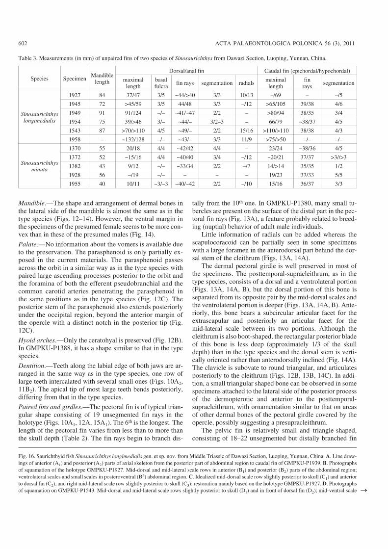
Table 3. Measurements (in mm) of unpaired fins of two species of Sinosaurichthys from Dawazi Section, Luoping, Yunnan, China.
Species Specimen Mandiblelength
Dorsal/anal fin Caudal fin (epichordal/hypochordal)
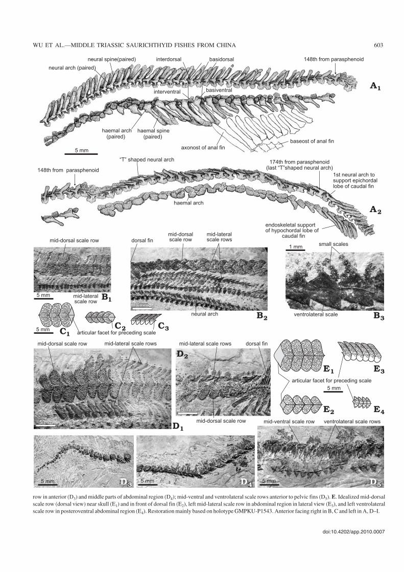
Fig. 16. Saurichthyid fish Sinosaurichthys longimedialis gen. et sp. nov. from Middle Triassic of Dawazi Section, Luoping, Yunnan, China. A. Line draw−ings of anterior (A1) and posterior (A2) parts of axial skeleton from the posterior part of abdominal region to caudal fin of GMPKU−P1939. B. Photographsof squamation of the holotype GMPKU−P1927. Mid−dorsal and mid−lateral scale rows in anterior (B1) and posterior (B2) parts of the abdominal region;ventrolateral scales and small scales in posteroventral (B3) abdominal region. C. Idealized mid−dorsal scale row slightly posterior to skull (C1) and anteriorto dorsal fin (C2), and right mid−lateral scale row slightly posterior to skull (C3); restoration mainly based on the holotype GMPKU−P1927. D. Photographsof squamation on GMPKU−P1543. Mid−dorsal and mid−lateral scale rows slightly posterior to skull (D1) and in front of dorsal fin (D2); mid−ventral scale �
doi:10.4202/app.2010.0007
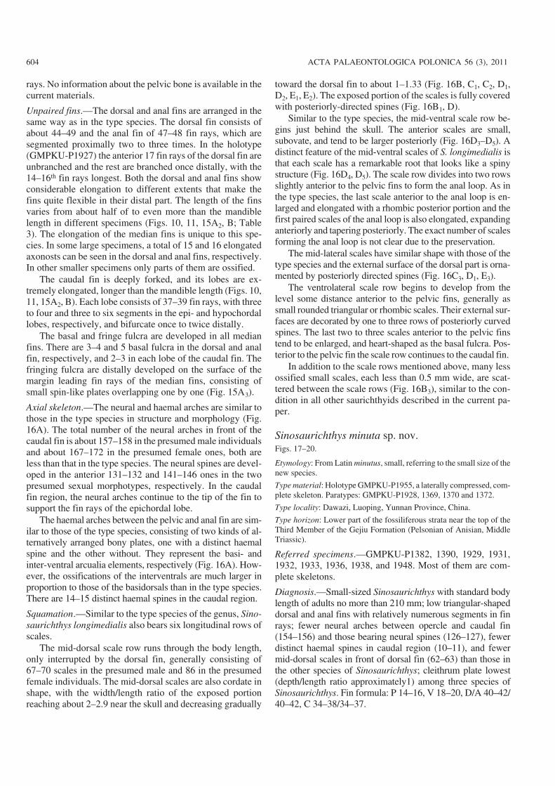
WU ET AL.—MIDDLE TRIASSIC SAURICHTHYID FISHES FROM CHINA 603
row in anterior (D3) and middle parts of abdominal region (D4); mid−ventral and ventrolateral scale rows anterior to pelvic fins (D5). E. Idealized mid−dorsalscale row (dorsal view) near skull (E1) and in front of dorsal fin (E2), left mid−lateral scale row in abdominal region in lateral view (E3), and left ventrolateralscale row in posteroventral abdominal region (E4). Restoration mainly based on holotype GMPKU−P1543. Anterior facing right in B, C and left in A, D–I.
rays. No information about the pelvic bone is available in thecurrent materials.
Unpaired fins.—The dorsal and anal fins are arranged in thesame way as in the type species. The dorsal fin consists ofabout 44–49 and the anal fin of 47–48 fin rays, which aresegmented proximally two to three times. In the holotype(GMPKU−P1927) the anterior 17 fin rays of the dorsal fin areunbranched and the rest are branched once distally, with the14–16th fin rays longest. Both the dorsal and anal fins showconsiderable elongation to different extents that make thefins quite flexible in their distal part. The length of the finsvaries from about half of to even more than the mandiblelength in different specimens (Figs. 10, 11, 15A2, B; Table3). The elongation of the median fins is unique to this spe−cies. In some large specimens, a total of 15 and 16 elongatedaxonosts can be seen in the dorsal and anal fins, respectively.In other smaller specimens only parts of them are ossified.
The caudal fin is deeply forked, and its lobes are ex−tremely elongated, longer than the mandible length (Figs. 10,11, 15A2, B). Each lobe consists of 37–39 fin rays, with threeto four and three to six segments in the epi− and hypochordallobes, respectively, and bifurcate once to twice distally.
The basal and fringe fulcra are developed in all medianfins. There are 3–4 and 5 basal fulcra in the dorsal and analfin, respectively, and 2–3 in each lobe of the caudal fin. Thefringing fulcra are distally developed on the surface of themargin leading fin rays of the median fins, consisting ofsmall spin−like plates overlapping one by one (Fig. 15A3).
Axial skeleton.—The neural and haemal arches are similar tothose in the type species in structure and morphology (Fig.16A). The total number of the neural arches in front of thecaudal fin is about 157–158 in the presumed male individualsand about 167–172 in the presumed female ones, both areless than that in the type species. The neural spines are devel−oped in the anterior 131–132 and 141–146 ones in the twopresumed sexual morphotypes, respectively. In the caudalfin region, the neural arches continue to the tip of the fin tosupport the fin rays of the epichordal lobe.
The haemal arches between the pelvic and anal fin are sim−ilar to those of the type species, consisting of two kinds of al−ternatively arranged bony plates, one with a distinct haemalspine and the other without. They represent the basi− andinter−ventral arcualia elements, respectively (Fig. 16A). How−ever, the ossifications of the interventrals are much larger inproportion to those of the basidorsals than in the type species.There are 14–15 distinct haemal spines in the caudal region.
Squamation.—Similar to the type species of the genus, Sino−saurichthys longimedialis also bears six longitudinal rows ofscales.
The mid−dorsal scale row runs through the body length,only interrupted by the dorsal fin, generally consisting of67–70 scales in the presumed male and 86 in the presumedfemale individuals. The mid−dorsal scales are also cordate inshape, with the width/length ratio of the exposed portionreaching about 2–2.9 near the skull and decreasing gradually
toward the dorsal fin to about 1–1.33 (Fig. 16B, C1, C2, D1,D2, E1, E2). The exposed portion of the scales is fully coveredwith posteriorly−directed spines (Fig. 16B1, D).
Similar to the type species, the mid−ventral scale row be−gins just behind the skull. The anterior scales are small,subovate, and tend to be larger posteriorly (Fig. 16D3–D5). Adistinct feature of the mid−ventral scales of S. longimedialis isthat each scale has a remarkable root that looks like a spinystructure (Fig. 16D4, D5). The scale row divides into two rowsslightly anterior to the pelvic fins to form the anal loop. As inthe type species, the last scale anterior to the anal loop is en−larged and elongated with a rhombic posterior portion and thefirst paired scales of the anal loop is also elongated, expandinganteriorly and tapering posteriorly. The exact number of scalesforming the anal loop is not clear due to the preservation.
The mid−lateral scales have similar shape with those of thetype species and the external surface of the dorsal part is orna−mented by posteriorly directed spines (Fig. 16C3, D1, E3).
The ventrolateral scale row begins to develop from thelevel some distance anterior to the pelvic fins, generally assmall rounded triangular or rhombic scales. Their external sur−faces are decorated by one to three rows of posteriorly curvedspines. The last two to three scales anterior to the pelvic finstend to be enlarged, and heart−shaped as the basal fulcra. Pos−terior to the pelvic fin the scale row continues to the caudal fin.
In addition to the scale rows mentioned above, many lessossified small scales, each less than 0.5 mm wide, are scat−tered between the scale rows (Fig. 16B3), similar to the con−dition in all other saurichthyids described in the current pa−per.
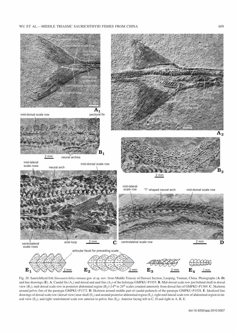
Sinosaurichthys minuta sp. nov.Figs. 17–20.
Etymology: From Latin minutus, small, referring to the small size of thenew species.
Type material: Holotype GMPKU−P1955, a laterally compressed, com−plete skeleton. Paratypes: GMPKU−P1928, 1369, 1370 and 1372.
Type locality: Dawazi, Luoping, Yunnan Province, China.
Type horizon: Lower part of the fossiliferous strata near the top of theThird Member of the Gejiu Formation (Pelsonian of Anisian, MiddleTriassic).
Referred specimens.—GMPKU−P1382, 1390, 1929, 1931,1932, 1933, 1936, 1938, and 1948. Most of them are com−plete skeletons.
Diagnosis.—Small−sized Sinosaurichthys with standard bodylength of adults no more than 210 mm; low triangular−shapeddorsal and anal fins with relatively numerous segments in finrays; fewer neural arches between opercle and caudal fin(154–156) and those bearing neural spines (126–127), fewerdistinct haemal spines in caudal region (10–11), and fewermid−dorsal scales in front of dorsal fin (62–63) than those inthe other species of Sinosaurichthys; cleithrum plate lowest(depth/length ratio approximately1) among three species ofSinosaurichthys. Fin formula: P 14–16, V 18–20, D/A 40–42/40–42, C 34–38/34–37.
604 ACTA PALAEONTOLOGICA POLONICA 56 (3), 2011
doi:10.4202/app.2010.0007
WU ET AL.—MIDDLE TRIASSIC SAURICHTHYID FISHES FROM CHINA 605
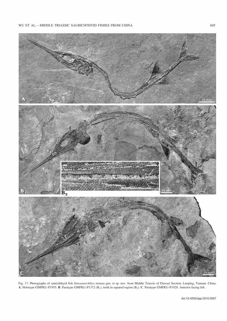
Fig. 17. Photographs of saurichthyid fish Sinosaurichthys minuta gen. et sp. nov. from Middle Triassic of Dawazi Section, Luoping, Yunnan, China.A. Holotype GMPKU−P1955. B. Paratype GMPKU−P1372 (B1), teeth in squared region (B2). C. Paratype GMPKU−P1928. Anterior facing left.
Description
General appearance.—The body is long and slender (Fig.17A, B1, C), with a standard body length varying between100–210 mm. The skull length is about 33–37% of the stan−dard body length and the rostrum makes up about 60–63.5%of the skull length and about 68.7–72.7% of the mandiblelength (Table 1). The pectoral fins lie above the midline ofthe body, close behind the opercle. The pelvic fins are placednearer to the caudal fin than to the opercle. The dorsal andanal fins are symmetrically arranged, much closer to the pel−vic fins than to the caudal fin (Table 2).
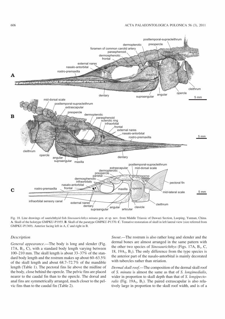
Snout.—The rostrum is also rather long and slender and thedermal bones are almost arranged in the same pattern withthe other two species of Sinosaurichthys (Figs. 17A, B1, C,18, 19A1, B1). The only difference from the type species isthe anterior part of the nasalo−antorbital is mainly decoratedwith tubercles rather than striation.
Dermal skull roof.—The composition of the dermal skull roofof S. minuta is almost the same as that of S. longimedialis,wider in proportion to skull depth than that of S. longipecto−ralis (Fig. 19A1, B1). The paired extrascapular is also rela−tively large in proportion to the skull roof width, and is of a
606 ACTA PALAEONTOLOGICA POLONICA 56 (3), 2011
Fig. 18. Line drawings of saurichthyid fish Sinosaurichthys minuta gen. et sp. nov. from Middle Triassic of Dawazi Section, Luoping, Yunnan, China.A. Skull of the holotype GMPKU−P1955. B. Skull of the paratype GMPKU−P1370. C. Tentative restoration of skull in left lateral view (size referred fromGMPKU−P1369). Anterior facing left in A, C and right in B.
rounded triangular shape, almost completely separates theposttemporal−supracleithrum from the dermopterotic, and indirect contact with the first mid−dorsal scale posteromedially(Figs. 18B, C, 19C).
Cheek and opercular series.—The orbit and the dermalcheek bones are highly consistent with those in S. longi−medialis in shape and arrangement (Figs. 17–19B1) and
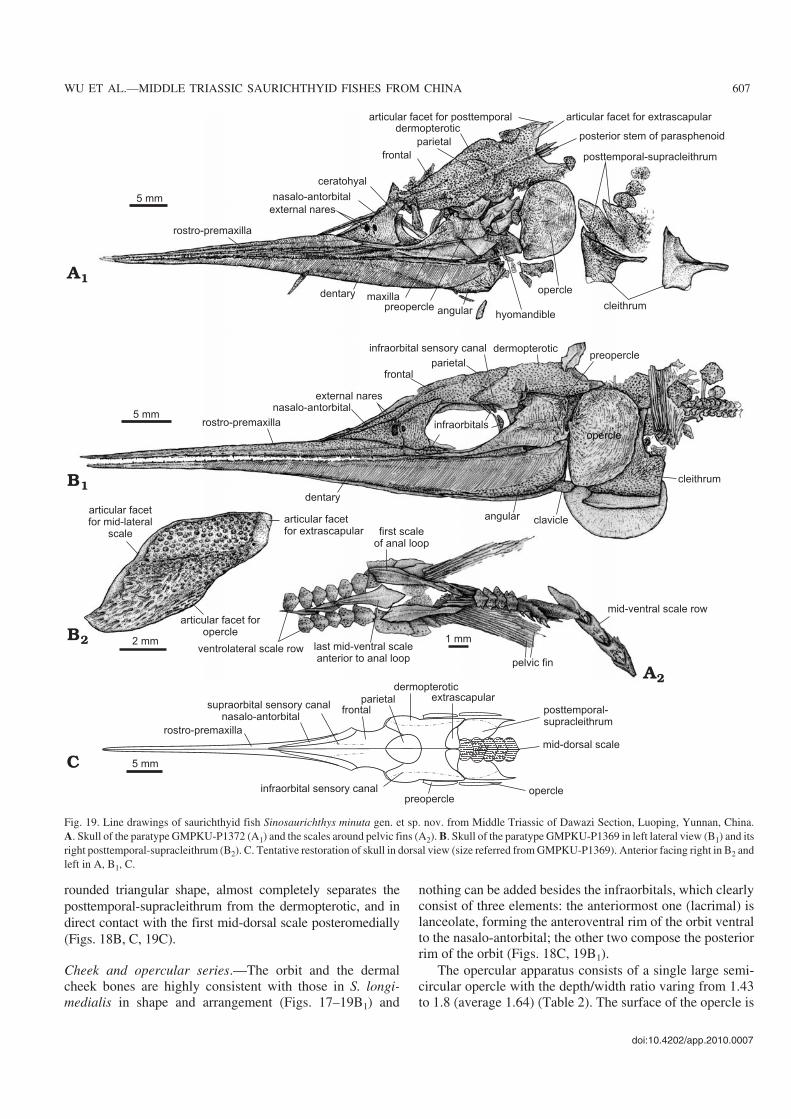
nothing can be added besides the infraorbitals, which clearlyconsist of three elements: the anteriormost one (lacrimal) islanceolate, forming the anteroventral rim of the orbit ventralto the nasalo−antorbital; the other two compose the posteriorrim of the orbit (Figs. 18C, 19B1).
The opercular apparatus consists of a single large semi−circular opercle with the depth/width ratio varing from 1.43to 1.8 (average 1.64) (Table 2). The surface of the opercle is
doi:10.4202/app.2010.0007
WU ET AL.—MIDDLE TRIASSIC SAURICHTHYID FISHES FROM CHINA 607
Fig. 19. Line drawings of saurichthyid fish Sinosaurichthys minuta gen. et sp. nov. from Middle Triassic of Dawazi Section, Luoping, Yunnan, China.A. Skull of the paratype GMPKU−P1372 (A1) and the scales around pelvic fins (A2). B. Skull of the paratype GMPKU−P1369 in left lateral view (B1) and itsright posttemporal−supracleithrum (B2). C. Tentative restoration of skull in dorsal view (size referred from GMPKU−P1369). Anterior facing right in B2 andleft in A, B1, C.
ornamented mainly with fine and dense concentric lines andtubercles. As in the other two species of Sinosaurichthys, thegular and branchiostegal rays are absent.
Mandible.—The shape and pattern of dermal bones in thelateral side of the mandible is almost the same with that of thetwo species described above (Fig. 18).
Palate.—Because of the preservation only part of the para−sphenoid can partially been seen, similar to the situation in S.longimedialis. This includes the long anterior stem across theventral part of the orbit, a posteriorly forked posterior stembetween the dermopterotic and opercle, and a large paired as−cending process posterior to the orbit (Figs. 18A, B, 19A1),but suggestive for the same morphology and construction tothat in the type species of the genus.
Hyoid arches.—The hyomandible is more or less hockey−stick shaped (Fig. 19A1), with a horizontally extended dorsalportion and a posteroventrally inclined ventral portion,slightly different from that in the type species in shape.
Dentition.—The teeth along the labial edge of both jaws arearranged in the same way and shape as in the other species ofSinosaurichthys (Fig. 17B2). The large teeth are quite smalland some of them curved posteriorly, similar to those in S.longimedialis (Fig. 17B2).
Paired fins and girdles.—The pectoral fin is roughly trian−gle−shaped, consisting of 14–16 unsegmented fin rays. Thelength of the pectoral fin varies from slightly less than tomore than the skull depth (Table 2), much shorter than that ofthe type species of the genus.
No radials or endoskeletal elements of the pectoral girdlecan be discerned, but the dermal pectoral girdle is well pre−served in most of the specimens and its elements are consis−tent with those in S. longimedialis in shape and configuration(Figs. 18, 19A1, B1, B2).
The pelvic fins are relatively small and triangular in shape(Fig. 20C), each consisting of about 18–20 unsegmented finrays. Little information about the pelvic bone is available inthe current materials.
Unpaired fins.—The dorsal and anal fins are arranged in thesame way as in the other species of Sinosaurichthys. Theyare triangular−shaped, much lower than those in the two spe−cies described above, with the depth almost equal to width(Fig. 20A2). The dorsal fin consists of about 41–50 and theanal fin of 42–48 fin rays, with a maximal segmentation ofthree to four times in the dorsal and four times in the anal fin.
Only the anterior radials of the dorsal and anal fins are os−sified, eight and seven to ten radials can be distinguished inthe dorsal and anal fin respectively.
The caudal fin is deeply forked, generally with 35–38 finrays in each lobe (Figs. 17A, B1, C, 20A1, Table 2). The finrays are generally segmented three to four times proximally,occasionally five times, and branched distally once or twice.
In the holotype, two basal fulcra are seen at the origin ofthe dorsal fin and three of the anal fin. The fringing fulcra, asin S. longimedialis, are present in all unpaired fins, consisting
of small spine−like elements overlapping one by one in thedistal part of the leading edge of the marginal fin rays.
Axial skeleton.—The axial skeleton consists of the neuraland haemal arches similar in structure to those in the otherspecies of Sinosaurichthys. The neural arches between theopercle and caudal fin number about 154–156 and the ante−rior 126–127 ones bear distinct neural spines, slightly fewerthan that in S. longimedialis. In the caudal fin region, thereare about 13–14 neural arches supporting the fin rays of theepichordal lobe of the caudal fin. Distinct haemal spines aredeveloped in the initial 10 to 11 haemal arches in the caudalregion, the fewest among the three species of the genus.
Squamation.—Similar to the two species described above,Sinosaurichthys minuta also bears six longitudinal scale rows.
There are 62–63 mid−dorsal scales in front of the dorsal fin.These scales are also cordate in the posterior exposed portion(Fig. 20B, E1, E2), wider than those in the type species and nar−rower than in S. longimedialis, with the width/length ratioabout 2.1–2.3 near the skull and 1–1.5 near the dorsal fin, andare decorated with spine−like tubercles (Fig. 20B).
The mid−ventral scale row divides into two rows slightlyanterior to the pelvic fins to form the anal loop and eachbranch consists of four scales (Figs. 19A2, 20A2, C). As in thetype species, the last scale anterior to the anal loop is en−larged and elongated with a rhombic posterior portion andthe first of paired scales of the anal loop is also elongated, ex−panding anteriorly and tapering posteriorly. In the caudalpeduncle, the mid−ventral scales are similar with the mid−dorsal ones in both shape and ornamentation, bearing somestrong posteriorly pointed tubercles (Fig. 20D).
The mid−lateral scales have a similar shape to that of typespecies and the dorsal part of the external surface is deco−rated with a few posteriorly−directed spines (Fig. 20E3).
The ventrolateral scale row commences a short distanceposterior to the skull and generally consists of small semicir−cular to rhombic scales, decorated with a few posteriorlypointed tubercles, and the scales are evidently enlarged nearthe origin of the pelvic fins (Fig. 19A2). Posterior to the pel−vic fins, the scale row continues backwards to the caudal fin(Fig. 20A1).
Remarks.—S. minuta is very similar to S. longimedialis in thegeneral morphology. The lack of substantial differences be−tween the two species, except for the body size and the shapeof the unpaired fins, makes it easy to regard S. minuta as the ju−venile of S. longimedialis. However, the degree of the seg−mentation in the unpaired fins between the two species doesnot support the juvenile−adult relationship because the numberof the segments of the fin rays usually increases accompany−ing with the growth of fin rays during ontogeny. It would becontradictory that the segmentation of the fin rays is fewer inadults than in juveniles. The number of rays in the pectoral finand ossified haemal spines in the caudal region is also differ−ent between the two species. Thus, here we treat S. minuta as aseparate species from S. longimedialis. Additionally, the twospecies occur in different stratigraphic levels. Field excavation
608 ACTA PALAEONTOLOGICA POLONICA 56 (3), 2011
doi:10.4202/app.2010.0007
WU ET AL.—MIDDLE TRIASSIC SAURICHTHYID FISHES FROM CHINA 609
Fig. 20. Saurichthyid fish Sinosaurichthys minuta gen. et sp. nov. from Middle Triassic of Dawazi Section, Luoping, Yunnan, China. Photographs (A–D)and line drawings (E). A. Caudal fin (A1) and dorsal and anal fins (A2) of the holotype GMPKU−P1955. B. Mid−dorsal scale row just behind skull in dorsalview (B1), mid−dorsal scale row in posterior abdominal region (B2) (14th to 24th scales counted anteriorly from dorsal fin) of GMPKU−P1369. C. Skeletonaround pelvic fins of the paratype GMPKU−P1372. D. Skeleton around middle part of caudal peduncle of the paratype GMPKU−P1928. E. Idealized linedrawings of dorsal scale row (dorsal view) near skull (E1) and around posterior abdominal region (E2), right mid−lateral scale row of abdominal region in lat−eral view (E3), and right ventrolateral scale row anterior to pelvic fins (E4). Anterior facing left in C, D and right in A, B, E.
reveals that S. longimedialis is restricted to the upper part ofthe fossiliferous strata, about 3–5 m above the level containingS. minuta in the Dawazi Section.
Comparison of the new genus withSaurichthysAs the type species of Saurichthys, S. apicalis, was estab−lished on the basis of a broken snout and isolated teeth(Agassiz 1834), the type material of the genus shows no cleardiagnostic features except the elongated, slender jaws andconical penetrating teeth, which caused a big confusion inthe subsequent studies. Stensiö (1925) and Rieppel (1985)tried to clarify the diagnosis of Saurichthys based on theircomprehensive work on the relatively well−preserved materi−als. However, the dozens of species attributed to the genus,mostly described based on rather poor and fragmentary ma−terials or even isolated teeth and showing great morphologicvariation, made it very difficult to get clear and appropriatediagnostic features for the genus. Under this situation, onlysome species erected on the basis of relatively well−pre−served materials are chosen as representatives of Saurichthysfor comparison with the current new genus.
Sinosaurichthys is very similar to Saurichthys in the gen−eral morphology. However, Sinosaurichthys differs clearlyfrom Saurichthys in having peculiar pectoral girdles, dor−sally inserted pectoral fins, highly elevated posterior stem ofthe parasphenoid, and relatively dorsally positioned axialskeleton in the abdominal region of the body, as well as dif−ferent configuration in squamation.
The pectoral girdle of the new genus connects with theskull through a large dermal element (here termed as post−temporal−supracleithrum). In Saurichthys the pectoral girdlemay connect with the skull through two elements (independ−ent posttemporal and supracleithrum), such as in Saurichthysornatus and Saurichthys madagascariensis (Stensiö 1925;Rieppel 1980), or through a small supracleithrum sensuRieppel (1985). The cleithrum of the pectoral girdle of Sino−saurichthys also differs from that of Saurichthys in shape.The posterior blade of this bone in Sinosaurichthys is ex−panded to a rectangular plate, much deeper than the anteriorarm, making the cleithrum look like a boot in lateral view.The cleithrum in Saurichthys is generally triradiate or in−verted “T” shaped with a low, round posterior blade that isgenerally as deep as or even lower than the anterior arm ofthis bone (see Stensiö 1925; Lehman et al. 1959; Griffith1962, 1977; Rieppel 1985, 1992; Mutter et al. 2008; Wu et al.2009). Only one specimen (GMUH VP992) attributed toSaurichthys cf. ornatus Stensiö, 1925, was mentioned tohave L−shaped cleithrum (Mutter et al. 2008: 118) but with−out further detailed information. The pectoral girdle in Sino−saurichthys shows more or less similar trend of specializa−tion to that in the hatchetfish (Gasteropelecidae) of SouthAmerica, the only extant fishes able to actively flap their ex−
tended pectoral fins in the air to obtain thrust (Rayner 1986)and whose posttemporal and supracleithrum are fused into asingle large bone with the pectoral girdle extremely ex−panded (Brousseau 1976; Weitzman and Palmer 2003). Thefusion of the posttemporal and supracleithrum in the hatchetfish is considered as a peculiar osteological feature related tospecialization of their pectoral girdle and fins for jumpingand flying behavior (Wiest 1995). Although it is hard to saythat Sinosaurichthys had obtained similar jumping and flyingability, it is undoubted that the similarity in the modificationof the pectoral girdle in Sinosaurichthys and the hatchetfishreflects a functional convergence to enhance the pectoral gir−dle supporting stronger pectoral musculatures.
Although the pectoral fins in both Sinosaurichthys andSaurichthys are inserted between the dorsal stem and poste−rior blade of their cleithrum, the different shape and depth ofthe cleithrum makes the insertion of the pectoral fins abovethe midpoint of the body depth in Sinosaurichthys and dis−tinctly below that in Saurichthys. The pectoral fins are alsodifferent in shape between the two genera. It is triangular tosickle shaped and relatively long in Sinosaurichthys and issmall fan shaped in the Middle–Late Triassic Saurichthysfrom Europe (Griffith 1959, 1962, 1977; Rieppel 1985). Theposterior stem of the parasphenoid in Sinosaurichthysis ele−vated much high above the mid−point of the skull depth andcorrespondingly the axial skeleton in the abdominal region isalso situated in the level above the mid−point of the bodydepth.
The information about the parasphenoid was recordedonly in a few species of Saurichthys, e.g., S. ornatus, S.hamiltoni, and S. wimani from Spitsbergen (Stensiö 1925), S.madagascarensis from Madagascar (Beltan 1968), S. obru−tchevi and S. proximus from Central Asia (Minich 1981) andS. curionii from Monte San Giorgio (Rieppel 1985). All ofthem show that the posterior stem of the parasphenoid is justslightly elevated and the ventral crista is rather low and theirvertebral column is therefore situated close to the mid−pointof the body depth. The differences in the dermal pectoral gir−dle, the insertion of the pectoral fins and the position of theaxial skeleton in the abdominal region between the two gen−era are apparently related to their difference in locomotionbehavior and ecological adaptation (see discussion below).
As for the squamation, the different configuration of themid−dorsal and mid−ventral scales of Sinosaurichthys alsomakes it distinguishable from Saurichthys. In the species ofSinosaurichthys, the mid−dorsal scales anterior to the dorsalfin are so broad that they are much wider than narrowmid−ventral scales. This unique feature is different from themajority of Saurichthys with the narrow, lanceolate mid−dor−sal and mid−ventral scales and few species with mid−ventralscales as broad as the wide mid−dorsal ones in the abdominalregion, such as S. madagarscariensis, S. gigas, and S. dayi(Woodward 1890; Piveteau 1945; Rieppel 1980; Mutter et al.2008). This feature makes sense for it suggests that Sino−saurichthys probably has a bodly form of an inverted drop inthe transverse section, i.e., the dorsal ridge comparatively
610 ACTA PALAEONTOLOGICA POLONICA 56 (3), 2011
broad and ventral edge narrow, which consequently means arelatively dorsally positioned center of gravity, mechanicallycorrelated with the elevation of its axial skeleton and the highinsertion of the pectoral fin.
Other important features distinguishing Sinosaurichthysfrom Saurichthys are related to the branchiostegal rays andposition of the pelvic and unpaired fins. In all known saurich−thyids, the branchiostegal rays are highly reduced. Amongthe relatively well−known species of Saurichthys the bran−chiostegal rays are generally reduced to one pair (Stensiö1925; Rieppel 1985), though two pairs occur in a species ofSaurichthys under description from the Panxian fauna (Wu2009) and possibly three pairs in Saurichthys madagasca−riensis (Lehman 1952). However, among the over one hun−dred specimens of Sinosaurichthys we checked, none showsany trace of branchiostegal rays.
Among Saurichthys, the position of the pelvic and un−paired fins in the trunk is recorded in quite a few species, e.g.,S. gigas from Australia (Woodward 1890), S. krambegeri andS. calcaratus from Austria (Griffith 1962; 1977), S. curionii,S. macrocephalus, and S. costasquamosus from Monte SanGiorgio (Rieppel 1985), S. dayi and S. toxolepis from Canada(Mutter et al. 2008), and a. dawaziensis from South China(Wu et al. 2009). Although the pelvic fins in certain species,such as S. gigas, S. dayi, and S. costasquamosus, are posi−tioned in similar place as in Sinosaurichthys, i.e., behind themid−point of the distance between the opercle and caudal fin,the anal fin of them is exclusively situated in place closer to thepelvic than to the caudal fin. The available information indi−cates that the positions of the anal fin in species of Saurichthysare generally closer to the caudal than to the pelvic fins or nearthe mid−point between the pelvic and caudal fins.
Thus Sinosaurichthys is easily distinguished from allknown species of Saurichthys based on the features dis−cussed above.
Ecology and functional morphologyAs noted above, one of the most distinguishable morphologi−cal features between Sinosaurichthys gen. nov. and Saurich−thys is the position and shape of their pectoral fins, which arelong, triangular to sickle shaped and dorsally inserted inSinosaurichthys and small, fan shaped and ventrolaterallypositioned in Saurichthys. The pectoral fins of fish are pri−marily responsible for controlling body stability, turning andbraking while swimming, especially for the forms usingbody/caudal fin propulsion. The morphologic similarity tothe extant needlefish suggests that the locomotion mode ofthese saurichthyids should resemble that of the belonids, rep−resenting anguilliform (Liao 2002) to carangiform body/cau−dal fin propulsion type (sensu Lindsey 1978). For fishes withpectoral fins situated low on the body, Breder (1926) sup−posed that the reaction of the braking force to the center ofbody mass will exert a substantial pitching or “somersault−ing” movement due to lack of a vertically oriented lift com−
ponent in the braking force because the center of pressure ofthe pectoral fin lies below the center of the body mass. Forfishes whose pectoral fins extend higher up on the body, Har−ris (1938) suggested that the braking force is comprised ofboth a horizontal and vertical component so that the reactionforce vector intersects the center of body mass, eliminatingthe destabilizing pitching moment. Modern experimentalstudy also reveals that high positioned pectoral fins can gen−erate higher forces for turning and direct braking forcescloser to the center of the body mass and will tend to havea greater ability to control body rotation during braking(Drucker and Lauder 2002), which may be of advantage forfish to maintain the body balance and agility in sustainedswimming. Among extant actinopterygians, some rapid andpersistent swimmers, such as the scombrids, belonids, andexocoetids, have relatively long and high inserted pectoralfins. Saurichthys had originally been considered as a kind ofpelagic predator, like the extant needlefish (Stensiö 1925;Beltan and Janvier 1978). This conclusion is supported bythe presumed absence of an opercular process on the hyo−mandible suggesting a rather weakly developed operclularpump (Stensiö 1925; Rieppel 1992). However, the relativelysmall and lower positioned pectoral fins seem to excludeSaurichthys from the category of the persistent and rapidswimmers that can chase their prey long distances. It is likelythat Saurichthys is an ambush or Esox−like predator (Rieppel1985, 1992; Tintori 1990), using its fast−start capability towaylay rather than pursue its prey. On the other hand, al−though Sinosaurichthys also possessed a typical “accelera−tion specialist” morphology like Saurichthys and the extantpike (Family Esocidae), the distinct resemblance in the bodyshape and fin arrangement to the needlefish (Fig. 21) sug−gests that Sinosaurichthys was a typical belonid−like predatorand probably had similar locomotion behavior and ecologi−cal habits as the needlefish. Most of the needlefish are swiftsurface−cruising predators, primarily inhabiting marine envi−ronments and feeding largely on small fishes which theycatch sideways in their long jaws. Some were reported tohave a similar migratory pattern as the mackerels (Muus andNielsen 1999). Thus, we believe that Sinosaurichthys repre−sents a lineage of saurichthyids derived from Saurichthys toadapt to a surface−cruising life style similar to the extantneedlefish, and the dorsally positioned pectoral fins mightsuggest the acquisition of sustained swimming ability in thisgenus. Field excavation at the two fossil sites revealed thatthe specimens of Sinosaurichthys discovered are much moreabundant than those of Saurichthys and always clustered to−gether in certain mass death layers. This tends to support theinterpretation that Sinosaurichthys is a surface−cruisingswimmer and Saurichthys is an ambush predator, becausethe cruising swimmers generally swim in schools contrary tothe solitary ambush predators, and thus have more individu−als than the latter in a living community. The taphonomic re−cords of the mass death events can more or less reflect thedifference of the relative individual abundance of each taxonin a natural local community.
doi:10.4202/app.2010.0007
WU ET AL.—MIDDLE TRIASSIC SAURICHTHYID FISHES FROM CHINA 611
It is interesting to note that three species of Sinosaurich−thys from the two fossil sites show different trends in the de−velopment of their fins. In the type species of the genus, Sino−saurichthys longipectoralis from Panxian of Guizhou, thepaired fins are extremely elongated, whereas in the two spe−cies from Luoping of Yunnan, one (S. minuta) keeps normalsized fins, the other (S. longimedialis) has extremely elon−gated median fins. This difference in fin morphology likelyreflects a further differentiation in locomotion behavior andhabits.
For Sinosaurichthys longipectoralis, the configuration ofits paired fin is more or less similar to that of the extant flyingfish (Family Exocoetidae) (Fig. 21), a close relative of theneedlefish. Exocoetids are known as having the capability ofleaping out of the water and gliding for long distances abovethe water surface to escape from predators (Gibbs 1978;Kutschera 2005), in which both pectoral and sometimes pel−vic fins are evidently extended to act as “wings” when glid−ing (Kutschera 2005). However, the distal part of the fin raysof the pectoral fins in S. longipectoralis seems too fine tosupport flying fish−like gliding. Possibly, this species pos−sessed only simple leaping ability like some extant needle−
fishes (Collette 1977; Helfman et al. 1997). However, thisspecies has high aspect ratio median fins. The aspect ratio(defined as depth2/area) of its caudal fin is about 4–5, higherthan that of other saurichthyids (Fig. 21) and within the lowerrange of typical thunniform swimmers like recent mackerelor tunas (aspect ratio 4.5 to 7.2; Sambilay 1990), which ren−ders this species one of the fast swimming fishes during thattime. A high aspect ratio is thought to be characteristic of pe−lagic marine fishes that have enhanced cruising speed, en−abling them to travel for long distance in search for food andbreeding locality (Webb 1994). Thus, we believe that theelongation of the paired fins in S. longipectoralis probablyrepresents a trend of adaptation to enhance the rapid swim−ming performance and, possibly, for a low−efficiency glidingability. Field observation and excavation revealed that nobenthonic organism was associated with this species exceptthin−shelled bivalves. The presence of other actinopterygianfishes and ichthyopterygian reptile Mixosaurus panxianensis(Jiang et al. 2006) suggests a relatively open offshore ratherthan a near−shore coastal habitat. The enhancement of rapidswimming performance is no doubt advantageous for thespecies to search for food and escape from larger predators(such as Birgeria and Mixosaurus) in an open pelagic envi−ronment. In addition to using the long pectoral fins to keepthe body in balance and enhance swimming ability (evengliding), it is also possible that the species uses the pectoralfins to drive its prey, like recent sailfish using the sail−likedorsal fin to hunt (Holland 2008).
For S. longimedialis from the Luoping of Yunnan, theelongation of the median fins is obviously a disadvantage forrapid swimming, as compared to S. longipectoralis. Althoughno solid answer can be put forward for why this species hassuch elongated median fins, a possible reason is that it lived ina more structurally complicated, near−coastal habitat, in whichpersistent rapid swimming performance may not be as impor−tant as in an open pelagic environment. This interpretation issupported by the taphonomic evidence that many benthonicinvertebrates, such as shrimps, echinoderms, gastropods, andeven freshwater−related isopods, were associated with thisspecies in the upper part of the fossiliferous strata in theDawazi Section.
ConclusionsSinosaurichthys is a new genus of the Saurichthyidae differ−ing from Saurichthys in having a peculiar pectoral girdle andhigh−inserted pectoral fin, and some other related features.These differences reflect the variation in the locomotionmode and ecological adaptation of saurichthyids. The newgenus represents a lineage closely related with Saurichthysbut adapted to achieve improved sustained swimming forcruising in surface water, like the extant needlefishes. Thedifferent fin morphology between the species of the new ge−nus might suggest a further differentiation in locomotion be−havior and ecologic habitats.
612 ACTA PALAEONTOLOGICA POLONICA 56 (3), 2011
Fig. 21. Morphological comparison of saurichthyids with extant needlefishand flying fish. A. Sinosaurichthys longipectoralis. B. Sinosaurichthyslongimedialis. C. Sinosaurichthys minuta. D. Saurichthys dawaziensis fromMiddle Triassic of Dawazi Section, Luoping, Yunnan, China (based on Wuet al. 2009). E. Atlantic needlefish Strongylura marina (Family Belonidae).F. Bluntnose flyingfish Prognichthys gibbifrons (Family Exocoetidae).E and F redrawed from images at http://www.fishbase.org/images/speciesthat were originally from Cervigón et al. (1992).
AcknowledgementsSpecial thanks to Mee−mann Chang (Institute of Vertebrate Paleontologyand Paleoanthropology, Beijing, China) for her kind encouragement andhelpful comments. We also thank Andrea Tintori (Universita degli Studidi Milano, Italy) for reading an earlier draft of the manuscript and provid−ing helpful suggestion. Comments and corrections provided by ToniBürgin (Naturmuseum St. Gallen, Switzerland), Brian M. Davis (Univer−sity of Oklahoma, Norman, USA), and Ilja Kogan (Freiberg Universityof Mining and Technology, Germany) greatly helped to improve thequality of the manuscript. This work is supported by the NSFC (grantsnos. 40572004, 40702001, and 41102011) and the State Key Laboratoryof Palaeobiology and Stratigraphy (NIGP, CAS) (grant no. 123109).
ReferencesAgassiz, L. 1834–1935. Recherches sur les Poissons Fossiles, Vol. 2. 336
pp. Imprimerie de Petitpierre, Neuchâtel.Aldinger, H. 1937. Permische Ganoidfische aus Ostgrönland. Meddelelser
om Grønland 102: 1–392.Arratia, G. 2008. Identifying patterns of diversity of the actinopterygian ful−
cra. Acta Zoologica (Stockholm) 90 (Supplement 1): 220–235.Arratia, G., Schultze, H.P., and Casciotta, J. 2001. Vertebral column and as−
sociated elements in Dipnoans and comparison with other fishes: devel−opment and homology. Journal of Morphology 250: 101–172.
Bellotti, C. 1857. Description di alcune nuove specie di fossili di Perledo e dialter località Lombarde. In: A. Stoppani (ed.), Studii Geologici ePaleontologici sulla Lombardia, 419–438. C. Turati, Milano.
Beltan, L. 1968. La faune ichthyologique de l’Eotrias du N. W. de Madagas−car: le neurocrane. 135 pp. C.N.R.S., Paris.
Beltan, L. 1972. La faune ichthyologique du Muschelkalk de la Catalogne.Memorias de la Real Academia de Ciencias y Artes de Barcelona, Bar−celona 41 (10): 281–352.
Beltan, L. and Janvier, P. 1978. Un nouveau Saurichthyidae (Pisces, Actino−pterygii), Saurichthys nepalensis n. sp., du Trias inférieur des Anna−purnas (Thakkhola, Nepal) et sa signification Paléobiogéograhique.Cybium 3: 17–28.
Beltan, L. and Tintori, A. 1980. The genus Saurichthys (Pisces, Actino−pterygii) during the Gondwana period. Proceedings of the Fifth Interna−tional Gomdwana Symposium, Wellington, New Zealand, 11–16 Feb−ruary, 1980, 53–59. A.A. Balkema, Rotterdam.
Breder, C.M. 1926. The locomotion of fishes. Zoologica 4: 159–297.Bronn, H.G. 1858. Beiträge zur triassischen Fauna und Flora der bituminösen
Schiefer von Raibl. Neues Jahrbuch für Mineralogie , Geognosie, Geo−logie und Petrefaktenkunde 1: 1–63.
Brousseau, R.A. 1976. The pectoral anatomy of selected Ostariophysi. 1.The Characiniformes. Journal of Morphology 148: 89–136.
Cartanyà, J. 1999. An overview of the Middle Triassic actinopterygiansfrom Alcover, Mont−ral and El Pinetell (Catalonia, Spain). In: G.Arratia and H.P. Schultze (eds.), Mesozoic Fishes 2—Systematics andFossil Record, 535–551. Verlag Dr. F. Pfeil, München.
Cervigón, F., Cipriani, R., Fischer, W., Garibaldi, L., Hendrickx, M., Lemus,A.J., Márquez, R., Poutiers, J.M., Robaina, G., and Rodriguez, B. 1992.Fichas FAO de identificación de especies para los fines de la pesca. Guíade campo de las especies comerciales marinasy de aquas salobres de lacosta septentrional de Sur América. Preparado con el financiamento de laComisión de Comunidades Europeas y de NORAD. 513 pp. FAO, Rome.
Collette, B.B. 1977. Belonidae. In: W. Fischer (ed.), FAO Species Identifi−cation Sheets for Fishery Purposes: Western Central Atlantic (FishingArea 31), Vol. 1, 14. FAO, Rome.
Costa, A. 1862. Descrizione de alcuni annelidi del Golfo di Napoli. Annuariodel Museo Zoologico della Reale Università di Napoli 1: 82–90.
Drucker, E.G. and Lauder, G.V. 2002. Wake dynamics and locomotor func−
tion in fishes: interpreting evolutionary patterns in pectoral fin design.Integrative and Comparative Biology 42: 997–1008.
Gardiner, B.G. 1960. A review of certain actinopterygian and coelacanthfishes, chiefly from the Lower Lias. Bulletin of the British Museum(Natural History), Geology 4 (7): 239–384.
Gardiner, B.G. 1984. The relationships of the palaeoniscid fishes, a reviewbased on new specimens of Mimia and Moythomasia from the UpperDevonian of Western Australia. Bulletin of the British Museum (NaturalHistory), Geology 37: 173–428.
Gardiner, B.G. and Schaeffer, B. 1989. Interrelationships of lower actino−pterygian fishes. Zoological Journal of the Linnean Society 97: 135–187.
Gibbs, R.H., Jr. 1978. Exocoetidae. In: W. Fischer (ed.), FAO Species Iden−tification Sheets for Fishery Purposes. Western Central Atlantic (Fish−ing Area 31), Volume 2. 32 pp. FAO, Rome.
Gozzi, E. 2004. Analisi Tassonomica e morfo−funzionale di Saurichthys eBirgeria (Osteichthyes, Actinopterygii). 379 pp. Unpublished Ph.D.thesis, Università degli Studi di Milano, Milano.
Grande, L. and Bemis, W.E. 1998. A comprehensive phylogenetic study ofamiid fishes (Amiidae) based on comparative skeletal anatomy. An em−pirical search for interconnected patterns of natural history. Memoir(Society of Vertebrate Paleontology) 4: 1–690.
Griffith, J. 1959. On the anatomy of two saurichthyids fishes, Saurichthysstriolatus (Bronn) and S. curionii (Bellotii). Proceedings of the Zoologi−cal Society of London 132 (4): 587–606.
Griffith, J. 1962. The Triassic fish Saurichthys krambergeri Schlosser.Palaeontology 5: 344–354.
Griffith, J. 1977. The Upper Triassic fishes from Polzberg bei Lunz, Austria.Zoological Journal of the Linnean Society 60: 1–93.
Griffith, J. 1978. A fragmentary specimen of Saurichthys sp. from the UpperBeaufort series of South Africa. Annals of the South African Museum 76(8): 299–307.
Harris, J.E. 1938. The role of the fins in the equilibrium of the swimmingfish. II. The role of the pelvic fins. Journal of Experimental Biology 16:32–47.
Helfman, G.S., Collette, B.B., and Facey, D.E. 1997. The Diversity ofFishes. 528 pp. Blackwell Science, Malden, MA.
Holland, J.S. 2008. Sailfish in the whirl. National Geographic September2008: 70–79. http://www. nationalgeographic.com /2008/09/sailfish/holland−text.
Jiang, D.Y., Schmitz, L., Hao, W.C., and Sun, Y.L. 2006. A new mixosauridichthyosaur from the Middle Triassic of China. Journal of VertebratePaleontology 26: 60–69.
Kogan, I., Schönberger, K., Fischer, J., and Voigt, S. 2009. A nearly completeskeleton of Saurichthys orientalis (Pisces, Actinopterygii) from the Mady−gen Formation (Middle to Late Triassic, Kyrgyzstan, Central Asia)—pre−liminary results. Freiberger Forschungshefte C532: 139–152.
Kutschera, U. 2005. Predaor−driven macroevolution in flyingfishes inferredfrom behavioural studies: historical controversies and a hypothesis. An−nals of the History and Philosophy of Biology 10: 59–77.
Lehman, J.P. 1952. Etude complémentaire des poissons de l’Eotrias deMadagascar. Kungliga Svenska Vetenskapsakademiens Handlingar, 4Series 2: 1–201.
Lehman, J.P., Chateau, C., Laurain, M., and Nauche, M. 1959. Paléontologiede Madagascar. XXVIII. Les poisons de la Sakamena Moyenne. Annalesde Paléontologie Vertébres 45: 177–219.
Liao, J.C. 2002. Swimming in needlefish (Belonidae): anguilliform locomo−tion with fins. Journal of Experimental Biology 205: 2875–2884.
Lindsey, C.C. 1978. Form, function, and locomotory habits in fish. In: W.S.Hoar and D.J. Randall (eds.), Fish Physiology 2. 100 pp. AcademicPress, New York.
Liu, X.T. and Wei, F. 1988. A new saurichthyid from the Upper Permian ofZhejiang, China [in Chinese, with English abstract]. Vertebrata Pal−asiatica 26 (2): 77–89.
Minich, A.V. 1981. Saurichthys species from the Triassic of the USSR [inRussian]. Paleontologičeskij žurnal 15: 105–113.
Mutter, R.J., Cartanyà, J., and Susan, A.U. 2008. New evidence of Saurich−thys from the Lower Triassic with an evaluation of early saurichthyid di−
doi:10.4202/app.2010.0007
WU ET AL.—MIDDLE TRIASSIC SAURICHTHYID FISHES FROM CHINA 613
versity. In: G. Arratia, H.P. Schultze, and M.V.H. Wilson (eds.), Meso−zoic Fishes 4—Homology and Phylogeny, Proceedings of the interna−tional meeting Miraflores de la Sierra, 2005, 103–127. Verlag Dr.Friedrich Pfeil, München.
Muus, B.J. and Nielsen, J.G. 1999. Sea Fish. Scandinavian Fishing YearBook. 340 pp. Hedehusene, Denmark.
Neuman, A.G. and Wilson, M.V.H. 1985. A fossil fish of the familySauricthyidae from the Lower Jurassic of western Alberta, Canada. Ca−nadian Journal of Earth Sciences 22: 1158–1162.
Nielsen, E. 1942. Studies on Triassic fishes from East Greenland I. Glauco−lepis and Boreosomus. Meddelser om Grønland 138: 1–403.
Nielsen, E. 1949. Studies on Triassic fishes from East Greenland. II.Australosomus and Birgeria. Meddelelser om Grønland 146: 1–309.
Patterson, C. 1975. The braincase of pholidophorid and leptolepid fishes,with a review of the actinopterygian braincase. Philosophical Transac−tions of Royal Society of London 269: 275–579.
Piveteau, J. 1945. Les Poissons du Trias Inférieur la Famille das Saurich−thyidés.Paléontologie de Madagascar, XXV. Annales de Paléontologie31: 79–87.
Rayner, J.M.V. 1986. Pleuston: animals which move in water and air.Endeavour 10: 58–64.
Rieppel, O. 1980. Additional specimens of Saurichthys madagascariensisPiveteau, from the Eotrias of Madagascar. Neues Jahrbuch für Geologieund Paläontologie, Monatshefte 1980 (1): 43–51.
Rieppel, O. 1985. Die Triasfauna der Tessiner Kalkalpen.XXV. Die GattungSaurichthys (Pisces, Actinopterygii) aus der mittleren Trias des MonteSan Giorgio, Kanton Tessin. Schweizerische Paläontologische Abhand−lungen 108: 1–103.
Rieppel, O. 1992. A new species of the genus Saurichthys (Pisces: Actino−pterygii) from the Middle Triassic of Monte San Giorgio (Switzerland),with comments on the phylogenetic interrelationships of the genus.Palaeontographica A 221: 63–94.
Sambilay, V.C. Jr. 1990. Interrelationships between swimming speed, cau−dal fin aspect ratio and body length of fishes. Fishbyte 8: 16–20.
Stensiö, E. 1925. Triassic Fishes from Spitzebergen, Part II. KungligaSvenska Vetenskapsakademiens Handlinga 2: 1–126.
Sun, Z.Y., Sun, Y.L., Hao, W.C., and Jiang, D.Y. 2006. Conodont evidencefor the age of the Panxian Fauna, Guizhou, China. Acta GeologicaSinica, English Edition 80: 621–630.
Sun, Z.Y., Tintori, A., Cristina, L., Jiang, D.Y., Wu, F.X., Hao, W.C., Sun,Y.L., and Rusconi, M. 2008. A new species of Colobodus (Actino−pterygians) from the Pelsonian (Anisian, Middle Triassic) of Guizhou,South China. Rivista Italiana di Paleontologia e Stratigrafia 114:363–376.
Sun, Z.Y., Tintori, A., Jiang, D.Y., Lombardo, C., Rusconi, M., Hao, W.C.,and Sun, Y.L. 2009. A new perleidiform (Actinopterygii, Osteichthyes)from the Middle Anisian (Middle Triassic) of Yunnan, South China.Acta Geologica Sinica, English Edition 83: 460–470.
Thies, D. 1985. Funde von Acidorhynchus brevirostris (Woodward 1895)aus dem Posidonienschiefer (Unter−Toarcium) NW−Deutschlands. Palae−ontographica A 187: 183–203.
Tintori, A. 1990. The vertebral column of the Triassic fish Saurichthys(Actinopterygii) and its stratigraphical significance. Rivista Italiana diPaleontologia e Stratigrafia 96: 93–102.
Tintori, A., Sun, Z.Y., Lombardo, C., Jiang, D.Y, Sun, Y.L., and Rusconi,M. 2008. New specialized basal neopterygians (Actinopterygii) fromTriassic of the Tethys Realm. Geologia Insubrica 10: 13–20.
Webb, P.W. 1994. The biology of fish swimming. In: L. Maddock, Q. Bone,and J.M.V. Rayner (eds.), Mechanics and Physiology of Animal Swim−ming, 45–62. Cambridge University Press, Cambridge.
Weitzman, S.H. and Palmer, L. 2003. Family Gasteropelecidae (freshwaterhatchetfishes). In: R.E. Reis, S.O. Kullander, and S.J. Ferraris (eds.),Check List of the Freshwater Fishes of South and Central America,101–103. EDIPUCRS, Porto Alegre.
Wiest, F.C. 1995. The specialized locomotory apparatus of the freshwaterhatchetfish family Gasteropelecidae. Journal of Zoology 236: 571–592.
Woodward, A.S. 1890. The fossil fishes of the Hawkesbury Series atGosford. Memoirs of the Geological Survey of New South Wales 4:1–55.
Woodward, A.S. 1891. Catalogue of the Fossil Fishes in the British Mu−seum (Natural History). Vol. 2. 567 pp. British Museum (Natural His−tory), London.
Wu, F.X. 2009. The Middle Triassic Saurichthyid Fishes from SouthwesternChina [in Chinese, with English abstract]. 106 pp. Unpublished Ph.D.thesis, Peking University, Beijing.
Wu, F.X., Sun, Y.L., Hao, W.C., Jiang, D.Y., and Tintori, A. 2008. New fos−sils and morpho−ecologic diversity of Saurichthyidae (Actinopterygian)from the Middle Triassic of Southwestern China. Journal of VertebratePaleontology 28: 162A.
Wu, F.X., Sun, Y.L., Hao, W.C., Jiang, D.Y., Xu, G.H., Sun, Z.Y., and Tintori,A. 2009. A new species of Saurichthys (Actinopterygii: Saurichthyidae)from Middle Triassic (Anisian) of Yunnan Province, China. Acta Geo−logica Sinica, English Edition 83: 440–450.
Zhang, Q.Y., Zhou, C.Y., Lü, T., and Bai, J.K. 2010. Discovery of MiddleTriassic Saurichthys in the Luoping area, Yunnan, China [in Chinese,with English abstract]. Geological Bulletin of China 29: 26–30