Page 1
NEWLY DESIGNED HNO TRIGGERED PRODRUGS
BY
WENRUI SU
A Thesis Submitted to the Graduate Faculty of
WAKE FOREST UNIVERSITY GRADUATE SCHOOL OF ARTS AND SCIENCES
in Partial Fulfillment of the Requirements
for the Degree of
MASTER OF SCIENCE
Chemistry
August 2016
Winston-Salem, North Carolina
Approved By:
Stephen B. King, Ph. D., Advisor
Mark E. Welker, Ph. D., Chair
Paul B. Jones, Ph. D.
Page 2
II
Acknowledgments
I am grateful to my advisor, Dr. S. B. King, whose experience, knowledge make it
possible for me to keep working on the study of prodrugs. Since my English is not good,
he also helped me a lot in revising this thesis with patience. I really learned a lot from
him in this two-year study. It was my pleasure to work with him.
Thanks to Zhengrui Miao in helping me with many experiments since I was a
beginner in organic chemistry. He also helped me a lot in proposing the basic idea of this
thesis. Without his help, I can’t finish the research.
I would thank Mu Yang for helping me in daily life. Since I was new in the U. S., he
taught me a lot about how to live in the strange country.
I would also thank Mr. Tom Poole, Dr. Colin Douglas, Dr. Rajeswari Mukherjee and
Dr. Salwa Elkazaz. They also helped me in experiments so that I could complete the
research.
At last, I would express my sincere gratitude to my parents. When I feel disappointed
and frustrated, they always encouraged me and made me feel optimistic to future. They
supported any decisions I made and helped me in achieving the goals. I might not be able
to finish my study without their encouragement and support.
Page 3
III
Table of Contents
List of Figures .................................................................................................................... V
Abstract .......................................................................................................................... VIII
Chapter 1. Background .....................................................................................................1
1.1 NO ..............................................................................................................................1
1.1.1 Biological Properties .........................................................................................1
1.1.2 Chemical Properties ..........................................................................................3
1.2 HNO ...........................................................................................................................4
1.2.1 Chemical Properties ..........................................................................................4
1.2.2 Biological Properties .........................................................................................6
1.2.3 Biosynthesis of HNO..........................................................................................8
1.3 HNO Detection ..........................................................................................................8
1.4 HNO Donor .............................................................................................................14
1.4.1 General Ways to Form HNO ..........................................................................14
1.4.2 Biological HNO Donor ....................................................................................16
1.5 Prodrugs ..................................................................................................................17
Chapter 2. Results and Discussion .................................................................................22
2.1 Prodrug Synthesis ..................................................................................................22
2.2 Reaction with HNO ................................................................................................25
Page 4
IV
2.2.1 Wintergreen Ester (3)......................................................................................27
2.2.2 Acetaminophen Ester (5) ................................................................................30
2.2.3 Metronidazole Ester (7) ..................................................................................32
2.2.4 Mechanism Discussion ....................................................................................35
2.3 Kinetic Analysis ......................................................................................................36
2.4 Selectivity ................................................................................................................40
2.5 Conclusion ...............................................................................................................44
Chapter 3. Experimental .................................................................................................46
3.1 General Chemistry .................................................................................................46
3.2 Synthetic Procedure ...............................................................................................46
3.3 Treatment of Prodrugs with HNO ........................................................................49
3.4 Kinetic Analysis of the Reaction of 3 with HNO .................................................50
3.5 Selectivity ................................................................................................................51
References .........................................................................................................................52
Appendix ...........................................................................................................................56
Curriculum Vitae .............................................................................................................76
Page 5
V
List of Figures
Figure 1. Physiology of NO in vasolidation ........................................................................2
Figure 2. The energy gap between different spin states of HNO .........................................6
Figure 3. Structure of [CuII(BOT1)Cl].................................................................................9
Figure 4. Structure of CoII(P) ............................................................................................10
Figure 5. Scheme of CoII(P) reacting with HNO ..............................................................10
Figure 6. Structure of an organic phosphine HNO probe ..................................................11
Figure 7. Scheme of organic probe reacting with HNO ....................................................12
Figure 8. Reaction mechanism of 13 with HNO................................................................13
Figure 9. Scheme of Nef reaction ......................................................................................14
Figure 10. HNO formation from nitrosative cleavage .......................................................15
Figure 11. HNO formation from retro-Diels Alder reaction ..............................................15
Figure 12. Decomposition of N-phosphinoylhydroxylamines...........................................15
Figure 13. Decomposition of Piloty’s Acid to HNO .........................................................16
Figure 14. Decomposition of Angeli’s Salt .......................................................................17
Figure 15. Mechanism of organophosphine-based HNO triggered prodrug .....................19
Figure 16. Methyl salicylate ..............................................................................................19
Figure 17. Acetaminophen .................................................................................................19
Figure 18. Metronidazole ...................................................................................................20
Figure 19. Metronidazole prodrug decomposition in the presence of HNO ......................21
Page 6
VI
Figure 20. Synthesis of 3 ...................................................................................................22
Figure 21. Synthesis of 5 ...................................................................................................23
Figure 22. Synthesis of 7 ...................................................................................................23
Figure 23. Reaction Mechanism ........................................................................................25
Figure 24. Reaction of 3 with HNO ...................................................................................27
Figure 25. Crude 31P-NMR of the reaction of 3 with HNO and comparison ....................28
Figure 26. LC-MS of the mixture of 3 and HNO ..............................................................29
Figure 27. Acetaminophen ester reacts with HNO ............................................................30
Figure 28. Crude 31P-NMR of the reaction of 5 with HNO and comparison ....................31
Figure 29. Reaction of metronidazole (7) with HNO ........................................................32
Figure 30. Crude 31P-NMR of the reaction of 7 with HNO and comparison ...................33
Figure 31. LC-MS of the mixture of 7 and HNO .............................................................34
Figure 32. Hydrolysis of ylide and phosphine oxide .........................................................36
Figure 33. Kinetic study .....................................................................................................38
Figure 34. Concentration of 3 vs. Time after Angeli’s Salt addition .................................39
Figure 35. Structure of biological reducing and oxidizing agents .....................................40
Figure 36. 31P-NMR of selectivity experiments ................................................................41
Figure 37. 31P-NMR of the reaction of 7 with NaNO2 ......................................................42
Figure 38. 31P-NMR of the reaction of 7 with DEANO ....................................................43
Figure 39. Summary of the reaction study .........................................................................45
Page 7
VII
Figure 40. Cell experiment of the prodrug.........................................................................46
Page 8
VIII
Abstract
Nitric oxide (NO) is an important species in many biological processes. The one-
electron reduced and protonated derivative of NO, HNO has also been determined to play
important biological roles. Because of HNO’s high reactivity, its detection has been
difficult and many methods including fluorescent copper compounds and electrochemical
methods have been devised. Our group has studied HNO for many years and recently
developed a detection method based on HNO’s reaction with organophosphorus
compunds.
Inflammation is associated with many pathological disorders, such as cancer and
infection and production of reactive oxygen species (ROS), including H2O2, is central to
many inflammatory diseases. ROS can react with NO to generate reactive nitrogen
species (RNS) including HNO and HNO-triggered prodrugs might be used for targeting
inflammatory sites.
In this thesis, we describe new HNO-triggered prodrugs based on our previously
described HNO detection methods. Specifically, three esters of the drugs wintergreen,
acetaminophen and metronidazole with an organo-phosphine were prepdared and
characterized. These compounds were treated with HNO, as generated by Angeli’s salt,
and evaluated for their release of drug and other predicted byproducts. In all cases, the
expected drug was recovered but the reactions of the wintergreen and acetaminophen
derivatives were relatively complicated by numerous side products. These side products
could result from various hydrolytic pathways or direct reactions of HNO with the
prodrugs at sites besides the phosphorus atom. The kinetics of the the process were
determined using the wintergreen prodrug and shown to follow the kinetics of Angeli’s
Page 9
IX
salt decomposition indicating the prodrugs rapidly react with HNO. The prodrugs do not
react with biological reducing agents but oxidize to the corresponding phosphine oxide
upon treatment with NO or nitrite. Such reactivity may limit the usefulness of these
HNO-triggered prodrugs.
The results are discussed in context of future plans regarding cellular experiments and
the need to further define the mechanisms of the HNO-based reactions with these
compounds.
Page 10
1
Chapter 1. Background
1.1 NO
NO, a diatomic free radical, is one of several oxides of nitrogen. Under
standard conditions, it is a colorless gas. It is an important intermediate in the
chemical industry and is unavoidably produced during combustion of fossil fuels.
In early times, the biggest concern about NO was its role in air pollution. Since
the 1980s, much research has been done on NO as NO also plays an important
biological role in vivo. [1] NO has a wide range of functions which include the
regulation of neurotransmission, blood clotting, blood pressure and the ability to
destroy tumor cells. Diverse fields such as neurobiology, immunology and
cardiovascular pharmacology have focused on the study of NO. [1]
1.1.1 Biological Properties
NO can be synthesized in vivo via nitric oxide synthases (NOSs). There are 3
isoforms of the NOS enzyme: endothelial (eNOS), neuronal (nNOS), and
inducible (iNOS) with different functions. [2] Several factors including shear stress,
acetylcholine, and cytokines stimulation can induce NO by endothelial nitric oxide
synthase (eNOS). NOS produce NO from the terminal guanidine-nitrogen of L-
arginine and oxygen. After NO synthesis in vivo, NO diffuses into smooth muscle
cells of the blood vessel and activates soluble guanylate cyclase (sGC) that
catalyzes the production of the second messenger cyclic guanosine
monophosphate (cGMP) from guanosine triphosphate (GTP). After that, cGMP
activates cyclic nucleotide-dependent protein kinase G (cGKI) which is a kinase
Page 11
2
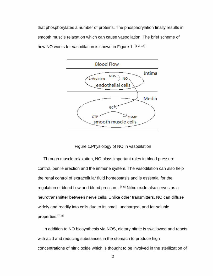
that phosphorylates a number of proteins. The phosphorylation finally results in
smooth muscle relaxation which can cause vasodilation. The brief scheme of
how NO works for vasodilation is shown in Figure 1. [1-3, 14]
Figure 1.Physiology of NO in vasodilation
Through muscle relaxation, NO plays important roles in blood pressure
control, penile erection and the immune system. The vasodilation can also help
the renal control of extracellular fluid homeostasis and is essential for the
regulation of blood flow and blood pressure. [4-6] Nitric oxide also serves as a
neurotransmitter between nerve cells. Unlike other transmitters, NO can diffuse
widely and readily into cells due to its small, uncharged, and fat-soluble
properties.[7, 8]
In addition to NO biosynthesis via NOS, dietary nitrite is swallowed and reacts
with acid and reducing substances in the stomach to produce high
concentrations of nitric oxide which is thought to be involved in the sterilization of
Page 12
3
swallowed food, preventing food poisoning, and maintaining gastric mucosal
blood flow. [7, 8]
1.1.2 Chemical Properties
Nitric oxide has N in the formal +2 oxidation state. Since the oxidation state of
nitrogen ranges from -3 to +5, nitric oxide can both be oxidized and reduced.
Several ways exist to prepare nitric oxide.
In a commercial setting, NO is produced by oxidation of ammonia with
platinum as catalyst.[9]
The uncatalyzed endothermic reaction of O2 and N2 can generate NO at a
very high temperature
In the laboratory, nitric oxide can be generated by the reduction of nitric acid.
In aerobic conditions, nitric oxide can be oxidized to NO2 which is a brown
toxic gas and considered as a major air pollutant.
Moreover, NOx reacts with volatile organic compounds in the presence of
sunlight to form ozone which can cause adverse effects such as damage to lung
Page 13
4
tissue and reduction in lung function. [10] Based on this toxicity, NO has
historically been considered as a major concern of air pollution which also can be
produced by the use of fuel.
1.2 HNO
HNO (Hydrogen oxonitrate, also called nitroxyl, nitrosyl hydride, nitroso
hydrogen, monomeric hyponitrous acid) has been found as an intermediate in a
variety of thermal and photochemical reactions since the early 1900s. [11] Since
then, much research has been done focusing on the intermediacy of HNO in
combustion of nitrogen-containing fuels, in the atmosphere in interstellar
chemistry and in bacterial denitrification. [11] Recently, the study of HNO has been
more concerned with the pharmacological effects and potential physiological
functions of HNO. [14]
1.2.1 Chemical Properties
HNO is the one-electron reduced and protonated derivative of NO. [14] It is a
very reactive species that undergoes a rapid dimerization. [12, 13] The dimerization
rate constant is about 8 × 10⁶ M−1 s−1. [49]
Also, HNO can be treated as the conjugate acid of NO- which, in turn, can be
seen as the conjugate base of HNO. But, these species are not simply related
through an acid-base relationship. Their different spins make the acid-base
relationship of HNO and NO- complicated.[11]
Page 14
5
Experiments show that 1HNO is about 18-19 kcal/mol more stable than 3HNO.
[11] Also, the protonation of 3NO- always produces 3NOH, the isomer of 3HNO.
The energy gap between the 3NOH and 1HNO is about 20-23 kcal/mol and 1HNO
is more stable. [15, 16] The energy gap between 1NO- and 3NO- is about 16-21
kcal/mol. The energy relationship between different spin states are shown in
Figure 2. [11]
HNO to NOH is thermally inaccessible under biological condition. However,
discrete deprotonation of HNO and NOH could lead to acid-base equilibria as
convoluted as depicted in Figure 2. [17]
Figure 1. Different spins states of HNO
The pKa of HNO was first determined to be 4.7 by Gratzel, and then
Shafirovich and Lymar updated the pKa to 11.4 in 2002.[27, 50]
Page 15
6
Figure 2. The energy gap between different spin states of HNO
1.2.2 Biological Properties
Until the mid1980s, attention to nitrogen oxide was generally limited to
environmental concerns. In 1980, vasodilation was determined to be actively
mediated by an unidentified species, which was labeled the endothelium-derived
relaxing factor (EDRF).[18] After chemical and biological research, the results led
to the conclusion that NO was EDRF. [19, 20] However, some certain dissimilarities
between the effects exerted by NO and EDRF were observed that led to the
speculation that this species may be HNO. [21] Angeli’s Salt (Na2N2O3) and
Piloty’s acid (benzenesulphonydraoxamic acid) are both bioactive HNO donors.
When administered intraperitoneally or intraarterially to mice or rats, these
donors result in vasorelaxation. [11]
Page 16
7
The vasoactivity is accompanied by increased cyclic guanosine
monophosphate (cGMP) production. [22] However, the vasoactivity is generally
less effective than that elicited by NO donors, which indicates that HNO is
converted to NO in vivo and that HNO is an intermediate form of EDRF. [23]
Fukuto et al. [24] demonstrated that HNO can be easily oxidized to NO in the
presence of SOD (superoxide dismutase). Interestingly, coinfusion of Angeli’s
salt and the electron paramagnetic resonance (EPR) trap diethyldithiocarbonate
(DETC) suggested that HNO was only minimally oxidized to NO in vivo (<5%)
which also raises the speculation that HNO only serves as an intermediate when
converted to NO in vivo. [11]
In vitro, scientists noticed that Angeli’s Salt, an HNO donor, enhanced
oxidative stress by peroxides while NO was protective under the same
conditions. [25] The cytotoxicity of Angeli’s salt was dependent on an aerobic
environment and enhanced by the absence of glutathione (GSH). [25] This
research provides the evidence that HNO could affect cellular function by
changing the redox status of the cell. Also, HNO can either associate with GSH
or scavenge GSH, which may affect the activity of enzymes containing critical
thiols. [11]
The biological function of HNO in vivo is not fully understood. More research
still needs to be done in the future to give a better understanding of HNO.
Page 17
8
1.2.3 Biosynthesis of HNO
No unequivocal evidence for the endogenous generation of HNO in
mammalian systems currently exists. However, some chemical and biochemical
processes have been shown to be possible ways of endogenous HNO formation.
For example, the reaction of S-nitrosothiols with other thiols can generate HNO
and difsulfide. [46]
N-Hydroxy-L-arginine (NOHA), a NO biosynthetic intermediate, can be
oxidized to HNO. [46] HNO can also be generated from L-arginine or NOHA by the
presence of NOS [46, 51] especially when it is deplete of one of its prosthetic
groups. [46] Another possible endogenous path way to HNO is the reaction of NO
and H2S. NO and H2S can enter a redox reaction with each other that lead to the
formation of HNO.[52]
1.3 HNO Detection
While both physiological and pathological roles of NO in vivo have been
deeply studied, HNO has been much less thoroughly investigated. Much
evidence shows that HNO plays important biological roles in potential
pharmacological applications distinct from those of NO and developing efficient
detection methods for HNO in vivo is very important. [11]
As stated before, HNO reacts rapidly with itself to form a dimer. Also, HNO
can convert to NO in vivo. So an efficient probe must have a high rate constant
for reacting with HNO and a high selectivity of reaction. For HNO, the most
Page 18
9
efficient methods include Cu-based fluorescent probes and HNO-specific
electrodes. This section will discuss some detection methods which have been
developed in recent years. [29, 30]
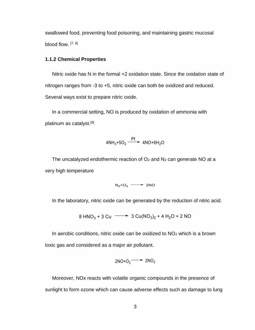
Rosenthal and Lippard developed a copper based fluorescent probe in 2010.
The [CuII(BOT1)Cl] structure is shown in Figure 3. [28]
Figure 3. Structure of [CuII(BOT1)Cl]
Just as SOD(CuII) reacts with HNO which generates NO and reduced
SODCuI, [CuII(BOT1)Cl] also reacts with HNO and forms [CuI(BOT1)Cl]. While
[CuII(BOT1)Cl] has no emission, [CuI(BOT1)Cl] fluoresces at 500-650nm. The
probe shows increased fluorescence when treated with Angeli’s Salt. Meanwhile,
treating the probe with other reactive nitrogen species (RNS) or reactive oxygen
species (ROS) including NO-, NO2-, NO3
-, ONOO-, H2O2, OCl- and NO do not
cause fluorescence showing a high selectivity of this probe. [28]
Another important detection method is a HNO-specific electrode whose
structure is shown in Figure 4 developed by Martí and Doctorovich that detects
HNO in a time resolved fashion at low nanomolar concentration. [29]
Page 19
10
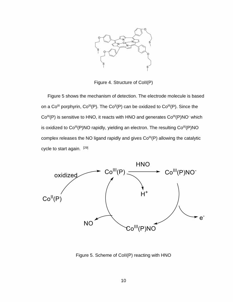
Figure 4. Structure of CoII(P)
Figure 5 shows the mechanism of detection. The electrode molecule is based
on a CoIII porphyrin, CoIII(P). The CoII(P) can be oxidized to CoIII(P). Since the
CoIII(P) is sensitive to HNO, it reacts with HNO and generates CoIII(P)NO- which
is oxidized to CoIII(P)NO rapidly, yielding an electron. The resulting CoIII(P)NO
complex releases the NO ligand rapidly and gives CoIII(P) allowing the catalytic
cycle to start again. [29]
Figure 5. Scheme of CoII(P) reacting with HNO
Page 20
11
In the mechanism described above, the current can be detected in the
presence of HNO due to the production of an electron. Also, the sensitivity and
selectivity of this electrode are both good. The Co(P) electrode exhibits a linear
response in transient HNO concentrations from 1 to 1000nM. The presence of
oxygen and other reactive nitrogen and oxygen species (RNOS) don’t affect
electrode performance. [29]
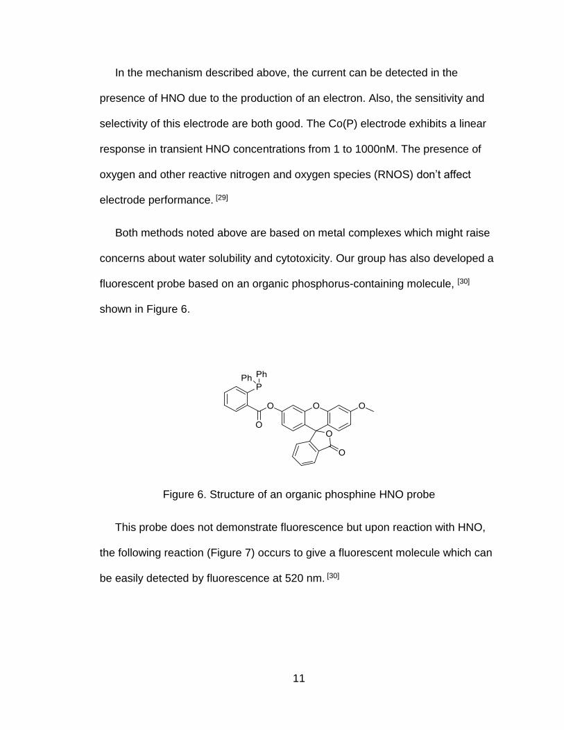
Both methods noted above are based on metal complexes which might raise
concerns about water solubility and cytotoxicity. Our group has also developed a
fluorescent probe based on an organic phosphorus-containing molecule, [30]
shown in Figure 6.
Figure 6. Structure of an organic phosphine HNO probe
This probe does not demonstrate fluorescence but upon reaction with HNO,
the following reaction (Figure 7) occurs to give a fluorescent molecule which can
be easily detected by fluorescence at 520 nm. [30]
Page 21
12
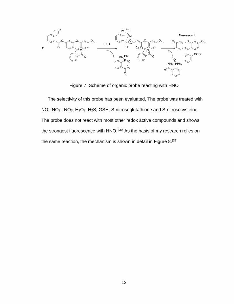
Figure 7. Scheme of organic probe reacting with HNO
The selectivity of this probe has been evaluated. The probe was treated with
NO-, NO2-, NO3, H2O2, H2S, GSH, S-nitrosoglutathione and S-nitrosocysteine.
The probe does not react with most other redox active compounds and shows
the strongest fluorescence with HNO. [30] As the basis of my research relies on
the same reaction, the mechanism is shown in detail in Figure 8.[31]
Page 22
13
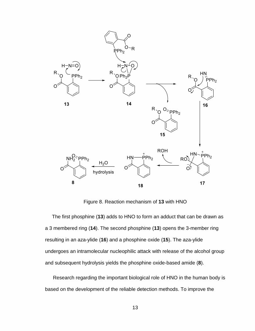
Figure 8. Reaction mechanism of 13 with HNO
The first phosphine (13) adds to HNO to form an adduct that can be drawn as
a 3 membered ring (14). The second phosphine (13) opens the 3-member ring
resulting in an aza-ylide (16) and a phosphine oxide (15). The aza-ylide
undergoes an intramolecular nucleophilic attack with release of the alcohol group
and subsequent hydrolysis yields the phosphine oxide-based amide (8).
Research regarding the important biological role of HNO in the human body is
based on the development of the reliable detection methods. To improve the
Page 23
14
methods, sensitivity, selectivity and cytotoxicity must be taken into concern. The
organic probe developed by our group is a very good detection method which
operates through a very specific chemical pathway to release a fluorophore.
However, this chemistry allows other molecules besides detection compounds to
be released making new potential prodrugs.
1.4 HNO Donors
1.4.1 General Ways to Form HNO
HNO is highly reactive and its dimerization makes it impossible to store. [12, 13,
26] To study HNO, efficient HNO donors must be used. Several HNO donors exist
that can be divided into organic and inorganic classes.
For the inorganic pathways, the simplest route to HNO is the reduction of NO.
[32] The aerobic photolysis of ammonia also generates HNO. [33] A chain reaction
may happen in the presence of O2. Both of these pathways have atmospheric
importance but are not relevant in biological research.
Organic compounds form another category of HNO donors. The Nef reaction
produces HNO as shown in Figure 9. [34] The nitro group is hydrolyzed to give
HNO with the by-product ketone.
Figure 9. Scheme of Nef reaction
Page 24
15
Another pathway to HNO is the nitrosative cleavage of tertiary amines (Figure
10). [35] Nitrosation of tertiary amines generate HNO by elimination of the
adjacent H to give HNO.
Figure 10. HNO formation from nitrosative cleavage
Retro Diels-Alder reactions can also lead to HNO formation as shown in
Figure 11. [36]
Figure 11. HNO formation from retro-Diels Alder reaction
The retro Diels Alder reaction of appropriate cycloadducts will produce an acyl
nitroso compound that is easily hydrolyzed to give nitroxyl. [36]
Simple decomposition of N-phosphinoylhydroxylamines also yield HNO (Figure
12). [37]
Figure 12. Decomposition of N-phosphinoylhydroxylamines
Page 25
16
The hydroxylamine can be oxidized to a nitroso group which is easily
hydrolyzed to give HNO.
1.4.2 Biological HNO Donors
While these pathways give HNO, these compounds cannot always be used as
efficient donors due to various drawbacks. Some of these compounds are hard to
store and some cannot react in biological conditions. More efficient ways to yield
HNO are required for biological research. The most commonly used HNO donors
are Piloty’s acid and Angeli’s Salt.[11]
Piloty’s acid (N-hydroxybenzenesulfonamide or benzosulfohydroxamic acid) is
an organic compound which hydrolyzes in basic conditions to yield HNO and a
sulfinic acid. Piloty’s acid decomposes through a base-catalyzed deprotonation
mechanism followed by S-N bond heterolysis. [11, 38] The mechanism is shown in
Figure 13.
Figure 13. Decomposition of Piloty’s Acid to HNO
Piloty’s acid provides a convinient way to investigate HNO in both high and
neutral pH. However, Piloty’s acid can be oxidzied to the corresbonding nitroxide
which then releases NO rather than HNO. [11, 39] Consequently, Piloty’s acid must
be utilized in anaerobic and reducing environments. Indeed, the oxidation of
Piloty’s acid to give NO is suggested to be the primary pathway in physiological
conditions.
Page 26
17
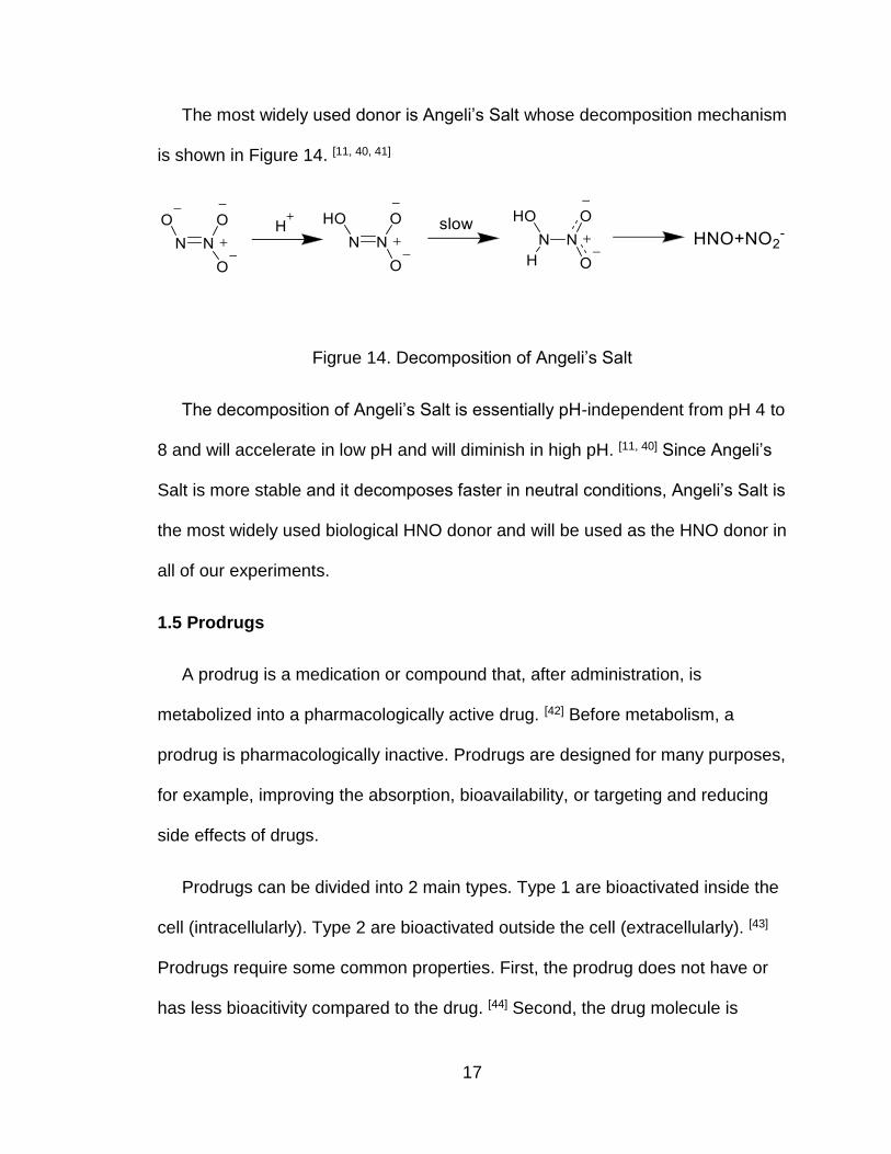
The most widely used donor is Angeli’s Salt whose decomposition mechanism
is shown in Figure 14. [11, 40, 41]
Figrue 14. Decomposition of Angeli’s Salt
The decomposition of Angeli’s Salt is essentially pH-independent from pH 4 to
8 and will accelerate in low pH and will diminish in high pH. [11, 40] Since Angeli’s
Salt is more stable and it decomposes faster in neutral conditions, Angeli’s Salt is
the most widely used biological HNO donor and will be used as the HNO donor in
all of our experiments.
1.5 Prodrugs
A prodrug is a medication or compound that, after administration, is
metabolized into a pharmacologically active drug. [42] Before metabolism, a
prodrug is pharmacologically inactive. Prodrugs are designed for many purposes,
for example, improving the absorption, bioavailability, or targeting and reducing
side effects of drugs.
Prodrugs can be divided into 2 main types. Type 1 are bioactivated inside the
cell (intracellularly). Type 2 are bioactivated outside the cell (extracellularly). [43]
Prodrugs require some common properties. First, the prodrug does not have or
has less bioacitivity compared to the drug. [44] Second, the drug molecule is
Page 27
18
connected to the carrier by a covalent bond that can be easily broken in vivo. [42]
Third, the rate of decomposition of the prodrug in vivo must be fast enough so
that the concentration of drug molecule can reach a certain level. Based on these
principles, one of the most common strategies to design a prodrug are through
an ester linkage.
HNO may play important roles in the human body and based on our current
HNO detection model that releases a fluorophore, we considered whether an
HNO triggered prodrug could be developed. Inflammation is associated with
many pathological disorders such as cancers and infections and production of
reactive oxygen species (ROS) including H2O2 is central to many inflammatory
diseases. [47] ROS can act as both a signaling molecule and a mediator of
inflammation. The ROS can also rapidly combine with NO to generate reactive
nitrogen species (RNS) including S-nitrosothiols peroxynitrite, and possibly
nitroxyl anion. [48] Based on these property, HNO might be a good signaling
marker for inflammation and an HNO-triggered prodrug can be efficient in
targeting the drug, especially on anti-inflammatory agent to an inflammation site.
In the presence of HNO, the prodrug would decompose and release a drug
molecule (Figure 15). The prodrug may improve the targeting properties of the
drug molecule but also would only be released in the presence of HNO which
could be of importance in redox biology. To determine whether the idea will work,
drugs with a hydroxyl group will be linked to 2-(diphenylphosphino) benzoic acid
(DPPBA) and our initial candidates include methyl salicylate, acetaminophen and
metronidazole.
Page 28
19
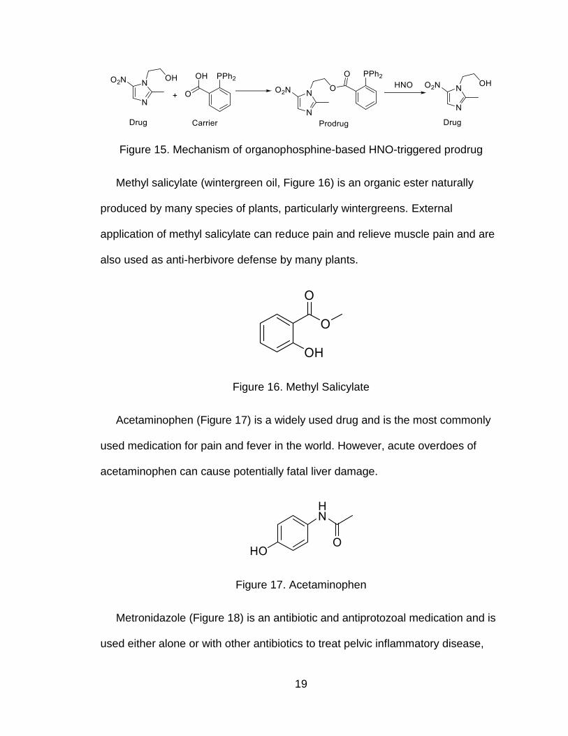
Figure 15. Mechanism of organophosphine-based HNO-triggered prodrug
Methyl salicylate (wintergreen oil, Figure 16) is an organic ester naturally
produced by many species of plants, particularly wintergreens. External
application of methyl salicylate can reduce pain and relieve muscle pain and are
also used as anti-herbivore defense by many plants.
Figure 16. Methyl Salicylate
Acetaminophen (Figure 17) is a widely used drug and is the most commonly
used medication for pain and fever in the world. However, acute overdoes of
acetaminophen can cause potentially fatal liver damage.
Figure 17. Acetaminophen
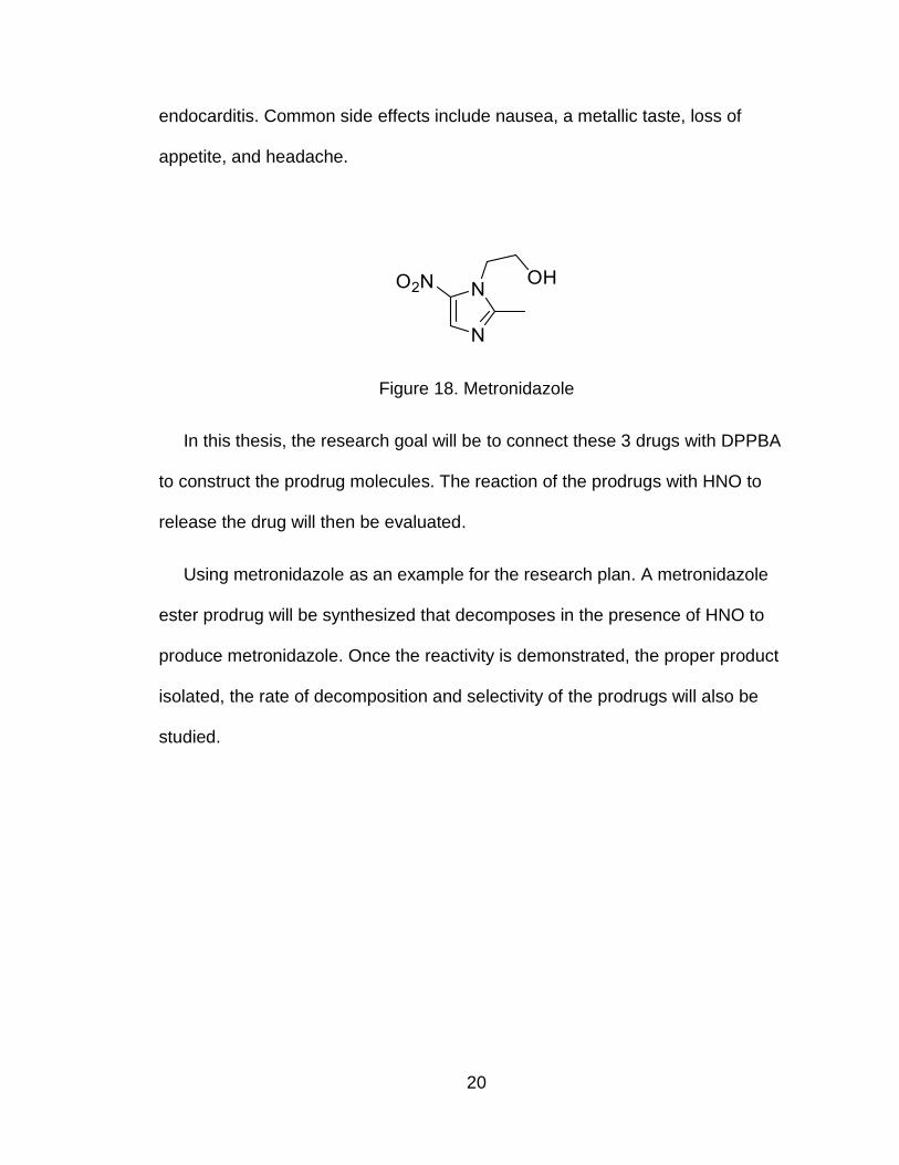
Metronidazole (Figure 18) is an antibiotic and antiprotozoal medication and is
used either alone or with other antibiotics to treat pelvic inflammatory disease,
Page 29
20
endocarditis. Common side effects include nausea, a metallic taste, loss of
appetite, and headache.
Figure 18. Metronidazole
In this thesis, the research goal will be to connect these 3 drugs with DPPBA
to construct the prodrug molecules. The reaction of the prodrugs with HNO to
release the drug will then be evaluated.
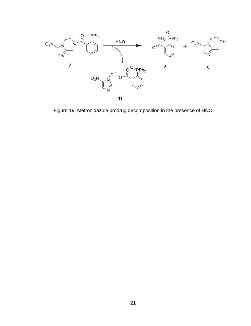
Using metronidazole as an example for the research plan. A metronidazole
ester prodrug will be synthesized that decomposes in the presence of HNO to
produce metronidazole. Once the reactivity is demonstrated, the proper product
isolated, the rate of decomposition and selectivity of the prodrugs will also be
studied.
Page 30
21
Figure 19. Metronidazole prodrug decomposition in the presence of HNO
Page 31
22
Chapter 2. Results and Discussion
2.1 Prodrug Synthesis
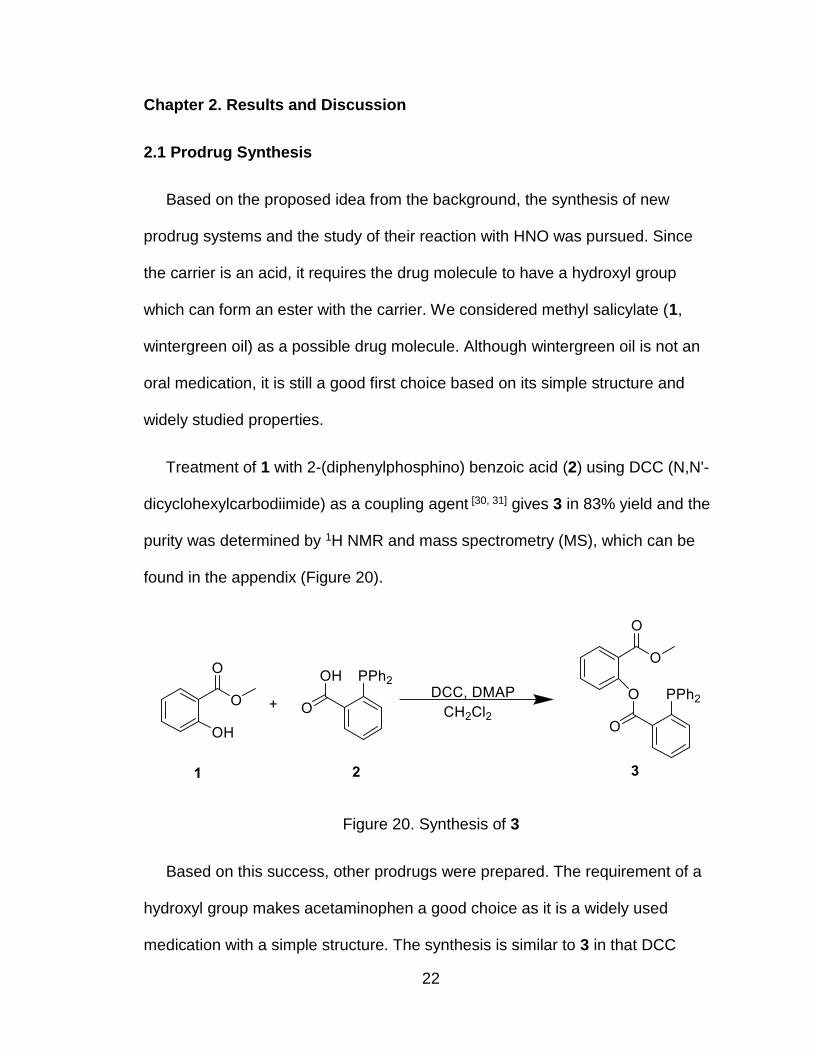
Based on the proposed idea from the background, the synthesis of new
prodrug systems and the study of their reaction with HNO was pursued. Since
the carrier is an acid, it requires the drug molecule to have a hydroxyl group
which can form an ester with the carrier. We considered methyl salicylate (1,
wintergreen oil) as a possible drug molecule. Although wintergreen oil is not an
oral medication, it is still a good first choice based on its simple structure and
widely studied properties.
Treatment of 1 with 2-(diphenylphosphino) benzoic acid (2) using DCC (N,N'-
dicyclohexylcarbodiimide) as a coupling agent [30, 31] gives 3 in 83% yield and the
purity was determined by 1H NMR and mass spectrometry (MS), which can be
found in the appendix (Figure 20).
Figure 20. Synthesis of 3
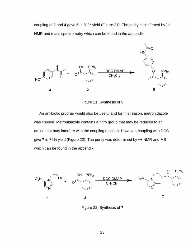
Based on this success, other prodrugs were prepared. The requirement of a
hydroxyl group makes acetaminophen a good choice as it is a widely used
medication with a simple structure. The synthesis is similar to 3 in that DCC
Page 32
23
coupling of 2 and 4 gave 5 in 81% yield (Figure 21). The purity is confirmed by 1H
NMR and mass spectrometry which can be found in the appendix.
Figure 21. Synthesis of 5
An antibiotic prodrug would also be useful and for this reason, metronidazole
was chosen. Metronidazole contains a nitro group that may be reduced to an
amine that may interfere with the coupling reaction. However, coupling with DCC
give 7 in 76% yield (Figure 22). The purity was determined by 1H NMR and MS
which can be found in the appendix.
Figure 22. Synthesis of 7
Page 33
24
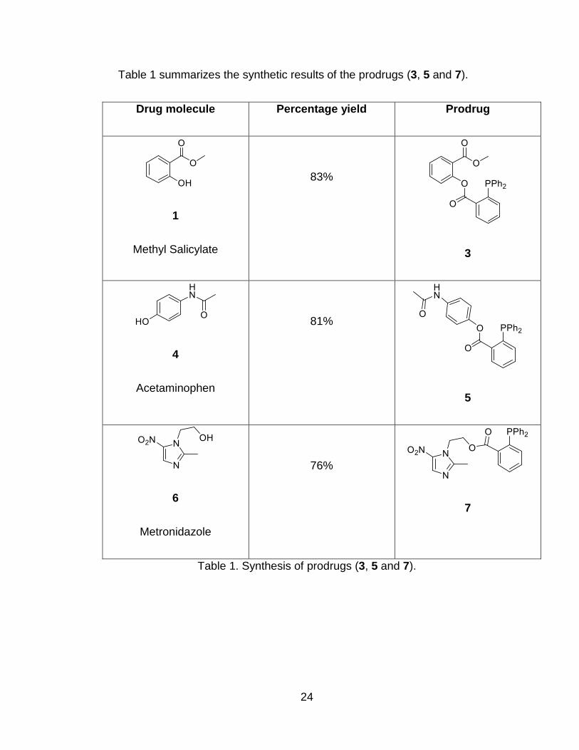
Table 1 summarizes the synthetic results of the prodrugs (3, 5 and 7).
Drug molecule Percentage yield Prodrug
1
Methyl Salicylate
83%
3
4
Acetaminophen
81%
5
6
Metronidazole
76%
7
Table 1. Synthesis of prodrugs (3, 5 and 7).
Page 34
25
2.2 Reaction with HNO
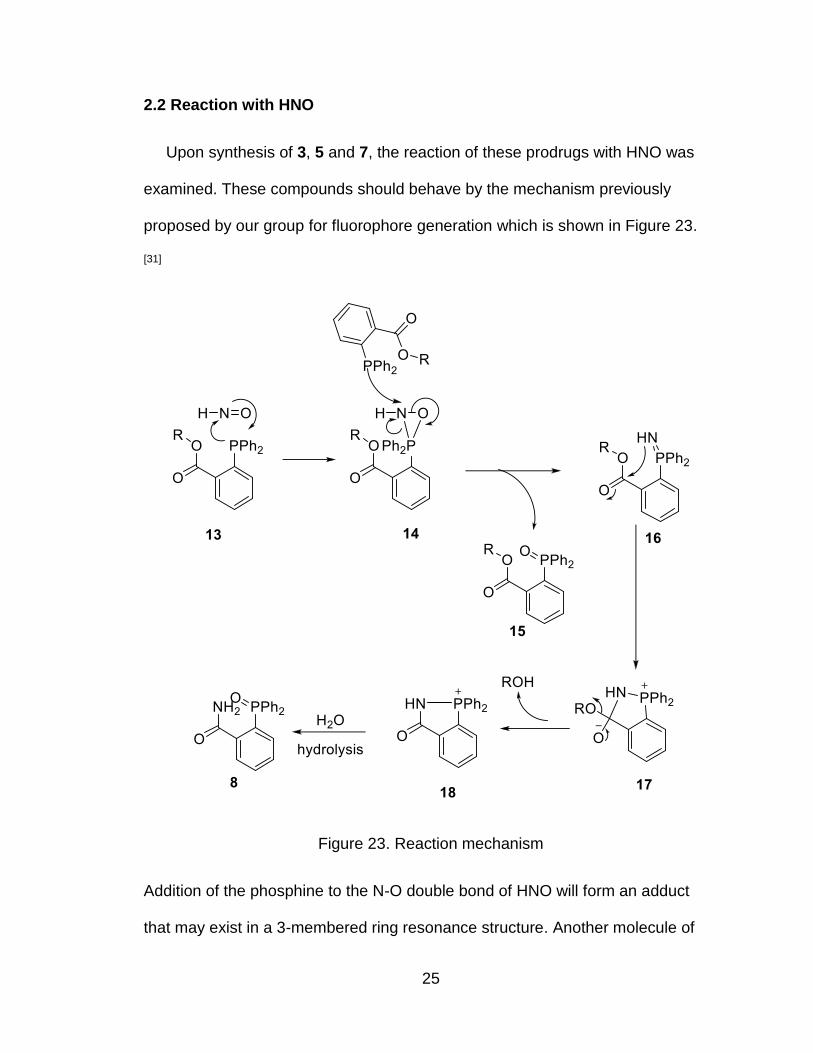
Upon synthesis of 3, 5 and 7, the reaction of these prodrugs with HNO was
examined. These compounds should behave by the mechanism previously
proposed by our group for fluorophore generation which is shown in Figure 23.
[31]
Figure 23. Reaction mechanism
Addition of the phosphine to the N-O double bond of HNO will form an adduct
that may exist in a 3-membered ring resonance structure. Another molecule of
Page 35
26
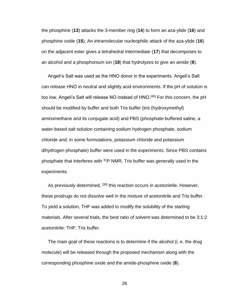
the phosphine (13) attacks the 3-member ring (14) to form an aza-ylide (16) and
phosphine oxide (15). An intramolecular nucleophilic attack of the aza-ylide (16)
on the adjacent ester gives a tetrahedral intermediate (17) that decomposes to
an alcohol and a phosphonium ion (18) that hydrolyzes to give an amide (8).
Angeli’s Salt was used as the HNO donor in the experiments. Angeli’s Salt
can release HNO in neutral and slightly acid environments. If the pH of solution is
too low, Angeli’s Salt will release NO instead of HNO.[45] For this concern, the pH
should be modified by buffer and both Tris buffer (tris (hydroxymethyl)
aminomethane and its conjugate acid) and PBS (phosphate-buffered saline, a
water-based salt solution containing sodium hydrogen phosphate, sodium
chloride and, in some formulations, potassium chloride and potassium
dihydrogen phosphate) buffer were used in the experiments. Since PBS contains
phosphate that interferes with 31P NMR, Tris buffer was generally used in the
experiments.
As previously determined, [30] this reaction occurs in acetonitrile. However,
these prodrugs do not dissolve well in the mixture of acetonitrile and Tris buffer.
To yield a solution, THF was added to modify the solubility of the starting
materials. After several trials, the best ratio of solvent was determined to be 3:1:2
acetonitrile: THF: Tris buffer.
The main goal of these reactions is to determine if the alcohol (i. e. the drug
molecule) will be released through the proposed mechanism along with the
corresponding phosphine oxide and the amide-phosphine oxide (8).
Page 36
27
2.2.1 Wintergreen Ester (3)
The wintergreen ester (3) was synthesized as noted. This white solid was
added to the solvent mixture, and 5 equivalents of Angeli’s Salt were added at
room temperature. After 24-hours, methyl salicylate was recovered in 76% yield
after chromatography as predicted by the proposed mechanism shown in Figure
24. The purity of methyl salicylate was determined by NMR and MS.
Figure 24. Reaction of 3 with HNO
Further, experiments to show that the phosphine oxide and the amide were
also produced as the proposed mechanism were performed. The isolation of 8
and 9 would support the proposed mechanism. Purification of the crude reaction
mixture by column chromatography, similar to the isolation of methyl salicylate,
unfortunately, did not yield 8 or 9.
Page 37
28
To determine what was generated in the reaction, a small scale reaction of 3
with Angeli’s Salt in deuterated acetonitrile was performed and the crude 31P
NMR was taken (Figure 25). For comparison, a standard of the phosphine oxide
(9) was prepared by adding hydrogen peroxide to the ester (3). In addition, a
standard of the amide (8) was prepared by the reaction of methyl 2-
(diphenylphosphanyl) benzoate (12) with HNO.
Figure 25. Crude 31P-NMR and comparison. Panel A is the crude 31P-NMR of the
reaction, Panel B is the 31P-NMR of 8 and Panel C is the 31P-NMR of 9. All the
NMR spectra are prepared in deuterated acetonitrile.
A
B
C
Page 38
29
In Figure 25, the peak in panel A (δ 33.47 ppm) aligns with C (δ 33.52 ppm) to
confirming that 9 forms in the reaction.
Moreover, a peak in Panel A (δ 32.37 ppm) aligns with a peak in Panel B (δ
32.59 ppm) confirming the formation of 8 in the reaction. Other peaks appear in A
that mean other byproducts exist other than the products the mechanism
predicts.
To further investigate this reaction, LC-MS (liquid chromatography-mass
spectrometry) of the mixture was used without any further purification (Figure
26).
Figure 26. LC-MS of the mixture
From the LC-MS results, the phosphine oxide (9) forms. However, other
impurities also form and it is difficult to determine the other compounds from LC-
MS. From the results, we can conclude that reaction of 3 with HNO forms 8 and 9
Page 39
30
and methyl salicylate, but also yields other products compared to the mechanism
we proposed previously.
Though impurities formed in the reaction of 3 with HNO, these results show
that this kind of prodrug system works as an HNO triggered prodrug. From the
analysis, the reason for multiple products might be that 2 ester groups exist in 3
which could be hydrolyzed or undergo other reactions. Based on this, we
considered an acetaminophen ester (5) as a better substrate as acetaminophen
does not have any other esters.
2.2.2 Acetaminophen Ester (5)
Treatment of the acetaminophen ester (5) with 5 equivalents of Angeli’s Salt in
the 3:1:2 acetonitrile: THF: Tris buffer at room temperature (Figure 27) gave
acetaminophen (4) in 90% yield and phosphine oxide (10) in 78% yield.
However, no evidence of 8 could be found.
Figure 27. Acetaminophen ester reacts with HNO
Page 40
31
Figure 28. 31P-NMR comparison. Panel A is the 31P-NMR of 8 and Panel B is the
crude 31P-NMR for the reaction of 5 with Angeli’s Salt.
Upon comparison of 31P-NMR spectra, a peak exists in Panel B (δ 32.15 ppm)
that aligns with Panel A (δ 32.10 ppm). From this comparison, we can conclude
that some 8 is produced in the reaction along with many other phosphorus-
containing compounds similar to the reaction of 3 with Angeli’s Salt. These
results suggest that this reaction does not occur cleanly and may undergo other
mechanisms than the originally proposed.
Considering the prodrugs work well in drug releasing, experiments with cells
may be useful. For such a goal, a drug with antibiotic properties would provide
A
A
B
Page 41
32
further evidence by killing bacterial cells after drug release. The final prodrug is
based on the antibiotic, metronidazole.
2.2.3 Metronidazole Ester
Metronidazole is a good choice for a new prodrug molecule since it has a
hydroxyl group and is a widely studied antibiotic. Furthermore, the structure of
metronidazole is not complicated, which means we can analyze the reaction
easily.
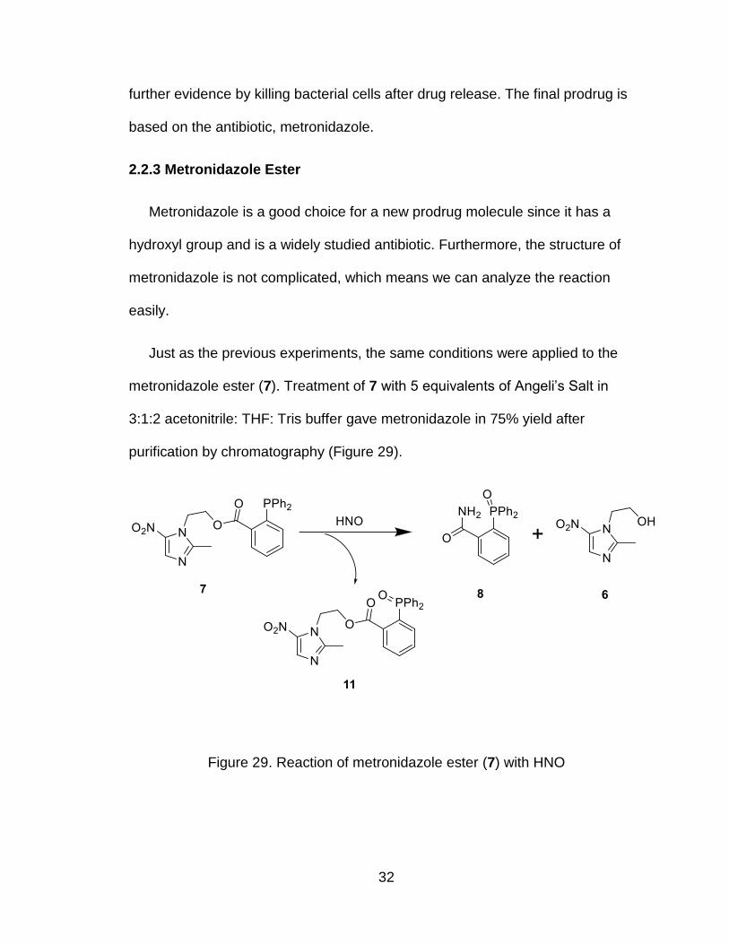
Just as the previous experiments, the same conditions were applied to the
metronidazole ester (7). Treatment of 7 with 5 equivalents of Angeli’s Salt in
3:1:2 acetonitrile: THF: Tris buffer gave metronidazole in 75% yield after
purification by chromatography (Figure 29).
Figure 29. Reaction of metronidazole ester (7) with HNO
Page 42
33
Unfortunately, no other compounds could be separated by chromatography. To
establish the presence of the amide (8) and the phosphine oxide (11), LC-MS
and 31P-NMR were used.
Figure 30. 31P-NMR comparison. Panel A is the 31P-NMR of 11, Panel B is the
31P-NMR of the reaction of metronidazole ester with HNO and Panel C is the 31P-
NMR of 8.
As Figure 30 shows, the crude reaction in panel B shows only 2 peaks which
means only 2 compounds with phosphorous are in the mixture after reaction. In
addition, the starting material (7) has a negative chemical shift which is absent in
A
A
B
A
C
Page 43
34
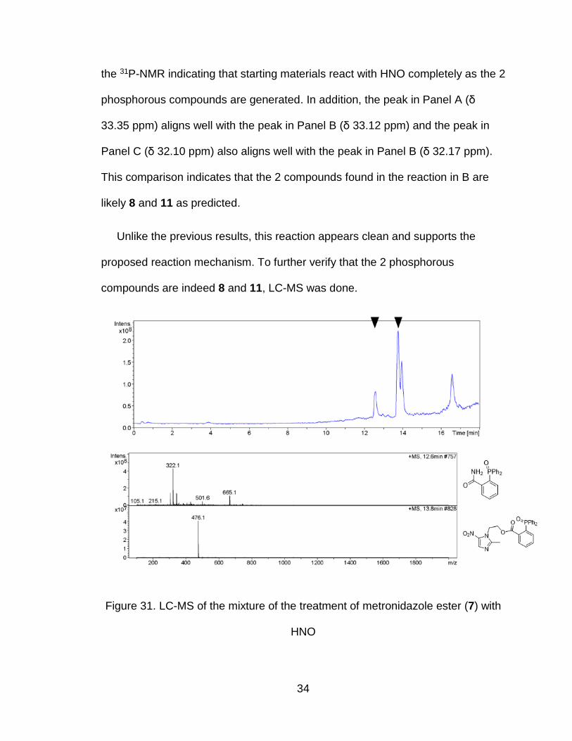
the 31P-NMR indicating that starting materials react with HNO completely as the 2
phosphorous compounds are generated. In addition, the peak in Panel A (δ
33.35 ppm) aligns well with the peak in Panel B (δ 33.12 ppm) and the peak in
Panel C (δ 32.10 ppm) also aligns well with the peak in Panel B (δ 32.17 ppm).
This comparison indicates that the 2 compounds found in the reaction in B are
likely 8 and 11 as predicted.
Unlike the previous results, this reaction appears clean and supports the
proposed reaction mechanism. To further verify that the 2 phosphorous
compounds are indeed 8 and 11, LC-MS was done.
Figure 31. LC-MS of the mixture of the treatment of metronidazole ester (7) with
HNO
Page 44
35
From the LC-MS, two peaks are found that correspond to 8 and 11. The
combination of these results (31P-NMR and LC-MS) support the formation of 8
and 11 during the reaction of 7 with HNO.
2.2.4 Mechanism Discussion
As shown, except for 7, both 3 and 5 react with HNO to yield many products
which complicates our mechanistic interpretation. For both 3 and 5 evidence
exists that the amide (8) and the corresponding phosphine oxide from. In all of
the reactions, isolation of all products proved difficult suggesting other reactions
are occurring. As shown in Figure 23, the phosphine probe will first react with
HNO to form a phosphine oxide and an aza-ylide. Competition between the
hydrolysis of ylide and the nucleophilic attack can occur. If hydrolysis occurs, the
phosphine oxide forms without the corresponding amide (8), in addition, direct
oxidation of the probe would also lead to the phosphine oxide without 8. If the
phosphine oxide hydrolyzes, carboxylic acid (19) will be produced as shown in
Figure 32, and such a compound might be one of the impurities shown in the 31P-
NMR.
Page 45
36
Figure 32. Hydrolysis of ylide and phosphine oxide
Moreover, the prodrug of wintergreen has a second ester group and
acetaminophen has an amide group, so both 3 and 5 can in theory hydrolyze.
While the amide is difficult to hydrolyze under normal conditions, the presence of
HNO might aid the decomposition of the amide. Hydrolysis of these groups could
also explain the many impurities in the reaction of 3 and 5 with HNO. Finally,
HNO is a good electrophile and may undergo some reaction with the aromatic
portion of these molecules, giving a new set of products.
2.3 Kinetic Analysis
As stated in the background, prodrugs require the important property that they
need to be metabolized in vivo at a high reaction rate in order to achieve a
certain concentration in the human body. The results show that the prodrugs can
be activated by the presence of HNO. Another important part of proving the
efficiency of the prodrug is to analyze the kinetics of the reaction.
31P-NMR was used to study the kinetics of this reaction with HNO. The basic
idea was to take a 31P-NMR spectrum at regular intervals and to judge its
Page 46
37
concentration by integrating the peak of starting materials and determining the
amount the concentration changes by comparing the change of the integral.
Moreover, in order to compare the integral and make the results reliable,
phosphoric acid, which has a chemical shift of δ 0 ppm in 31P-NMR was added as
a standard.
Initially, 3 was dissolved in 3:1:2 acetonitrile: THF: Tris buffer and Angeli’s
Salt was added and the mixture was quickly transferred to an NMR tube along
with a tiny amount of phosphoric acid. 31P-NMR spectra were taken every 15
minutes until all of 3 disappeared. Controls included both pure 3 and 9, produced
by adding hydrogen peroxide to 3, and Figure 33 shows the results of these
experiments.
Page 47
38
Figure 33. Kinetic study. Panel A is the 31P-NMR of 9, Panel B is the 31P-NMR of
3. The other 9 are the 31P-NMR of the reaction mixture at the time labeled.
Figure 33 shows that over time, the peak of 3 (δ -4.95 ppm) decreases and
the peak of 9 (δ 33.90 ppm) increases which shows the formation of phosphine
oxide. In about 45 minutes, 3 is nearly gone, which means 3 reacts with HNO
completely within 1 hour.
Figure 34 shows this data graphically by setting the integral of phosphoric
acid as a standard and following the integral of 3 over time to give a relative
amount.
A
B
t=0 min
t=13 min
t=28 min
t=43 min
t=58 min
t=75 min
t=90 min
t=104 min
t=122 min
Page 48
39
Figure 34. Concentration of 3 vs. Time after Angeli’s Salt addition
From the graph, 3 decomposes quickly in the first 20 minutes, with almost
80% of 3 gone in the first 20 minutes. After 100 minutes, almost all of 3 is gone.
Actually, two reactions are occurring in the system, one is the decomposition
of Angeli’s Salt to release HNO and the other is the reaction of 3 with HNO. To
determine which reaction Figure 34 depicts, the observed rate constant was
calculated to be 5.6 × 10⁻⁴ s-1. This value closely matches the reported rate
constant for Angeli’s Salt decomposition at 25 ℃ of 6.8 × 10⁻⁴ s-1.[49] This rate
constant is also much slower than the reported rate constants for the reaction of
HNO with phosphines of 9×10⁻⁵ s-1 and 8.4×10⁶ M-1 s-1.[49] These results indicate
Figure 34 shows the slow decomposition of Angeli’s Salt that rapidly reacts with
0
0.02
0.04
0.06
0.08
0.1
0.12
0 20 40 60 80 100 120 140
Inte
gral
of
pro
du
ct 3
Time/min
Page 49
40
the phosphine prodrug and reveals that the prodrugs have a rapid reaction rate
with HNO as desired.
Based on these results, we can conclude that the newly designed prodrugs
rapidly react with HNO to release drug within 1 hour. As required for prodrugs,
the fast conversion meets the requirement of designing a prodrug.
2.4 Selectivity
With the basic reactivity and kinetic analysis established, a further need is to
examine the selectivity of these compounds for reacting with HNO. We have
shown that these prodrugs are activated by HNO but must show whether these
prodrugs react with other compounds such as glutathione (GSH), cysteine (cys)
or NO. To demonstrate the selectivity of the prodrug, experiments were done
with 7 using GSH, cysteine, NaNO2 and NO (DEANO as donor, Figure 35) whose
structures are shown in Figure 35.
Figure 35. Structure of biological reducing and oxidizing agents.
Compound 7 was treated with 5 equivalents of each compound and a control
was also included. After 24 hours, 31P-NMR spectra were taken to determine if a
reaction occurred. Figure 36 shows the results of experiments with GSH, cys and
a control.
Page 50
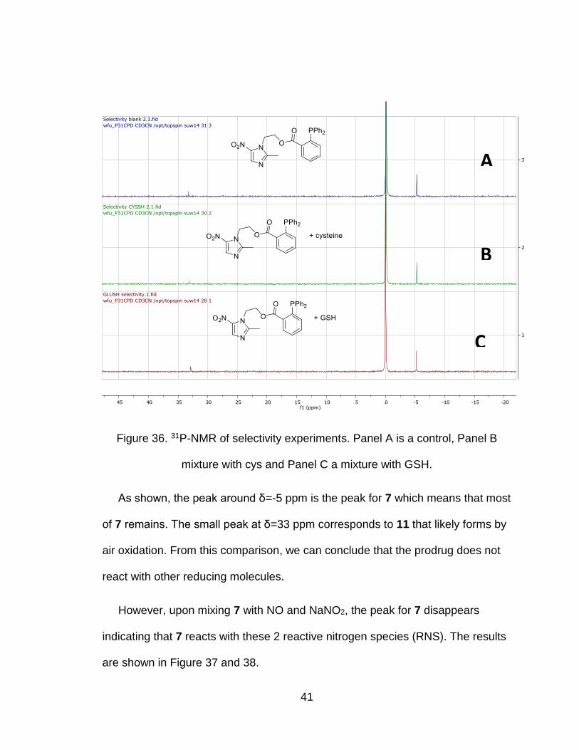
41
Figure 36. 31P-NMR of selectivity experiments. Panel A is a control, Panel B
mixture with cys and Panel C a mixture with GSH.
As shown, the peak around δ=-5 ppm is the peak for 7 which means that most
of 7 remains. The small peak at δ=33 ppm corresponds to 11 that likely forms by
air oxidation. From this comparison, we can conclude that the prodrug does not
react with other reducing molecules.
However, upon mixing 7 with NO and NaNO2, the peak for 7 disappears
indicating that 7 reacts with these 2 reactive nitrogen species (RNS). The results
are shown in Figure 37 and 38.
A
B
C
Page 51
42
Figure 37. 31P-NMR of the reaction of 7 with NaNO2. Panel A shows addition of
11 to the reaction, Panel B is the reaction of 7 with NaNO2.
The difference between this reaction and the blank is that the peak for 7
totally disappears. As only one peak forms, we conclude that NaNO2 can oxidize
7 to produce 11. To prove this, 11 was added to the mixture (Figure 37, Panel A),
and we see that there is still only one peak in the 31P-NMR. Based on that result,
we can conclude that 7 can react with NaNO2 and produce 11. This results
stands in contrast to previous experiments that show other triphenylphosphine
derivatives do not react with NaNO2. [49, 50]
A
B
Page 52
43
Figure 38. 31P-NMR of the reaction of 7 with DEANO, Panel A shows the addition
of 11 to the reaction, Panel B is the reaction of 7 reacting with DEANO.
Similar to NaNO2, the 31P-NMR of the reaction of 7 with NO is shown in Figure
38. Panel A shows the addition of 11 to the reaction mixture and Panel B is the
spectrum of the reaction of 7 with DEANO. These results are similar to previous
results that show NO oxidizes phosphine to phosphine oxide. [49]
Based on these selectivity experiments, the prodrug does not react with
reducing agents such as GSH and cysteine but does react with oxidants
including NaNO2, NO, and hydrogen peroxide. However, the reaction with the
A
B
Page 53
44
oxidants, only forms phosphine oxide (11) and no ylide forms and does not lead
to drug release.
Further, as stated, the decomposition of Angeli’s Salt will generate HNO and
sodium nitrite. Though, sodium nitrite can react with 7 and ruin its activity, 7 can
still be activated by Angeli’s Salt, which means the prodrug 7 has a higher
reaction rate with HNO than sodium nitrite and shows a high selectivity with
HNO.
2.5 Conclusion
Based on previous reactions described by our group (Figure 22), a type of
prodrug has been designed. Three prodrugs (3, 5 and 7) have been sythesized in
reasonable yield indicating that this type of prodrug can be easily produced.
The reaction of 3, 5 and 7 with HNO were studied. We recovered the drugs
from all three reactions in good yields meaning all 3 prodrugs worked well for
drug release. Though we assumed the drugs will be released by the proposed
mechanism, some impurities formed during the drug release process. Focusing
on those impurities, possible side reactions have also been proposed. The
summary is shown in Figure 39.
Page 54
45
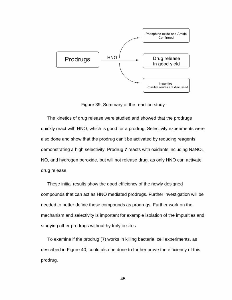
Figure 39. Summary of the reaction study
The kinetics of drug release were studied and showed that the prodrugs
quickly react with HNO, which is good for a prodrug. Selectivity experiments were
also done and show that the prodrug can’t be activated by reducing reagents
demonstrating a high selectivity. Prodrug 7 reacts with oxidants including NaNO2,
NO, and hydrogen peroxide, but will not release drug, as only HNO can activate
drug release.
These initial results show the good efficiency of the newly designed
compounds that can act as HNO mediated prodrugs. Further investigation will be
needed to better define these compounds as prodrugs. Further work on the
mechanism and selectivity is important for example isolation of the impurities and
studying other prodrugs without hydrolytic sites
To examine if the prodrug (7) works in killing bacteria, cell experiments, as
described in Figure 40, could also be done to further prove the efficiency of this
prodrug.
Page 55
46
Figure 40. Cell experiment of the prodrug
Chapter 3. Experiment
3.1 General Chemistry
Analytical thin-layer chromatography (TLC) was performed on Sorbtech Silica
G TLC plates with UV254, 200/pk. Proton NMR, Carbon-13 NMR and Phosphor-
31 NMR spectra were taken on a Bruker (300 MHz) multinuclear spectrometer.
LC-MS was performed on Agilent Technologies HPLC column. MS was done by
an Agilent ion trap (ESI) with HPLC. Organic solvents were distilled from drying
agents before use. Commercially available reagents were used without further
purification.
3.2 Synthetic Procedure
2-(methoxycarbonyl)phenyl 2-(diphenylphosphanyl)benzoate (3). 2-
(Diphenylphosphino) benzoic acid (0.92 g, 3.0 mmol) was added to a solution of
methyl salicylate (1, 0.46 g, 3.0 mmol) in dichloromethane (20 mL) to give a
yellow turbid solution. Dicyclohexylcarbodiimide (DCC, 0.93 g, 4.5 mmol) and 4-
dimethylaminopyridine (DMAP, 0.074 g, 0.61 mmol) were added to this solution
and stirred at room temperature. After 24 hours, the white mixture was filtered,
Page 56
47
the filtrate was concentrated and purified by column chromatography (1:1 ethyl
acetates: hexane) to give 3 as white solid (1.10 g, 2.4 mmol, 83%). 1H
NMR(CDCl3): δ8.29 (m, 1H); 7.87 (d, 1H); 7.33 (m, 3H); 7.15 (m, 11H); 6.90 (m,
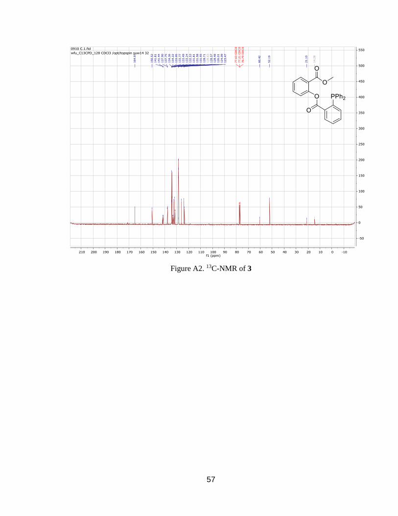
1H); 6.81 (d, 1H); 3.59 (s, 3H). 13C NMR (CDCl3): δ 164.97, 150.52, 141.44,
137.90, 134.36, 134.22, 133.5, 132.53, 131.81, 131.55, 128.57, 126.01, 124.09,
123.47, 77.63, 77.21, 76.79, 60.42, 52.19, 21.10, 14.28. 31P NMR (CDCl3) δ -
5.20.
4-Acetamidophenyl 2-(diphenylphosphanyl)benzoate (5). 2-
(Diphenylphosphino) benzoic acid (0.92 g, 3.0 mmol) was added to a solution of
N-(4-hydroxyphenyl) ethanamide (acetaminophen, 0.45 g, 3.0 mmol) in
dichloromethane (20 mL) to give a yellow turbid solution.
Dicyclohexylcarbodiimide (DCC, 0.93 g, 4.5 mmol) and 4-dimethylaminopyridine
(DMAP, 0.074 g, 0.61 mmol) were added to this solution and stirred at room
temperature. After 24 hours, the white mixture was filtered, the filtrate was
concentrated and purified by column chromatography (1:1 ethyl acetates:
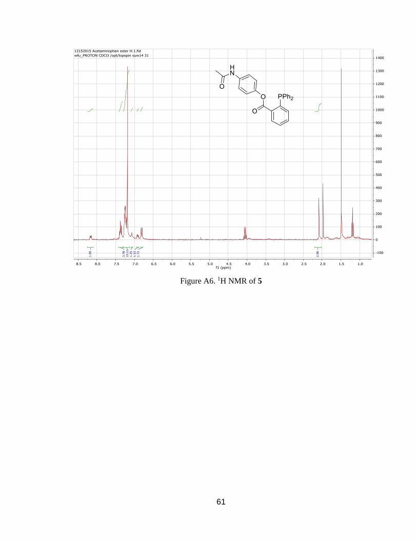
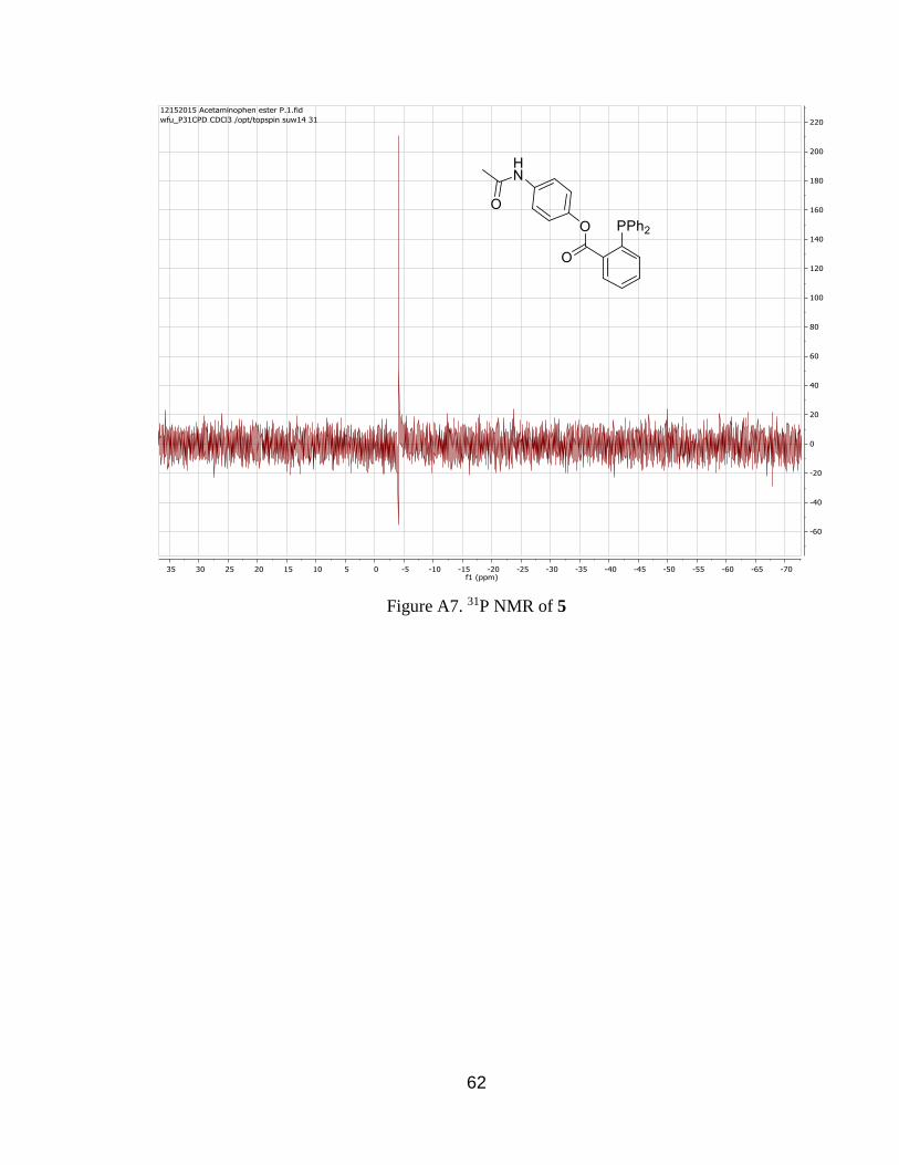
hexane) to give 5 as white solid (1.07 g, 2.4 mmol, 81%). 1H NMR(CDCl3): δ8.20
(m, 1H); 7.31 (m, 4H); 7.20 (m, 15H); 7.10 (m, 1H); 6.90 (m, 1H); 6.80 (d, 1H);
2.10 (s, 3H). 31P NMR (CDCl3) δ -4.90. MS (LC-MSD-Trap-SL, ESI): 441.1
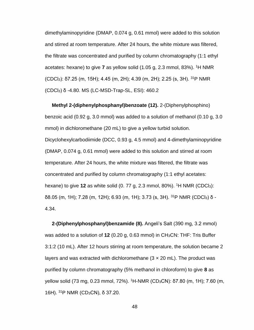
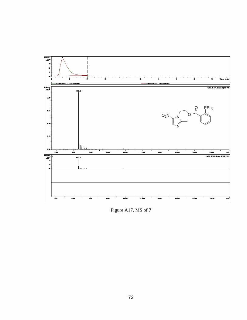
2-(2-Methyl-5-nitro-1H-imidazol-1-yl)ethyl 2-
(diphenylphosphanyl)benzoate (7). 2-(Diphenylphosphino) benzoic acid (0.92
g, 3.0 mmol) was added to a solution of 2-(2-methyl-5-nitro-1H-imidazol-1-yl)
ethanol (metronidazole, 0.51 g, 3.0 mmol) in dichloromethane (20 mL) to give a
yellow turbid solution. Dicyclohexylcarbodiimide (DCC, 0.93 g, 4.5 mmol) and 4-
Page 57
48
dimethylaminopyridine (DMAP, 0.074 g, 0.61 mmol) were added to this solution
and stirred at room temperature. After 24 hours, the white mixture was filtered,
the filtrate was concentrated and purified by column chromatography (1:1 ethyl
acetates: hexane) to give 7 as yellow solid (1.05 g, 2.3 mmol, 83%). 1H NMR
(CDCl3): δ7.25 (m, 15H); 4.45 (m, 2H); 4.39 (m, 2H); 2.25 (s, 3H). 31P NMR
(CDCl3) δ -4.80. MS (LC-MSD-Trap-SL, ESI): 460.2
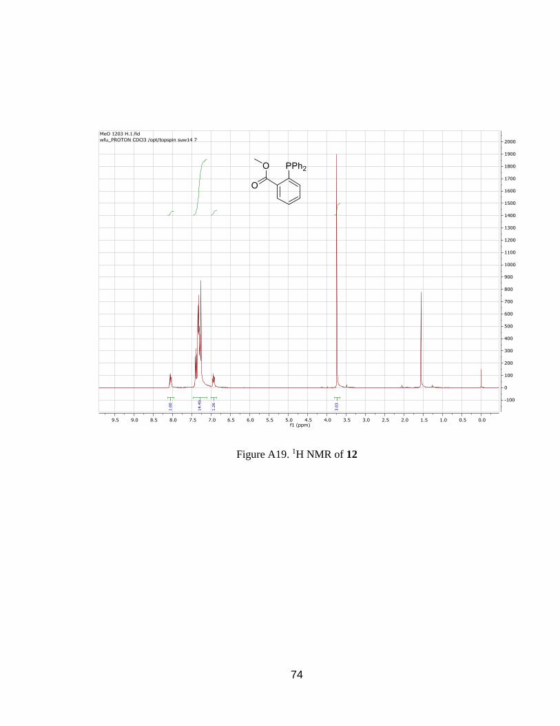
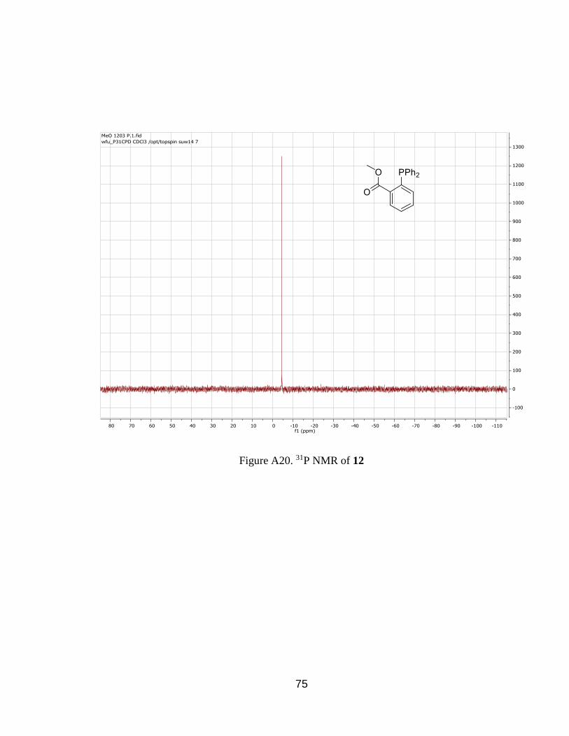
Methyl 2-(diphenylphosphanyl)benzoate (12). 2-(Diphenylphosphino)
benzoic acid (0.92 g, 3.0 mmol) was added to a solution of methanol (0.10 g, 3.0
mmol) in dichloromethane (20 mL) to give a yellow turbid solution.
Dicyclohexylcarbodiimide (DCC, 0.93 g, 4.5 mmol) and 4-dimethylaminopyridine
(DMAP, 0.074 g, 0.61 mmol) were added to this solution and stirred at room
temperature. After 24 hours, the white mixture was filtered, the filtrate was
concentrated and purified by column chromatography (1:1 ethyl acetates:
hexane) to give 12 as white solid (0. 77 g, 2.3 mmol, 80%). 1H NMR (CDCl3):
δ8.05 (m, 1H); 7.28 (m, 12H); 6.93 (m, 1H); 3.73 (s, 3H). 31P NMR (CDCl3) δ -
4.34.
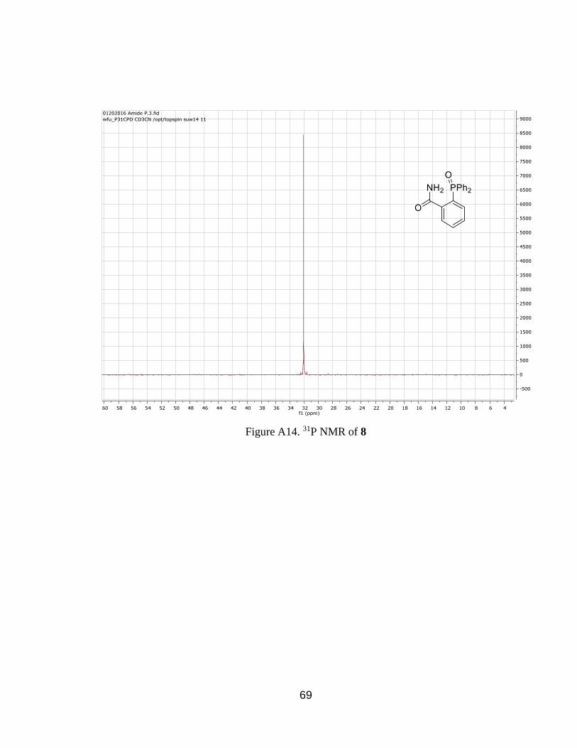
2-(Diphenylphosphanyl)benzamide (8). Angeli’s Salt (390 mg, 3.2 mmol)
was added to a solution of 12 (0.20 g, 0.63 mmol) in CH3CN: THF: Tris Buffer
3:1:2 (10 mL). After 12 hours stirring at room temperature, the solution became 2
layers and was extracted with dichloromethane (3 × 20 mL). The product was
purified by column chromatography (5% methanol in chloroform) to give 8 as
yellow solid (73 mg, 0.23 mmol, 72%). 1H-NMR (CD3CN): δ7.80 (m, 1H); 7.60 (m,
16H). 31P NMR (CD3CN), δ 37.20.
Page 58
49
Phosphine Oxide 9. Hydrogen peroxide (30%, 3 mL) was added to a solution
of 3 (0.02 g, 0.045 mmol) in a mixture of CD3CN: THF: Tris Buffer 3:1:2 (2mL).
After 12 hours stirring at room temperature. 31P NMR (CD3CN), δ 33.47.
Phosphine Oxide 10. Hydrogen peroxide (30%, 3 mL) was added to a
solution of 5 (0.02 g, 0.046 mmol) in a mixture of CD3CN: THF: Tris Buffer 3:1:2
(2mL). After 12 hours stirring at room temperature. 31P-NMR were taken. 1P NMR
(DMSO), δ 28.75.
Phosphine Oxide 11. Hydrogen peroxide (30%, 3 mL) was added to a
solution of 7 (0.02 g, 0.044 mmol) in a mixture of CD3CN: THF: Tris Buffer 3:1:2
(2mL). After 12 hours stirring at room temperature. 31P-NMR were taken. 1P NMR
(CD3CN), δ 33.34.
3.3 Treatment of Prodrugs with HNO
Treatment of 3 with HNO. Angeli’s Salt (600 mg, 4.9 mmol) was added to a
solution of prodrug 3 (0.440 g, 1.00 mmol) in a mixture of CH3CN: THF: Tris
Buffer 3:1:2 (20 mL). After 12 hours stirring at room temperature, the solution
became 2 layers and was extracted with dichloromethane (3 × 20 mL). The
product was purified by column chromatography (5% methanol in chloroform) to
give 1 as white solid (116 mg, 0.76 mmol, 76%). 1H-NMR (CD3Cl): δ10.67 (s,
1H); 7.72 (m, 1H); 7.32 (m, 1H); 6.85 (m, 1H); 6.75 (m, 1H); 3.80 (s, 1H). 13C
NMR (75 MHz, CDCl3) δ 170.57, 161.60, 135.69, 129.90, 119.15, 117.57,
112.39, 77.47, 77.05, 76.63, 52.26.
Page 59
50
Treatment of 5 with HNO. Angeli’s Salt (600 mg, 4.9 mmol) was added to a
solution of 5 (0.439 g, 1.00 mmol) in a mixture of CH3CN: THF: Tris Buffer 3:1:2
(20 mL). After 12 hours stirring at room temperature, the solution became 2
layers and was extracted with dichloromethane (3 × 20 mL). The product was
purified by column chromatography (5% methanol in chloroform) to give 4 as
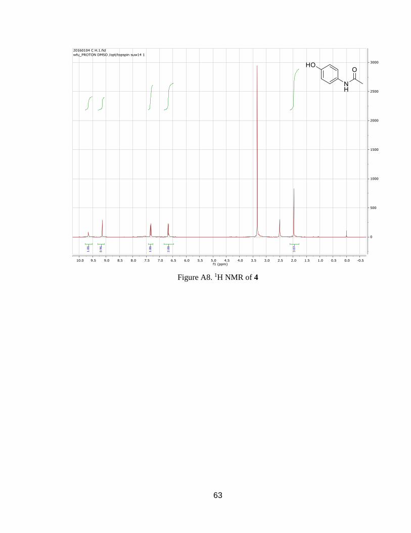
white solid (136 mg, 0.90 mmol, 90%). 1H-NMR (DMSO-d): δ9.70 (s, 1H); 9.14
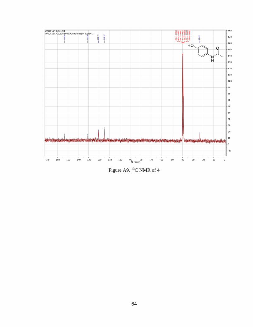
(s, 1H); 7.33 (d, 2H); 6.67 (d, 2H); 1.97 (s, 3H). 13C NMR (75 MHz, DMSO) δ
153.05, 130.98, 120.72, 114.92, 40.31, 40.03, 39.75, 39.48, 39.20, 38.92, 38.64,
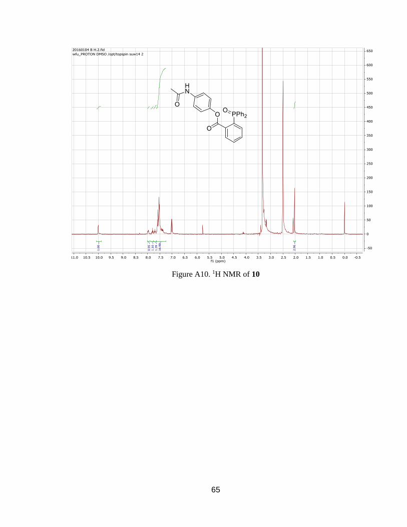
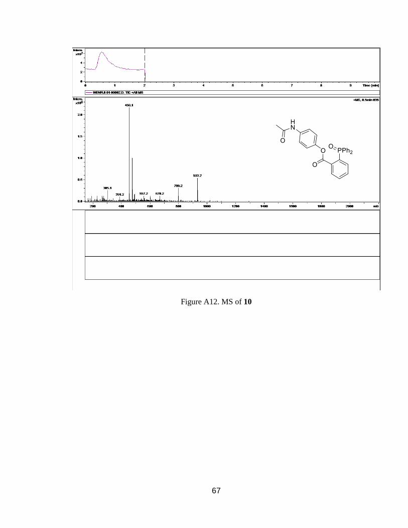
23.69 and phosphine oxide (10) as yellow solid (354 mg, 0.78 mmol, 78%). 1H-
NMR (CD3Cl): δ10.00 (s, 1H); 7.90 (m, 1H); 7.75 (m, 1H); 7.40 (m, 1H); 6.55 (m,
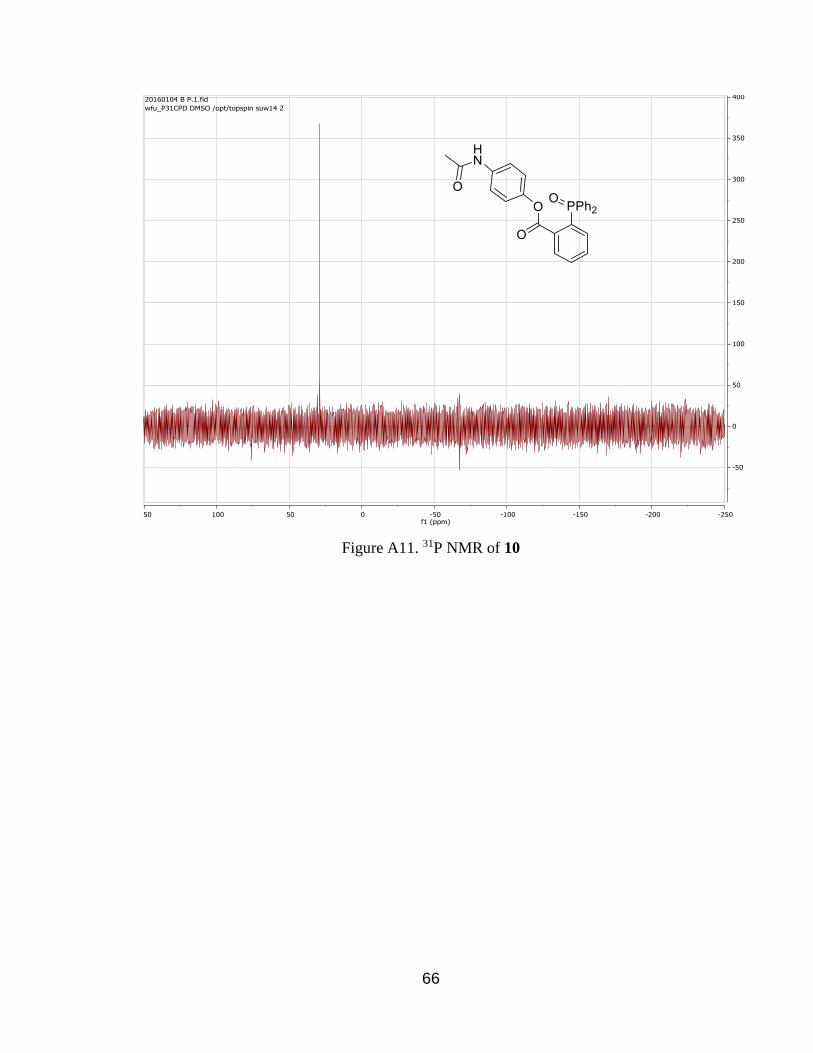
17H); 2.00 (s, 3H). 13P NMR (DMSO-d): 38.00. MS (LC-MSD-Trap-SL, ESI):
456.1.
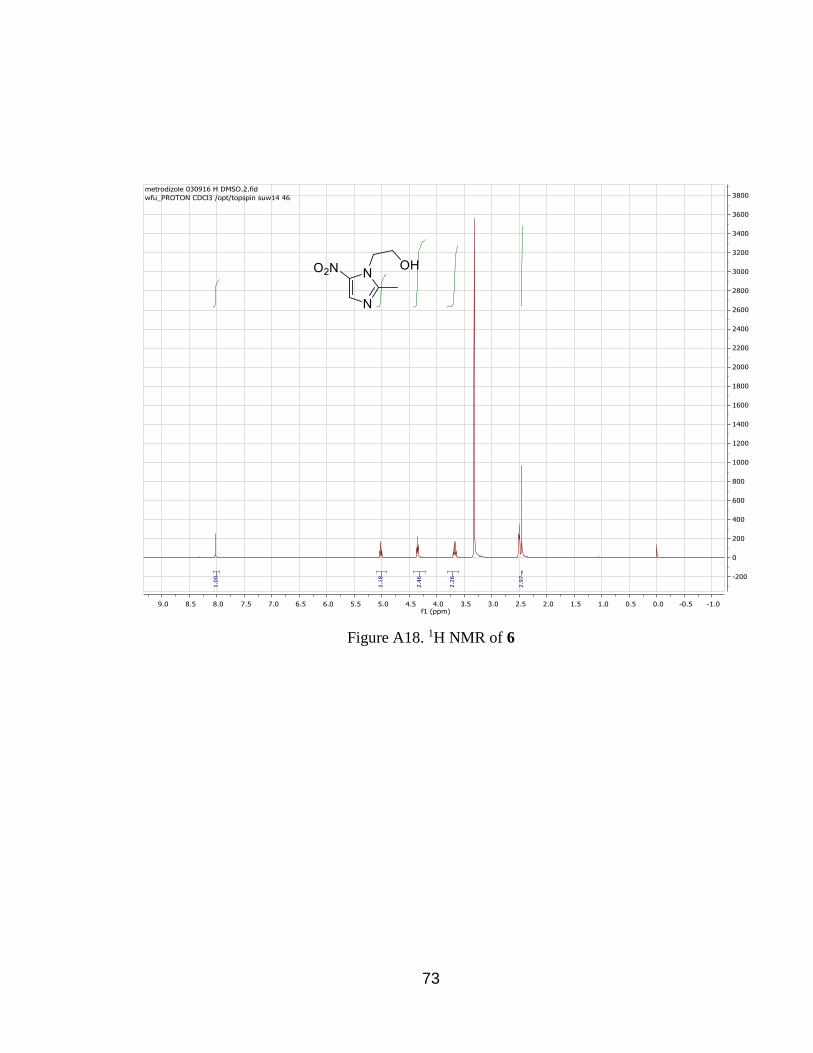
Treatment of 7 with HNO. Angeli’s Salt (300 mg, 2.5 mmol) was added to a
solution of 7 (0.230 g, 1.00 mmol) in a mixture of CH3CN: THF: Tris Buffer 3:1:2
(20 mL). After 12 hours stirring at room temperature, the solution became 2
layers and was extracted with dichloromethane (3×20 mL). The product was
purified by column chromatography (5% methanol in chloroform) to give 6 as
white solid (65 mg, 0.38 mmol, 75%). 1H-NMR (DMSO): δ12.79 (s, 1H); 9.77 (t,
1H); 9.12 (t, 2H); 8.45 (m, 2H); 7.23 (s, 3H). 13C-NMR (DMSO) δ 208.67, 192.24,
138.19, 64.96, 53.48, 19.48.
3.4 Kinetic Analysis of the Reaction of 3 and HNO.
Angeli’s Salt (12.6 mg, 0.103 mmol) was added to a solution of 3 (0.0104 g,
0.0236 mmol) in a mixture of CD3CN: THF: Tris Buffer 3:1:2 (600 μL) in an NMR
Page 60
51
tube. 31P-NMR spectra were taken every 15 minutes and overall 9 spectra were
taken. A phosphoric acid standard was also added for the 31P-NMR. By setting
the integral of the phosphoric acid as the standard, intergrals were calculated
from starting material 3 and compared. The time resolved graph was drawn and
an observed rate constant was calculated.
3.5 Selectivity
Compound 7 (138 mg, 0.30 mmol) was dissolved in a mixture of CD3CN: THF:
Tris Buffer 3:1:2 (12 mL). The mixture was separated into 5 vials and each vial
contained 2 mL of the mixture. GSH (0.47 mmol, 144 mg), cys (0.47 mmol, 57
mg), NaNO2 (0.47 mmol, 32 mg), DEANO (0.16 mmol, 20 mg) and a blank were
added. After 24 hours, 31P-NMR spectra were taken.
Page 61
52
References
1. Bredt, D. S.; Snyder, S. H. Annu. Rev. Biochem. 1994, 63, 175.
2. Ignarro, L. J. Nitric Oxide: Biology and Pathobiology. Academic Press, 2000: San
Diego.
3. Yoon, Y.; Song, U.; Hong, S.H.; Kim, J.Q. Clinic. Chem. 2000, 46, 1626.
4. Cannon, R. O. Clin. Chem. 1998, 44, 1809.
5. Moncada, S.; Palmer, R. M. J.; Higgs, E. A. Pharmacol. Rev. 1991, 43, 109.
6. Kerwin, J. F.; Lancaster, J. R.; Feldman, P. L. J. Med. Chem. 1995, 38, 4343.
7. Green, S.J. Nat. Med. 1995, 1, 515.
8. Lundberg, J. O.; Weitzberg, E.; Gladwin, M. T. Nat. Rev. Drug. Discov. 2008, 7,
156.
9. Bradley, J. M.; Hopkinson, A.; King, D. A. J. Phys. Chem., 1995, 99, 17032.
10. Fowler, D.; Flechard, C.; Skiba, U.; Coyle, M.; Cape, J. N. New. Phytol. 1998,
139, 11.
11. Miranda, K. M. Coord. Chem. Rev. 2005, 249, 433.
12. Smith, P. A. S.; Hein, G. E. J. Am. Chem. Soc. 1960, 82, 5731.
13. Kohout, F. C.; Lampe, F. W. J. Am. Chem. Soc. 1965, 87, 5795.
14. Irvine, J. C.; Ritchie, R. H.; Favaloro, J. L.; Andrews, K. L.; Widdop, R. E.;
Kemp-Harper, B. K. Trends Pharmacol. Sci. 2008, 29, 601.
15. Gaudagnini, R.; Schatz, G. C. J. Chem. Phys. 1995, 102, 774.
16. Bruna, P. J.; Marian, C. M., Chem. Phys. Lett. 1979, 67, 109.
17. Wink, D. A.; Hanbauer, I.; Krishna, M. C.; Degraff. W.; Gamson, J.; Mitchell, J.
B. Proc. Natl. Acad. Sci. U. S. A. 1993, 90 ,9813.
Page 62
53
18. Furchgott, R. F.; Zawadzki, J. V. Nature, 1980, 288, 373.
19. Murad, F.; Mittal, C. K.; Arnold, W. P.; Katsuki, S.; Kimura, H. Adv. Cyclic
Nucleotide Res. 1978, 9, 145.
20. Palmer, R. M. J.; Ferrige, A. G.; Moncada, S. Nature, 1987, 327, 524.
21. Dierks, E. A.; Burstyn, J. N. Biochem. Pharmacol. 1996, 51, 1593.
22. Ignarro, L. J. Nitric Oxide: Biology and Pathobiology. Academic Press, 2000: San
Diego.
23. Fukuto, J. M.; Chiang, K.; Hszieh, R.; Wong, P.; Chaudhuri, G.; J. Pharmacol.
Exp. Ther. 1992, 263, 546.
24. Fukuto, J. M.; Hobbs, A. J.; Ignarro, L. J. Biochem. Biophys. Res. Commun. 1993,
196, 707.
25. Wink, D. A.; Feelisch, M.; Fukuto, J.; Chistodoulou, D.; Jourd’heuil, D.;
Grisham, M. B.; Vodovotz, Y.; Cook, J. A.; Krishna, M.; Degraff, W. G.; Kim,
S.; Gamson, J.; Mitchell, J.B. Arch. Biochem. Biophys. 1998, 351, 66.
26. Bazylinski, D. A.; Hollocher, T. C. Inorg. Chem. 1985, 24, 4285.
27. Gratzel, M. Taniguchi, S. Henglein, A. Bunsen-Ges, B. Phys. Chem. 1970, 74,
1003.
28. Rosenthal, J.; Lippard, S. J. J. Am. Chem. Soc. 2010, 132, 5536.
29. Suárez, S. A.; Bikiel, D. E.; Wetzler, D. E.; Martí, M. A.; Doctorovich, F. Anal.
Chem. 2013, 85, 10262.
30. Zhengrui, M.; Reisz, J. A.; Mitroka, S. M.; Pan, J.; Xian, M.; King, S. B. Bioorg.
Meg. Chem. Lett. 2015, 25, 16.
31. Reisz, J. A.; Klorig, E. B.; Wright, M. W.; King, S. B. Org. Lett. 2009, 11, 2719.
Page 63
54
32. Smallwood, H. M.; J. Am. Chem. Soc. 1929, 15, 1985.
33. Cheskis, S. G.; Sarkisov, O. M.; Chem. Phys. Lett. 1979, 62, 72.
34. Hawthorne, M. F. J. Am. Chem. Soc. 1957, 79, 2510.
35. Smith, P. A. S.; Pars, H. G. J. Org. Chem. 1959, 24, 1325.
36. Xu, Y. P.; Alavanja, M. M.; Johnson, V. L.; Yasaki, G.; King, S. B. Tetrahedr.
Lett. 2000, 41, 4265.
37. Ware, R. W.; King, S. B. J. Am. Chem. Soc. 1999, 121, 6769.
38. Bonner, F. T.; Ko, Y. H. Inorg. Chem. 1992, 31, 2514.
39. Zamora, R.; Grzesiok, A.; Weber, H.; Feelisch, M. Biochem. J. 1995, 312, 333.
40. Bonner, F. T.; Ravid, B. Inorg. Chem. 1975, 14, 558.
41. Hendrickson, D. N.; Jolly, W. L. Inorg. Chem. 1969, 8, 693.
42. Wermuth, C. G.; Ganellin, C. R.; Lindberg, P; Mitscher, L. A. Pure Appl. Chem.
1998, 70, 1129.
43. Kuei-Meng, W.; Pharmaceuticals 2009, 2, 77.
44. Harper, H. J. J. Med. Chem. 1959, 1, 467.
45. Dumond, J. F.; King, S. B. Antioxid. Redox Signal. 2011, 14, 1637.
46. Fukuto, J. M.; Switzer, C. H.; Miranda, K. M.; Wink, D. A. Annu. Rev.
Pharmacol. Toxicol. 2005, 45, 335.
47. Chung, M.; Chia, W.; Wan, W.; Lin, Y.; Sung, H. J. Am. Chem. Soc. 2015, 137,
12462.
48. Mittal, M.; Siddiqui, M. R.; Tran, K.; Reddy, S. P.; Malik, A. B. Antioxid Redox
Signal. 2014, 20, 1126.
49. Reisz, J. A.; Zink, C. N.; King, S. B. J. Am. Chem. Soc. 2011, 133, 11675.
Page 64
55
50. Shafirovich, V.; Lymar, S. V. Proc. Natl. Acad. Sci. USA. 2002, 99, 7340.
51. Hobbs, A. J.; Fukuto, J. M.; Ignarro, L. J. Proc. Natl. Acad. Sci. USA. 1994, 91,
10992.
52. Eberhardt. M.; et. al. Nat. Commun. 2014, 5, 4381.
Page 65
56
Appendix
Figure A1. 1H-NMR of 3
Page 66
57
Figure A2. 13C-NMR of 3
Page 67
58
Figure A3. 31P-NMR of 3
Page 68
59
Figure A4. 1H-NMR for 1
Page 69
60
Figure A5. 13C-NMR of 1
Page 70
61
Figure A6. 1H NMR of 5
Page 71
62
Figure A7. 31P NMR of 5
Page 72
63
Figure A8. 1H NMR of 4
Page 73
64
Figure A9. 13C NMR of 4
Page 74
65
Figure A10. 1H NMR of 10
Page 75
66
Figure A11. 31P NMR of 10
Page 76
67
Figure A12. MS of 10
Page 77
68
Figure A13. 1H NMR of 8
Page 78
69
Figure A14. 31P NMR of 8
Page 79
70
Figure A15. 1H NMR of 7
Page 80
71
Figure A16. 31P NMR of 7
Page 81
72
Figure A17. MS of 7
Page 82
73
Figure A18. 1H NMR of 6
Page 83
74
Figure A19. 1H NMR of 12
Page 84
75
Figure A20. 31P NMR of 12
Page 85
76
Wen-Rui Su Curriculum Vitae
Personal Information
Name: Wenrui Su
Address: 1252 Brookwood Dr., Winston-Salem, NC 27106
Cell phone:336-655-7491
Email: [email protected]
Nationality: China
GPA: 3.10/4.00
Education
2010-2014 B. S. Chemistry Department, Peking University, Beijing, China
Major in Physical Chemistry (Advisor: Professor Kai Wu)
Minor in Mathematics and Applied Mathematics
2010-2014 M. S. Chemistry Department, Wake Forest University, U. S.
Major in Organic Chemistry (Advisor: Professor S. B. King)
Courses related
Linear Algebra, Real Analysis, Complex Analysis, Statistics, Probability Theory,
Quantitative Analysis, C language, Data Structure and Algorithm.
Working Experience
2014-2016 Teaching Assistant in Wake Forest University
I was T. A. for General Chemistry and Organic Chemistry and
gained strong ability of communication in English.
Jul.-Aug. 2015 Internship in Bank of China
Page 86
77
I was in the international settlement department and mainly
responsible for the issue of Letter of Credit.
Achievements
July, 2014 Ph. D. Program in Wake Forest University
I was admitted by Ph. D. program in Wake Forest University for full
scholarship. Based on the interest, I talked with my advisor to
change to M. S. program.
June, 2015 CFA level 1 exam
I passed the CFA level 1 exam at the first attempt. I got >70% in
Cooperate Finance, Equity Investment and Financial Reporting
Analysis and 50-70% in most other subjects.
Interests
-Mathematics
Mathematics is my most favorite subject especially analysis part. I did well in
Probability Theory and Statistic. I also audited the Applied Stochastic Process which
made me interest in modeling analysis.
-Quantitative Analysis
I took Quantitative Analysis about chemistry and audited the Applied Mathematical
Software (about the using of SAS) which made me interest in applying mathematical
and computational method in practical analysis.
-Communication
I’m outgoing and want to communicate with others. 2-year T. A. work helps me a lot
in learning how to communicate with others. Also, I enjoy that job and get lots of
positive comments from undergraduate students in Wake Forest University.
Additional Skills:
Technical:
-STM, AFM, UV-Vis spectrum, NMR, Mass Spectrum, X-Ray Diffraction.
Computing:
Page 87
78
-C, MS Windows, MS Office, Origin 8.0, SAS, SPSS, Matlab, MS Mathematics
Languages:
-Fluent in Chinese (Native) and English.