Niacin and biosynthesis of PGD 2 by platelet COX-1 in mice and humans Wen-Liang Song, … , Ellen Puré, Garret A. FitzGerald J Clin Invest. 2012;122(4):1459-1468. https://doi.org/10.1172/JCI59262. The clinical use of niacin to treat dyslipidemic conditions is limited by noxious side effects, most commonly facial flushing. In mice, niacin-induced flushing results from COX-1–dependent formation of PGD 2 and PGE 2 followed by COX-2– dependent production of PGE 2 . Consistent with this, niacin-induced flushing in humans is attenuated when niacin is combined with an antagonist of the PGD 2 receptor DP1. NSAID-mediated suppression of COX-2–derived PGI 2 has negative cardiovascular consequences, yet little is known about the cardiovascular biology of PGD 2 . Here, we show that PGD 2 biosynthesis is augmented during platelet activation in humans and, although vascular expression of DP1 is conserved between humans and mice, platelet DP1 is not present in mice. Despite this, DP1 deletion in mice augmented aneurysm formation and the hypertensive response to Ang II and accelerated atherogenesis and thrombogenesis. Furthermore, COX inhibitors in humans, as well as platelet depletion, COX-1 knockdown, and COX-2 deletion in mice, revealed that niacin evoked platelet COX-1–derived PGD 2 biosynthesis. Finally, ADP-induced spreading on fibrinogen was augmented by niacin in washed human platelets, coincident with increased thromboxane (Tx) formation. However, in platelet-rich plasma, where formation of both Tx and PGD 2 was increased, spreading was not as pronounced and was inhibited by DP1 activation. Thus, PGD 2 , like PGI 2 , may function as a homeostatic response to thrombogenic and hypertensive stimuli and may have particular relevance as a […] Research Article Cardiology Find the latest version: https://jci.me/59262/pdf
Transcript
Niacin and biosynthesis of PGD2 by platelet COX-1 in mice andhumans
Wen-Liang Song, … , Ellen Puré, Garret A. FitzGerald
The clinical use of niacin to treat dyslipidemic conditions is limited by noxious side effects, most commonly facial flushing.In mice, niacin-induced flushing results from COX-1–dependent formation of PGD2 and PGE2 followed by COX-2–dependent production of PGE2. Consistent with this, niacin-induced flushing in humans is attenuated when niacin iscombined with an antagonist of the PGD2 receptor DP1. NSAID-mediated suppression of COX-2–derived PGI2 hasnegative cardiovascular consequences, yet little is known about the cardiovascular biology of PGD2. Here, we show thatPGD2 biosynthesis is augmented during platelet activation in humans and, although vascular expression of DP1 isconserved between humans and mice, platelet DP1 is not present in mice. Despite this, DP1 deletion in mice augmentedaneurysm formation and the hypertensive response to Ang II and accelerated atherogenesis and thrombogenesis.Furthermore, COX inhibitors in humans, as well as platelet depletion, COX-1 knockdown, and COX-2 deletion in mice,revealed that niacin evoked platelet COX-1–derived PGD2 biosynthesis. Finally, ADP-induced spreading on fibrinogenwas augmented by niacin in washed human platelets, coincident with increased thromboxane (Tx) formation. However, inplatelet-rich plasma, where formation of both Tx and PGD2 was increased, spreading was not as pronounced and wasinhibited by DP1 activation. Thus, PGD2, like PGI2, may function as a homeostatic response to thrombogenic andhypertensive stimuli and may have particular relevance as a […]
TheJournalofClinicalInvestigation http://www.jci.org Volume 122 Number 4 April 2012 1459
Niacin and biosynthesis of PGD2 by platelet COX-1 in mice and humans
Wen-Liang Song,1,2 Jane Stubbe,1,2,3 Emanuela Ricciotti,1,2 Naji Alamuddin,1,2 Salam Ibrahim,1,2 Irene Crichton,4 Maxwell Prempeh,5 John A. Lawson,1,2 Robert L. Wilensky,5
Lars Melholt Rasmussen,6 Ellen Puré,4 and Garret A. FitzGerald1,2
1Institute for Translational Medicine and Therapeutics and 2Department of Pharmacology, University of Pennsylvania Perelman School of Medicine, Philadelphia, Pennsylvania, USA. 3Cardiovascular and Renal Research, Institute of Molecular Medicine, University of Southern Denmark, Odense, Denmark.
4The Wistar Institute, Philadelphia, Pennsylvania, USA. 5Cardiovascular Division of Hospital of the University of Pennsylvania, Philadelphia, Pennsylvania, USA. 6Department of Clinical Biochemistry and Pharmacology, Odense University Hospital, Odense, Denmark.
IntroductionPGD2 is formed from the PGH2 COX product of arachidonic acid by the action of either lipocalin-like PGD synthase (lPGDS) or hemopoietic PGD synthase (1). PGD2 mediates its effects via activation of 2 D prostanoid receptors (DPs), DP1 and DP2 (the latter also known as chemoattractant receptor–homologous mol-ecule expressed on Th2 cells [CRTH2]) (2–4). Suppression of PGD2 has been implicated in the bronchoconstriction of aspirin-evoked respiratory disease (5, 6), and release of PGD2 contributes to the vascular instability of systemic mastocytosis (7–9). DP1 depletion ameliorates allergen-induced airway inflammation in mice (10), and DP1 antagonism is being pursued as an effective treatment for allergic nasal congestion in humans (11, 12). DP1 is coupled to Gs-dependent adenylate cyclase activation (13) and is expressed on mast cells, in which PGD2 is the predominant product of COX metabolism (14). PGD2 also plays a pivotal role in the regulation of physiological sleep via the lPGDS/DP1 pathway (15).
PGD2 appears to derive in roughly equal amounts from COX-1 and COX-2 in liver macrophages in vitro under basal and LPS-stimulated conditions (16), whereas in mast cells, PGD2 is initially derived from secretory phospholipase A2 (PLA2) and COX-1, fol-lowed by sustained formation by cytoplasmic PLA2 and COX-2 (17). We previously reported that 11,15-Dioxo-9α-hydroxy-2,3,4,5-tetranorprostan-1,20-dioic acid (tetranor PGDM), an abundant
metabolite in urine, reflects modulated biosynthesis of PGD2 in humans and mice. We have shown that in healthy volunteers, 325 mg aspirin (which inhibits both COX-1 and COX-2), but not cele-coxib and rofecoxib (selective inhibitors of COX-2), suppresses PGD2 formation (18). This suggests that COX-1 is the dominant source of systemic PGD2 formation under physiological circum-stances in humans. However, there is no direct evidence that COX-1 inhibition results in PGD2 suppression or, if so, of the cellular source of its formation.
Mast cells are a major potential source of PGD2. Lesser amounts can be formed by other cells, including platelets, macrophages, and lymphocytes. For example, although PGD2 is a relatively minor product of platelet COX-1 in vitro, sufficient exogenous PGD2 can constrain platelet activation via DP1 (19–21). How-ever, it is not known whether platelet generation is a substantial contributor to actual biosynthesis or becomes a more important contributor to overall biosynthesis of PGD2 under conditions of perturbed vascular biology.
Morrow and colleagues first noted that PGD2 and its products appeared to mediate the cutaneous vasodilation that constrains the use of the hypolipidemic drug niacin (22, 23). Indeed, admin-istration of niacin to healthy volunteers results in formation of PGD2. PGD2 relaxes vascular smooth muscle cells in vitro, and its release by dermal dendritic cells contributes to facial flushing (23). In mice, niacin-induced flushing has been shown to result from an early phase of COX-1–dependent formation of PGD2 and PGE2 by such Langerhans cells, followed by delayed COX-2–dependent production of PGE2 by keratinocytes (24).
Authorshipnote: Wen-Liang Song and Jane Stubbe contributed equally to this work.
Conflictofinterest: Robert L. Wilensky reports equity interest in Johnson & Johnson.
1460 TheJournalofClinicalInvestigation http://www.jci.org Volume 122 Number 4 April 2012
Recent interest in PGD2 has been prompted by the use of DP1 blockade as an adjunct to niacin therapy (25) and by the potential role of PGD2 and its metabolites in the resolution of inflammation (26). Indeed, a combination of extended-release niacin and laropip-rant, a DP1 antagonist, has been approved in Europe; US approval awaits the outcome of a randomized trial. DP1 is expressed on human platelets and, like the I prostanoid receptor (IP), is coupled to adenylate cyclase activation (20, 21). Given the cardiovascular hazard from NSAIDs that results from suppression of COX-2–derived PGI2 (27), we sought to elucidate the cardiovascular biol-ogy of PGD2 and the potential implications of DP1 antagonism in patients with cardiovascular disease treated with niacin.
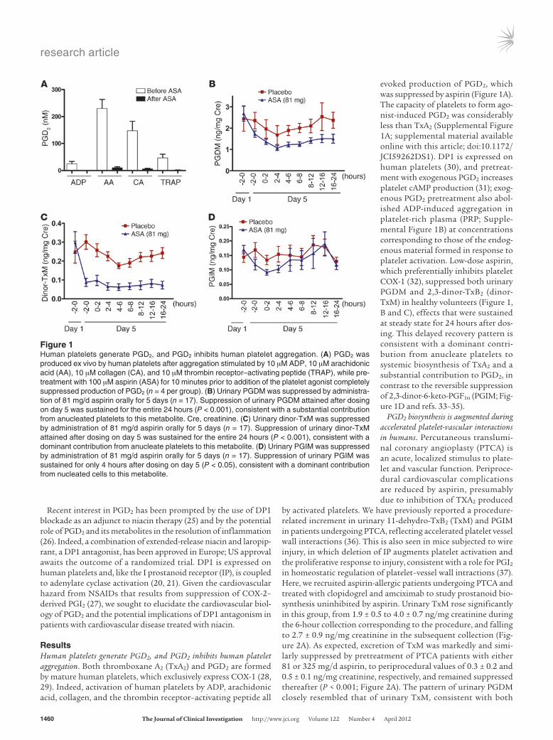
ResultsHuman platelets generate PGD2, and PGD2 inhibits human platelet aggregation. Both thromboxane A2 (TxA2) and PGD2 are formed by mature human platelets, which exclusively express COX-1 (28, 29). Indeed, activation of human platelets by ADP, arachidonic acid, collagen, and the thrombin receptor–activating peptide all
evoked production of PGD2, which was suppressed by aspirin (Figure 1A). The capacity of platelets to form ago-nist-induced PGD2 was considerably less than TxA2 (Supplemental Figure 1A; supplemental material available online with this article; doi:10.1172/JCI59262DS1). DP1 is expressed on human platelets (30), and pretreat-ment with exogenous PGD2 increases platelet cAMP production (31); exog-enous PGD2 pretreatment also abol-ished ADP-induced aggregation in platelet-rich plasma (PRP; Supple-mental Figure 1B) at concentrations corresponding to those of the endog-enous material formed in response to platelet activation. Low-dose aspirin, which preferentially inhibits platelet COX-1 (32), suppressed both urinary PGDM and 2,3-dinor-TxB2 (dinor-TxM) in healthy volunteers (Figure 1, B and C), effects that were sustained at steady state for 24 hours after dos-ing. This delayed recovery pattern is consistent with a dominant contri-bution from anucleate platelets to systemic biosynthesis of TxA2 and a substantial contribution to PGD2, in contrast to the reversible suppression of 2,3-dinor-6-keto-PGF1α (PGIM; Fig-ure 1D and refs. 33–35).
PGD2 biosynthesis is augmented during accelerated platelet-vascular interactions in humans. Percutaneous translumi-nal coronary angioplasty (PTCA) is an acute, localized stimulus to plate-let and vascular function. Periproce-dural cardiovascular complications are reduced by aspirin, presumably due to inhibition of TXA2 produced
by activated platelets. We have previously reported a procedure-related increment in urinary 11-dehydro-TxB2 (TxM) and PGIM in patients undergoing PTCA, reflecting accelerated platelet vessel wall interactions (36). This is also seen in mice subjected to wire injury, in which deletion of IP augments platelet activation and the proliferative response to injury, consistent with a role for PGI2 in homeostatic regulation of platelet–vessel wall intractions (37). Here, we recruited aspirin-allergic patients undergoing PTCA and treated with clopidogrel and amciximab to study prostanoid bio-synthesis uninhibited by aspirin. Urinary TxM rose significantly in this group, from 1.9 ± 0.5 to 4.0 ± 0.7 ng/mg creatinine during the 6-hour collection corresponding to the procedure, and falling to 2.7 ± 0.9 ng/mg creatinine in the subsequent collection (Fig-ure 2A). As expected, excretion of TxM was markedly and simi-larly suppressed by pretreatment of PTCA patients with either 81 or 325 mg/d aspirin, to periprocedural values of 0.3 ± 0.2 and 0.5 ± 0.1 ng/mg creatinine, respectively, and remained suppressed thereafter (P < 0.001; Figure 2A). The pattern of urinary PGDM closely resembled that of urinary TxM, consistent with both
Figure 1Human platelets generate PGD2, and PGD2 inhibits human platelet aggregation. (A) PGD2 was produced ex vivo by human platelets after aggregation stimulated by 10 μM ADP, 10 μM arachidonic acid (AA), 10 μM collagen (CA), and 10 μM thrombin receptor–activating peptide (TRAP), while pre-treatment with 100 μM aspirin (ASA) for 10 minutes prior to addition of the platelet agonist completely suppressed production of PGD2 (n = 4 per group). (B) Urinary PGDM was suppressed by administra-tion of 81 mg/d aspirin orally for 5 days (n = 17). Suppression of urinary PGDM attained after dosing on day 5 was sustained for the entire 24 hours (P < 0.001), consistent with a substantial contribution from anucleated platelets to this metabolite. Cre, creatinine. (C) Urinary dinor-TxM was suppressed by administration of 81 mg/d aspirin orally for 5 days (n = 17). Suppression of urinary dinor-TxM attained after dosing on day 5 was sustained for the entire 24 hours (P < 0.001), consistent with a dominant contribution from anucleate platelets to this metabolite. (D) Urinary PGIM was suppressed by administration of 81 mg/d aspirin orally for 5 days (n = 17). Suppression of urinary PGIM was sustained for only 4 hours after dosing on day 5 (P < 0.05), consistent with a dominant contribution from nucleated cells to this metabolite.
research article
TheJournalofClinicalInvestigation http://www.jci.org Volume 122 Number 4 April 2012 1461
metabolites deriving predominantly from platelets. Thus, in a setting of platelet activation, urinary PGDM increased in a pro-cedure-related manner in the aspirin-sensitive patients and was maximally suppressed by both aspirin regimens (Figure 2B). In contrast, periprocedural PGIM, reflecting its predominant vascu-lar origin, was dose-dependently suppressed by aspirin, being only partially inhibited by the 81-mg/d regimen (Figure 2C).
DP1 activation restrains the hypertensive and aneurysmal responses to Ang II in male mice. Unlike human platelets, mouse platelets lacked DP1 (Supplemental Figure 2, A and B). Thus, exogenous PGD2, unlike the PGI2 analog cicaprost, failed to inhibit platelet aggre-gation (data not shown) or disaggregate ADP-induced platelet aggregation (Supplemental Figure 2, C–H). Despite this, the sys-tolic hypertensive response to 4-week infusion of Ang II was sig-nificantly augmented in male hyperlipidemic ApoE KO mice by DP1 deletion (Supplemental Figure 3, A and B). By contrast, the elevation in systolic and mean arterial pressure evoked by 14 days on a 4% high-salt diet was unaltered by DP1 deletion (Supplemen-tal Figure 3, C and D). DP1 deletion significantly increased Ang II– induced abdominal aortic aneurysm (AAA) formation in the ApoE KO mice, whether measured by wet weight, external diameter, or blinded allocation of severity (Figure 3).
PGD2 restrains thrombogenesis in female mice. Despite the absence of DP1 on mouse platelets, its deletion accelerated the partial and complete thrombogenic occlusive response to a photochemical injury to the carotid artery in female mice (Figure 4).
Activation of DP1 restrains atherogenesis in female mice. The impact of DP1 deficiency on atherogenesis was assessed in LDL recep-tor KO mice. Lesion burden was measured en face at 3, 6, and 9 months on a high-fat diet. Lesion burden increased with time in both genders, and there was no significant impact of DP1 deletion on disease progression in male mice. However, lesion progression was accelerated modestly, but significantly, in female mice lacking DP1 (Supplemental Figure 4). In mice, DP1 immunoreactivity was localized within lesions to activated vascular smooth muscle cells in both the media of the underlying lesion and the neointima, as
identified morphologically together with expression of VCAM-1 detected in serial sections as previously described (38), and in areas of inflammatory myeloid infiltrates, as identified by expression of CD11b (Figure 5) and CD45 (data not shown). In humans, DP1 expression was detected in endothelial cells, and in human athero-matous tissue, additional expression was noted in intravascular endothelial cells, macrophages, and vascular smooth muscle cells (Supplemental Figure 5).
Niacin evokes platelet PGD2 biosynthesis in humans. To address the cellular contribution of platelets to PGD2 biosynthesis in humans, we randomized healthy volunteers to receive 5 daily doses of aspi-rin (81 mg/d) or placebo. Niacin (600 mg) was then administered orally either 30 minutes or 24 hours after the final dose of aspi-rin, on the assumption that a differential, partial recovery of the capacity for prostanoid biosynthesis will have occurred in nucle-ated cells, but not in platelets, with delayed niacin dosing (34). Niacin evoked a marked increase in urinary PGDM, TxM, PGIM, and PGEM under placebo conditions. Both urinary PGDM and TxM were completely suppressed when niacin was administered either 30 minutes or 24 hours after the last dose of aspirin (Figure 6, A and B). Again, this delayed pattern of recovery is consistent with both prostanoids deriving predominantly from platelets. In contrast, whereas urinary PGIM and PGEM were suppressed at the earlier time point, both had partially recovered when niacin was administered 24 hours after the last dose of aspirin (Figure 6, C and D), consistent with their predominant source being nucleated cells, such as those of vascular origin.
Niacin evokes platelet COX-1–dependent prostaglandin formation in mice. Niacin evoked an increase in urinary excretion of PGDM, TxM, PGEM, and PGIM in mice as in humans. We have previ-ously demonstrated that COX-1 knockdown (KD) mice exhibit an asymmetric effect on platelet COX-1, reminiscent of the effects of low-dose aspirin in humans (39). In the present study, COX-1 KD ablated the niacin-induced increment in urinary TxM observed in WT littermate controls (average suppression, 97.8% ± 11.9%; P < 0.001) and almost completely suppressed the increment in
Figure 2PGD2 biosynthesis is augmented during accelerated platelet-vascular interactions in humans. (A) Excretion of TxM in successive 6-hour urinary aliquots commencing 6 hours before PTCA. TxM excretion increased significantly in aspirin-allergic patients (n = 3; P < 0.05). Pretreatment with aspirin at either 81 mg/d (n = 3) or 325 mg/d (n = 17) in patients for a minimum of 5 days before the procedure suppressed TxM (P < 0.001) and prevented the procedure-related increase in TxM during PTCA (P < 0.001). (B) Excretion of PGDM in successive 6-hour urinary aliquots commencing 6 hours before PTCA. PGDM excretion increased significantly in aspirin-allergic patients (n = 3; P < 0.05). Pretreatment with aspirin (ASA) at either 81 mg/d (n = 3) or 325 mg/d (n = 17) in control patients for a minimum of 5 days before the procedure suppressed PGDM (P < 0.001) and prevented the increase in PGDM during PTCA (P < 0.001). (C) Excretion of PGIM in successive 6-hour aliquots commencing 6 hours before PTCA. Pretreatment with 325 mg aspirin reduced PGIM significantly in control patients before and during PTCA (P < 0.01); how-ever, 81 mg/d aspirin had no significant effect on urinary PGIM. While urinary PGIM increased significantly during PTCA only in the control group (P < 0.05), there was a significant difference (P < 0.05) among the 3 groups with respect to procedure-related maximal urinary PGIM values.
research article
1462 TheJournalofClinicalInvestigation http://www.jci.org Volume 122 Number 4 April 2012
urinary PGDM (average suppression, 91.0% ± 16.4%; P < 0.001; Supplemental Figure 6, A and B). COX-1 KD had a more modest impact on the niacin-evoked increments in urinary PGIM (average suppression, 70.0% ± 20.5%; P < 0.01) and PGEM (average suppres-sion, 61.3% ± 10.6%; P < 0.01). We used COX-2 KO mice to probe the contribution of COX-2 to the niacin-evoked prostanoid response (40). These experiments in the COX modified mice were not quan-titatively comparable, as the mice differ in genetic backgrounds (see Supplemental Methods). For example, the niacin-evoked TxM response in the COX-1 WT littermate controls was quantitatively less than in the COX-2 WT littermate controls (Supplemental Fig-ure 6). In this case, COX-2 deletion failed to alter significantly the niacin-evoked increments in all 4 prostanoid metabolites (Sup-
plemental Figure 6, E–H). Depletion of the platelet count from 8.1 × 10⁸ to 0.1 × 10⁸ platelets/ml was attained with intravenous anti-GPIbα (ref. 41 and Supplemental Methods). However, after antibody administration, there was a transient increase of urinary TxM and PGDM (Supplemental Figure 7), probably reflecting ini-tial platelet activation with subsequent depletion (42). At 24 hours after antibody administration, urine was collected for 12 hours, followed by intraperitoneal injection with niacin (300 mg/kg body weight) or vehicle and urine collection for another 12 hours for prostaglandin analysis. Antibody administration significantly depressed the niacin-evoked increment in urinary TxM (average suppression, 60.3% ± 28.2%; P < 0.05) and PGDM (average suppres-sion, 41.2% ± 15.8%; P < 0.05). No significant effect was observed
Figure 3DP1 deletion augments Ang II–induced aneu-rysm formation. (A) Representative images of abdominal aortas after 28 days of Ang II infusion. dKO, double KO. Scale bars: 1 mm. Abdominal aorta wet weights (B) and the outer diameter of abdominal aortas (C) were both significantly increased in DP1-ApoE double KO versus ApoE KO mice (n = 18–21; *P < 0.05). Each data point represents measurement from an individual mouse aorta displaying inter-group variation. The horizontal bars represent mean ± SEM within each group. (D) Distribu-tion of median AAA severity within both groups (n = 18–21), as classified previously (57).
research article
TheJournalofClinicalInvestigation http://www.jci.org Volume 122 Number 4 April 2012 1463
with either urinary PGEM or PGIM (Supplemental Figure 6, I–L). These data are consistent with a predominant contribution from platelet COX-1 to the niacin-evoked increment in urinary PGDM in mice, as in humans.
The niacin GPR109A receptor is expressed on human platelets. The GPR109A receptor for niacin was identified on human platelets by flow cytometry (Supplemental Figure 8A) and immunofluorescent staining, predominately of the cell membrane (Supplemental Figure 8, B and C). Niacin-induced degradation of its receptor, as observed in other cell types (43), was apparent on Western blotting of platelet cell membranes (Supplemental Figure 8D). Niacin dose-depend-ently decreased platelet cyclic AMP, as expected, upon activation of this Gi-linked receptor (ref. 44 and Supplemental Figure 8E).
Niacin-induced stimulation of PGD2 restrains activation of human platelets. Niacin alone failed to alter platelet prostanoid formation or spreading on a fibrinogen-coated plate (ref. 45 and Figure 7, A and B). However, when platelets were preincubated with niacin and then stimulated with ADP, the effects of niacin dif-fered depending on whether the platelets were bathed in plasma, a source of lPGDS (29). Indeed, immunodepletion of lPGDS signif-icantly suppressed PGD2 formation in PRP (Supplemental Figure 9). Preincubation with niacin in PRP evoked an increased response of both TxB2 and PGD2 to ADP (Fig-ure 7, C and D), whereas in washed platelets (WPs), niacin only evoked an increase in TxB2 (Figure 7, E and F). Despite this differential
impact on prostanoid formation, there was no effect of niacin on ADP-induced platelet aggregation in WP or PRP (data not shown). However, ADP-induced platelet spreading on fibrinogen (a more sensitive indicator of platelet activation; ref. 46) was significantly more pronounced in WPs, but not in PRP, after preincubation with niacin (Figure 7, G–J), consistent with a restraining effect on niacin-dependent platelet activation by endogenous PGD2. The eicosanoid acts via the DP1 to exert this effect. We conclude that a DP1 antago-nist blocks the inhibitory effect of niacin on human platelets acti-vated by a lower dose of ADP (Supplemental Figure 10).
DiscussionNiacin is currently the only approved treatment for dyslipidemia that not only decreases LDL cholesterol, but also elevates HDL cholesterol (47). While the mechanism by which niacin influ-ences HDL remains unclear, it appears to be independent of its activation of GPR109A (47). Formation of COX-1–dependent PGD2 and PGE2 by cutaneous dendritic cells, followed by COX-2– dependent formation of PGE2 by keratinocytes, results in flush-ing via activation of vascular DP1, EP2, and EP4 receptors (24), an adverse effect of niacin that compromises patient adherence (47). Niacin-dependent activation of GPR109A also restrains ath-erogenesis in mice, independent of its effects on lipids, by inducing ABCG1-dependent macrophage cholesterol efflux and inhibiting MIP-1–dependent macrophage recruitment to the vasculature (48). While elevated HDL correlates with cardiovascular health in epidemiological studies, the benefit of pharmacologically induced elevation of HDL remains controversial (49). Indeed, the prema-turely concluded AIM-HIGH study failed to detect any benefit with respect to cardiovascular outcomes from adding extended-release niacin to a statin with or without ezetimibe to lower LDL
Figure 4PGD2 restrains thrombogenesis in mice. (A and B) DP1 deletion short-ened the mean time to 50% and 100% vascular occlusion of the carotid artery in female mice (n = 16–18; *P < 0.05). (C and D) No effect on thrombogenesis was evident in male mice (n = 15–18).
Figure 5Expression of DP1 in atherosclerotic lesions of LDL receptor KO mice. Staining of atherosclerotic lesions in the (A) aortic root and (B) coronary artery with isotype control, anti-DP1, anti–VCAM-1, and anti-CD11b. Images were composited from approximately 20 images taken with a ×20 objective. Shown are representative composite images (n = 6). Scale bar: 300 μm.
research article
1464 TheJournalofClinicalInvestigation http://www.jci.org Volume 122 Number 4 April 2012
cholesterol (50). In that study, niacin was not combined with a DP1 antagonist, and there was a premature discontinuation rate of 25% in the niacin group, despite administration of a lower dose of niacin to the control group to diminish the effect of flushing on patient compliance. The effects of extended-release niacin have also been compared with those of ezetimibe added to a statin in a study of patients with coronary heart disease using a surro-gate endpoint. While niacin and ezetimibe had divergent effects on HDL cholesterol, a greater reduction in LDL cholesterol was attained with ezetimibe. Despite this, progression of atherosclero-sis, as reflected by the carotid intima/media ratio, was much less in the patients receiving niacin, leading to premature termination of this study (51). However, this surrogate endpoint has been of variable fidelity in predicting the impact of hypolipidemic drugs on clinical outcomes (49).
Given these observations, it was unsurprising that combination of extended release niacin with an antagonist of DP1, laropiprant, would reduce — but not eliminate — niacin-induced flushing in both mice and humans (25). It was postulated that this strategy would improve the tolerability of niacin, thereby exposing more patients to clinical benefit. While this combination has been approved for cardiovascular prevention in Europe, consideration for approval in the US has been delayed until the outcome of the HPS2-THRIVE trial (ClinicalTrials.gov NCT00461630), in which patients are randomized to receive extended-release niacin/laropip-
rant with simvistatin or ezetimibe, and cardiovascular events are record-ed. We performed the present studies to address the hypothesis that PGD2, like PGI2 (27), plays a homeostatic and protective role in the setting of accelerated platelet–vessel wall interactions and/or predisposition to hypertension in patients with car-diovascular disease.
Here we present evidence that PGD2 is formed by platelet COX-1 when platelets are activated by diverse agonists and that low-dose aspirin suppresses urinary PGDM in a manner consistent with a substan-tial, but not exclusive, contribution from platelets under physiological conditions in humans. PGD2 inhib-ited platelet aggregation in vitro by activating DP1, which, like IP, is coupled to adenylate cyclase activa-tion. Furthermore, biosynthesis of PGD2 was augmented in vivo in a human model of accelerated plate-let–vessel wall interactions: patients undergoing PTCA. This response was also observed with PGI2, which has a recognized role in vivo as a homeostatic regulator of platelet and vascular function. Unlike PGI2, the increment in PGDM was com-pletely suppressed by pretreatment with low-dose aspirin, again con-sistent with a platelet origin for the
procedure-related increment in this eicosanoid.As there are no clinical studies that address the cardiovascular
consequences of DP1 antagonism in humans, we turned to DP1-deficient mice to address this question. However, unlike IP, DP1 is absent from mouse platelets. Despite this, DP1 is expressed in the vasculature in both mice and humans; it is also expressed on immune cells, and lPGDS is upregulated in endothelial cells by laminar shear (52). Deletion of the DP1 had a clear, but less pronounced, adverse phenotypic impact than IP deletion on the cardiovascular system. Thus, although basal blood pressure and the response to a high-salt diet were unaltered, deletion of DP1 did augment the hypertensive and aneurysmal responses to Ang II in male mice and accelerate atherogenesis and the response to a thrombogenic stimulus in females. It is presently unknown wheth-er this interaction of genotype with gender reflects a mechanistic basis or the comparative ease of signal detection in the phenotypic screens used. For example, the effect of genotype on atherogenesis is often more readily detectable in less-developed lesions, as might be the case in this example in female mice (Supplemental Figure 4). Aside from this evidence consistent with PGD2, like PGI2, playing a role as a homeostatic regulator of cardiovascular insults in vivo, we also present evidence in both humans and mice that niacin evokes platelet COX-1–derived PGD2 formation. Experiments in mice relied on platelet depletion and COX-1 KD. While it is difficult to estimate what might be a low dose of aspirin in a mouse, we have
Figure 6Niacin evokes platelet PGD2 biosynthesis in humans. Healthy volunteers received placebo or 81 mg aspirin each for 5 days, then 600 mg niacin (NA) was administered either 30 minutes (day 5 NA; n = 9) or 24 hours (day 6 NA; n = 6) after the last dose. Niacin evoked a significant increase in excretion of all prostanoid metabolites under placebo-treated conditions (P < 0.001). Administration of aspirin suppressed excretion of all metabolites when niacin was administered 30 minutes after the last aspirin dose, but only suppressed urinary PGDM and TxM significantly when administered 24 hours after the last dose (P < 0.001).
research article
TheJournalofClinicalInvestigation http://www.jci.org Volume 122 Number 4 April 2012 1465
Figure 7Stimulation of PGD2 release restrains activation of human platelets by niacin. While niacin did not alter generation of TxB2 or PGD2 by human platelets in PRP under basal conditions (A and B; n = 4), formation of both eicosanoids was significantly increased in a dose-dependent man-ner by niacin when it was preincubated with platelets that were then stimulated with 10 μM ADP in PRP (C and D; n = 4). The trivial amount of PGD2 formed in WPs was unaltered by niacin, whereas niacin again significantly increased TxB2 formation (E and F; n = 4). Addition of 100 μM niacin to PRP did not alter platelet spreading, either with or without subsequent induction of spreading by stimulation with 40 μM ADP (G and H; n = 3). However, ADP-induced (40 μM) WP spreading was significantly augmented by preincubation with niacin (I and J; n = 4). Scale bars: 20 μm. *P < 0.05; **P < 0.01; ***P < 0.001.
research article
1466 TheJournalofClinicalInvestigation http://www.jci.org Volume 122 Number 4 April 2012
previously shown that the COX-1 KD mice exhibit the same asym-metric impact on platelet prostanoid formation as is observed with low-dose aspirin in humans. Here, COX-1 KD maximally sup-pressed the niacin-evoked increment in urinary TxM and PGDM and, to a much lesser extent, the increments in PGIM and PGEM. In the human studies, we administered niacin 30 minutes and 24 hours after the last dose of a steady-state regimen of low-dose aspi-rin. We made the assumption that covalent inactivation of platelet COX-1 would remain complete at the later time point, whereas prostanoid formation by nucleated cells would have begun to recover (34). Niacin was unable to evoke urinary PGDM at either time point, a result also observed with TxM, but not PGEM or PGIM, which likely reflects the transient blockade and subsequent recovery of COX function after low-dose aspirin exposure to nucle-ated cells. These results suggest that niacin acts on platelet COX-1 to generate PGD2 in both mice and humans. Niacin may theoreti-cally act via GPR109A to enhance platelet activation (by remov-ing a restraint on the platelet response to traditional agonists, like ADP) or to directly inhibit platelet activation (by causing COX-1– dependent PGD2 to act via the DP1 receptor; Supplemental Figure 11). Despite evoking release of both TxB2 (the hydrolysis product of TxA2) and PGD2 in PRP, niacin had no effect on high-dose ago-nist–induced platelet aggregation or on the more sensitive indi-cator of platelet activation, spreading on fibrinogen. However, formation of PGD2 relied on plasma lPGDS (as supported here by immunodepletion experiments; Supplemental Figure 9), and in its absence, niacin only stimulated formation of TxB2 in WPs, resulting in an augmented spreading response to ADP. Indeed, a direct effect of niacin-evoked PGD2 formation, acting to restrain platelet activation via the DP1, was evident when the concentra-tion of ADP agonist was reduced. Thus, the concomitant genera-tion of platelet-derived PGD2 limits the functional consequences of niacin-evoked platelet TxA2 generation.
In summary, platelet COX-1–derived PGD2, just like vascular PGI2, may serve as a homeostatic restraint on thrombogenic and hypertensive stimuli in vivo. This subtle phenotype in mice, reflec-tive of vascular DP1 expression, might be expected to be more pro-nounced in humans in whom DP1 is also extant on platelets. Thus, DP1 antagonism may be intrinsically undesirable in patients with coronary artery disease. Moreover, DP1 antagonism may restrain the antiinflammatory effects (47) and hence the putative athero-protective benefit of niacin; it may further undermine the efficacy of niacin by variously failing to mediate direct platelet-inhibitory effects or by removing a restraint on niacin-evoked platelet Tx for-mation (53). The cardioprotective properties of low-dose aspirin suggest that suppression of TxA2 trumps the coincident partial inhi-bition of PGI2 and complete inhibition of PGD2, such that cardio-vascular adverse effects of the niacin/laropiprant combination may only become manifest in aspirin-intolerant patients. On the other hand, low-dose aspirin may be just as effective as a DP1 antagonist in attenuating flushing and improving patient adherence to niacin therapy. Unfortunately, HPS2-THRIVE is not designed to address these hypotheses. If new trials are designed to address the usefulness of the niacin/laropiprant combination in statin-intolerant patients (54), they might usefully address these possibilities.
MethodsFurther information can be found in Supplemental Methods.
Clinical studies. 3 clinical studies were performed. In the first study (Clini-calTrials.gov NCT01275300), healthy volunteers were enrolled into a ran-
domized, double-blind, crossover comparison of placebo or 81 mg aspirin orally administered each day for 5 days. Urine was collected sequentially for analysis. In the second study (ClinicalTrials.gov NCT01001260), patients assigned for elective PTCA were considered for inclusion in the study. 3 groups of patients were enrolled. Patients who had a history of allergy to aspirin were treated with clopidogrel and abciximab, but not aspirin. Patients who had been on low-dose (81 mg/d) or 325 mg/d aspi-rin upon entry in the study were continued on the respective regimens. Urinary samples were collected 6 hours prior, during, and 6 hours after the procedure. In the third study (ClinicalTrials.gov NCT01275300), healthy volunteers were enrolled at University of Pennsylvania’s Clinical and Translational Research Center (CTRC) for entry into the randomized, double-blind, crossover comparison of placebo with aspirin (81 mg) for each of 5 days, with a single dose of niacin (600 mg) administered 30 min-utes (day 5) or 24 hours (day 6) after the last dose of aspirin or placebo. There was a 2-week washout period between each treatment. Urine was collected sequentially for analysis.
Photochemical injury of the carotid artery. DP1 KO mice together with WT mice underwent photochemically induced vascular injury in the carotid artery. Briefly, in anesthetized (sodium pentobarbital, 80 mg/kg) mice 12–16 weeks of age, the left common carotid artery was isolated, and a Doppler flow probe (model 0.5 VB; Transonic Systems Inc.) was applied. The probe was connected to a flowmeter (model T105; Transonic Sys-tems Inc.) and interpreted with a computerized data acquisition program (PowerLab; ADInstruments). Rose Bengal (Fisher Scientific International) was diluted in PBS and then injected into the jugular vein in a volume of 0.12 ml in a final concentration of 50 mg/kg. Vascular injury was induced by applying 1.5 mW green light laser (540 nm) (Melles Griot) at a distance of 5 cm from the desired site on the carotid artery. Blood flow was moni-tored for 120 minutes or until stable occlusion occurred. Stable occlusion was defined as a blood flow of 0 ml/min for 3 minutes. Mice that did not occlude within the 120-minute time course were excluded from the experi-ment. Complete and 50% occlusion time were determined.
AAA. In order to induce AAA, male littermates (ApoE KO and DP1-ApoE double KO) at 9 weeks of age were fed a Western diet (TD 88137; Teklad Harlan), and at 10 weeks of age, osmotic minipumps (model 2004; Alzet) were implanted subcutaneously under light anesthesia (ketamine/xylazine) in the neck and closed with 2 stitches. They were then left for 4 weeks subjected continuously to Ang II infusion (1 μg/kg∙min; Calbiochem) to induce a moderate increase in blood pressure, as previously described (55). Changes in systolic blood pressure were measured daily at 11 am by tail cuff measurement (BP-2000; Visitech Systems) throughout the experi-ment. After 4 weeks, aortas were carefully isolated and cleaned, and AAA incidence and size was determined by outer diameter and wet weight of abdominal aorta and classified for severity.
Platelet in vitro studies. Blood samples were centrifuged to obtain PRP, and platelet counts were determined with a Coulter counter. Platelet-poor plasma supernatant was used to adjust volumes for aggregation assays. Aggregation studies were performed under constant stirring at 37°C, and light transmittance was measured with a dual-channel aggregometer. Gel filtration (Sepharose 2B) was used to isolate WPs from PRP as previously described (45). Briefly, blood was drawn using acid citrate dextrose solu-tion as the anticoagulant. Following centrifugation at 150 g to obtain PRP, the platelets were gel filtered over Sepharose 2B in a modified Tyrode buffer to get the WPs.
The spreading assay was carried out as previous reported (45). Briefly, PRP or WPs were stimulated with or without ADP and spread on immobi-lized fibrinogen (100 μg/ml) on a glass coverslip. Platelets were visualized by reflection interference contrast microscopy using an inverted microscope (Axiovert 200; Carl Zeiss). Images were recorded using a charge-coupled
research article
TheJournalofClinicalInvestigation http://www.jci.org Volume 122 Number 4 April 2012 1467
device camera (Retiga Exi Fast Cooled Mono 12-bit camera 32-0082B-128; QIMAGING). DP1 antagonist MK-0524 (20 nM; catalog no. 10009835; Cayman Chemical) was added to PRP together with niacin (100 μM), and the system was incubated for 10 minutes before stimulation with ADP.
Prostaglandin measurements. Urinary prostaglandin metabolites and pros-tanoid formation by platelets were measured using mass spectrometry as previously reported (18, 56). Briefly, samples were spiked with isotope-labeled internal standards and were subjected to solid phase extraction prior to analysis by mass spectrometry.
Immunohistochemical analysis of mouse aortic and coronary lesions. Mouse hearts were embedded in optical coherence tomography compound, and 8-μm serial sections of the aortic root were mounted on masked slides (Carlson Scientific) for analysis of lesion morphology. Briefly, acetone-fixed, peroxidase-quenched sections were blocked with goat IgG (Jackson ImmunoResearch) and incubated with primary anti-DP1 antibody (Cay-man Chemical), followed by incubation with biotinylated goat anti-rab-bit (Vector Laboratories) secondary antibody. Other serial sections were blocked with rat IgG (Jackson ImmunoResearch), then incubated with biotinylated rat anti-mouse VCAM1/CD106 (BD Biosciences) or biotinyl-ated rat anti-mouse CD11b (BD Biosciences) as indicated. All reactions were amplified with Vectastain ABC avidin-biotin (Vector Laboratories) and developed with diaminobenzidine (DAB; Dako). All sections were counterstained with Gill’s Formulation No. 1 hematoxylin (Fisher Sci-entific). Isotype-matched controls were run in parallel and showed neg-ligible staining in all cases.
Statistics. Urinary prostaglandins are expressed after correction for uri-nary creatinine concentration. Statistical comparisons were performed initially using 2-way ANOVA, with subsequent 2-tailed comparisons as appropriate. Distribution-free approaches were used. Differences were judged significant for P values less than 0.05. All data are presented as mean ± SEM unless otherwise stated.
Study approval. The clinical study protocols were approved by the Institu-tional Review Board of the University of Pennsylvania and by the Advisory Council of the CTRC of the University of Pennsylvania. All subjects pro-vided informed consent prior to their participation in the studies. All stud-ies were registered on ClinicalTrials.gov. All animal studies were performed following protocol review and approval by the IACUC of the University of Pennsylvania. Knockout mice are described in Supplemental Methods.
AcknowledgmentsWe thank Catherine Steenstra, Helen Zou, Wenxuan Li, Zhiying Zou, Takashi Miwa, Alexander Zaslavsky, Zhou Yu, Weili Yan, Claire Morgan, Miao Wang, Georgios K. Paschos, Jennifer Bruce, Kenneth Andersen, Inger Nissen, Frederick Keeney, and Lei Zhao for technical help and advice. We thank Lavenia Banas, Illona Feld-man, Marie Farley, Susan Demarco, Kristina Valdez, and Carsten Skarke for help in clinical studies. This work is supported by NIH grants HL-83799, HL-62250, and UL1-RR-024134 (to G.A. FitzGer-ald) and by Clinical Research Program Award 09CRP2260567 from the American Heart Association (to W.L. Song). J. Stubbe received support from the Villum Kann Rasmussens Fund and Danish Research Council (FSS; 271-07-0711).
Received for publication June 2, 2011, and accepted in revised form January 25, 2012.
Address correspondence to: Garret A. FitzGerald, Institute for Translational Medicine and Therapeutics, Translational Research Center, Room 110-11, 34th and Civic Center Boulevard, Univer-sity of Pennsylvania School of Medicine, Philadelphia, Pennsylva-nia 19104, USA. Phone: 215.898.1185; Fax: 215.573.9135; E-mail: [email protected].
1. Urade Y, Hayaishi O. Prostaglandin D synthase: struc-ture and function. Vitam Horm. 2000;58:89–120.
2. Hirai H, et al. Gene structure and functional proper-ties of mouse CRTH2, a prostaglandin D2 receptor. Biochem Biophys Res Commun. 2003;307(4):797–802.
3. Kabashima K, Narumiya S. The DP receptor, aller-gic inflammation and asthma. Prostaglandins Leukot Essent Fatty Acids. 2003;69(2–3):187–194.
4. Sawyer N, et al. Molecular pharmacology of the human prostaglandin D2 receptor, CRTH2. Br J Pharmacol. 2002;137(8):1163–1172.
5. O’Sullivan S, Dahlen B, Dahlen SE, Kumlin M. Increased urinary excretion of the prostaglandin D2 metabolite 9 alpha, 11 beta-prostaglandin F2 after aspirin challenge supports mast cell activa-tion in aspirin-induced airway obstruction. J Allergy Clin Immunol. 1996;98(2):421–432.
6. Bochenek G, Nagraba K, Nizankowska E, Szczek-lik A. A controlled study of 9alpha,11beta-PGF2 (a prostaglandin D2 metabolite) in plasma and urine of patients with bronchial asthma and healthy con-trols after aspirin challenge. J Allergy Clin Immunol. 2003;111(4):743–749.
7. Awad JA, Morrow JD, Roberts LJ 2nd. Detection of the major urinary metabolite of prostaglandin D2 in the circulation: demonstration of elevated levels in patients with disorders of systemic mast cell acti-vation. J Allergy Clin Immunol. 1994;93(5):817–824.
8. Morrow JD, Guzzo C, Lazarus G, Oates JA, Roberts LJ 2nd. Improved diagnosis of mastocytosis by mea-surement of the major urinary metabolite of prosta-glandin D2. J Invest Dermatol. 1995;104(6):937–940.
9. Morrow JD, et al. Increased formation of throm-boxane in vivo in humans with mastocytosis. J Invest Dermatol. 1999;113(1):93–97.
10. Matsuoka T, et al. Prostaglandin D2 as a mediator of allergic asthma. Science. 2000;287(5460):2013–2017.
11. Lai E, et al. Pharmacokinetics, pharmacodynamics, and safety of a prostaglandin D2 receptor antago-nist. Clin Pharmacol Ther. 2008;83(6):840–847.
12. Van Hecken A, et al. The effect of MK-0524, a prosta-glandin D(2) receptor antagonist, on prostaglandin D (2)-induced nasal airway obstruction in healthy volunteers. Eur J Clin Pharmacol. 2007;63(2):135–141.
13. Hata AN, Zent R, Breyer MD, Breyer RM. Expres-sion and molecular pharmacology of the mouse CRTH2 receptor. J Pharmacol Exp Ther. 2003; 306(2):463–470.
14. Lewis RA, Holgate ST, Roberts LJ 2nd, Oates JA, Austen KF. Preferential generation of prostaglan-din D2 by rat and human mast cells. Kroc Found Ser. 1981;14:239–254.
15. Qu WM, et al. Lipocalin-type prostaglandin D syn-thase produces prostaglandin D2 involved in regu-lation of physiological sleep. Proc Natl Acad Sci U S A. 2006;103(47):17949–17954.
16. Dieter P, Scheibe R, Jakobsson PJ, Watanabe K, Kolada A, Kamionka S. Functional coupling of cyclooxygenase 1 and 2 to discrete prostanoid syn-thases in liver macrophages. Biochem Biophys Res Commun. 2000;276(2):488–492.
17. Reddy ST, Herschman HR. Prostaglandin syn-thase-1 and prostaglandin synthase-2 are coupled to distinct phospholipases for the generation of prostaglandin D2 in activated mast cells. J Biol Chem. 1997;272(6):3231–3237.
18. Song WL, et al. Tetranor PGDM, an abundant urinary metabolite reflects biosynthesis of prosta-glandin D2 in mice and humans. J Biol Chem. 2008; 283(2):1179–1188.
19. Whittle BJ, Moncada S, Vane JR. Comparison of the effects of prostacyclin (PGI2), prostaglandin E1 and D2 on platelet aggregation in different species. Prostaglandins. 1978;16(3):373–388.
20. Oelz O, Oelz R, Knapp HR, Sweetman BJ, Oates JA. Biosynthesis of prostaglandin D2. 1. Formation of prostaglandin D2 by human platelets. Prostaglan-dins. 1977;13(2):225–234.
21. Bushfield M, McNicol A, MacIntyre DE. Inhibition of platelet-activating-factor-induced human plate-let activation by prostaglandin D2. Differential sensitivity of platelet transduction processes and functional responses to inhibition by cyclic AMP. Biochem J. 1985;232(1):267–271.
22. Morrow JD, Awad JA, Oates JA, Roberts LJ 2nd. Iden-tification of skin as a major site of prostaglandin D2 release following oral administration of niacin in humans. J Invest Dermatol. 1992;98(5):812–815.
23. Morrow JD, Parsons WG 3rd, Roberts LJ 2nd. Release of markedly increased quantities of prostaglandin D2 in vivo in humans following the administration of nicotinic acid. Prostaglandins. 1989;38(2):263–274.
24. Hanson J, et al. Nicotinic acid- and monomethyl fuma-rate-induced flushing involves GPR109A expressed by keratinocytes and COX-2-dependent prostanoid for-mation in mice. J Clin Invest. 2010;120(8):2910–2919.
25. Cheng K, et al. Antagonism of the prostaglandin D2 receptor 1 suppresses nicotinic acid-induced vasodilation in mice and humans. Proc Natl Acad Sci U S A. 2006;103(17):6682–6687.
26. Rajakariar R, et al. Hematopoietic prostaglandin D2 synthase controls the onset and resolution of acute inflammation through PGD2 and 15-deoxyDelta12 14 PGJ2. Proc Natl Acad Sci U S A. 2007; 104(52):20979–20984.
27. Grosser T, Yu Y, FitzGerald GA. Emotion recollect-ed in tranquility: lessons learned from the COX-2 saga. Annu Rev Med. 2010;61:17–33.
28. Catella F, Healy D, Lawson JA, FitzGerald GA. 11-Dehydrothromboxane B2: a quantitative index of thromboxane A2 formation in the human circula-
research article
1468 TheJournalofClinicalInvestigation http://www.jci.org Volume 122 Number 4 April 2012
tion. Proc Natl Acad Sci U S A. 1986;83(16):5861–5865. 29. Watanabe T, Narumiya S, Shimizu T, Hayaishi O.
Characterization of the biosynthetic pathway of prostaglandin D2 in human platelet-rich plasma. J Biol Chem. 1982;257(24):14847–14853.
30. Whittle BJ, Hamid S, Lidbury P, Rosam AC. Speci-ficity between the anti-aggregatory actions of pros-tacyclin, prostaglandin E1 and D2 on platelets. Adv Exp Med Biol. 1985;192:109–125.
31. Monneret G, Li H, Vasilescu J, Rokach J, Powell WS. 15-Deoxy-delta 12,14-prostaglandins D2 and J2 are potent activators of human eosinophils. J Immunol. 2002;168(7):3563–3569.
32. Capone ML, et al. Clinical pharmacology of plate-let, monocyte, and vascular cyclooxygenase inhibi-tion by naproxen and low-dose aspirin in healthy subjects. Circulation. 2004;109(12):1468–1471.
33. Heavey DJ, Barrow SE, Hickling NE, Ritter JM. Aspirin causes short-lived inhibition of bradyki-nin-stimulated prostacyclin production in man. Nature. 1985;318(6042):186–188.
34. Ritter JM, Cockcroft JR, Doktor HS, Beacham J, Barrow SE. Differential effect of aspirin on throm-boxane and prostaglandin biosynthesis in man. Br J Clin Pharmacol. 1989;28(5):573–579.
35. Patrono C, Garcia Rodriguez LA, Landolfi R, Baigent C. Low-dose aspirin for the prevention of athero-thrombosis. N Engl J Med. 2005;353(22):2373–2383.
36. Braden GA, Knapp HR, FitzGerald GA. Suppres-sion of eicosanoid biosynthesis during coronary angioplasty by fish oil and aspirin. Circulation. 1991; 84(2):679–685.
37. Cheng Y, et al. Role of prostacyclin in the cardiovas-cular response to thromboxane A2. Science. 2002; 296(5567):539–541.
38. Cuff CA, et al. The adhesion receptor CD44 pro-
motes atherosclerosis by mediating inflammatory cell recruitment and vascular cell activation. J Clin Invest. 2001;108(7):1031–1040.
39. Yu Y, et al. Differential impact of prostaglandin H synthase 1 knockdown on platelets and parturi-tion. J Clin Invest. 2005;115(4):986–995.
40. Cheng Y, Wang M, Yu Y, Lawson J, Funk CD, FitzGerald GA. Cyclooxygenases, microsomal pros-taglandin E synthase-1, and cardiovascular func-tion. J Clin Invest. 2006;116(5):1391–1399.
41. Petri B, et al. von Willebrand factor promotes leuko-cyte extravasation. Blood. 2010;116(22):4712–4719.
42. Bergmeier W, Rackebrandt K, Schroder W, Zirngibl H, Nieswandt B. Structural and functional char-acterization of the mouse von Willebrand factor receptor GPIb-IX with novel monoclonal antibod-ies. Blood. 2000;95(3):886–893.
43. Li G, et al. Internalization of the human nicotinic acid receptor GPR109A is regulated by G(i), GRK2, and arrestin3. J Biol Chem. 2010;285(29):22605–22618.
44. Walters RW, et al. beta-Arrestin1 mediates nico-tinic acid-induced flushing, but not its antilipolytic effect, in mice. J Clin Invest. 2009;119(5):1312–1321.
45. Stalker TJ, et al. Endothelial cell specific adhesion molecule (ESAM) localizes to platelet-platelet con-tacts and regulates thrombus formation in vivo. J Thromb Haemost. 2009;7(11):1886–1896.
46. Zou Z, Chen H, Schmaier AA, Hynes RO, Kahn ML. Structure-function analysis reveals discrete beta3 integrin inside-out and outside-in signaling path-ways in platelets. Blood. 2007;109(8):3284–3290.
47. Bodor ET, Offermanns S. Nicotinic acid: an old drug with a promising future. Br J Pharmacol. 2008; 153 suppl 1:S68–S75.
48. Lukasova M, Malaval C, Gille A, Kero J, Offermanns S. Nicotinic acid inhibits progression of atherosclerosis
in mice through its receptor GPR109A expressed by immune cells. J Clin Invest. 2010;121(3):1163–1173.
49. Rich JD, Cannon CP, Murphy SA, Qin J, Giugliano RP, Braunwald E. Prior aspirin use and outcomes in acute coronary syndromes. J Am Coll Cardiol. 2010; 56(17):1376–1385.
50. Francois H, et al. Prostacyclin protects against elevated blood pressure and cardiac fibrosis. Cell Metab. 2005;2(3):201–207.
51. Taylor AJ, et al. Extended-release niacin or ezeti-mibe and carotid intima-media thickness. N Engl J Med. 2009;361(22):2113–2122.
52. Taba Y, et al. Fluid shear stress induces lipocalin-type prostaglandin D(2) synthase expression in vascular endothelial cells. Circ Res. 2000;86(9):967–973.
53. Jacobson TA. A “hot” topic in dyslipidemia man-agement--”how to beat a flush”: optimizing niacin tolerability to promote long-term treatment adher-ence and coronary disease prevention. Mayo Clin Proc. 2010;85(4):365–379.
54. Maningat P, Breslow JL. Needed: pragmatic clini-cal trials for statin-intolerant patients. N Engl J Med. 2011;365(24):2250–2251.
55. Wang M, et al. Microsomal prostaglandin E syn-thase-1 deletion suppresses oxidative stress and angiotensin II-induced abdominal aortic aneurysm formation. Circulation. 2008;117(10):1302–1309.
56. Song WL, Lawson JA, Wang M, Zou H, FitzGerald GA. Noninvasive assessment of the role of cyclooxy-genases in cardiovascular health: a detailed HPLC/MS/MS method. Methods Enzymol. 2007;433:51–72.
57. King VL, Trivedi DB, Gitlin JM, Loftin CD. Selec-tive cyclooxygenase-2 inhibition with celecoxib decreases angiotensin II-induced abdominal aortic aneurysm formation in mice. Arterioscler Thromb Vasc Biol. 2006;26(5):1137–1143.