Sweat testing to evaluate autonomic function Ben M.W. Illigens, MD and Christopher H. Gibbons, MD MMSc Department of Neurology, Beth Israel Deaconess Medical Center, Harvard Medical School, Boston, Massachusetts, U.S.A. Abstract Sudomotor dysfunction is one of the earliest detectable neurophysiologic abnormalities in distal small fiber neuropathy. Traditional neurophysiologic measurements of sudomotor function include thermoregulatory sweat testing (TST), quantitative sudomotor axon reflex testing (QSART), silicone impressions, the sympathetic skin response (SSR), and the recent addition of quantitative direct and indirect axon reflex testing (QDIRT). These testing techniques, when used in combination, can detect and localized pre- and postganglionic lesions, can provide early diagnosis of sudomotor dysfunction and can monitor disease progression or disease recovery. In this article, we review the common tests available for assessment of sudomotor function, detail the testing methodology, review the limitations and provide examples of test results. Keywords Sudomotor; Sweat testing; Autonomic Testing Introduction Changes in peripheral autonomic nervous system function may be the earliest manifestation of distal small fiber neuropathy [30]. Dysfunction of the sudomotor system may result in an increase or decrease in sweat production, resulting in disturbances in thermoregulation. Human thermoregulation is a complex and tightly controlled homeostatic system. Central thermoreceptors, in the preoptic anterior hypothalamus, and peripheral thermoreceptors, in the skin, viscera and spinal cord, provide information from the body core and body shell to the central thermoregulatory center located in the hypothalamus [22,35]. The hypothalamus integrates the thermal information with the non-thermal references (mainly changes in fluid volume and electrolyte concentrations) resulting in thermoregulatory activity [2,22]. An increase in body temperature can be achieved by shivering or non-shivering thermogenesis or through reduced convective heat loss via sympathetically controlled cutaneous vasoconstriction. Heat dissipation can be augmented by increased cutaneous blood flow resulting in convective heat loss or through an increase in sweating causing evaporative heat loss. Sweating as a way to regulate body temperature is unique to humans and primates and is mediated through eccrine sweat glands [12,34]. The parasympathetic influence on sudomotor function is negligible, while the sympathetic influence is derived from the hypothalamus through preganglionic cholinergic neurons that synapse in the paravertebral ganglia with postganglionic sympathetic cholinergic sudomotor axons [26,27,29]. Tests of Address correspondence to: Christopher H. Gibbons M.D., Autonomic and Peripheral Nerve Laboratory, Department of Neurology, Beth Israel Deaconess Medical Center, 1 Deaconess Road, Boston, MA 02215, U.S.A., Phone: (617) 632-8454, FAX: (617) 632-0852, [email protected]. NIH Public Access Author Manuscript Clin Auton Res. Author manuscript; available in PMC 2011 March 1. Published in final edited form as: Clin Auton Res. 2009 April ; 19(2): 79–87. doi:10.1007/s10286-008-0506-8. NIH-PA Author Manuscript NIH-PA Author Manuscript NIH-PA Author Manuscript
Transcript
Sweat testing to evaluate autonomic function
Ben M.W. Illigens, MD and Christopher H. Gibbons, MD MMScDepartment of Neurology, Beth Israel Deaconess Medical Center, Harvard Medical School,Boston, Massachusetts, U.S.A.
AbstractSudomotor dysfunction is one of the earliest detectable neurophysiologic abnormalities in distalsmall fiber neuropathy. Traditional neurophysiologic measurements of sudomotor function includethermoregulatory sweat testing (TST), quantitative sudomotor axon reflex testing (QSART),silicone impressions, the sympathetic skin response (SSR), and the recent addition of quantitativedirect and indirect axon reflex testing (QDIRT). These testing techniques, when used incombination, can detect and localized pre- and postganglionic lesions, can provide early diagnosisof sudomotor dysfunction and can monitor disease progression or disease recovery. In this article,we review the common tests available for assessment of sudomotor function, detail the testingmethodology, review the limitations and provide examples of test results.
IntroductionChanges in peripheral autonomic nervous system function may be the earliest manifestationof distal small fiber neuropathy [30]. Dysfunction of the sudomotor system may result in anincrease or decrease in sweat production, resulting in disturbances in thermoregulation.Human thermoregulation is a complex and tightly controlled homeostatic system. Centralthermoreceptors, in the preoptic anterior hypothalamus, and peripheral thermoreceptors, inthe skin, viscera and spinal cord, provide information from the body core and body shell tothe central thermoregulatory center located in the hypothalamus [22,35]. The hypothalamusintegrates the thermal information with the non-thermal references (mainly changes in fluidvolume and electrolyte concentrations) resulting in thermoregulatory activity [2,22]. Anincrease in body temperature can be achieved by shivering or non-shivering thermogenesisor through reduced convective heat loss via sympathetically controlled cutaneousvasoconstriction. Heat dissipation can be augmented by increased cutaneous blood flowresulting in convective heat loss or through an increase in sweating causing evaporative heatloss.
Sweating as a way to regulate body temperature is unique to humans and primates and ismediated through eccrine sweat glands [12,34]. The parasympathetic influence onsudomotor function is negligible, while the sympathetic influence is derived from thehypothalamus through preganglionic cholinergic neurons that synapse in the paravertebralganglia with postganglionic sympathetic cholinergic sudomotor axons [26,27,29]. Tests of
Address correspondence to: Christopher H. Gibbons M.D., Autonomic and Peripheral Nerve Laboratory, Department of Neurology,Beth Israel Deaconess Medical Center, 1 Deaconess Road, Boston, MA 02215, U.S.A., Phone: (617) 632-8454, FAX: (617) 632-0852,[email protected].
NIH Public AccessAuthor ManuscriptClin Auton Res. Author manuscript; available in PMC 2011 March 1.
Published in final edited form as:Clin Auton Res. 2009 April ; 19(2): 79–87. doi:10.1007/s10286-008-0506-8.
NIH
-PA Author Manuscript
NIH
-PA Author Manuscript
NIH
-PA Author Manuscript
sudomotor function aid in localization, diagnosis and monitoring disease progression inneurologic disorders associated with autonomic neuropathy [24].
A direct sweat response can be obtained by stimulation of M3 muscarinic receptors on sweatglands via iontophoresis of cholinergic agonists, such as acetylcholine, pilocarpine ormethacholine [25]. However, this stimulation also provokes a sudomotor axon reflexthrough binding of the cholinergic agents to the nicotinic receptors on sudomotor nerveterminals [25]. The evoked impulse travels antidromically along the postganglionicsympathetic sudomotor neuron. At a branch point this impulse travels orthodromically untilit reaches another population of eccrine sweat glands causing an indirect axon-reflexmediated sweat response (Figure 1) [33].
Traditional neurophysiologic measurements of sudomotor function include thermoregulatorysweat testing (TST), quantitative sudomotor axon reflex testing (QSART), siliconeimpressions and sympathetic skin response (SSR), and the recent addition of quantitativedirect and indirect axon reflex testing (QDIRT) [8,11,25]. Each of these tests has benefitsand drawbacks; we describe the individual testing techniques and limitations for each ofthese tests of sudomotor function.
Thermoregulatory Sweat TestingThermoregulatory sweat testing (TST) is used to evaluate the integrity of central andperipheral sympathetic sudomotor pathways from the CNS to the cutaneous sweat glands[9,28]. The core body temperature is raised by increasing the ambient room temperaturewhich in turn raises blood and skin temperature. The degree and extent of sweat productionis then visualized with an indicator dye.
MethodologyTST is performed in a temperature and humidity controlled room or chamber (Figure 2). Thetemperature is adjusted to 45–50 °C with a relative humidity of 35–40%. The subject liessupine on a table and is covered with an indicator that changes color in the presence ofmoisture. Sweat produces a change in local pH resulting in the indicator dye changing colorand marking the location of sweat production (sweat has a pH of 4.5–5.5 at low sweat ratesof 15–100nL/gland per hour). Two common indicators include alizarin red powder (alizarinred, corn starch, sodium carbonate, 1:2:1) and iodine corn starch. Skin and oral temperatureprobes are mounted to measure body surface and core temperature. Mean skin temperatureis kept between 38.5 – 39.5 °C using overhead infrared heaters. Oral temperature must riseat least 1.0°C above baseline temperature or to 38 °C (whichever is higher). Maximalsweating is achieved within 30–65 minutes. Heating time should not exceed 70 minutes toavoid a decline in total sweat production and hyperthermia [8]. The mean rise during astandard TST is 1.2 °C and the average heating time is 45 minutes. Sweating causes theindicator to change its color (from yellow to dark red for alizarin red and from brown topurple with iodine). Digital photographs are taken and a sweat density map is generated onstandard anatomical drawings. Data are expressed as TST% which is the measured area ofanhidrosis divided by the area of the anatomic figure, multiplied by 100 (Figure 3).
ResultsNormal sweating patterns are generally symmetric but vary in quantity (Figure 3A).Asymmetric sweat patterns and anhidrotic areas (focal, segmental, regional, length-dependent) are noted. The TST% can provide a general index of severity of the autonomicfailure (Figure 3B–E).
Illigens and Gibbons Page 2
Clin Auton Res. Author manuscript; available in PMC 2011 March 1.
NIH
-PA Author Manuscript
NIH
-PA Author Manuscript
NIH
-PA Author Manuscript
LimitationsTST can localize specific areas of sudomotor dysfunction but can not differentiatepreganglionic from postganglionic lesions [24]. In combination with a test measuringpostganglionic sudomotor function (QSART, QDIRT, silicone impression) the site of alesion can be separated: preganglionic lesions show an abnormal TST, while the QSART,QDIRT or silicone imprints are normal. A postganglionic lesion will be abnormal in all tests[9,28]. Although the test has tremendous clinical utility, the TST is not routinely performedexcept in highly specialized testing centers because it is time-consuming, requires specialequipment, a large clinical space and special preparation and treatment of the patient [8].Unfortunately, poor reimbursement is likely the largest obstacle to more widespread clinicaluse of this test.
Quantitative sudomotor axon reflex testQuantitative sudomotor axon reflex test (QSART) is used to evaluate postganglionicsympathetic cholinergic sudomotor function by measuring the axon-reflex mediated sweatresponse over time and has achieved widespread clinical use. Sweat glands are stimulatedvia iontophoresis of a cholinergic agent and the sweat production is measured as an increaseof humidity through a hygrometer [25].
MethodologyStimulation and recording are done through a multi-compartmental sweat capsule. Standardtesting sites are forearm, proximal leg, distal leg and dorsum of the foot. The capsule isplaced on the skin and the outer ring is filled with 10% acetylcholine (or the drug of choice).The inner ring has nitrogen gas flowing across the skin with the outflow humidity measuredby a hygrometer (Figure 4). Once a stable baseline has been reached, iontophoresis of theacetylcholine is started at a 2mA current for 5 minutes. Humidity is continuously recordedfrom baseline to 15 minutes post stimulation. The output of the sweat production ismeasured by the change in humidity. Results are analyzed by area under the curve, maximalsweat production and sweat onset latency (Figure 5). Equipment is calibrated by injectingknown volumes of water onto a small square of filter paper contained within thesudorometer: 0.5, 1, 2, 5 and 10µl volumes of water are injected sequentially and the area ofthe difference in relative humidity (∆RH) time curve is regressed against the test volumes.
ResultsIn normal individuals, the sweat output starts with a delay of 1–2 minutes. The sweat outputincreases for up to 5 minutes after stimulation until it reaches the inflection point anddecreases slowly. While males and females have similar latency the sweat output differs.Mean sweat output for males is 2–3 µl/cm2 (approximate range 0.7–5.4 µl/cm2) and forfemales 0.25–1.2 µl/cm2 (approximate range 0.2–3 µl/cm2) with some variation dependingon the site of stimulation [10]. Sweat response can be absent, decreased or increased. Alonger latency of the sweat onset can be seen as well as a lack of recovery, the “hung up”response (Figure 5C) [22]. Increased sweat production is often a sign of axonal excitability,seen in conditions such as diabetic neuropathy, reflex sympathetic dystrophy and other smallfiber neuropathies. In diabetic neuropathy, especially during early stages, a length-dependentpattern of sweat reduction can be seen [8].
LimitationsQSART measures the postganglionic sudomotor response and will be unable to detectpreganglionic lesions. QSART is also time-consuming, requires special equipment and is notwidely available. In most settings only the axon-mediated response is measured. There aremodifications of the QSART reported, where the multi-compartmental cell is replaced with
Illigens and Gibbons Page 3
Clin Auton Res. Author manuscript; available in PMC 2011 March 1.
NIH
-PA Author Manuscript
NIH
-PA Author Manuscript
NIH
-PA Author Manuscript
another unloaded cell which measures direct and indirect sweat response simultaneously, butthis adds to the complexity of the test [17].
Silicone ImpressionsThe silicone impression method is used to evaluate the postganglionic sympatheticcholinergic sudomotor function by measuring the direct and axon-reflex mediated sweatresponse at specific time points. Sweat glands are stimulated by iontophoresis ofacetylcholine, pilocarpine or methacholine, followed by application of a thin layer ofmoldable material on the skin [37]. Sweat droplets formed by activated sweat glandsdisplace the silicone material during polymerization resulting in permanent impressions thatcan be quantified by various methods. Dental impression material is typically used, whichhas a working time of approximately 2 minutes and fully polymerizes within 5 minutes(depending on the temperature). Polyvinyl siloxane is the standard material for dentalimpression formulation. It is a two part system with a base and an accelerator which mixedin equal parts causes cross-linking of the vinyl and silane terminal groups after activation viaa platinum salt catalyst [40].
MethodologyAcetylcholine (or other cholinergic agonist) is iontophoresed into the skin at a constantcurrent of 2mA for 5 minutes. At the end of stimulation the drug delivery probe is removed,the skin is blotted dry and the compound is applied in a thin layer either immediately or atselected time points up to 20 minutes after iontophoresis (the two compounds of the dentalimpression kit are mixed thoroughly in even parts one minute before the target time point ofapplication). In order to apply pressure more evenly, silicone material can be rolled out on atransparency sheet, placed over the testing region and applied by pulling down on the edgesof the transparency. After polymerization (usually within 5 minutes), the silicone imprintsare analyzed for number, distribution and droplet size either directly under a lightmicroscope (by counting the more translucent sweat impressions) or through computerassisted analysis (the imprints can be stained with black toner and copied by scanning ordigital photography) followed by computer aided quantitation of droplet number and size[11]. Data are reported as droplet number, size and distribution. Volume of sweat productioncan be estimated by assuming the droplets form a hemisphere [1].
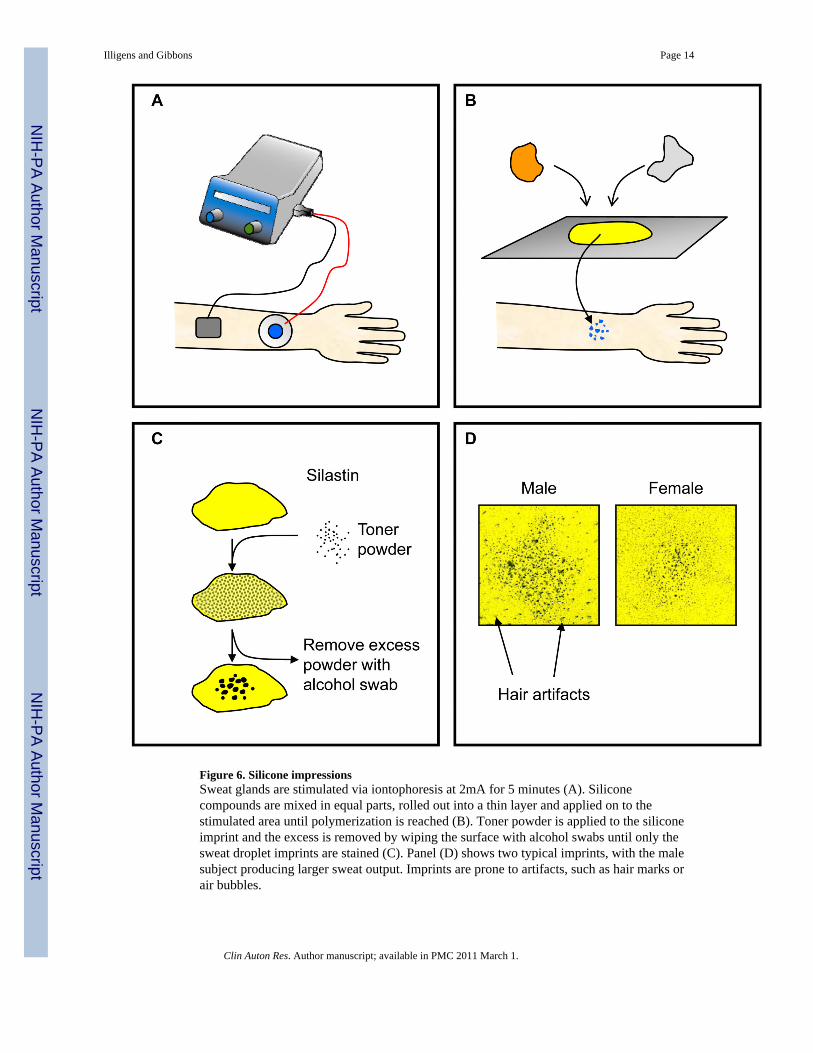
ResultsNormal individuals have 311±38 sweat droplets/cm2 in the hand (lower limit 255) and281±38 sweat droplets/cm2 in the foot (lower limit 235) [15,16]. Abnormal impressions canbe seen in postganglionic lesions, when the sweat duct is occluded, when the sweat glandsthemselves are damaged or when sweat glands are completely absent (Figure 6).
LimitationsAlthough the silicone impression method is probably the easiest method to conduct, it isprone to artifacts left by hairs, dirt, skin surface texture and air bubbles. Caution should alsobe taken when wearing rubber examination gloves. Traces of zinc diethyl dithiocarbamate,an accelerator used in the manufacture of certain hospital gloves, react with the platinumcatalyst in the polyvinyl siloxane and delay or totally inhibit polymerization, which willdramatically alter testing results. Dental impression material has been modified over the pastseveral decades. The original materials used were hydrophobic condensation-silicones.Modifications to the base polymer generated hydrophilic addition-silicones, which werefurther altered by adding compounds such as surfactants to increase water displacement,leading to a reduction in impression defects [31]. While these improvements have enhancedthe quality of dental molds they reduced the ability of sweat droplets to create impressions.
Illigens and Gibbons Page 4
Clin Auton Res. Author manuscript; available in PMC 2011 March 1.
NIH
-PA Author Manuscript
NIH
-PA Author Manuscript
NIH
-PA Author Manuscript
Elasticon, a condensation-silicone, was the imprint material of choice for almost twodecades. Elasticon generated better impressions of sweat droplets than polyvinyl siloxanebased materials because of its hydrophobicity (but for that specific reason also lead to itsdiscontinuation). A description of newer silicones that are currently available has beenreported [40].
Quantitative direct and indirect axon reflex testingQuantitative direct and indirect axon reflex testing is a novel technique to evaluate thepostganglionic sympathetic cholinergic sudomotor function by measuring the direct andaxon-reflex mediated sweat response in a dynamic fashion. Sweat glands are stimulated byacetylcholine iontophoresis and sweat is displayed via an activator dye followed by digitalphotographs over time [11].
MethodologyAcetylcholine (or other cholinergic agonist) is iontophoresed into the skin at a constantcurrent of 2mA for 5 minutes. After iontophoresis the drug delivery probe is quicklyremoved, the area is blotted dry, a thin layer of alizarin red mixture is applied (alizarin red,corn starch, sodium carbonate, 1:2:1) and digital pictures are taken every 15 seconds for 7minutes. Images of QDIRT are then uploaded on a computer as a sequence and processed(Image Pro Plus, Media Cybernetics, Bethesda, MD) with automated image stabilization andgraphical thresholding. A full description of the image processing procedure has beenpublished online as an accompaniment to the paper introducing QDIRT [11]. Sweat dropletsare quantified by number, size and percent area over the area of interest, separating betweendirect and indirect sweat production. An entire test can be finished in approximately 15minutes (Figure 7).
ResultsQDIRT is a new technique and requires further study in disorders of the autonomic nervoussystem. QDIRT is simple, inexpensive and can be used by clinicians without moresophisticated autonomic laboratories.
LimitationsThis technique has been tested in selected patients with small fiber neuropathy, but has notbeen used to study other disorders of the autonomic nervous system [11].
Sympathetic skin responseSympathetic skin response (SSR, also referred to as galvanic skin response) is a measure ofelectrodermal activity and provides a surrogate measure of sympathetic cholinergicsudomotor function. Perturbation of the autonomic nervous system, through rapidinspiration or electrical stimulation, induces a change in skin potential [6,7,14,18,19,36].Changes in skin potential are also seen in response to emotion or attention and have beenwidely studied in psychiatry and law enforcement as the “lie detector” [4,32]. The source ofthe skin potential is presumed to be the sweat glands and the epidermis, although it ispresent in subjects with congenital absence of sweat glands [5,10]. SSR measures apolysynaptic reflex with a spinal, a bulbar and a suprabulbar component [39]. Although thisis not a test of “sweat” function, it is often included in this category as a measure ofsudomotor activity.
Illigens and Gibbons Page 5
Clin Auton Res. Author manuscript; available in PMC 2011 March 1.
NIH
-PA Author Manuscript
NIH
-PA Author Manuscript
NIH
-PA Author Manuscript
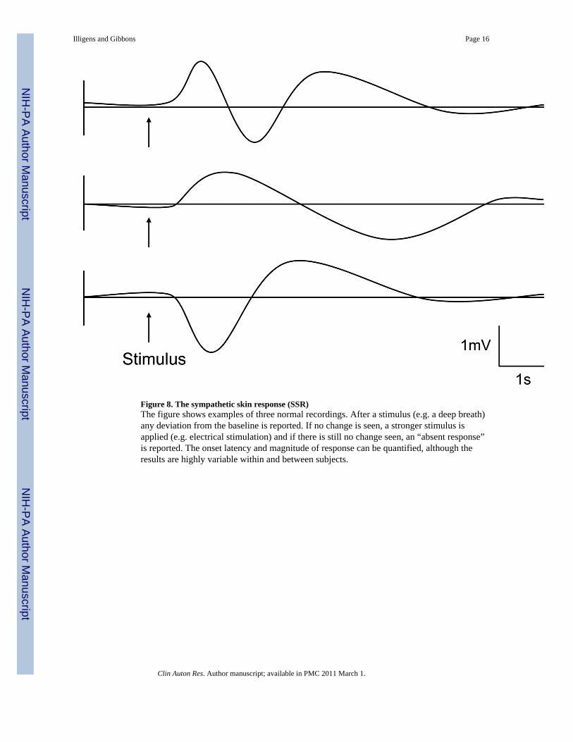
MethodologyRecording electrodes are placed on the dorsal and ventral surface of the hand, medialforearm, proximal leg, distal leg or proximal foot. Recordings are taken by any standardEMG, with low frequency filters set to 0.5Hz or below to prevent attenuation of thepotential. The response can be triggered by an inspiratory gasp, forceful expiration, startleresponse, or electrical stimulation [20]. SSR’s are reported as present or absent and foramplitude and latency (Figure 8). Absent potentials can occur due to inadequate stimulationand habituation.
ResultsThe SSR in the hands traditionally has larger amplitudes and shorter latencies than the feet(hands: 1.5 seconds latency, 0.5–1.3mV amplitude, feet: 1.9–2.1 seconds latency, 0.15 –0.8mV amplitude) [10]. Abnormalities of SSR’s are seen in many disease states, includinggeneral autonomic failure, peripheral neuropathy and even CNS degeneration, such asAlzheimer’s disease [3,13,21].
LimitationsAlthough this method is extremely easy to perform, there is high variability within andbetween subjects. SSR declines with age, and may not be seen in many subjects over 50[10]. It should also be noted that this method is only a surrogate measure of sudomotorfunction; patients with congenital absence of sweat glands (ectodermal anhidrotic dysplasia)will still have a response [38].
SummaryQuantitative assessment of sudomotor function is an important component to autonomictesting. Sudomotor abnormalities can confirm a diagnosis of autonomic dysfunction,monitor disease progression and identify the success of treatment. The choice of which testto perform is made on the availability of testing equipment, lesion localization and the typeof suspected disease. The TST provides an assessment of pre- and postganglionic functionover the whole body. TST can be combined with any test of postganglionic function(QSART, silicone impression, QDIRT) to separate pre- and postganglionic lesions. QSARTidentifies the axon reflex sudomotor response by change in relative humidity and onsetlatency while silicone impressions identify sweat droplets in both direct and indirect regions,but only at a single time point [25,37]. QDIRT combines many aspects of QSART andsilicone impressions, but requires further validation for clinical use [11]. All tests ofsudomotor function are prone to artifacts and confounding variables. Ambient temperature,hydration status, medication use, age and gender can alter sudomotor responses [24].Previous exposure of the skin to alcohol, repeated testing over the same region andapplication of moisturizing creams can also alter the results [23]. In order to obtain a highlevel of reproducibility and reliability all of these factors should be controlled as tightly aspossible. Despite these limitations and restrictions, neurophysiologic assessment of thesudomotor system complements other measures of autonomic function, and remains one ofthe most sensitive and specific means to detect distal small fiber neuropathy [30].
References1. Berghoff M, Kilo S, Hilz MJ, Freeman R. Differential impairment of the sudomotor and nociceptor
axon-reflex in diabetic peripheral neuropathy. Muscle Nerve. 20062. Boulant JA. Hypothalamic mechanisms in thermoregulation. Federation Proceedings 1981;40:2843–
2850. [PubMed: 6273235]
Illigens and Gibbons Page 6
Clin Auton Res. Author manuscript; available in PMC 2011 March 1.
NIH
-PA Author Manuscript
NIH
-PA Author Manuscript
NIH
-PA Author Manuscript
3. Caccia MR, Dezuanni E, Salvaggio A, Osio M, Bevilacqua M, Norbiato G, Mangoni A.Sympathetic skin response versus maximum motor and sensory conduction velocity to detectsubclinical neuropathy in non- insulin-dependent diabetics. Acta Neurologica Belgica 1991;91:213–222. [PubMed: 1746243]
4. Dawson ME, Rissling AJ, Schell AM, Wilcox R. Under what conditions can human affectiveconditioning occur without contingency awareness? Test of the evaluative conditioning paradigm.Emotion 2007;7:755–766. [PubMed: 18039045]
5. Edelberg R. Relation of electrical properties of skin to structure and physiologic state. The Journalof investigative dermatology 1977;69:324–327. [PubMed: 894073]
6. Elie B, Guiheneuc P. Sympathetic skin response: normal results in different experimentalconditions. Electroencephalography and clinical neurophysiology 1990;76:258–267. [PubMed:1697257]
7. Fagius J, Wallin BG. Sympathetic reflex latencies and conduction velocities in normal man. Journalof the neurological sciences 1980;47:433–448. [PubMed: 7420119]
8. Fealey RD, Low PA, Thomas JE. Thermoregulatory sweating abnormalities in diabetes mellitus.Mayo Clinic Proceedings 1989;64:617–628. [PubMed: 2747292]
9. Fealey RD, Low PA, Thomas JE. Thermoregulatory sweating abnormalities in diabetes mellitus.Mayo Clinic proceedings 1989;64:617–628. [PubMed: 2747292]
10. Gibbons C, Freeman R. The evaluation of small fiber function-autonomic and quantitative sensorytesting. Neurologic Clinics 2004;22:683–702. vii. [PubMed: 15207880]
11. Gibbons CH, Illigens BM, Centi J, Freeman R. QDIRT: quantitative direct and indirect test ofsudomotor function. Neurology 2008;70:2299–2304. [PubMed: 18541883]
12. Guttman L, Silver J, Wyndham CH. Thermoregulation in spinal man. Journal of Physiology1958;142:406–419. [PubMed: 13576444]
13. Hilz MJ, Stemper B, Axelrod FB. Sympathetic skin response differentiates hereditary sensoryautonomic neuropathies III and IV. Neurology 1999;52:1652–1657. [PubMed: 10331694]
14. Hoeldtke RD, Davis KM, Hshieh PB, Gaspar SR, Dworkin GE. Autonomic surface potentialanalysis: assessment of reproducibility and sensitivity. Muscle & nerve 1992;15:926–931.[PubMed: 1495508]
15. Kennedy WR, Sakuta M, Sutherland D, Goetz FC. Quantitation of the sweating deficiency indiabetes mellitus. Annals of neurology 1984;15:482–488. [PubMed: 6732196]
16. Kennedy WR, Sakuta M, Sutherland D, Goetz FC. The sweating deficiency in diabetes mellitus:methods of quantitation and clinical correlation. Neurology 1984;34:758–763. [PubMed: 6539438]
17. Kihara M, Opfer-Gehrking TL, Low PA. Comparison of directly stimulated with axon reflex-mediated sudomotor responses in human subjects and in patients with diabetes. Muscle Nerve1993;16:655–660. [PubMed: 8502263]
18. Knezevic W, Bajada S. Peripheral autonomic surface potential. A quantitative technique forrecording sympathetic conduction in man. Journal of the neurological sciences 1985;67:239–251.[PubMed: 3981220]
19. Knezevic W, Bajada S. Peripheral autonomic surface potential: a quantitative technique forrecording autonomic neural function in man. Clinical and experimental neurology 1985;21:201–210. [PubMed: 3843220]
20. Lader MH, Montagu JD. The psycho-galvanic reflex: a pharmacological study of the peripheralmechanism. Journal of neurology, neurosurgery, and psychiatry 1962;25:126–133.
21. Linden D, Berlit P. Sympathetic skin responses (SSRs) in monofocal brain lesions: topographicalaspects of central sympathetic pathways. Acta Neurol. Scand 1995;91:372–376. [PubMed:7639067]
23. Low, PA. Clinical autonomic disorders: evaluation and management. Boston: Little, Brown andCompany; 1993. Pitfalls in autonomic testing; p. 355-365.
Clin Auton Res. Author manuscript; available in PMC 2011 March 1.
NIH
-PA Author Manuscript
NIH
-PA Author Manuscript
NIH
-PA Author Manuscript
25. Low PA, Caskey PE, Tuck RR, Fealey RD, Dyck PJ. Quantitative sudomotor axon reflex test innormal and neuropathic subjects. Annals of neurology 1983;14:573–580. [PubMed: 6316835]
26. Low PA, Dyck PJ. Splanchnic preganglionic neurons in man. III. Morphometry of myelinatedfibers of rami communicantes. Journal of Neuropathology & Experimental Neurology1978;37:734–740. [PubMed: 739274]
27. Low PA, Dyck PJ. Splanchnic preganglionic neurons in man: II. Morphometry of myelinatedfibers of T7 ventral spinal root. Acta Neuropathologica 1977;40:219–225. [PubMed: 602685]
30. Low VA, Sandroni P, Fealey RD, Low PA. Detection of small-fiber neuropathy by sudomotortesting. Muscle Nerve 2006;34:57–61. [PubMed: 16718689]
31. Mandikos MN. Polyvinyl siloxane impression materials: an update on clinical use. Australiandental journal 1998;43:428–434. [PubMed: 9973714]
32. Meijer EH, Smulders FT, Johnston JE, Merckelbach HL. Combining skin conductance and forcedchoice in the detection of concealed information. Psychophysiology 2007;44:814–822. [PubMed:17584188]
33. Namer B, Bickel A, Kramer H, Birklein F, Schmelz M. Chemically and electrically inducedsweating and flare reaction. Auton. Neurosci 2004;114:72–82. [PubMed: 15331047]
34. Sato K, Dobson RL. Regional and individual variations in the function of the human eccrine sweatgland. The Journal of investigative dermatology 1970;54:443–449. [PubMed: 5446389]
35. Schmidt KD, Chan CW. Thermoregulation and fever in normal persons and in those with spinalcord injuries. Mayo Clinic Proceedings 1992;67:469–475. [PubMed: 1405774]
36. Shahani BT, Halperin JJ, Boulu P, Cohen J. Sympathetic skin response--a method of assessingunmyelinated axon dysfunction in peripheral neuropathies. Journal of neurology, neurosurgery,and psychiatry 1984;47:536–542.
37. Stewart JD, Nguyen DM, Abrahamowicz M. Quantitative sweat testing using acetylcholine fordirect and axon reflex mediated stimulation with silicone mold recording; controls versusneuropathic diabetics. Muscle Nerve 1994;17:1370–1377. [PubMed: 7969237]
38. Uncini A, Pullman SL, Lovelace RE, Gambi D. The sympathetic skin response: normal values,elucidation of afferent components and application limits. Journal of the neurological sciences1988;87:299–306. [PubMed: 2850351]
39. Vetrugno R, Liguori R, Cortelli P, Montagna P. Sympathetic skin response: basic mechanisms andclinical applications. Clin Auton Res 2003;13:256–270. [PubMed: 12955550]
40. Vilches JJ, Navarro X. New silicones for the evaluation of sudomotor function with the impressionmold technique. Clin. Auton. Res 2002;12:20–23. [PubMed: 12102444]
Illigens and Gibbons Page 8
Clin Auton Res. Author manuscript; available in PMC 2011 March 1.
NIH
-PA Author Manuscript
NIH
-PA Author Manuscript
NIH
-PA Author Manuscript
Figure 1. The sudomotor axon reflexCholinergic agonists (such as acetylcholine) applied through iontophoresis (shown with theblack arrow) bind to muscarinic receptors causing local sweat production (dashed arrow).The cholinergic agonist simultaneously binds to nicotinic receptors on nerve terminals ofsudomotor fibers and an impulse travels antidromically. At branch points this impulsetravels orthodromically to a neighboring population of eccrine sweat glands causing anindirect axon mediated sweat response (dotted arrows).
Illigens and Gibbons Page 9
Clin Auton Res. Author manuscript; available in PMC 2011 March 1.
NIH
-PA Author Manuscript
NIH
-PA Author Manuscript
NIH
-PA Author Manuscript
Figure 2. Thermoregulatory sweat testing (TST)A TST chamber is temperature and humidity controlled. Ceiling mounted infrared heaterscontrol the patient’s temperature. The patient is placed in the supine position. Oral andcutaneous temperature probes are attached. During the application of the indicator dye, thepatient’s eyes, nose and mouth should be protected. To achieve even distribution of theindicator powder, an atomizer should be used. The test is started by increasing the roomtemperature. Oral temperature must rise at least 1.0°C above baseline temperature or to 38°C (whichever is higher). At the end of the test pictures are taken and used to generate atopographical map of the sweat pattern.
Illigens and Gibbons Page 10
Clin Auton Res. Author manuscript; available in PMC 2011 March 1.
NIH
-PA Author Manuscript
NIH
-PA Author Manuscript
NIH
-PA Author Manuscript
Figure 3. Thermoregulatory sweat test (TST) resultsNormal sweat patterns show a sweat response present over the entire body that may bevariable in intensity (A). In (B), a length dependent neuropathy from diabetes with stockingand glove distribution loss is seen. A patient with a complete myelopathy at T9 is shown in(C). Lesions to individual nerves can show focal or dermatomal sweat defects. A patientwith a right T10 radiculopathy and a left lateral femoral cutaneous neuropathy can beidentified in (D). A patient with complete anhidrosis secondary to pure autonomic failure isseen in (E).
Illigens and Gibbons Page 11
Clin Auton Res. Author manuscript; available in PMC 2011 March 1.
NIH
-PA Author Manuscript
NIH
-PA Author Manuscript
NIH
-PA Author Manuscript
Figure 4. Quantitative sudomotor axon reflex test (QSART)An overview of the QSART testing procedure: A multi-compartmental sweat capsule has anouter ring (A, 1.5mm wide) for iontophoretic stimulation and an inner compartment (C, 1cmdiameter) for measuring humidity. The stimulation and recording sites are separated by asmall compartment (B, 1.5mm wide) to prevent direct stimulation of the sweat glands andleakage of the iontophoresis fluid. Dry nitrogen gas is released at a steady rate of flow(typically 100 cc/min) controlled through a flow meter. The gas flows through a temperaturecontrolled heat exchanger and into the sweat capsule (C). Upon exiting the capsule the gasflows back through the heat exchanger and to a hygrometer, where changes in humidity arerecorded on a computer.
Illigens and Gibbons Page 12
Clin Auton Res. Author manuscript; available in PMC 2011 March 1.
NIH
-PA Author Manuscript
NIH
-PA Author Manuscript
NIH
-PA Author Manuscript
Figure 5. QSART resultsPanel (A), after a baseline is reached (1) recording is started for at least 2 minutes.Iontophoresis is started (2) at 2mA for 5 minutes (3). Recording is continued to monitorrecovery for at least another 10 minutes (4) (A). Panel (B) shows a normal response. Theexample in (C) shows a “hung-up” response typically seen if sweat production is excessiveand no recovery is reached. Panel (D) shows a reduced sweat response with a delayed onsetof sweat production.
Illigens and Gibbons Page 13
Clin Auton Res. Author manuscript; available in PMC 2011 March 1.
NIH
-PA Author Manuscript
NIH
-PA Author Manuscript
NIH
-PA Author Manuscript
Figure 6. Silicone impressionsSweat glands are stimulated via iontophoresis at 2mA for 5 minutes (A). Siliconecompounds are mixed in equal parts, rolled out into a thin layer and applied on to thestimulated area until polymerization is reached (B). Toner powder is applied to the siliconeimprint and the excess is removed by wiping the surface with alcohol swabs until only thesweat droplet imprints are stained (C). Panel (D) shows two typical imprints, with the malesubject producing larger sweat output. Imprints are prone to artifacts, such as hair marks orair bubbles.
Illigens and Gibbons Page 14
Clin Auton Res. Author manuscript; available in PMC 2011 March 1.
NIH
-PA Author Manuscript
NIH
-PA Author Manuscript
NIH
-PA Author Manuscript
Figure 7. QDIRT: Quantitative direct and indirect reflex testing of sudomotor functionSweat glands are stimulated via iontophoresis at 2mA for 5 minutes (A). Alizarin redpowder is applied in a thin layer onto the stimulated area (B). Immediately, digital picturesare taken every 15 seconds for 7 minutes (C). The results can be quantified by dropletnumber, size, location and response latency in both direct and indirect testing regions.
Illigens and Gibbons Page 15
Clin Auton Res. Author manuscript; available in PMC 2011 March 1.
NIH
-PA Author Manuscript
NIH
-PA Author Manuscript
NIH
-PA Author Manuscript
Figure 8. The sympathetic skin response (SSR)The figure shows examples of three normal recordings. After a stimulus (e.g. a deep breath)any deviation from the baseline is reported. If no change is seen, a stronger stimulus isapplied (e.g. electrical stimulation) and if there is still no change seen, an “absent response”is reported. The onset latency and magnitude of response can be quantified, although theresults are highly variable within and between subjects.
Illigens and Gibbons Page 16
Clin Auton Res. Author manuscript; available in PMC 2011 March 1.