Gender Differences of Airway Dimensions in AnatomicallyMatched Sites on CT in Smokers
Yu-Il Kim1,2, Joyce Schroeder3, David Lynch3, John Newell3, Barry Make1, AdamFriedlander1, Raúl San José Estépar4, Nicola A. Hanania5, George Washko6, James R.
NIH Public AccessAuthor ManuscriptCOPD. Author manuscript; available in PMC 2013 July 07.
Published in final edited form as:COPD. 2011 August ; 8(4): 285–292. doi:10.3109/15412555.2011.586658.
NIH
-PA
Author M
anuscriptN
IH-P
A A
uthor Manuscript
NIH
-PA
Author M
anuscript
Murphy7, Carla Wilson7, John E. Hokanson8, Jordan Zach3, Kiel Butterfield1, Russell P.Bowler1, and COPDGene® Investigators8
1Department of Medicine, National Jewish Health, Denver, CO, USA2Department of Internal Medicine, Chonnam National University Hospital, Gwangju, Korea3Department of Radiology, National Jewish Health, Denver, CO, USA4Department of Radiology, Brigham and Women’s Hospital, Boston, MA, USA5Department of Medicine, Baylor College of Medicine, Houston, TX, USA6Department of Medicine, Brigham and Women’s Hospital, Boston, MA, USA7Biostatistics and Bioinformatics, National Jewish Health, Denver, CO, USA8Department of Epidemiology, Colorado School of Public Health, University of Colorado, Denver,CO, USA
AbstractRationale and Objectives—There are limited data on, and controversies regarding genderdifferences in the airway dimensions of smokers. Multi-detector CT (MDCT) images wereanalyzed to examine whether gender could explain differences in airway dimensions ofanatomically matched airways in smokers.
Materials and Methods—We used VIDA imaging software to analyze MDCT scans from 2047smokers (M:F, 1021:1026) from the COPDGene® cohort. The airway dimensions were analyzedfrom segmental to subsubsegmental bronchi. We compared the differences of luminal area, innerdiameter, wall thickness, wall area percentage (WA%) for each airway between men and women,and multiple linear regression including covariates (age, gender, body sizes, and other relevantconfounding factors) was used to determine the predictors of each airway dimensions.
Results—Lumen area, internal diameter and wall thickness were smaller for women than men inall measured airway (18.4 vs 22.5 mm2 for segmental bronchial lumen area, 10.4 vs 12.5 mm2 forsubsegmental bronchi, 6.5 vs 7.7 mm2 for subsubsegmental bronchi, respectively p < 0.001).However, women had greater WA% in subsegmental and subsubsegmental bronchi. Inmultivariate regression, gender remained one of the most significant predictors of WA%, lumenarea, inner diameter and wall thickness.
Conclusion—Women smokers have higher WA%, but lower luminal area, internal diameter andairway thickness in anatomically matched airways as measured by CT scan than do male smokers.This difference may explain, in part, gender differences in the prevalence of COPD and airflowlimitation.
INTRODUCTIONSmoking is a major risk factor for chronic obstructive pulmonary disease (COPD) andairflow obstruction. However, only a minority of smokers develop COPD, and therelationship between smoking history and the severity of airflow obstruction is weak (1).Thus, there is a new appreciation that COPD may be a heterogeneous disorder of smokingwith many phenotypes (2). Some of the factors that are associated with the severity ofairflow obstruction include: age, height, race, gender, genetic susceptibility, air pollution,and airway dimensions (3–7). Within the past several decades, there has also been a
Kim et al. Page 2
COPD. Author manuscript; available in PMC 2013 July 07.
NIH
-PA
Author M
anuscriptN
IH-P
A A
uthor Manuscript
NIH
-PA
Author M
anuscript
demographic shift in gender distribution of individuals with COPD. In 2000 there were morewomen diagnosed with COPD than men in the United States (8). Some have postulated thatwomen may be more susceptible to the damaging effects of smoking and may be at greaterrisk of smoking-induced lung function impairment (4, 9–11).
Besides spirometry, chest CT has recently been used as a valuable tool to assess lungdamage from smoking. Advances in CT imaging have permitted more detailed analysis ofairway dimensions (12). It has been suggested that these CT measurements has potentialpower to represent histological dimension changes in the airway (13, 14). Although CTmeasurements of airway dimensions are predominantly of medium-sized airways, they couldbe representative of the degree of remodeling in small airways determined by pathology(13). Aysola et al. (14) reported that the airway thickness on endobronchial biopsy samplesfrom individuals with asthma and healthy subjects correlated with wall area percentage (WA%).
Histologically, Martinez et al. (15) reported that women exhibited smaller airway lumenswith disproportionately thicker airway walls than men in patients with severe COPD. Threeother publications (7, 16, 17) that reported sex differences associated with CT airwaythickness found that female smokers did not show increased wall thickness compared tomen; however, a recent publication found that the square root of the wall thickness of ahypothetical airway of internal perimeter of 10 mm (SQRTWA@pi10) was higher in menthan women (16).
None of these studies has reported gender difference of anatomically matched, specifiedairway wall. Furthermore, most studies used only a single population of subjects for airwaymeasurement. To overcome these limitations and to evaluate whether there are genderdifferences in airway dimensions even when including confounding variables, we used theCOPDGene® (the Genetic epidemiology of COPD) cohort (http://www.copdgene.org/) (18)to determine whether gender could explain the differences in airway dimensions ofanatomically matched airways in smokers.
MATERIALS AND METHODSStudy Populations
The COPDGene® Study is an ongoing multicenter investigation of the geneticepidemiology of smoking-related lung disease (18). The first 2,047 smokers withquantitative CT data from the COPDGene® cohort were included in this study. All subjectswere studied after obtaining the consent of study under protocols approved by localInstitutional Review Board (IRB) and with guidelines recommended by the NationalInstitutes of Health. Subjects were men and women; non-Hispanic whites or African-Americans aged 45 to 80 years with a smoking history of at least 10 pack years.
Previously proposed exclusion criteria (18) were applied in the cohort (18): exclusioncriteria are a pregnant woman, a history of other lung disease except asthma (e.g.,pulmonary fibrosis, extensive bronchiectasis, cystic fibrosis), previous surgical excision ofat least one lung lobe (or lung volume reduction procedure), active cancer under treatment,suspected lung cancer (large or highly suspicious lung mass), metal in the chest, recentexacerbation of COPD treated with antibiotics or steroids, recent eye surgery, MI, othercardiac hospitalization, recent chest or abdominal surgery, inability to use albuterol, multipleself-described racial categories, history of chest radiation therapy, and first- or second-degree relative already enrolled in the study. Smokers who have an unclassified pattern byGOLD (Global initiative for chronic Obstructive Lung Disease) criteria on spirometry,denoted as GOLD U (normal FEV1/FVC but reduced FEV1) and GOLD 0 (smokers with
Kim et al. Page 3
COPD. Author manuscript; available in PMC 2013 July 07.
normal spirometry) are eligible for the study. Each subject underwent a spirometry andmulti-detector CT (MDCT). The COPDGene® cohort includes nineteen clinical centers inthe United States (18) (see center and investigator list in Acknowledgments).
Quantitative CT AnalysisAnalysis of COPDGene cohort using VIDA software—In COPDGene® study, allMDCT (at least 16 detector channels) of the chest used a tube potential of 120 kVp and aneffective mAs of 200 (Supplemental Tables S1-A, B and C) (18). Submillimeter nearisotropic MDCT scans without contrast were acquired at end inspiration. The images werereconstructed with slice thicknesses of 0.625, 0.75 or 0.9 mm depending on the CTmanufacturer (General Electric Medical Systems, Siemens and Philips) (18). The optimalreconstruction kernel for a given model of CT scanner for the VIDA software program wasused to segment the lungs, lobes and airway tree. The image matrix size was 512 × 512pixels, and the pixel sizes ranged from x: 0.55 to 0.78 mm, y: 0.55 to 0.78. Other detailedCT protocols were the same with the previous report (18).
Airway dimensions were measured using automated, quantitative software that was designedto label and quantify the bronchial tree (Pulmonary Workstation+ VIDA Diagnostics; IowaCity, IA. www.vidadiagnostics.com, Supplemental Figure 1) (19, 20). These airways wereas follows: right upper apical segmental, subsegmental and subsubsegmental brochi, rightmiddle lateral segmental, subsegmental and subsubsegmental bronchi, right posterior basalsegmental, subsegmental and subsubsegmental bronchi, left upper apical segmental,subsegmental and subsubsegmental bronchi, left superior lingular segmental, subsegmentaland subsubsegmental bronchi, and left posterior basal segmental, subsegmental andsubsubsegmental bronchi.
These airway indices were measured from the centerline to the airway edge in each slice ofthe 3D image set. Reported airway dimensions represented the average of all themeasurements collected along the middle third of each individual airway segment. For eachindividual, the segmental, subsegmental and subsubsegmental airway data were averaged toprovide a mean value for each level of branching. Structural measurements of airwaydimensions included the lumen area (Ai), inner diameter, airway wall thickness, wall areapercentage (WA%) and SQRTWA@pi10 in each anatomically matched airway. Theperimeters of the airway lumen and of the adventitia subtended two areas: Ai (luminal area)and Ao (total area). WA% was calculated as (Ao – Ai)/Ao × 100. SQRTWA@pi10 wascalculated for each subject by fitting a linear relationship between Pi and SQRTWA of eachmeasured bronchus (16, 21).
For determining the extent of emphysema, quantitative densitometric analysis wasperformed with VIDA and areas of CT emphysema were defined as low attenuation areas(LAA) [<–950 Hounsfield units, HU]. Then, the percentage of LAA (LAA% −950HU) wasdetermined for the entire lung. Region growing of airway tree was performed by researchassistants under the training and supervision of the Imaging core of the COPDGene® study(list in Acknowledgements). The stability of CT measurements for each scanner ismonitored by monthly scanning using a custom COPDGene phantom (18).
Statistical AnalysisGender differences were evaluated using t-tests for continuous variables and χ2 tests forcategorical variables. Data that were not normally distributed (e.g., LAA% −950 HU andpacks years of smoking (PYs)) were also analyzed after log transformation. Regressionanalysis was used to determine predictors of WA%. Multivariate analysis was performedusing linear regression models for WA% adjusted for subject’s age, sex, height, weight,
Kim et al. Page 4
COPD. Author manuscript; available in PMC 2013 July 07.
PYs, race, smoking status (current/former smoker), LAA% −950HU and total lung capacity(TLC% predicted) to adjust demographic and body size differences, and confounding factorsthat could affect WA%. Clinical centers and CT scanner types were also included as avariable to adjust those differences in multicenter COPDGene® cohort. Correlationsbetween lung function (FEV1% pred.) and airway parameters were determined usingparametric testing methods with Pearson correlation coefficients. P values less than 0.05were considered statistically significant. Statistical software (SPSS, version 17.0; Chicago,IL) was used for analysis.
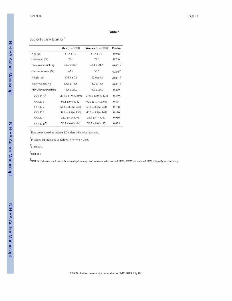
RESULTSDemographics, smoking history and lung function for the 2047 COPDGene® subjects whowere included in the study are shown in Table 1. Subjects were predominantly White, butthere were no ethnic differences between genders (Caucasian 78.0% in male, 77.3% infemale, p = 0.706). Subjects had a heavy smoking history and PYs was higher in males(M:F, 49.9: 42.1 PYs, p < 0.01). Current smokers were more frequently male (M:F, 42.8% :36.6%, p = 0.004). Height and body weight were smaller in women. There were nosignificant differences in lung function between genders and mean FEV1% (pred.) results(M:F, 72.5%: 73.9%) were consistent with GOLD stage II disease (812 smokers withoutevidence of airway obstruction, 146 smokers with GOLD-I, 486 smokers with GOLD-II,294 smokers with GOLD-III, 158 smokers with GOLD-IV, and 151 smokers with GOLDU).
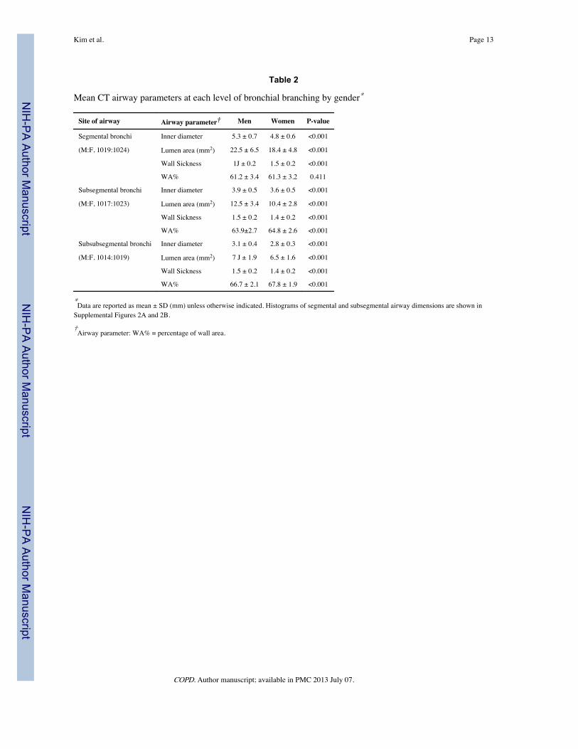
Most airway measurements (inner diameter, wall thickness and lumen area) were lower inwomen compared to men (Table 2 and Supplemental Figs. 2A and 2B). The numbers ofobtainable measurements were slightly decreased as the bronchial branches go more distal(n = 2043 for segmental bronchi, 2040 for subsegmental, 2033 for subsubsegmental).However, women had higher WA% in the subsegmental and subsubsegmental bronchi. Insubgroup analyses using subjects with or without airflow obstruction, gender differences ofairway dimensions were consistent (Supplemental Table S2-A and B). SQRTWA@pi10 wasnot significantly different between men and women (Supplemental Table S3).
Emphysema (LAA% < −950 HU) was more extensive and CT measured lung volume (TLC% predicted) was lower in men than women (Supplemental Table S3). Univariate analysiswas used to determine which factors might be associated with WA% for different airways(Supplemental Table S4). There were significant associations between WA% and mostvariables (age, gender, race, pack-years, smoking status, height, weight, emphysema score,TLC, study center, and scanner type). Male gender (t ratios −7.8, −12.2), height (t ratios−10.4, −13.3) and LAA% (t ratios 9.1, 7.9) were stronger predictors than other variables insubsegmental and subsubsegmental bronchi while body weight and TLC% were morepowerful in segmental bronchi compared to other variables.
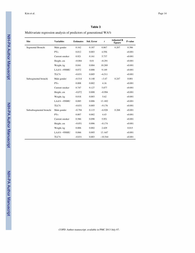
In multivariate analysis including all of these variables (Table 3: shown for several keyvariables, and Supplemental Table S5 shown for all variables), PYs, smoking status (currentsmoker), height, weight and TLC% showed consistent and significant associations with WA% from all airways, from segmental to subsubsegmental bronchi. Male gender wasnegatively associated with subsegmental and subsubsegmental WA% (t = −3.47, −6.9; p =0.001, <0.001, repectively) while there was no significant associations between gender andsegmental WA%.
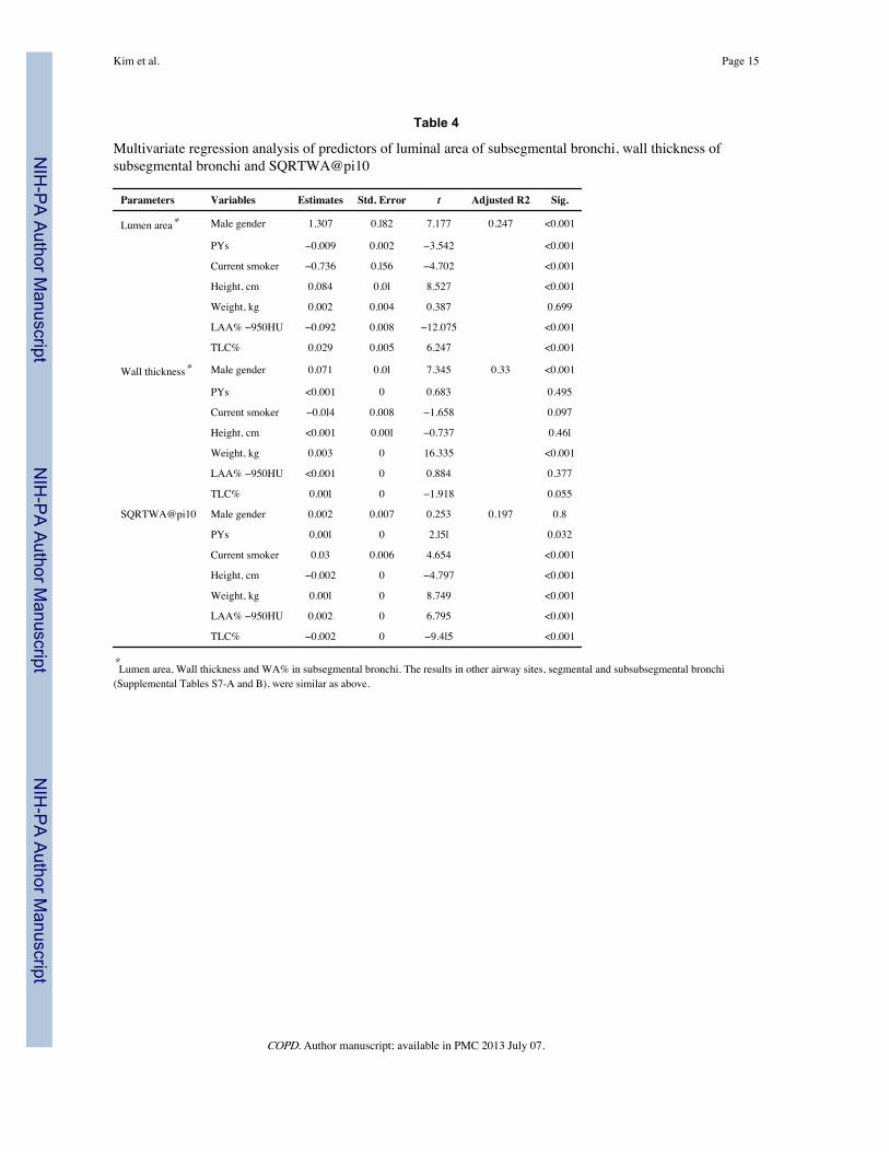
Among the above variables, height, LAA% and TLC% were more powerful predictors thanother demographic predictors in the subsegmental and subsubsegmental paths. Inmultivariate analysis for other airway parameters such as lumen area and wall thickness
Kim et al. Page 5
COPD. Author manuscript; available in PMC 2013 July 07.
NIH
-PA
Author M
anuscriptN
IH-P
A A
uthor Manuscript
NIH
-PA
Author M
anuscript
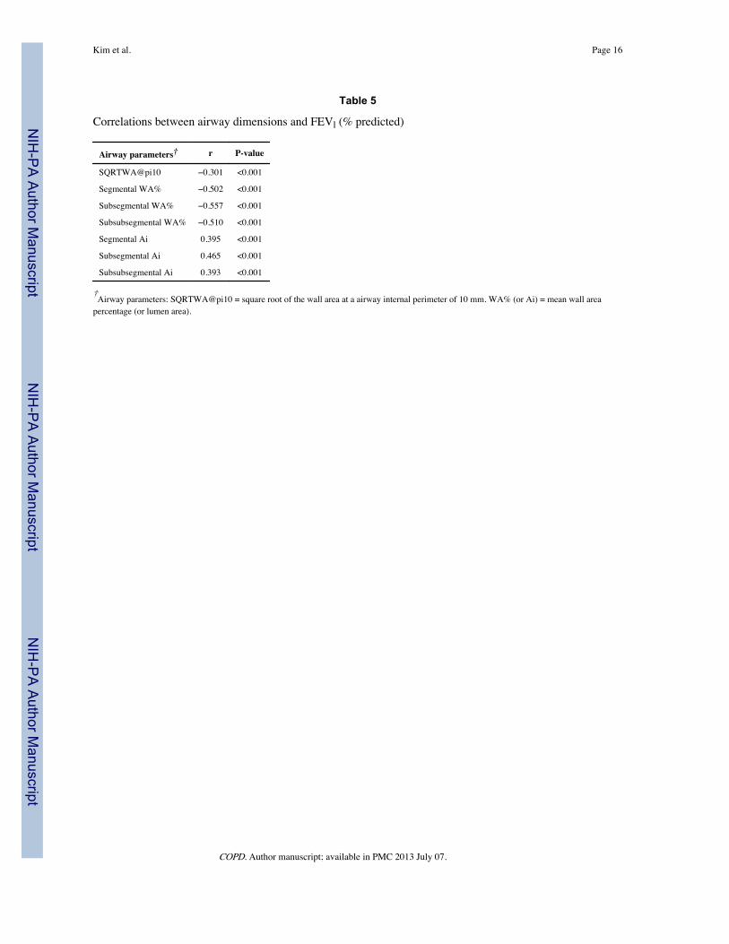
(Table 4: shown for several key variables, Supplemental Table S6, S7-A and B), gender wasone of the significant and powerful determinants for each quantitative CT parameter.However, gender was not a significant predictor of SQRTWA@pi10 in the multivariateanalysis. WA%, lumen area and SQRTWA@pi10 were significantly correlated with FEV1%predicted (Table 5).
DISCUSSIONComputed tomography is becoming a useful, non-invasive tool to evaluate the airwaydimensions. There are several different investigational methods used to express themorphologic characteristics of airway wall. These include the two most frequently usedmetrics: WA% and SQRTWA@Pi10 (21–23). It should be noted that these two metrics arenot directly measured, but are derived from other airway measurements. Directly measuredmetrics include luminal area, inner diameter and wall thickness. These computationaldifferences have led to different investigators reporting apparently paradoxical conclusionsregarding gender differences and have led to confusion in interpretation of CT derivedairway measurement. For example, WA% is a deceptive measure of wall thickness becauseas airways become smaller, the WA% becomes larger (24).
Thus, WA% is affected not only by airway thickness, but also by airway size.SQRTWA@Pi10 is a useful method to correct for differences in airway size; however, theconcept of a hypothetical airway is less relevant when one can measure actual airways thathave been anatomically matched. Using the SQRTWA@Pi10 also discounts the importanceof airway size on airflow. We speculated that this is why WA% and luminal area had bettercorrelation with FEV1 (% predicted) than SQRTWA@Pi10 (Table 5).
To our knowledge, this report is the largest investigation of airway dimensions measured byMDCT and the only report of gender differences in airway dimensions classified accordingto bronchial branching order. A novel finding is that in anatomically matched sites,especially in distal airways such as subsegmental and subsubsegmental bronchi, femalesmokers have higher WA% compared to male smokers. However, they have lower luminalarea, airway thickness, and internal diameter of airway in anatomically matched airwaysthan do male smokers. The significance of reduced luminal area in women is particularlyimportant to physiology because the smaller size of women’s lungs is associated with lowerflow rates (25).
Furthermore, airflow limitation in COPD is more closely related to the dimensions of thedistal (small) airways than proximal (large) airways (23). The diameters of subsubsegmentalbronchi in our study were around 3 mm, which is thought to be more representative ofairflow limitation (26). Thus, the direct measurement of anatomically matched airway lumenalso has an important physiologic relevance to airflow. The smaller lumen area and thehigher WA% of these distal airways in women could explain why women have a higherprevalence of COPD and may also explain gender differences in the presentation andpathophysiology of airflow obstruction and COPD.
SQRTWA@Pi10 is a hypothetical airway parameter that is obtained by fitting a linearrelationship between Pi and SQRTWA (21). Other studies (7, 16, 17) have come to differentconclusions from our results; namely that airways are thicker in men compared to women interms of SQRTWA@Pi10. In our study, there were no significant differences ofSQRTWA@Pi10 between genders. In the subgroup analysis according to GOLD stage,there was significant difference of SQRTWA@Pi10 between genders (men : women, 3.84 :3.80 mm, p = 0.02) only among severe COPD (GOLD 3 and 4) like the other previous report(7). But, the differences of SQRTWA@Pi10 between genders are very small. In the other
Kim et al. Page 6
COPD. Author manuscript; available in PMC 2013 July 07.
NIH
-PA
Author M
anuscriptN
IH-P
A A
uthor Manuscript
NIH
-PA
Author M
anuscript
studies (7, 16, 17), the differences of SQRTWA@Pi10 between genders (around 0.2–0.3mm) were also very small.
We postulated that these small differences could be easily obscured by other hiddenconfounding factors such as different airway measurement algorithm in each study. Incontrast to SQRTWA@Pi10, women had the higher WA% in the subsegmental andsubsubsegmental paths through all GOLD stages. Thus, in regarding whether airways arethicker in women compared to men, it is important to consider which definitions of airwaydimensions are reported. However, further study will be needed to clarify thesediscrepancies and its contribution to clinical relevance, and to evaluate which airwayparameter could be more important to clinical settings.
The other major difference between this study and the recently published studies of airwaymeasurements (7, 16, 17) is the methodology for determining airway wall thickness. Mostpublications have used the Full-Width-At-Half-Maximum (FWHM) method to measure thedimensions. We used an optimal surface algorithm (VIDA) to determine airway boundaries.The results from VIDA have showed better subpixel accuracy for the inner border andequivalent results for the outer wall border compared with those of the FWHM method (20).The segmentation algorithm of VIDA retrieves a significantly higher count of airwaybranches compared with a commonly used region growing segmentation algorithm (20).However, the numbers of obtainable measurements were decreased as the branches go moredistal. This suggests that some difficulties including reproducibility are still remained tomeasure airway dimensions especially in the small airways with the VIDA software eventhough this is a more updated and automatically operated software that has been validatedpreviously (28).
Also, long-term reproducibility should be validated in a longitudinal future study usingVIDA. But, a strength of this study includes the large sample of airway measurements withaveraging data for each generational path. The problem of reproducibility of themeasurements could be weakened, at least, to some extent by averaging the values of eachdifferent airway from a large number of subjects. Furthermore, parallel imaging analyses ofLAA% were done using Airway Inspector (www.airwayinspector.org) and 3D Slicer (http://www.slicer.org/) for all subjects and, airway dimensions were also measured using 3D slicerfor more than 80 subjects. The results of gender differences from 3D slicer were similar toVIDA’s (data not shown).
A secondary finding in this study was that age, smoking (status and amount), body sizes(height and weight), emphysema and other various factors affect airway measurements.Smoking status, body sizes and TLC% showed significant associations with WA% from allthe airways. Gender effects for WA% were present in the subsegmental andsubsubsegmental paths, not in the segmental bronchi. Among the variables for WA%,height, LAA% and TLC% were more powerful predictors than other demographic predictorsin the subsegmental and subsubsegmental paths. For other airway parameters, gender is oneof the powerful predictors for luminal area and wall thickness, but not for [email protected] suggests that each variable could affect different airway metrics with different intensityand different location, and this might be associated with the heterogeneity of COPD and theimportance of airway measurements in anatomically matched sites.
There were small gender differences (around 1%) in WA% that could affect the smallphysiologic relevance. However, the small changes in each variable should be considered tobetter understand the heterogeneity of COPD because the factors of airflow obstruction andCOPD are multifactorial. Additionally, gender differences in WA% were exaggerated incurrent smokers with COPD (Supplemental Table S8). This could suggest that women’s
Kim et al. Page 7
COPD. Author manuscript; available in PMC 2013 July 07.
airway may be more susceptible than men’s to the airway damaging effects of currentsmoking. However, to clarify these heterogeneous relationships between each airwayparameter and other variables, and smoking effects in gender, a longitudinal study is neededin the future.
We found other sources of variability in airway wall measurements including clinical center,CT scanner type, and location of airways. However, the magnitudes of gender effect forluminal area (t ratio, around 7.2, Table 4) and wall thickness (t-ratio, around 7.2, Table 4) inall airway paths were higher than those of CT scanner type (t-ratio, around 1.0, data notshown) or clinic center (t-ratio, around 2.0, data not shown). The magnitudes of gendereffect for WA% in subsegmental and subsubsegmental paths were also higher than theywere for scanner or center. This suggests gender differences are consistent irrespective ofscanner type or clinic center. However, center or scanner type could affect the quantitativemeasurements as a noise to some extent.
These findings indicate that gender is just one factor for airway wall, that a complexbackground of airway dimensions exists and that failure to take into account other clinicalvariables may weaken any observed differences in CT derived airway wall measurements.The most likely explanations for gender differences in airways are biological andenvironmental determinants (30). Several studies suggest that genetic interactions may beimportant to the gender differences and CT phenotypes associated with COPD (4, 31, 32).However, the precise mechanism and determinants of gender differences on airwaydimension remains unknown. This study also suggests that other causes of variability inairway measurements need further investigation.
The limitations of this study are similar to those of the parent study (COPDGene®). First, itis cross-sectional and may not account for changes in airway dimension over time. Second,nonsmokers were not included. Thus, we cannot fully evaluate the effects of smoking per seon airway dimensions. Additionally, a fundamental limitation of airway measurements is thespatial resolution (voxel size: x, y, z = slice thickness) of the underlying CT image data. Forexample, for a CT acquisition field-of-view of 35 cm, typical for the COPDGene® cohort,the 512 × 512 image size translates to an x, y pixel dimension of 0.68 mm. Slice thickness(z) ranged from 0.625 to 0.9 mm depending on the CT scanner manufacturer. Therefore, tohave two pixels on a feature of interest (e.g., an airway lumen or airway wall) as suggestedfrom Nyquist sampling theory would require a feature of approximately 1.37 mm size orgreater (33).
Airway wall thickness may be near the spatial resolution limit in subsegmental andsubsubsegmental airways in the COPDGene cohort. This requires further study andcomparison to CT phantom results. Note that larger field-of-view dimensions and largerslice thicknesses will further decrease spatial resolution. These technical issues may explain,at least in part, the variation of airway dimensions by clinical center and scanner type. Last,we used a convenience cohort from COPDGene cohort that was obtained to look for geneticfactors in COPD. The cohort is a heterogeneous mix of individuals with varied smokinghistories and a range of airflow obstruction. It was not ideally recruited to answer thequestion about gender differences. More studies will be needed only for gender differencesof airway dimensions to confirm these differences.
In conclusion, female smokers have disproportionately higher WA%, but lower luminal areaand airway thickness in anatomically matched sites, subsegmental and subsubsegmentalbronchi as measured by CT scan than do male smokers. This difference may explain, in part,gender differences in the heterogeneity of COPD and airflow obstruction. Awareness of the
Kim et al. Page 8
COPD. Author manuscript; available in PMC 2013 July 07.
NIH
-PA
Author M
anuscriptN
IH-P
A A
uthor Manuscript
NIH
-PA
Author M
anuscript
gender difference in airway dimensions should be considered in future investigations ofairway related disease.
Supplementary MaterialRefer to Web version on PubMed Central for supplementary material.
AcknowledgmentsThe authors wish to thank COPDGene® investigators (below) and Christina Schnell—including her invaluablesecretarial support—for data collection and their support of writing this manuscript.
Funding Support: This study was supported by National Heart, Lung and Blood Institute (NHLBI RO1HL 095432,U01 HL089856, U01 HL089897); UL1 RR025780 from NCRR/HIH; and K25HL104085 (SJE).
ABBREVIATIONS
COPD chronic obstructive pulmonary disease
FEV1% pred. % Predicted Forced Expiratory Volume in 1 Second
LAA% percentage of low attenuation areas
SQRTWA@pi10 square root of the wall area at a airway internal perimeter of 10 mm
WA% percentage of wall area
REFERENCES1. Pauwels RA, Buist AS, Calverley PM, Jenkins CR, Hurd SS. Global strategy for the diagnosis,
management, and prevention of chronic obstructive pulmonary disease. NHLBI/WHO globalinitiative for chronic obstructive lung disease (gold) workshop summary. Am J Respir Crit CareMed. 2001; 163:1256–1276. [PubMed: 11316667]
3. Rabe KF, Hurd S, Anzueto A, Barnes PJ, Buist SA, Calverley P, Fukuchi Y, Jenkins C, Rodriguez-Roisin R, van Weel C, Zielinski J. Global strategy for the diagnosis, management, and prevention ofchronic obstructive pulmonary disease: Gold executive summary. Am J Respir Crit Care Med.2007; 176:532–555. [PubMed: 17507545]
5. Orozco-Levi M, Garcia-Aymerich J, Villar J, Ramirez-Sarmiento A, Anto JM, Gea J. Wood smokeexposure and risk of chronic obstructive pulmonary disease. Eur Respir J. 2006; 27:542–546.[PubMed: 16507854]
6. Liou TG, Kanner RE. Spirometry. Clinic Rev Allerg Immunol. 2009; 37:137–152.7. Kim WJ, Silverman EK, Hoffman E, Criner GJ, Mosenifar Z, Sciurba FC, Make BJ, Carey V,
Estepar RS, Diaz A, Reilly JJ, Martinez FJ, Washko GR. Ct metrics of airway disease andemphysema in severe COPD. Chest. 2009; 136:396–404. [PubMed: 19411295]
9. Dransfield MT, Davis JJ, Gerald LB, Bailey WC. Racial and gender differences in susceptibility totobacco smoke among patients with chronic obstructive pulmonary disease. Respir Med. 2006;100:1110–1116. [PubMed: 16236491]
10. de Torres JP, Casanova C, Hernandez C, Abreu J, Aguirre-Jaime A, Celli BR. Gender and copd inpatients attending a pulmonary clinic. Chest. 2005; 128:2012–2016. [PubMed: 16236849]
Kim et al. Page 9
COPD. Author manuscript; available in PMC 2013 July 07.
NIH
-PA
Author M
anuscriptN
IH-P
A A
uthor Manuscript
NIH
-PA
Author M
anuscript
11. Chapman KR. Chronic obstructive pulmonary disease: Are women more susceptible than men?Clin Chest Med. 2004; 25:331–341. [PubMed: 15099893]
12. Coxson HO. Quantitative computed tomography assessment of airway wall dimensions: Currentstatus and potential applications for phenotyping chronic obstructive pulmonary disease. Proc AmThorac Soc. 2008; 5:940–945. [PubMed: 19056721]
13. Nakano Y, Wong JC, de Jong PA, Nagao T, Coxson HO, Elliott WM, Hogg JC, Pare PD. Theprediction of small airway dimensions using computed tomography. Am J Respir Crit Care Med.2005; 171:142–146. [PubMed: 15516531]
14. Aysola RS, Hoffman EA, Gierada D, Wenzel S, Cook-Granroth J, Tarsi J, Zheng J, SchechtmanKB, Ramkumar TP, Cochran R, Xueping E, Christie C, Newell J, Fain S, Altes TA, Castro M.Airway remodeling measured by multidetector ct is increased in severe asthma and correlates withpathology. Chest. 2008; 134:1183–1191. [PubMed: 18641116]
15. Martinez FJ, Curtis JL, Sciurba F, Mumford J, Giardino ND, Weinmann G, Kazerooni E, MurrayS, Criner GJ, Sin DD, Hogg J, Ries AL, Han M, Fishman AP, Make B, Hoffman EA, MohsenifarZ, Wise R. Sex differences in severe pulmonary emphysema. Am J Respir Crit Care Med. 2007;176:243–252. [PubMed: 17431226]
16. Grydeland TB, Dirksen A, Coxson HO, Pillai SG, Sharma S, Eide GE, Gulsvik A, Bakke PS.Quantitative computed tomography: Emphysema and airway wall thickness by sex, age andsmoking. Eur Respir J. 2009; 34:858–865. [PubMed: 19324952]
17. Camp PG, Coxson HO, Levy RD, Pillai SG, Anderson W, Vestbo J, Kennedy SM, Silverman EK,Lomas DA, Pare PD. Sex differences in emphysema and airway disease in smokers. Chest. 2009;136:1480–1488. [PubMed: 19617404]
18. Regan EA, Hokanson JE, Murphy JR, Make B, Lynch DA, Beaty TH, Curran-Everett D,Silverman EK, Crapo JD. Genetic epidemiology of copd (copdgene) study design. COPD. 2010;7:32–43. [PubMed: 20214461]
19. Palagyi K, Tschirren J, Sonka M. Quantitative analysis of intrathoracic airway trees: Methods andvalidation. Inf Process Med Imaging. 2003; 18:222–233. [PubMed: 15344460]
20. Tschirren J, Hoffman EA, McLennan G, Sonka M. Segmentation and quantitative analysis ofintrathoracic airway trees from computed tomography images. Proc Am Thorac Soc. 2005; Vol2:484–487. [PubMed: 16352753]
21. Patel BD, Coxson HO, Pillai SG, Agusti AG, Calverley PM, Donner CF, Make BJ, Muller NL,Rennard SI, Vestbo J, Wouters EF, Hiorns MP, Nakano Y, Camp PG, Nasute Fauerbach PV,Screaton NJ, Campbell EJ, Anderson WH, Pare PD, Levy RD, Lake SL, Silverman EK, LomasDA. Airway wall thickening and emphysema show independent familial aggregation in chronicobstructive pulmonary disease. Am J Respir Crit Care Med. 2008; 178:500–505. [PubMed:18565956]
22. Nakano Y, Muro S, Sakai H, Hirai T, Chin K, Tsukino M, Nishimura K, Itoh H, Pare PD, HoggJC, Mishima M. Computed tomographic measurements of airway dimensions and emphysema insmokers. Correlation with lung function. Am J Respir Crit Care Med. 2000; 162:1102–1108.[PubMed: 10988137]
23. Hasegawa M, Nasuhara Y, Onodera Y, Makita H, Nagai K, Fuke S, Ito Y, Betsuyaku T, NishimuraM. Airflow limitation and airway dimensions in chronic obstructive pulmonary disease. Am JRespir Crit Care Med. 2006; 173:1309–1315. [PubMed: 16556695]
24. Coxson HO, Mayo J, Lam S, Santyr G, Parraga G, Sin DD. New and current clinical imagingtechniques to study chronic obstructive pulmonary disease. Am J Respir Crit Care Med. 2009;180:588–597. [PubMed: 19608719]
25. Hankinson JL, Odencrantz JR, Fedan KB. Spirometric reference values from a sample of thegeneral U.S. Population. Am J Respir Crit Care Med. 1999; 159:179–187. [PubMed: 9872837]
26. Hogg JC, Macklem PT, Thurlbeck WM. Site and nature of airway obstruction in chronicobstructive lung disease. N Engl J Med. 1968; 278:1355–1360. [PubMed: 5650164]
27. Estepar RS, Washko GG, Silverman EK, Reilly JJ, Kikinis R, Westin CF. Accurate airway wallestimation using phase congruency. Med Image Comput Comput Assist Interv Int Conf MedImage Comput Comput Assist Interv. 2006; 9:125–134.
Kim et al. Page 10
COPD. Author manuscript; available in PMC 2013 July 07.
NIH
-PA
Author M
anuscriptN
IH-P
A A
uthor Manuscript
NIH
-PA
Author M
anuscript
28. Hu S, Hoffman EA, Reinhardt JM. Automatic lung segmentation for accurate quantitation ofvolumetric x-ray ct images. IEEE Trans Med Imaging. 2001; 20:490–498. [PubMed: 11437109]
29. Tschirren J, McLennan G, Palagyi K, Hoffman EA, Sonka M. Matching and anatomical labeling ofhuman airway tree. IEEE Trans Med Imaging. 2005; 24:1540–1547. [PubMed: 16353371]
30. Becklake MR, Kauffmann F. Gender differences in airway behaviour over the human life span.Thorax. 1999; 54:1119–1138. [PubMed: 10567633]
31. DeMeo DL, Hersh CP, Hoffman EA, Litonjua AA, Lazarus R, Sparrow D, Benditt JO, Criner G,Make B, Martinez FJ, Scanlon PD, Sciurba FC, Utz JP, Reilly JJ, Silverman EK. Geneticdeterminants of emphysema distribution in the national emphysema treatment trial. Am J RespirCrit Care Med. 2007; 176:42–48. [PubMed: 17363767]
32. Hizawa N, Makita H, Nasuhara Y, Hasegawa M, Nagai K, Ito Y, Betsuyaku T, Konno S,Nishimura M. Functional single nucleotide polymorphisms of the ccl5 gene andnonemphysematous phenotype in COPD patients. Eur Respir J. 2008; 32:372–378. [PubMed:18385174]
33. Shannon CE. Communication in the presence of noise. Proc IRE. 1949; 37:10–21.
Kim et al. Page 11
COPD. Author manuscript; available in PMC 2013 July 07.
NIH
-PA
Author M
anuscriptN
IH-P
A A
uthor Manuscript
NIH
-PA
Author M
anuscript
NIH
-PA
Author M
anuscriptN
IH-P
A A
uthor Manuscript
NIH
-PA
Author M
anuscript
Kim et al. Page 12
Table 1
Subject characteristics*
Men (n = 1021) Women (n = 1026) P-value
Age (yr) 61.7 ± 9.3 61.7 ± 9.1 0.968
Caucasian (%) 78.0 77.3 0.706
Pack years smoking 49.9 ± 29.3 42.1 ± 24.5 <0.001‡
*Data are reported as mean ± SD (mm) unless otherwise indicated. Histograms of segmental and subsegmental airway dimensions are shown inSupplemental Figures 2A and 2B.
†Airway parameter: WA% = percentage of wall area.
COPD. Author manuscript; available in PMC 2013 July 07.
NIH
-PA
Author M
anuscriptN
IH-P
A A
uthor Manuscript
NIH
-PA
Author M
anuscript
Kim et al. Page 14
Table 3
Multivariate regression analysis of predictors of generational WA%
Site Variables Estimates Std. Error t Adjusted RSquare P-value
Segmental Bronchi Male gender 0.162 0.187 0.867 0.207 0.386
PYs 0.012 0.003 4.556 <0.001
Current smoker 0.921 0.161 5.737 <0.001
Height, cm −0.084 0.01 −8.291 <0.001
Weight, kg 0.041 0.004 10.269 <0.001
LAA% −950HU 0.072 0.008 9.149 <0.001
TLC% −0.031 0.005 −6.511 <0.001
Subsegmental bronchi Male gender −0.514 0.148 −3.47 0.247 0.001
PYs 0.008 0.002 4.16 <0.001
Current smoker 0.747 0.127 5.877 <0.001
Height, cm −0.072 0.008 −8.994 <0.001
Weight, kg 0.018 0.003 5.62 <0.001
LAA% −950HU 0.085 0.006 13. 692 <0.001
TLC% −0.031 0.005 −9.176 <0.001
Subsubsegmental bronchi Male gender −0.794 0.115 −6.928 0.268 <0.001
PYs 0.007 0.002 4.43 <0.001
Current smoker 0.586 0.098 5.951 <0.001
Height, cm −0.051 0.006 −8.174 <0.001
Weight, kg 0.006 0.002 2.429 0.015
LAA% −950HU 0.066 0.005 13. 647 <0.001
TLC% −0.031 0.003 −10.544 <0.001
COPD. Author manuscript; available in PMC 2013 July 07.
NIH
-PA
Author M
anuscriptN
IH-P
A A
uthor Manuscript
NIH
-PA
Author M
anuscript
Kim et al. Page 15
Table 4
Multivariate regression analysis of predictors of luminal area of subsegmental bronchi, wall thickness ofsubsegmental bronchi and SQRTWA@pi10
Parameters Variables Estimates Std. Error t Adjusted R2 Sig.
Lumen area* Male gender 1.307 0.l82 7.177 0.247 <0.001
PYs −0.009 0.002 −3.542 <0.001
Current smoker −0.736 0.l56 −4.702 <0.001
Height, cm 0.084 0.0l 8.527 <0.001
Weight, kg 0.002 0.004 0.387 0.699
LAA% −950HU −0.092 0.008 −12.075 <0.001
TLC% 0.029 0.005 6.247 <0.001
Wall thickness* Male gender 0.071 0.0l 7.345 0.33 <0.001
PYs <0.001 0 0.683 0.495
Current smoker −0.0l4 0.008 −1.658 0.097
Height, cm <0.001 0.00l −0.737 0.46l
Weight, kg 0.003 0 16.335 <0.001
LAA% −950HU <0.001 0 0.884 0.377
TLC% 0.00l 0 −1.918 0.055
SQRTWA@pi10 Male gender 0.002 0.007 0.253 0.197 0.8
PYs 0.00l 0 2.l5l 0.032
Current smoker 0.03 0.006 4.654 <0.001
Height, cm −0.002 0 −4.797 <0.001
Weight, kg 0.00l 0 8.749 <0.001
LAA% −950HU 0.002 0 6.795 <0.001
TLC% −0.002 0 −9.4l5 <0.001
*Lumen area, Wall thickness and WA% in subsegmental bronchi. The results in other airway sites, segmental and subsubsegmental bronchi(Supplemental Tables S7-A and B), were similar as above.
COPD. Author manuscript; available in PMC 2013 July 07.
NIH
-PA
Author M
anuscriptN
IH-P
A A
uthor Manuscript
NIH
-PA
Author M
anuscript
Kim et al. Page 16
Table 5
Correlations between airway dimensions and FEVl (% predicted)
Airway parameters† r P-value
SQRTWA@pi10 −0.301 <0.001
Segmental WA% −0.502 <0.001
Subsegmental WA% −0.557 <0.001
Subsubsegmental WA% −0.510 <0.001
Segmental Ai 0.395 <0.001
Subsegmental Ai 0.465 <0.001
Subsubsegmental Ai 0.393 <0.001
†Airway parameters: SQRTWA@pi10 = square root of the wall area at a airway internal perimeter of 10 mm. WA% (or Ai) = mean wall areapercentage (or lumen area).
COPD. Author manuscript; available in PMC 2013 July 07.


![AHLUWALIA CONTRACTS (INDIA) LIMITED [ACIL] - acilnet.com Investor PPT Nov.pdf · ACIL is an integrated construction company with many milestones underlining its growth odyssey: 5+decades](https://static.documents.pub/doc/80x56/5e106d990793ad4d077bbd82/ahluwalia-contracts-india-limited-acil-investor-ppt-novpdf-acil-is-an-integrated.jpg)


![AHLUWALIA CONTRACTS (INDIA) LIMITED [ACIL] - … Investor PPT Nov... · AHLUWALIA CONTRACTS (INDIA) LIMITED [ACIL] ... management and ethos, which is being ... Aluminum Smelter Plant](https://static.documents.pub/doc/80x56/5aa80d967f8b9acf258b5b39/ahluwalia-contracts-india-limited-acil-investor-ppt-novahluwalia-contracts.jpg)


