Characterization of third-body media particles and their effect onin vitro composite wear
Nathaniel C. Lawson1, Deniz Cakir2, Preston Beck2, Mark S. Litaker3, and John O. Burgess2
1Department of Biomedical Engineering, University of Alabama, Birmingham Alabama, USA2Department of Prosthodontics, University of Alabama, Birmingham Alabama, USA3Department of General Dental Sciences, University of Alabama, Birmingham Alabama, USA
AbstractObjectives—The purpose of this study was to compare four medium particles currently used forin vitro composite wear testing (glass and PMMA beads and millet and poppy seeds).
Methods—Particles were prepared as described in previous wear studies. Hardness of mediumparticles was measured with a nano-indentor, particle size was measured with a particle sizeanalyzer, and the particle form was determined with light microscopy and image analysissoftware. Composite wear was measured using each type of medium and water in the Alabamawear testing device. Four dental composites were compared: a hybrid (Z100), flowablemicrohybrid (Estelite Flow Quick), micromatrix (Esthet-X), and nano-filled (Filtek SupremePlus). The test ran for 100,000 cycles at 1.2Hz with 70N force by a steel antagonist. Volumetricwear was measured by non-contact profilometry. A two-way analysis of variance (ANOVA) andTukey's test was used to compare both materials and media.
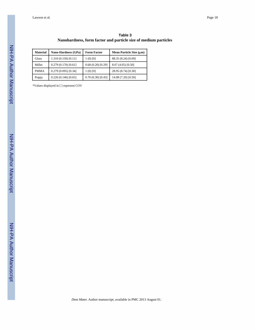
Results—Hardness values (GPa) of the particles are (glass, millet, PMMA, poppy respectively):1.310(0.150), 0.279(.170), 0.279(0.095), and 0.226(0.146). Average particle sizes (μm) are (glass,millet, PMMA, poppy respectively): 88.35(8.24), 8.07(4.05), 28.95(8.74), and 14.08(7.20). Glassand PMMA beads were considerably more round than the seeds. During composite wear testing,glass was the only medium that produced more wear than the use of water alone. The rankordering of the materials varied with each medium, however, the glass and PMMA bead mediumallowed better discrimination between materials.
Significance—PMMA beads are a practical and relevant choice for composite wear testingbecause they demonstrate similar physical properties as seeds but reduce the variability of wearmeasurements.
Publisher's Disclaimer: This is a PDF file of an unedited manuscript that has been accepted for publication. As a service to ourcustomers we are providing this early version of the manuscript. The manuscript will undergo copyediting, typesetting, and review ofthe resulting proof before it is published in its final citable form. Please note that during the production process errors may bediscovered which could affect the content, and all legal disclaimers that apply to the journal pertain.
NIH Public AccessAuthor ManuscriptDent Mater. Author manuscript; available in PMC 2013 August 01.
Published in final edited form as:Dent Mater. 2012 August ; 28(8): e118–e126. doi:10.1016/j.dental.2012.04.021.
NIH
-PA Author Manuscript
NIH
-PA Author Manuscript
NIH
-PA Author Manuscript
IntroductionWhile wear of dental composites is a prolifically studied subject [1], there is great variabilityin testing methods [2]. The 2001 International Standards Organization report “Wear by twoand or three body contact” describes eight methods for measuring in vitro wear. Amongother variables, the report describes three different food-simulating media for three-bodywear including: millet seed, PMMA beads, and poppy seed [3]. The adoption of these mediaparticles originated from a 1986 publication from de Gee, who compared wear ratesproduced with different seeds and polymethyl methacrylate (PMMA) powder in the ACTAwear testing device. He determined that using a mixture of 80% millet seeds and 20%PMMA powder most closely correlated in vivo wear data [4]. Later, Leinfelder and Suzukiused PMMA alone as a third-body medium because it does not degrade like millet and alsoexpedites the wear process [5,6]. Condon and Ferracane introduced poppy seed as areplacement to millet seed in de Gee's original mixture [7,8]. Since that time, additionalthird body particles have been examined including hydroxyapetite, green carborundum [9]and calcium diphosphate [10]. Glass microbeads have also been used as a third-bodymedium in industry protocol to expedite wear testing.
Although the effect of media particle selection on composite wear has not been directlystudied, various test methods which incorporate different particles have been compared.Two studies by Heintze et al compared the ACTA, Alabama and OHSU wear testingmethods, which incorporate millet seed, PMMA, and poppy seed media respectively. Thesestudies determined that relative wear ranking of composite materials varied significantlybetween testing methods [11,12]. Among those testing methods, there is variation in manyother factors (such as the methods of masticatory force application and tooth slidingreproduction) [13], so it is not possible to attribute the discrepancy in wear ranking tovariation in media particles alone. The aim of this study is to compare the wear of fourcomposites in the Alabama wear testing device with four currently used third-body mediumparticles (millet seed, poppy seed, PMMA beads, and glass microbeads) and water. The nullhypothesis is that the ranking of materials will be similar for all medium used.
Measuring the physical properties of the abrasive particles is critical for understanding thewear-producing mechanisms that differentiate each medium. Theoretically, a particle will bemore abrasive if: 1. it is harder than the surface it is indenting and 2. the size and form of theparticle allow it to penetrate through composite filler particles to the wear-prone resinmatrix. The hardness, size and shape of each abrasive medium particle will be measured inthis study, as these properties have been identified as critical parameters in tribologicaltesting [14].
Materials and methodsMedia particle preparation
Millet seed was prepared, as described by Nihei [15], by grinding 50g of seeds in a rotatingblade grinder for 5 seconds. Poppy seed was prepared as described by Condon andFerracane [7] by grinding 3g of poppy seed with 100 strokes of mortar and pestle. PMMAbeads (Dentsply Caulk, Milford, DE, USA) and soda lime glass microbeads (Size 270,Unibrite Corporation, Port Washington, NY, USA) were obtained from their manufacturer.
Nano-hardness measurementThe medium particles were embedded in a 95% methyl methacrylate / 5% nButylembedding epoxy (Fischer Scientific, Pittsburgh, PA, USA) before testing. The glass andPMMA beads were first stained with methylene blue to aid in their visualization. A thin coatof each type of medium particle was dispersed on the surface of a cup half-filled with set
Lawson et al. Page 2
Dent Mater. Author manuscript; available in PMC 2013 August 01.
NIH
-PA Author Manuscript
NIH
-PA Author Manuscript
NIH
-PA Author Manuscript
epoxy. The specimens were then covered with a layer of unset epoxy which polymerizedunder ultraviolet light for 48 hours. The surface of the specimens were wet polished with asuccession of 320 grit, 800 grit and 1200 grit paper on a surface parallel plane grinder(400CS, Exakt Technologies Inc, Oklahoma City, OK, USA) to reveal a layer of sectionedparticles. The nano-hardness of the exposed surfaces of the medium particles was measuredwith a nano-indentation tester (G200, MTS, Oak Ridge, TN, USA). Indentions were depthcontrolled to 0.5μm and performed with a diamond Berkovitch pyramid-shaped stylus(diameter = 40nm). A 4×4 grid of indents (5μm spacing between indents) was selected onthree millet and poppy seeds. Fifteen individual glass and PMMA beads were selected fortesting. Indents were examined after testing and hardness values that were obtained fromindenting the epoxy were discarded.
Composite specimens were prepared in a silicone mold (1cm diameter × 4mm) and lightpolymerized at 2mm increments with a Coltolux LED curing light (Coltene/Whaledent,Cuyahoga Falls, OH, USA) (583mW/cm2). They were then polished using 600 and 1200 gritsilicon carbon paper followed by 0.5μm alumina slurry on a polishing wheel (Metallurgicalpolisher, Buehler Ltd., Evanston, IL, USA) at 80 rotations/sec and 20N of force. Nano-hardness of the composites was determined by creating a 4×4 grid of indents (5μm spacingbetween indents) at two locations on the composite surface. The same testing parameterswere used as described above.
Particle size measurementThe medium particles were mixed with distilled water in a 3:1 ratio. A 3mL sample of eachmedium was measured in a LASER light diffraction optical particle size analyzer (Microtrac3500, Microtrac Inc., York, PA, USA) operated between the size range of 24nm and2800μm. Three measurements were taken of each sample, and media were sonicated for 2minutes between measurements to prevent agglomeration.
Particle imaging and shape measurementParticles were randomly dispersed on a glass slide. The particles were imaged with 1000×optical magnification using digital light microscopy (VHX-600, Keyence Co., Osaka, Japan)as described in Table 1 of ASTM standard F1877-05 [16]. Three images of each mediumwere collected, and within the images, the perimeter (P) and area (A) of the outline of eachparticle was recorded with image analysis software (ImageJ, NIH, Bethesda, MD, USA).
The form of the particles was determined using the form factor (FF) equation: [14].Form factor gives an indication of the roughness or roundness of a particle's outline;particles with a circular outline have a FF = 1. The average of the form factor of all particlesfrom each media type was reported.
Wear testing of composite materialsFour commercially available light-cured composites were studied: a hybrid (Z100, 3M Co.,St Paul, MN, USA), a flowable microhybrid (Estelite Flow Quick, Tokuyama, Tokyo,Japan), a micromatrix (Esthet-X, Caulk Dentsply, Milford, DE, USA), and a nano-filled(Filtek Supreme Plus, 3M ESPE). The materials were chosen to represent a range of fillerconcentrations (71%-85%) and filler particle sizes (0.005-3.5 μm). Their properties arelisted in Table 1.
Specimens (n=8) were fabricated in silicone molds (1cm diameter × 4mm depth) and lightpolymerized at 2mm increments with a Coltolux LED curing light (Coltene/Whaledent)(583mW/cm2). After production, the specimens were set in brass holders with acrylic(Dentsply Repair Material, Dentsply Caulk) and polished using 600 and 1200 grit silicon
Lawson et al. Page 3
Dent Mater. Author manuscript; available in PMC 2013 August 01.
NIH
-PA Author Manuscript
NIH
-PA Author Manuscript
NIH
-PA Author Manuscript
carbon paper followed by 0.5μm alumina slurry on a polishing wheel (Metallurgicalpolisher, Buehler Ltd.) at 80 rotations/sec and 20N of force. Specimens were stored in waterat 37°C for 48 hours. The spring containing antagonist pistons were load calibrated beforetesting with a universal testing device (Model 4411, Instron, Norwood, MA, USA) to ensurea maximum force of 70N. Specimens were placed into the Alabama wear device. whichoperates by pressing the spring-loaded pistons into the composite specimens followed by a30° rotation of the antagonist. The antagonists on the pistons contact the specimens for400ms applying a maximum force of 70N and counter-rotate 30° prior to lifting off thespecimens (Figure 1). 2g of the medium prepared as described above was mixed with 3mLof distilled water. This ratio was chosen based on previous studies [4,15]. The mediummixture was stirred and vibrated until the particles fully mixed with the water. Due to thedifferences in densities of each type of particle, some wells contained more particles thanothers, however, all specimens were completely covered with medium. The medium wasthen poured into the individual wells above the composite specimens created by the brassrings (Figure 2). A fifth group was prepared in which the specimen wells were filled withdistilled water. The test was run for 100,000cycles at 1.2Hz. A new stainless steel antagonistball (Ra = 4.7μm) was used for every test. Following the test, specimens were ultrasonicallycleaned for 1 minute. The surface of each specimen was scanned with a non-contact opticalprofilometer (Scantron 2000, Scantron Industrial Products, Tauton, England) with a 20μm ×20μm resolution. The scans were analyzed with superimposition software (Pro-Form,Scantron Industrial Products) to determine volumetric wear.
Following testing, representative wear specimens were removed from the brass holders andcoated with Au-Pd in a sputter coater (Hummer X, Anatech, Union City, CA, USA). Theirsurfaces were examined by secondary-electron SEM (Model 40, International ScientificInstruments, Milpitas, CA, USA). Samples of glass and PMMA beads were removed fromthe wells following testing and examined using digital light microscopy (VHX-600,Keyence Co).
Statistical analysisThe study design was a two-way layout, with groups defined by material and medium. Theprimary analysis technique utilized two-way analysis of variance (ANOVA). Pairwisecomparisons among group means were conducted using Tukey's test. A rank transformationof the data was used, due to significant nonhomogeneity of variance among the groups (p <0.0001, Levene's Test), and substantial asymmetry of the sample distributions. Separate one-way ANOVA analyses were conducted for materials within each medium and mediumwithin each material group in order to evaluate the significant interactions.
ResultsThe nano-hardness of the composite materials is given in Table 2. The mean, standarddeviation and coefficient of variation (COV) of the nano-hardness, form factor and size foreach type of particle is given in Table 3. The COV was calculated using the formula COV =mean/standard deviation. The COVs of nano-hardness, form factor and size values of thePMMA and glass beads were less than those of the millet and poppy seeds. The lower COVof PMMA and glass beads indicated a more homogenous composition of these particles. Thesize distribution of particles as distributed by number of particles is graphed in Figure 3.Images of the media particles are presented in Figure 4.
The mean, standard deviation, and coefficient of variance of volumetric wear data is givenin Table 4. The results of a 2-way ANOVA revealed that there was a significant material bysolution interaction (p < 0.0001), as well as significant main effects for material (p = 0.004)and solution (p < 0.0001). The individual analyses by medium type reveal that materials can
Lawson et al. Page 4
Dent Mater. Author manuscript; available in PMC 2013 August 01.
NIH
-PA Author Manuscript
NIH
-PA Author Manuscript
NIH
-PA Author Manuscript
be grouped into significantly different groups for all medium types except millet seeds. Theindividual analyses by material type reveal that there was significantly more wear on allmaterials when glass was used as a medium than in water alone and that there was less wearon all materials when poppy seeds, millet seeds and PMMA beads were used as mediumthan in water alone (Table 4).
SEM images are presented for the worn surface of Filtek Supreme Plus as representativeexamples for all materials (Figure 5). The figure shows that wear track that is produced fromthe contact and 30° rotation of the spherical stainless steel antagonist. The wear track in theglass group (Figure 5A) shows deep gauging and spalling of the composite surface and it islarger than the tracks from all other groups. The edges of the wear track appear jagged frommaterial that has chipped off from contact with the hard glass particles. The wear track onthe water group shows deep, smooth gauging in the wear track (Figure 5E). The smoothnessof the tracks from the water group compared to the glass group can be attributed to thesmaller dimensions of the asperity heights on the steel antagonist (∼4.7μm) than size of theglass beads (∼88μm). The PMMA, millet and poppy groups (Figure 5 B-D) show superficialscratching of the composite surface. These scratches may have occurred from the three-bodyabrasion of medium particles or periods of direct two-body contact between the steelantagonist and the composite surface.
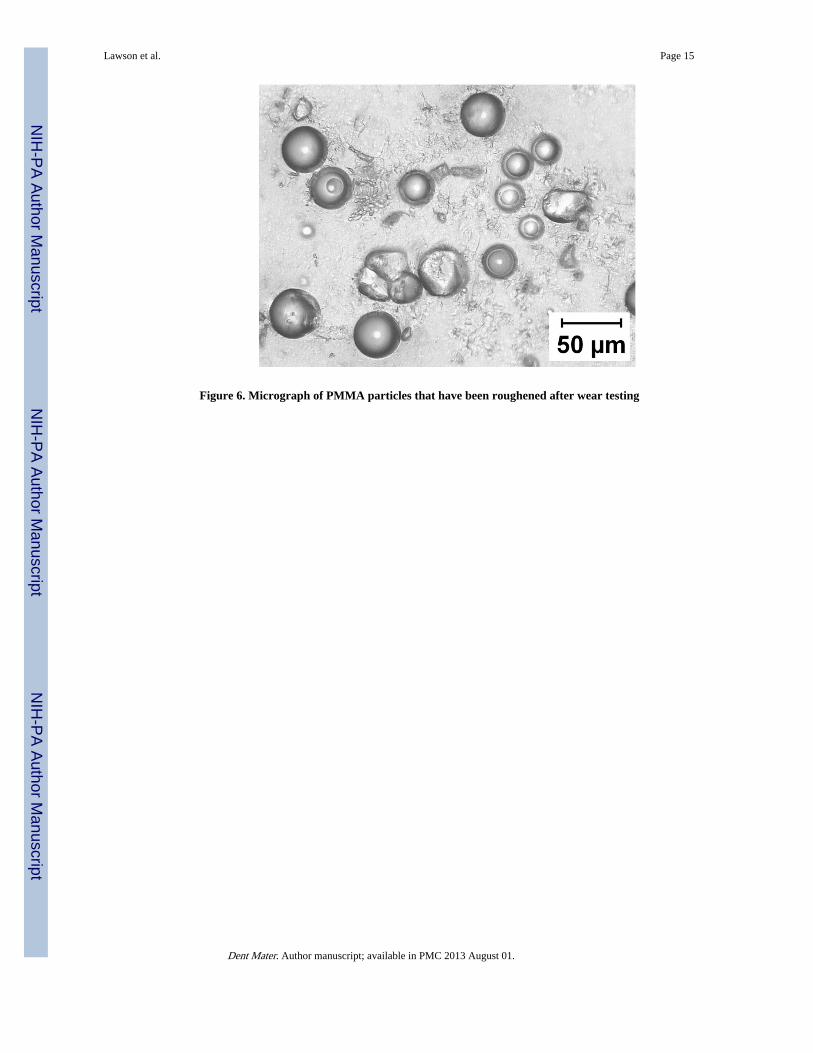
A micrograph of PMMA beads following wear testing (Figure 6) demonstrates surfaceroughening of several of the beads. Roughening was not noted on the glass beads and nodifference could be visibly discerned between the seeds before and after wear testing(however degradation of the seeds is suspected).
DiscussionIn vitro wear testing should predict the wear performance of a material in vivo. To date, onlyone study has reported a correlation between the amount of wear produced in the Alabamawear testing device and clinical wear measurements [17]. Due to limited clinical wear data,it is more practical to utilize in vitro wear testing to determine the relative rankings ofmaterials than quantify an expected amount of wear. Another practical utility of an in vitrowear test is its ability to discriminate between materials. Variation among specimens shouldbe attributed to the differences in the physical properties of the material not the variation in acomponent of the wear testing system, such as the food simulating medium. It is important,however, to include a food simulating medium in wear testing as previous studies haveshown both increased and decreased wear in the presence of a third-body medium [18,19].
Based on the results of this experiment, material ordering with regard to wear was dependenton the medium used. Individual analyses by medium type revealed that significantdifferences could be seen between materials using glass, PMMA and poppy seeds but notmillet seeds. Therefore, the null hypothesis cannot be accepted. Additionally, the use ofglass beads produced more composite wear than with water alone and the use of all othermedium particles resulted in less wear than the use of water alone. An analysis of themedium particle properties will be used to try to explain differences in relative wear values,discriminating ability of the different media, and material ordering.
The most obvious difference between medium particles was the hardness of the glass beads.A recent study examined the nano-hardness of several composite materials [20]. In thatstudy, both the hardness of the resin matrix and filler particles were measured. The materialstested were similar to the materials tested in this study, containing both dimethcrylate resinmatrix and silica-and zirconia-based filler particles. The reported resin matrix hardnessranged from 0.3-0.5GPa and the filler particle hardness ranged from 2-4GPa. Based on the
Lawson et al. Page 5
Dent Mater. Author manuscript; available in PMC 2013 August 01.
NIH
-PA Author Manuscript
NIH
-PA Author Manuscript
NIH
-PA Author Manuscript
measured hardness of the medium particles in this study, the PMMA, millet and poppyparticles are slightly softer than the resin matrix and much softer than the filler particles ofcomposite materials. The glass beads, however, are much harder than the resin matrix andonly slightly softer than the composite filler particles. The ability of the glass beads toabrasively wear the resin matrix and possibly even the filler particles of the compositesexplains the relatively large amount of composite wear using this abrasive medium.
The size and shape of abrasive particles and the interparticle spacing in a dental compositewill determine the ability of a medium particle to penetrate to the resin matrix of acomposite [21]. Jorgensen postulated that an interparticle spacing of less than 0.1μm wouldallow filler particles to protect the resin matrix from food abrasion. His theory was based onclinical observations of composite restorations [22]. Bayne et al theorized a method fordetermining the interparticle spacing of microfilled composites based on the volume fractionof filler particles. His calculations are based on .02μm filler particles that are either evenlydispersed or agglomerated in resin matrix. The theorized maximum inter-particle or inter-cluster spacing was determined for various filler volume fractions [23]. The materials in thisstudy range in volume fraction of filler from 53%-66% and particle sizes range from 0.01–3.5μm, however, all materials contain some particles below 0.02μm. Assuming all materialscontain at least 10% vol of filler particles smaller than 0.02μm, the theoretical maximuminterparticle spacing would still be below 1μm. Therefore, abrasive particles larger than1μm would not be expected to penetrate through the interparticle spacing.
The mean size of all medium particles used in this study are larger than 1μm, however, it isplausible that asperities on the surfaces of the medium particles could penetrate a 1μmspace. Asperities are projections from a surface and the amount of asperities on the surfaceof a particle can be approximated by measuring its form factor. The PMMA and glassparticles are spherical and do not have asperities (form factor of 1). The millet seeds andpoppy seeds, however, have rough surfaces (form factor of 0.68 and 0.70 respectively) withasperities capable of penetrating through the interparticle spacing. The roughness of the seedparticles may explain why most composites showed more wear with seed particles thanPMMA beads, despite all three having similar hardness values. Micrographs of wornPMMA showed roughening of its surface, which may have created asperities and graduallyincreased the abrasivity of the PMMA beads.
The rank ordering of materials varied based on the type of medium particle used, however,due to the multifactorial nature of the wear processes and broad differences in materials,explaining the exact reasoning for the different rank orders is difficult. Several generalitiesregarding the wear rankings can be proposed based on the physical and mechanicalproperties of the composite materials and medium particles. Using glass as a third-bodymedium, the amount of composite wear was inversely related to the hardness and percentagefiller content of the composite materials. This paradoxical relationship is explained by thefact that the glass beads have hardness values greater than the average bulk hardness of allthe composites tested. The glass beads were twenty times the size of the largest fillerparticles and might have seen the composite surface as a bulk material. So even if the glassbeads were not harder than the individual composite filler particles, they may have been ableto fracture off asperities of bulk composite material. Since the glass was harder than the bulksurface of all composite materials, it could non-specifically wear the filler particles and resinmatrix regardless of their hardness or percentage filler content. Therefore, the hardness ofthe composites relative to each other would not be useful for discriminating betweenmaterials. This theory is substantiated by the fact that SEM images of the worn compositeshows gouging and spalling of the composite deep into the bulk of the material.
Lawson et al. Page 6
Dent Mater. Author manuscript; available in PMC 2013 August 01.
NIH
-PA Author Manuscript
NIH
-PA Author Manuscript
NIH
-PA Author Manuscript
When PMMA was used as a third-body medium, the wear ranking was approximatelyrelated to the hardness and percentage filler content of the composite materials. Thisrelationship suggests that wear testing with PMMA is affected by contribution of fillerparticles in the dental composite. The average PMMA particle size was about ten times thesize of the largest filler particles. Therefore, it is unlikely that the entire PMMA beadpenetrated the inter-particle composite spacing and selectively wore the resin matrix. ThePMMA beads are softer than the bulk composite materials and much softer than thecomposite filler particles. During the wear process, the composite filler particles likelyabraded the PMMA beads. Micrographs of the PMMA beads at the end of wear testing showthat their surface becomes roughened. Possibly, the asperities on the roughened PMMAsurface penetrated the interparticle spacing and preferentially wore the resin matrix.Therefore, composites with lower filler content would be less able to protect against thispreferential wear. SEM images of the composite worn by PMMA media showed scratchingof the composite surface, supporting the theory of preferential matrix wear.
The use of millet and poppy seeds in the third-body medium provided limited ability todiscriminate between materials. The variation in wear volumes between specimens of thesame material and seed type is evident by high values of the coefficient of variation. HighCOV is also present in the size and shape measurements of millet and poppy seeds. Sinceseed particles were stored in individual wells above each specimen during wear testing, thedistribution of sizes and shapes of seed particles was different for every specimen. Thisvariation in seed particles affected the abrasivity of the medium for each specimen andcreated variability in the resulting wear. Another source of variation using the seed particlesis the differential breakdown of individual seed particles through mechanical deformationand water degradation. As the seed particles degrade through the wear process, they willbecome reduced in dimension. A study by Pallav et al showed that as medium particles arereduced from 3μm to 1μm, direct contact occurs between a composite and antagonistmaterial [24]. Another study by Kunzelmann showed that larger crushed millet particles aremore abrasive than smaller particles [25]. Therefore, the abrasive or lubricant properties of abatch of seeds are dependent on the individual breakdown of the seed particles in that batch.The tendency for the seed particles to degrade and allow direct two-body wear between thecomposite and the antagonist may explain the relatively higher wear with seen with a seedmedium than a PMMA medium. SEM evaluation of the composite worn with millet andpoppy seeds reveals scratching of the composite surface, similar in appearance as the surfaceworn by PMMA beads. These scratches may either be a result of harder seed particlesscratching the resin in the composite or direct contact of the steel antagonist throughdegraded seed particles.
A limitation of this study was that it was difficult to explain differences between compositematerials due to the compositional variability of commercially available materials. Morematerials would need to be included in this analysis to make a definitive conclusion of themost relevant third-body medium particle. Future studies should consider using materialsstandardized for resin and filler composition and filler particle size and size distribution tobetter understand the wear process. An additional limitation of this study is that these resultsare only applicable to the Alabama wear testing device and the sizes of PMMA and glassbeads used in this protocol.
ConclusionBased on the analysis of medium particles and composite wear, the PMMA used in thisstudy seems to be a practical and relevant material to use in a third-body medium forcomposite wear testing. It has a similar hardness as natural seed materials. It is slightlylarger than seed particles, however, the size distributions have overlap. The seeds are
Lawson et al. Page 7
Dent Mater. Author manuscript; available in PMC 2013 August 01.
NIH
-PA Author Manuscript
NIH
-PA Author Manuscript
NIH
-PA Author Manuscript
rougher than the PMMA initially, but PMMA roughens through the wear process.Composite wear using a PMMA abrasive produces volumetric wear on the same order ofmagnitude as wear with natural seeds. Finally, SEM evaluation of wear surfaces appearsimilar with PMMA and natural seeds. The advantage of using PMMA as a third-bodymedium is that it produced less variation between specimens of the same material than seedsand therefore a greater ability to discriminate between different materials. For these reasons,the authors recommend using the PMMA used in this study for in vitro three-body weartesting.
AcknowledgmentsThe authors would like to thank Dr. Xiaoming Xu at LSUHSC School of Dentistry for conducting particle sizeanalysis of the medium particles. Additionally, Sarah Syklawer and Courtney Michelson should be acknowledgedfor their assistance with specimen preparation.
References1. Ferracane JL. Is the wear of dental composites still a clinical concern? Is there still a need for in
2. Heintze SD. How to qualify and validate wear simulation devices and methods. Dent Mater. 2006;22:712–734. [PubMed: 16574212]
3. ISO. Dental Material, Guidance on testing of wear. Part 2. Wear by two-and/or three-body contact.Technical Specification 2001, No. 14569-2.
4. de Gee AJ, Pallav P, Davidson CL. Effect of abrasion medium on wear of stress bearing compositesand amalgam in vitro. J Dent Res. 1986; 65(5):654–658. [PubMed: 3457819]
5. Leinfelder KF, Suzuki S. In vitro wear device for determining posterior composite wear. JADA.1999; 130:1347–1353. [PubMed: 10492543]
6. Leinfelder KF, Beaudreau RW, Mazer RB. An in vitro device for predicting clinical wear.Quintessence Int. 1989; 20(10):755–761. [PubMed: 2639390]
7. Condon J, Ferracane J. Evaluation of composite wear with a new multi-mode oral wear simulator.Dent Mater. 1996; 12:218–226. [PubMed: 9002838]
8. Condon JR, Ferracane JL. Factors effecting dental composite wear in vitro. J Biomed Mater Res.1997; 38:303–313. [PubMed: 9421751]
9. Satou N, Khan AM, Satou K, Satou J, Shintani H, Wakasa K, Yamaki M. In-vitro and in-vivo wearprofile of composite resins. J Oral Rehabil. 1992; 19(1):31–37. [PubMed: 1316435]
10. Kakuta K, Ogura H. Effects of abrasive and fiber components in medium on wear of compositeresins. Dent Mater J. 2008; 27(5):716–722. [PubMed: 18972789]
11. Heintz SD, Sappini G, Rousson V. Wear of ten dental restorative materials in five wear simulators– Results of a round robin test. Dent Mater. 2005; 21:304–317. [PubMed: 15766577]
12. Heintze SD, Barkmeier WW, Latta MA, Rousson V. Round robin test: wear of nine dentalrestorative materials in six different wear simulators - supplement to the round robin test of 2005.Dent Mater. 2011; 27(2):e1–9. [PubMed: 20888629]
13. Lambrechts P, Debels E, Van Landuyt K, Peumans M, Van Meerbeek B. How to simulate wear?Overview of existing methods. Dent Mater. 2006; 22(8):693–701. [PubMed: 16712913]
14. Hutchings, IM. Tribology. London: Arnold; 1992.
15. Nihei T, Dabanoglu A, Teranaka T, Kurata S, Ohashi K, Kondo Y, Yoshino N, Hickel R,Kunzelmann KH. Three-body-wear resistance of the experimental composites containing fillertreated with hydrophobic silane coupling agents. Dent Mater. 2008; 24(6):760–764. [PubMed:17964643]
16. ASTM. Annual book of ATM standards, Medical devices and standards. Standard practice forcharacterization of particles. Technical Specification 2007, F 1877-05.
17. Barkmeier WW, Latta MA, Erickson RL, Lambrechts P. Comparison of laboratory and clinicalwear rates of resin composites. Quintessence Int. 2004; 35(4):269–274. [PubMed: 15119711]
Lawson et al. Page 8
Dent Mater. Author manuscript; available in PMC 2013 August 01.
NIH
-PA Author Manuscript
NIH
-PA Author Manuscript
NIH
-PA Author Manuscript
18. Sajewicz E. Effect of saliva viscosity on tribological behaviour of tooth enamel. Tribol Int. 2009;42(2):327–332.
19. Condon JR, Ferracane JL. Factors effecting dental composite wear in vitro. J Biomed Mater Res.1997; 38(4):303–313. [PubMed: 9421751]
20. Drummond JL. Nanoindentation of dental composites. J Biomed Mater Res B Appl Biomater.2006; 78(1):27–34. [PubMed: 16278844]
21. Mortensen, A. Concise Encyclopedia of Composite Materials. 2nd. Amsterdam: Elsevier; 2007.
22. Jorgensen KD. Occlusal abrasion of a composite resin with ultra-fine filler – an initial study.Quintessence Int. 1978; 6:73–78. [PubMed: 288100]
23. Bayne SC, Taylor DF, Heymann HO. Protection hypothesis for composite wear. Dent Mater. 1992;8:305–309. [PubMed: 1303372]
24. Pallav P, de Gee AJ, Werner A, Davidson CL. Influence of shearing action of food on contactstress and subsequent wear of stress-bearing composites. J Dent Res. 1993; 72(1):56–61.[PubMed: 8418108]
25. Kunzelmann KH, Hickel R. The influence of different abrasion media on three-body-wear ofcomposites. J Dent Res. 1995; 74(A):625.
Lawson et al. Page 9
Dent Mater. Author manuscript; available in PMC 2013 August 01.
NIH
-PA Author Manuscript
NIH
-PA Author Manuscript
NIH
-PA Author Manuscript
Figure 1. Schematic of Alabama wear testing device
Lawson et al. Page 10
Dent Mater. Author manuscript; available in PMC 2013 August 01.
NIH
-PA Author Manuscript
NIH
-PA Author Manuscript
NIH
-PA Author Manuscript
Figure 2. Brass wells of medium particles suspended over composite specimens
Lawson et al. Page 11
Dent Mater. Author manuscript; available in PMC 2013 August 01.
NIH
-PA Author Manuscript
NIH
-PA Author Manuscript
NIH
-PA Author Manuscript
Figure 3. Micrographs of 4 medium particles (A) glass, (B) millet, (C) PMMA, (D) poppy
Lawson et al. Page 12
Dent Mater. Author manuscript; available in PMC 2013 August 01.
NIH
-PA Author Manuscript
NIH
-PA Author Manuscript
NIH
-PA Author Manuscript
Figure 4. Particle size (by number) of 4 medium particles (A) glass, (B) millet, (C) PMMA, (D)poppy
Lawson et al. Page 13
Dent Mater. Author manuscript; available in PMC 2013 August 01.
NIH
-PA Author Manuscript
NIH
-PA Author Manuscript
NIH
-PA Author Manuscript
Figure 5. SEM images of worn surfaces of Filtek Supreme using different medium particles (A)glass, (B) millet, (C) PMMA, (D) poppy, (E) water
Lawson et al. Page 14
Dent Mater. Author manuscript; available in PMC 2013 August 01.
NIH
-PA Author Manuscript
NIH
-PA Author Manuscript
NIH
-PA Author Manuscript
Figure 6. Micrograph of PMMA particles that have been roughened after wear testing
Lawson et al. Page 15
Dent Mater. Author manuscript; available in PMC 2013 August 01.
NIH
-PA Author Manuscript
NIH
-PA Author Manuscript
NIH
-PA Author Manuscript
NIH
-PA Author Manuscript
NIH
-PA Author Manuscript
NIH
-PA Author Manuscript
Lawson et al. Page 16
Tabl
e 1
Mat
eria
ls u
sed
in t
his
stud
y
Mat
eria
lC
lass
ific
atio
nL
ot N
o.M
atri
xF
iller
Tot
al f
iller
con
tent
Est
elite
Flo
w Q
uick
Flow
able
Mic
rohy
brid
UE
401
236
Bis
-EM
A, T
EG
DM
A, 1
-6bi
s(m
etha
cryl
eth
ylox
ycar
bony
lam
ino
)tri
met
hyl h
exan
eSi
lica/
titan
ia a
nd s
ilica
/zir
coni
a pa
rtic
les:
0.04
-0.6μ
m71
% (
wt)
53%
(vo
l)
Est
het X
Mic
rom
atri
x06
0329
Ure
than
e m
odif
ied
bis-
GM
AB
ariu
mal
umin
o fl
uoro
boro
silic
ate
glas
s:0.
02-2
.5μ
m S
ilica
: 10-
20nm
77%
(w
t) 6
0% (
vol)
Filte
k Su
prem
e Pl
usN
ano-
fille
d20
0606
06B
is-E
MA
6, U
DM
A, B
is-G
MA
, TE
GD
MA
Silic
a: 5
–20n
m n
anop
artic
le Z
irco
nia/
silic
a: 0
.6–1
.4μ
m n
anoc
lust
er78
.5%
(w
t) 5
7.7%
(vo
l)
Z10
0H
ybri
d6K
MB
is-G
MA
, TE
GD
MA
, 2-b
enzo
tria
zoly
lmet
hylp
heno
lZ
irco
nia/
silic
a: 0
.01–
3.5μ
m85
% (
wt)
66%
(vo
l)
Bis
-EM
A =
Bis
phen
ol A
pol
yeth
oxy
met
hacr
ylat
e, T
EG
DM
A =
Tri
ethy
lene
gly
col d
imet
hacr
ylat
e, B
isE
MA
6 =
Bis
phen
ol A
pol
yeth
ylen
e gl
ycol
die
ther
dim
etha
cryl
ate,
UD
MA
= D
iure
than
edi
met
hacr
ylat
e, B
is-G
MA
= B
isph
enol
A d
igly
cidy
l eth
er d
imet
hacr
ylat
e
Dent Mater. Author manuscript; available in PMC 2013 August 01.
NIH
-PA Author Manuscript
NIH
-PA Author Manuscript
NIH
-PA Author Manuscript
Lawson et al. Page 17
Table 2Nano-hardness of composite materials used in this study
Material Nano-hardness (GPa)
Estelite Flow Quick 0.303 (0.062)
Esthet X 0.657 (0.076)
Filtek Supreme Plus 0.873 (0.107)
Z100 1.101 (0.256)
Dent Mater. Author manuscript; available in PMC 2013 August 01.
NIH
-PA Author Manuscript
NIH
-PA Author Manuscript
NIH
-PA Author Manuscript
Lawson et al. Page 18
Table 3Nanohardness, form factor and particle size of medium particles
Material Nano-Hardness (GPa) Form Factor Mean Particle Size (μm)
Dent Mater. Author manuscript; available in PMC 2013 August 01.
NIH
-PA Author Manuscript
NIH
-PA Author Manuscript
NIH
-PA Author Manuscript
Lawson et al. Page 19
Table 4Volumetric wear of composites using 4 medium particles and water
Volumetric wear × 10-3 (mm3) reported as mean (SD) [COV]
Material Estelite Flow Esthet X Filtek Supreme Z100
Glass 868.88(72.94)a C [0.08] 1183.12(145.31)b C [0.12] 1343.51(140.02)b,c D [0.10] 1359.02(81.87)c E [0.06]
Millet 30.96 (57.82)a A [1.87] 14.51(32.68)a A [2.25] 56.86(49.32)a B,C [0.87] 1.94(2.90)a B [1.50]
PMMA 1.86(1.06)b A [0.57] 3.74(1.95)c A [0.52] 1.86(0.93)b A [0.50] 0.37(0.07)a A [0.19]
Poppy 2.64(3.21)a A [1.21] 10.30(8.28)b A [0.80] 17.34(29.42)b A,B [1.70] 17.88(16.79)b C [0.94]
Water 94.75(11.07)c B [.011] 80.88(9.00)b,c B [0.11] 46.88(6.51)a C [0.14] 70.00(22.73)b D [0.32]
*Values displayed in [ ] represent COV
#Similar lowercase superscripts represent statistically similar groups in each row and similar uppercase superscripts represent statistically similargroups in each column
Dent Mater. Author manuscript; available in PMC 2013 August 01.