31
NATIONAL TOXICOLOGY PROGRAM PROPOSAL TO LIST NICKEL NANOPARTICLES IN FUTURE EDITIONS OF THE ROC Comments of the Nickel Producers Environmental Research Association October 17, 2013
NATIONAL TOXICOLOGY PROGRAM PROPOSAL TO LIST NICKEL NANOPARTICLES
IN FUTURE EDITIONS OF THE ROC
Comments of the Nickel Producers Environmental Research Association
October 17, 2013
INC. Nickel
Producers COMMENTS ON THE NTP PROPOSAL Environmental Research TO LIST NICKEL NANOPARTICLES IN Association FUTURE EDITIONS OF THE ROC
Table of Contents
Page
1. Executive Summary ............................................................................................................... 3
2. Data on current production, use patterns, and human exposure...................................... 4
2.1 Production volume of nickel nanoparticles in the United States........................................ 4
2.2 Use patterns...................................................................................................................... 5
2.3 Human exposure ...............................................................................................................6
3. Published studies related to evaluating carcinogenicity ................................................... 8
3.1 Hazard comparison of nano- and micron-sized particles of the same materials ............... 8 3.1.1 Biodurable substances............................................................................................ 8 3.1.2 Nickel substances ................................................................................................... 9 3.1.3 Summary of all substances ..................................................................................... 9
3.2 Summary of toxicological data for nickel-containing nanoparticles ................................... 9 3.2.1 Possible toxicological effects of nickel-containing nanoparticles compared to
micron-sized nickel particles ................................................................................... 9 3.2.2 Possible carcinogenicity effects of nickel-containing nanoparticles compared to
micron-sized nickel particles ................................................................................. 13
4. Scientific issues important for assessing the carcinogenicity of nickel nanoparticles 16
5. Conclusions .......................................................................................................................... 18
6. Scientist(s) with expertise or knowledge of nickel ........................................................... 18
7. References ............................................................................................................................ 19
8. Appendix ............................................................................................................................... 27
Appendix, Table 1. Nickel-containing nanoparticles tested in in vivo toxicological studies .... 27
Appendix, Table 2. Inhalation studies..................................................................................... 28
Appendix, Table 3. Instillation or oropharyngeal aspiration (OPA) studies ............................ 29
Appendix, Table 4. Injection and implantation studies ........................................................... 31
Page 2 of 31
INC. Nickel
Producers COMMENTS ON THE NTP PROPOSAL Environmental Research TO LIST NICKEL NANOPARTICLES IN Association FUTURE EDITIONS OF THE ROC
1. EXECUTIVE SUMMARY
The Nickel Producers Environmental Research Association (NiPERA) is pleased to submit these Comments to the NTP regarding the nomination for possible review and listing of nickel nanoparticles in a future edition of the Report on Carcinogens (RoC). Please note that NiPERA is responding on behalf of the global nickel mining and refining companies as well as bulk nickel producers. Although NiPERA does not represent industries involved in the downstream production of nickel nanoparticles, we appreciate the opportunity to submit comments.
The nomination to review nickel nanoparticles as a potential cancer hazard for people in the United States does not appear to be justified based on the following:
Nickel compounds are already listed in the RoC as “known human carcinogens” (dating from the 9th RoC in 2000), and that listing applies to all forms of nickel compounds (nanoparticles included).
Nickel metal is already listed in the RoC as “reasonably anticipated to be a human carcinogen” (dating from the 10th RoC in 2002), and that listing applies to all forms of nickel metal (nanoparticles included).
Based on the weight of evidence of current toxicological data and using proper dose metrics, the lung toxicity and carcinogenicity hazards of nanoparticles (including nickel nanoparticles) do not appear to be different from those of micron-sized particles of the same materials. With the publication of a negative carcinogenicity study of nickel metal powder in 2008 and the lack of positive human epidemiology data, the evidence for nickel metal being a carcinogen is weaker now than it was in 2002, with stronger evidence that it is not a carcinogen. There is at present no basis for distinguishing between nickel micron-sized particles and nickel nanoparticles in this regard.
Information obtained from several sources indicate that nickel nanomaterials do not have a high production volume in the United States (<100 tons/year) and the number of people with the potential for significant inhalation, oral or dermal exposure to nickel nanomaterials in the U.S. is very low (~300 people).
Altogether, there does not appear to be any demonstrable justification (based on exposure or hazard information) to consider a separate listing for nickel nanoparticles in NTP’s RoC.
Recommendation: Nickel nanoparticles appear to present the same hazard as micron-sized nickel particles. Since nanomaterials of nickel compounds and nickel metal are already covered in the 9th and 10th RoC, respectively, there is no need for a separate listing.
Page 3 of 31
INC. Nickel
Producers COMMENTS ON THE NTP PROPOSAL Environmental Research TO LIST NICKEL NANOPARTICLES IN Association FUTURE EDITIONS OF THE ROC
NiPERA’s comments include information in support of our recommendation as per NTP’s formal Request for Information.
2. DATA ON CURRENT PRODUCTION, USE PATTERNS, AND HUMAN EXPOSURE
2.1 Production volume of nickel nanoparticles in the United States
Although precise volumes of nickel nanoparticles are not known, estimates can be made from available data of multiple sources. Nanoparticles, in general, are manufactured worldwide for use in several consumer product categories, such as health and fitness, home and garden, automobile, food and beverage, cross cutting (coatings), electronics and computers, and appliances. According to the Project on Emerging Nanotechnologies online inventory (2013) of nanotechnology-based consumer products on the market, approximately 45% (741 out of 1628 products) of those nanotechnology-containing products are made in the U.S., with Europe (27%) and East Asia (17%) producing most of the remaining products. The most common nanocomponents referenced in this online inventory are silver, carbon, titanium, silicon/silica, zinc, and gold nanomaterials. Nickel was not specifically mentioned as one of the primary nanomaterials used in these products. A separate database, Nanowerk Nanomaterial Database (2013), reports 32 suppliers of nickel nanomaterials worldwide, with only nine of these suppliers (28%) located in the U.S. However, some additional companies that produce nickel nanoparticles did not provide information for the database and thus were not represented.
Approximately 10 to 20 companies produce nickel nanoparticles in the U.S.1 However, a few additional companies were reported to produce nickel nanoparticle in surveys from 2008 and 2009 that are no longer producing nickel nanoparticles. The annual nickel nanoparticle production volume of some of these companies is only in the gram to kilogram range each, while a few other companies may produce ~1 ton per year, as confirmed by several experts in the field. For example, one U.S. company that produces nickel metal nanoparticles, QuantumSphere, indicates in its website2 that it has 8 reactors that can generate up to a combined total of ~6 tons of nanoparticles of iron, silver, copper, nickel and manganese per year. Although the specific volume of nickel metal nanoparticles is not listed, it could be predicted to be no greater than ~1 ton a year for QuantumSphere. Based on this information, a high estimate of the total U.S. production of nickel nanoparticles is ~20 tons per year, assuming each company produced the high limit of 1 ton annually, although this is unlikely since several companies only produce nickel nanoparticles in kilogram quantities.
An inference of U.S. nanonickel production can be made from recent REACH registrations in the European Union. The REACH regulatory initiative requires registration of all chemicals manufactured in or imported to the European Union. As of May 2013, all chemicals placed on
1 NiPERA does not represent downstream producers of nickel nanoparticles. However, we are in the process of developing a list of the U.S. producers as a result of the NTP nomination for possible review and listing of nickel nanoparticles in the RoC. This information can be made available upon request. 2 QuantumSphere website: http://www.qsinano.com/products_nanomaterials.html
Page 4 of 31
INC. Nickel
Producers COMMENTS ON THE NTP PROPOSAL Environmental Research TO LIST NICKEL NANOPARTICLES IN Association FUTURE EDITIONS OF THE ROC
the European Union market had to be registered if manufactured or imported at more than 100 tons/year by a manufacturer(s) or importer(s). As of October 2013, there were 23 registrations that covered nanomaterials or nanoforms of substances. Of those 23 registrations, no nickel nanomaterials were listed, indicating that any nickel nanomaterials produced in Europe would be less than 100 tons/year.3 The registration information from REACH is not only evidence of low production volumes of nickel nanoparticles in Europe (<100 tons/year), it also suggests low volumes of imported nickel nanoparticles coming from the U.S. (<100 tons/year).
Data is scarce with regard to the production volume of nickel nanoparticles and specific producers in the U.S. However, as mentioned above, nickel nanoparticles are not as frequently produced as other types of nanoparticles. The information regarding producers of nickel nanoparticles in the U.S. and information gathered from REACH registrations indicate a rough estimate of 1 to 100 tons/year of total nickel nanoparticles produced in the U.S.4
2.2 Use patterns
Nickel nanoparticles have many commercial and industrial uses. Several worldwide nickel nanoparticle producers5 report the use of these small particles in:
Additives in ceramics, lubricants, and sintering Alloys Batteries Capacitor materials Catalysis reactions Ceramic and diamond tool production Electrical conductors / Conductive paste Fuel cell applications Fuel combustion Magnetic materials Metallic conductive coatings Pigmentations Uranium purification
The specific uses for nickel nanoparticles suggest that occupational exposure (via inhalation and dermal routes) is of primary concern. The various tasks involved with the production and handling of these nanoparticles needs to be considered with regard to the exposure of workers
3 Multi walled carbon nanotubes (MWCNTs), some of which may contain small amounts of nickel impurities, were registered with a production volume of 100-1000 tons/year (much less in terms of nickel). 4 Carbon nanotubes (CNTs) are estimated to have a U.S production of ~75 tons/year (with ~278 tons/year worldwide) but there was no information on the proportion of CNTs that are catalyzed by nickel versus those catalyzed by other chemicals (Eklund et al., 2007). 5 The uses identified in the text were obtained from information provided from the websites of several producers of nickel nanoparticles: SkySpring Nanomaterials, Sun Innovations, NanoMaterial, Applied Nanotech Inc., QuantumSphere, MTI Corporation, US Research Nanomaterials Inc., CVMR Powders, NANONI, and EPRUI Nanoparticles and Microspheres Co.
Page 5 of 31
INC. Nickel
Producers COMMENTS ON THE NTP PROPOSAL Environmental Research TO LIST NICKEL NANOPARTICLES IN Association FUTURE EDITIONS OF THE ROC
in manufacturing and in research and development laboratories. The general public is typicallynot exposed to nickel nanoparticles through the use of consumer products. Nanoparticles are generally tightly-bound or well-integrated into most end-products for consumer use (Biskos anSchmidt-Ott, 2012; Chaudhry et al., 2009), thus greatly reducing the potential for inhalation or dermal exposure to nickel nanoparticles on the part of the general public. 2.3 Human exposure
For all nanomaterials, the routes of exposure of concern for carcinogenicity are inhalation, oraland dermal.6 Inhalation is the primary route of concern for carcinogenicity in workers exposedto nickel-containing materials, as only respiratory tract tumors have been consistently associated with exposure to nickel-containing compounds in animal (in studies via inhalation and oral routes) and epidemiological studies.
The dermal route of exposure is important when evaluating dermal sensitization for occupationexposure and consumer uses of nickel metal nanoparticles, nickel compound nanoparticles, anickel-containing carbon nanotubes. Dermal exposure to sufficient amounts of nickel ions fromnickel-releasing materials can trigger de novo dermal sensitization or elicit dermatitis in individuals who already are nickel-sensitized. For workers, there is also a possibility of oral exposure via the perioral region, but the absorbed oral dose of nickel will be low at these levelof exposure, and no toxicity is expected to occur.
Currently, data on the number of people exposed to nickel metal nanoparticles, nickel compound nanoparticles, and nickel-containing carbon nanotubes (CNTs) in the U.S. are not available. The commercial and industrial uses identified in Section 2.2 above indicate that workers in these industries would have the highest potential risk of inhalation and dermal exposure during the handling and processing of the raw materials. A rough estimate of U.S. workers exposed to nickel nanoparticles is ~300, with ~150 comprised of industrial workers an~150 comprised of university workers.
The general public is expected to have limited exposure to nickel metal nanoparticles, nickel compound nanoparticles, and nickel-containing CNTs, especially when the nickel nanoparticleare contained in end-use products that are used by only a small subset of the general population. At most, their contact with these particles could be related to possible dermal exposures associated with handling batteries, possible physical contact with various types of surface coatings applied to consumer goods, and improper disposal methods. Even in those situations, the risk of general population exposure is expected to be minimal, as nickel
d
,
al nd
s
d
s
nanocomponents will likely be tightly bound, no longer nano-sized, or otherwise wholly
6 Currently, no information is available to indicate that nickel nanoparticles are used in orthopedic or vascular devices. However, there has been some interest in the development of nickel nanoparticles for drug delivery via injection (Xu et al., 2006; Klostergaard and Seeney, 2012). Biomedical use of nickel nanoparticle-containing drug delivery systems is experimental only and not approved by the FDA. However, if this application were to become FDA approved, it would only involve a small number of exposed people. Yet, because of the possibility of immunological reactions, this application should be very carefully monitored and it may be determined to be an inappropriate application for these materials.
Page 6 of 31
INC. Nickel
Producers COMMENTS ON THE NTP PROPOSAL Environmental Research TO LIST NICKEL NANOPARTICLES IN Association FUTURE EDITIONS OF THE ROC
integrated into most consumer end-use products, thereby reducing direct physical contact with the nickel nanoparticles themselves (Biskos and Schmidt-Ott, 2012; Chaudhry et al., 2009; Meyer et al., 2009).
In air, nanoparticles can exist as unbound, agglomerate, or aggregate particles. It is quite difficult for nanoparticles to remain in the unbound state, which is the most toxic form. Under normal environmental conditions, nanoparticles tend to agglomerate or aggregate, unless the nanoparticles are dispersed in specific media or have specific surface modifications designed to prevent such interactions (Card et al, 2008). Agglomerate and aggregate nanoparticles that have a total particle size over 100 nanometers will have different deposition fractions in the respiratory tract and most likely no longer exhibit nano-specific properties.
Nickel nanoparticles, primarily nickel metal and nickel oxide, can be used as catalysts in the formation of CNTs as well as coatings for CNTs (Chaudhry et al., 2009). Although these specific uses in CNTs are listed above (e.g., catalysis reactions and coatings) in Section 2.2, the issue of nickel nanocomponents in CNTs should be addressed briefly in light of recent publications that link nickel impurities to toxicity effects associated with CNTs in laboratory studies (e.g., Morimoto et al., 2013).
CNTs that could contain nickel nanoparticle impurities, as a result of their role as catalysts for the production of CNTs, are used often in textiles, plastics and electronics (e.g., clothing, epoxy resins, batteries, electronic components, sporting equipment, airplane components, etc.). Overall, the general population’s exposure to nickel nanoparticle impurities contained in end-use products is expected to be negligible because CNTs are typically tightly bound or embedded within these consumer products (Chaudhry et al., 2009; Mueller and Nowack 2008). However, improper use and disposal methods (e.g., incomplete incineration) as well as CNT-coated textiles are scenarios with potential dermal (and under some extreme circumstances inhalation) exposures. Research on CNTs is becoming increasingly popular in industrial and university settings, with workers in these research and development laboratories having a potential for inhalation and dermal exposure, especially during CNT synthesis. These workers represent a small subpopulation of the general public (e.g., U.S. researchers in university and industrial research laboratories testing the safety or the applications of nickel-containing CNTs) and the exposure to nickel from these CNTs is likely to be very low (especially if workers utilize proper safety precautions), thus indicating that widespread exposure to CNTs containing nickel nanoparticles is limited (Chaudhry et al., 2009).
In summary, manufacturing workers as well as industrial and university researchers have the highest potential risk of exposure to nickel nanoparticles. However, the number of exposed workers in the U.S. is unlikely to be higher than 300. Negligible or very low exposure (e.g., CNT-coated textiles) is expected for the general population from consumer products.
Page 7 of 31
INC. Nickel
Producers COMMENTS ON THE NTP PROPOSAL Environmental Research TO LIST NICKEL NANOPARTICLES IN Association FUTURE EDITIONS OF THE ROC
3. PUBLISHED STUDIES RELATED TO EVALUATING CARCINOGENICITY
3.1 Hazard comparison of nano- and micron-sized particles of the same materials
In recent years, significant efforts have been made to clarify the hazard profiles of nano- and micron-sized particulates, especially for pulmonary effects. At the same mass concentration, nano-sized particles have a greater surface area (SA) and particle number than micron-sized particles. The anatomy of the respiratory tract of animals and humans and their respective respiratory parameters together with the aerodynamic particle size distributions of the aerosols will determine in which part of the respiratory tract, and to what extent, the inhaled particles will be deposited (Geiser and Kreyling, 2010). The deposition of particles in the various regions of the respiratory tract is primarily controlled by physical mechanisms such as sedimentation, inertial impaction and diffusion. While the first two mechanisms are relevant for particles larger than 0.5 µm, the latter governs the lung deposition of nanoparticles (< 0.1 µm). Equal exposure levels (mass/volume) of nano- and micron-sized particles are expected to result in different deposited masses (as well as SA and number of particles) in various regions of the respiratory tract. However, it should be noted that nanoparticles may readily agglomerate, and thus then have a deposition pattern like micron-sized particles (Card et al., 2008). Furthermore, the clearance of nano- versus micron-sized particles will play a role in determining the ultimate retained dose. Differential uptake and bioavailability of the nano- and micron-sized particles in the target cells will influence the ultimate effective dose.
3.1.1 Biodurable substances
Oberdörster et al. (1994) performed a sub-chronic inhalation study with nano (20 nm) and fine (250 nm) anatase titanium dioxide (TiO2) at similar mass concentrations (23.5 and 22.3 mg/m³ respectively). Lung inflammatory responses and changes in lung morphology were significantly more severe after exposure to nano TiO2. Thus, the authors suggested that surface area (SA) and not mass or volumetric load was the more appropriate dose metric for nanoparticles in correlation with the examined endpoints. Along similar lines, Tran et al. (2000) analyzed the results from TiO2, barium sulphate (BaSO4), and carbon black inhalation studies (Cullen et al., 2000; Driscoll et al., 1996; and Oberdörster et al., 1994). The number of inflammatory cells in the lung in relation to the corresponding lung particle burdens expressed in terms of mass, SA, and particle numbers were compared. The surface-area burden was the most likely of the three measures to explain the difference in the numbers of inflammatory cells among the three different dusts. Stoeger et al. (2006) also found that pulmonary inflammation caused by six different carbonaceous nanoparticle types also correlated with SA over organic content and primary particle size.
Further comparisons were performed by Sager et al. (2008) and Sager and Castranova (2009) who examined the lung response of nano-sized versus micron-sized TiO2 and carbon black after intratracheal instillation to rats. This administration technique bypasses deposition differences between nano- and micron-sized particles and thus delivers an equivalent dose of each to the deep lung. They compared the dose-response relationship of nano- and micron-sized material either on a mass-based or a surface-based dose metric. They observed that on a mass dose
Page 8 of 31
INC. Nickel
Producers COMMENTS ON THE NTP PROPOSAL Environmental Research TO LIST NICKEL NANOPARTICLES IN Association
UTURE DITIONS OF THE O
basis, nano-sized particles gave a 30–100-fold higher pulmonary response than the micron-sized particles of the same composition. However, when the dose was normalized to SA, the difference of the same sets of parameters was about an order of magnitude lower (e.g., 3–10fold, Sager et al., 2008).
Analysis of rat inhalation carcinogenicity studies with nano- and micron-sized particles of the same biodurable materials (e.g., talc, toner, titanium dioxide, diesel emissions) concluded that differences in carcinogenic potency between the two forms of these materials was low (e.g., 2– 2.5-fold, using mass concentration as the dose-metric) (Gebel, 2012).
A number of studies over the past 15 years on biodurable particles lacking specific toxicity suggest that the smaller the particle (e.g. the greater the SA dose), the greater the induced pulmonary inflammatory response. Additionally, the studies indicate that inflammatory processes are responsible for the pathogenic lung responses to these particles, and that mechanistically no difference between nano- or micron-sized particles exists, as both follow the same mode of action. There is no evidence to indicate that particles below 100 nm show any kind of step-change in their hazard status and for the onset of any novel nano-specific hazard (Donaldson and Poland, 2012).
3.1.2 Nickel substances
When nano nickel oxide particles were compared to micron-sized nickel oxide particles, the authors concluded that in terms of SA, the toxicity of the materials was similar. Importantly, when doses are expressed in terms of SA, nano- and micron-sized particles fit the same dose-response curve and did not display different hazard profiles (Mizuguchi et al., 2013).
3.1.3 Summary of all substances
A higher biological activity of smaller particles is not necessarily to be expected and, notwithstanding their smaller size, nanoparticles are no more hazardous than conventional particles. Normal toxicological principles can therefore be applied equally, and conventional particle toxicology data are useful and relevant to the determination of nanoparticle hazard evaluation. The OECD determined, after a six-year review, that existing international and national chemical regulatory frameworks can adequately manage the risk of nanomaterials, and “that the approaches for the testing and assessment of traditional chemicals are in general appropriate for assessing the safety of nanomaterials, but may have to be adapted to the
F E R C
specificities of nanomaterials” regarding dosimetry (OECD, 2013).
3.2 Summary of toxicological data for nickel-containing nanoparticles
3.2.1 Possible toxicological effects of nickel-containing nanoparticles compared to micron-sized nickel particles
As discussed in Section 3.1, evidence is mounting that to compare the effects elicited by nanoforms of metal and metal compounds to those elicited by micron-sized particles, it is critical
Page 9 of 31
INC. Nickel
Producers COMMENTS ON THE NTP PROPOSAL Environmental Research TO LIST NICKEL NANOPARTICLES IN Association FUTURE EDITIONS OF THE ROC
to consider the most appropriate metrics. Comparison of inhalation effects based on mass may not be appropriate, as equal masses of nano- and micron-sized particles will differ greatly in the number of particles present, the SA of the particles, and their deposition in the respiratory tract. In turn, the SA of particles will influence their interactions with macrophages and other lung cells as well as their release of metal ions in the different biological fluids and the clearance of the particles.
Studies done with poorly soluble particles in nano- and micron-sized ranges demonstrate that when equal masses of nano- and micron-sized particles are compared, nano forms show higher toxicity after instillation or inhalation in vivo. However, when doses are expressed in terms of equal number of particles or equal SA, the results are comparable or differ only by a few-fold (see Section 3.1). These results strongly indicate that the hazard of the two forms does not change, only the potency may differ as articulated by Donaldson and Poland (2012). Likewise, an examination of rat inhalation carcinogenicity studies with nano- and micron-sized particles of the same biodurable materials (e.g., talc, toner, titanium dioxide, diesel emissions) concluded that differences in carcinogenic potency between the two forms of these materials was low (e.g., 2–2.5-fold, using mass concentration as the dose-metric) (Gebel, 2012).
The following discussion examines in vivo studies comparing lung toxicity effects (and carcinogenicity when available) of nickel-containing nanoparticles and micron-sized particles of the same substance administered by routes relevant to human exposure (instillation and inhalation). Also considered are a few studies with nickel metal nanomaterials conducted by non-relevant routes of exposure. Emphasis is given to evidence related to the carcinogenicity of nickel metal.
Nickel-containing nanomaterials tested
The most common form of nickel nanoparticles examined in inhalation-instillation studies are nickel oxides. Fewer studies looked at the toxicity of nanoforms of nickel metal, nickel hydroxide, and nickel sulfate. Another group of studies looked at the toxicity of carbon nanoparticles (e.g., single or multiwall nanotubes, fullerenes) some of which, depending on the manufacturing process, may contain nickel metal or nickel oxide impurities (Appendix, Table 1).
Types of in vivo studies with nickel-containing nanomaterials
The majority of the in vivo studies examined the lung effects of exposure to nickel-containing nanomaterials via inhalation or instillation. Often the responses elicited by the two routes of exposure were compared and/or the responses between nano- and micron-sized nickel particles of the same substances were contrasted. Sometimes the in vivo responses were compared to in vitro results.7 In other studies, lung deposition and clearance of nickel nanoparticles were studied. Many of these studies included other metal nanomaterials in an effort to identify signature responses or differences in toxic potency for various metals. Nickel oxide nanoparticles were by far the most studied of the nickel-containing nanomaterials. A
7 Note: Exclusive in vitro studies with nickel nanoparticles were not systematically reviewed.
Page 10 of 31
INC. Nickel
Producers COMMENTS ON THE NTP PROPOSAL Environmental Research TO LIST NICKEL NANOPARTICLES IN Association FUTURE EDITIONS OF THE ROC
summary of the inhalation studies with nickel-containing nanoparticles is provided in Appendix, Table 2. No inhalation studies with nickel metal nanoparticles were identified.
The inhalation studies with nickel oxide nanoparticles demonstrated higher toxicity compared to nanoparticles of TiO2 or carbon fullerenes (C60) at equal particle number (Morimoto et al., 2011b, Fujita et al., 2009; Ogami et al., 2011). This is expected based on similar differences in toxicity observed in two year inhalation studies with micron-sized particles of nickel oxide (Dunnick et al., 1995) compared to micron-sized particles of TiO2 or carbon black (Lee et al., 1985; Mauderly et al., 1994).
In a series of inhalation toxicity studies with nickel oxide nanoparticles, a range of lung inflammation responses was observed after 4 weeks of repeated exposure to the same concentration of micron-sized nickel oxide particles of what appears to be the same or very similar material. (Oyabu et al., 2007; Fujita et al., 2009; Ogami et al., 2011; Morimoto et al., 2011b; Mizuguchi et al., 2013). In general, the toxicity of nano nickel oxide in these studies was described as not severe and did not progress after exposure. When nano nickel oxide particles were compared to micron-sized nickel oxide particles (Mizuguchi et al., 2013), the authors concluded that in terms of SA, the toxicity of the materials was similar. This is consistent with previous reports for low solubility and low toxicity particles indicating nano- and micron-sized particles have similar hazard profiles when comparing exposures in terms of particle number or SA (Section 3.1).
The studies conducted with nickel hydroxide nanoparticles demonstrated toxicity similar to that of nickel sulfate, and these results are consistent with the high solubility of this particular sample of nickel hydroxide. However, it is unclear if the high solubility of this sample is due to the process of synthesis of the hydroxide or to the particle size. No micron-sized particles of nickel hydroxide were tested in parallel. Nickel hydroxide samples can vary in composition [e.g., BetaNiO(OH) and Ni3O2(OH)4, content of Ni(III) and Ni (VI), etc.] (Gmelin, 1966). Therefore, the present results do not allow us to conclude whether the effects of nickel hydroxide nanoparticles are any different from those of micron-sized particles of the same material.
A summary of the instillation or oropharyngeal aspiration (OPA) studies with nickel-containing nanomaterials is provided in Appendix, Table 3. As observed for inhalation, instillation studies with nickel oxide nanoparticles demonstrate higher toxicity compared to nanoparticles of the other metal oxides tested (see Appendix, Table 3) at equal SA doses. Considering both particle size/SA dose as well as surface characteristics, Duffin et al. (2007) tested the pulmonary toxicity of different particles indicated by polymorphonuclear leukocyte (PMN) content in bronchioalveolar lavage fluid (BALF) of rats after instillation. Low-toxicity biodurable materials, metal nanoparticles (metallic nickel and cobalt), and quartz as an example of a particle with a highly reactive surface were compared. Low-toxicity biodurable particles produced a greater inflammatory response as particle SA dose increased. Compared to these materials, quartz, at the same SA dose, induced a far greater level of inflammation. The inflammatory response of the metals fell midway between the effect of the low-toxicity biodurable particles and the quartz particles. Thus the specific characteristics of nanoparticles are determined by SA and functionalization (Duffin et al. 2007).
Page 11 of 31
INC. Nickel
Producers COMMENTS ON THE NTP PROPOSAL Environmental Research TO LIST NICKEL NANOPARTICLES IN Association FUTURE EDITIONS OF THE ROC
The results obtained by Hamilton et al. (2012) with multiwall carbon nanotubes (MWCNT) containing 3–5% of nickel as an impurity are unexpected in light of other results. The authors attributed the observed toxicity of the materials administered to mice by OPA (i.e., mild to extensive inflammation) to nickel metal, and they reported a dose-response for toxicity with increasing nickel content of the MWCNT. However, instillation exposure to nickel metal nanoparticles at 10-fold higher mass than present in Hamilton’s MWCNT, using the same OPA method (Poland et al., 2012), did not seem to elicit as great a toxic response in mice as observed with MWCNT. The Hamilton et al. (2012) results with MWCNT (0.15 mg/mouse) are different from those obtained by Lam et al. (2004) with single wall carbon nanotubes (SWCNT). These researchers found that at equally instilled mass doses, SWCNT without nickel resulted in a higher incidence of inflammation and fibrosis than SWCNT with 26% nickel content, when observed 90 days after instillation. The SWCNT with 26% nickel content showed granulomas and high mortality at the high dose (0.5 mg/mouse) but had no adverse effects at the low dose (0.1 mg/mouse), and no incidence of lung inflammation at either exposure level.
Several investigators looked at the importance of route of exposure for toxic effects (Ogami et al., 2011; Fujita et al., 2009; Mizuguchi et al., 2013). While instillation of nickel nanoparticles can recapitulate the same toxicity effects of inhalation, it took repeated inhalation exposure for 4 weeks at relatively high levels of nickel oxide nanoparticles (0.2 mg/m3) to elicit the same toxic effects as a single bolus dose of 0.2 mg/rat via instillation. In terms of SA/lung, the same PMN effects could be seen at lower doses of nano nickel oxide particles after repeated exposure (5h/d x 5d/w x 4 w) via inhalation (1/10) than via a single instillation. These researchers also compared the effect on BALF PMNs of instillation of nickel oxide particles of different particle size (nano- and micron-sized). Importantly, when doses are expressed in terms of SA, nano- and micron-sized particles fit the same dose-response curve and did not display different hazard profiles (Mizuguchi et al., 2013).
Cho et al., (2012a,b) compared the in vivo effects of nano nickel oxide to other metal oxide nanoparticles (e.g., zinc oxide, copper oxide, cobalt oxide, and chromium oxide) at similar SA doses. Different profiles of BALF cells were found. Researchers concluded that the in vivo results cannot be predicted from the in vitro tests. Nickel oxide nano-sized particles at 150 cm2/rat caused an immune response, inflammation and proteinosis. Nickel substances in micron-sized particles have also been shown to induce proteinosis after repeated exposure via inhalation (Benson et al., 1995; Oller et al., 2008). Cho et al. also compared the effects of nano nickel oxide particles to their aqueous extract. The extracts had no toxicity. These results are not surprising as researchers showed that dissolution of this nano nickel oxide sample at neutral pH (used to generate extracts) was low (1–2%). By contrast, release of nickel ion after 4 weeks at pH 5.5 (as could be found in lysosomes in vivo) was 34%.
Page 12 of 31
INC. Nickel
Producers COMMENTS ON THE NTP PROPOSAL Environmental Research TO LIST NICKEL NANOPARTICLES IN Association FUTURE EDITIONS OF THE ROC
3.2.2 Possible carcinogenicity effects of nickel-containing nanoparticles compared to micron-sized nickel particles
The study of Hansen et al. (2006) (also reported in Gatti et al., 2008) is the only study which specifically looked at the ability of nickel metal nanoparticles (compared to massive forms of nickel metal) to induce tumors (Appendix, Table 4). Both samples produced rhabdomyosarcomas in rats but did not induce tumors when injected into sheep knee joints. In rats, tumors were generally surrounded by fibrous capsules. The bulk material (discs) implantation sites showed a central cystic component and no particles. The authors noted that the presence of calcium (Ca) and phosphorus (P) precipitates in spherical shape seems to be part of the evolution of the pathology. There are some issues with this study, but most importantly, the authors themselves stressed that rat implantation tumors are not a good model for humans (Hansen et al., 2006). No difference in cancer hazard between nano and massive forms of nickel metal could be made based on the results from this study.
Existing data on the carcinogenicity of nickel metal
Intratracheal instillation of bolus doses of nickel metal micron-sized particles have been shown in early studies to cause lung tumors (e.g., Ivankovic et al., 1988; Pott et al., 1987). Injection studies of metallic nickel at non-physiological doses by non-relevant routes of exposure have also shown induction of tumors (see IARC, 1990 and Sivulka et al., 2005, for complete list). These studies provided the basis for the 2002 listing of nickel metal as “reasonably anticipated to be a human carcinogen” in NTP’s 10th RoC. The 10th RoC entry regarding nickel metal states:
“A variety of carcinogenicity studies in rodents indicate that metallic nickel powder can produce tumors when given by intratracheal instillation or subcutaneous, intramuscular, or intraperitoneal injection. Intratracheal instillation of metallic nickel powder induces primarily adenocarcinoma, whereas injection most frequently induces sarcoma, demonstrating that metallic nickel can induce both epithelial and connective-tissue tumors. Tumors have been produced by metallic nickel exposure in both rats and hamsters (IARC 1990). The available data from human studies of metallic nickel exposures are less informative. The available epidemiological studies of workers are limited by inadequate exposure information, low exposures, short follow-up periods, and small numbers of cases.”
A review of the numerous studies investigating excess respiratory cancer risk in the nickel refining and nickel-using industries generally suggests that exposures to nickel metal do not increase workers’ respiratory cancer risk. Epidemiological studies have found an increased respiratory cancer risk among workers involved in refining and processing sulfidic nickel ores where there were mixed inhalation exposures to both water soluble nickel compounds (e.g., nickel sulfate or nickel chloride) and water insoluble nickel compounds (e.g., nickel subsulfides, oxides or mixed nickel-copper oxides).8 However, no association between metallic nickel exposure and respiratory cancer risk was found in several studies (ICNCM, 1990; Egedahl et al., 2001; Egedahl and Collins, 2009; Goldberg, 1994; Sorahan and Williams, 2005). Only two studies analyzing the data from the Clydach and Kristiansand nickel refineries show hints of
8 Epidemiological studies have consistently identified the inhalation route and respiratory tract tumors as the only relevant route and sites associated with exposure to nickel compounds. This is why in the EU, nickel compounds are classified as Category 1A carcinogens by the inhalation route only (Carc. 1A; H350i via inhalation, EC, 2009).
Page 13 of 31
INC. Nickel
Producers COMMENTS ON THE NTP PROPOSAL Environmental Research TO LIST NICKEL NANOPARTICLES IN Association FUTURE EDITIONS OF THE ROC
possible statistical correlations between excess cancer risk and nickel metal exposure (Easton et al., 1992; Grimsrud et al., 2002). However, these associations were either not reproduced or lost statistical significance after accounting for confounding exposures. Moreover, no association between inhalation exposure to any nickel species and increased respiratory cancer risk has been found outside of nickel refineries (e.g., in nickel alloy industries or barrier manufacturing), although, clearly, metallic nickel would have been present (Cragle et al., 1984; Arena et al., 1998; Moulin et al., 2000; Sivulka, 2005; Sorahan, 2004; Sivulka and Seilkop, 2009).
Overall, there is little, if any, evidence to suggest that exposures to metallic nickel increase respiratory cancer risks in workers employed in either the nickel-producing or nickel–using industry sectors, encompassing > 80,000 workers. However, as recognized in the 10th RoC, the epidemiological studies have limitations related to the power of each study to detect an effect. Thus, in studies where nickel metal exposures were high, the number of exposed workers generally has been low; while a high proportion of the workers in most studies have been exposed to relatively low concentrations of nickel metal. Therefore, in order to arrive at a more definitive conclusion regarding the carcinogenicity of nickel metal, it is necessary to consider the epidemiological data together with information from animal studies by a relevant route of exposure and knowledge of mode of action for the carcinogenicity of nickel substances.
A post-2002 carcinogenicity study by a relevant route of exposure (inhalation) with nickel metal powder did not show induction of respiratory tumors (Oller et al., 2008). This 30-month inhalation study (OECD guideline and GLP compliant study) with Wistar rats investigated the potential carcinogenic responses to inhaled nickel metal powder (i.e., MMAD =1.8 µm; purity = 99.9% purity) over a period of exposure of up to two years, with a follow-up senescence period of 6 months. The study demonstrated that exposure levels up to 0.4 mg/m3 of nickel metal (maximum tolerated dose, MTD) did not induce respiratory tumors in male and female Wistar rats. However, these exposures induced significant lung inflammation and resulted in a retained nickel burden of 60 µg/lung at the MTD.9 Despite the presence of chronic lung inflammation in these animals, no respiratory tract tumors were observed.
A dose-related increase of adrenal gland pheochromocytomas in male rats and a dose-related increase for combined adenomas/carcinomas of the adrenal cortex in female rats were the only neoplastic findings in this study. In both cases, statistical significance was achieved only in the 0.4 mg/m3 exposure group. These findings are considered to be treatment-related (secondary to lung toxicity) in the case of pheocromocytomas (Ozaki et al., 2002), or within historical
9 The highest lung burden that can be achieved in Wistar rats at the MTD in a chronic inhalation study with nickel metal powder (60 µg/lung) is much lower than the burden achieved by Pott et al. (1987) in the intratracheal instillation studies at which local tumors were detected (single instillation of 300 or 900 µg nickel metal, 10 instillations totaling 3,000 and 9,000 µg of cumulative doses) in the same strain of rats. Similarly, Ivankovic et al. (1988) observed a statistically significant increase in tumors in hamster instilled with 40,000 but not 10,000 µg of nickel metal powder. Intratracheal studies will result not only in higher dose rates but also in higher local doses that could never be achieved by inhalation. Instillation produces hotspots and more centralized particle deposition than inhalation. In studies where the lung burden achieved by intratracheal instillation is massive, there is a potential for affecting lung defense mechanisms and affecting the animal’s ability to eliminate the material. These conditions can lead to false positive results when extrapolated to realistic inhalation exposure levels. Muhle et al. (1992) did not observe significant tumor induction in hamsters instilled with a cumulative dose of 10,000 µg nickel metal.
Page 14 of 31
INC. Nickel
Producers COMMENTS ON THE NTP PROPOSAL Environmental Research TO LIST NICKEL NANOPARTICLES IN Association FUTURE EDITIONS OF THE ROC
background levels (in the case of cortical tumors) (Bomhard, 1992), but not nickel-related.10
Increased incidence of either type of adrenal tumors was not observed in a rat oral carcinogenicity study with nickel sulfate (Heim et al., 2007), in which blood nickel levels were much higher than in the nickel metal study. If the adrenal tumors were related to systemic exposure to nickel ion, the oral study would have replicated the results from the inhalation study (MTD in oral study = 50 mg nickel sulfate/kg).
Therefore, the information from epidemiological and animal carcinogenicity studies is consistent in not identifying a carcinogenic potential associated with exposure to nickel metal by relevant routes. Differences in carcinogenic potential between nickel metal and nickel compounds can be attributed to their distinct physico-chemical and surface properties.
The bioavailability model for lung cancer induction by nickel compounds has been proposed by many researchers (e.g., Haber et al., 2000; Costa et al., 2003; Hack et al., 2007) and has been recently articulated in Goodman et al., (2011). This model proposes that lung tumors will occur when sufficient amounts of bioavailable nickel ion can reach the nucleus of target respiratory cells. This bioavailability will depend on the characteristics of the nickel exposure such as: retained particle dose, uptake of particles into the cells and intracellular dissolution. In the case of micron-sized nickel metal powder, the maximum lung levels that can be tolerated are limited by the high respiratory toxicity. The resulting relatively low retained dose, combined with very poor intracellular uptake (also observed in vitro with several micron-sized metals in elemental state, Costa et al., 1981) and low intracellular dissolution (i.e., the particles need to be oxidized as reflected in their relatively low in vitro cell transformation potency) results in a low predicted nuclear bioavailability in vivo. This is consistent with the lack of respiratory tumors observed in the Oller et al. (2008) rat study.
In a 2011 review of existing animal, human and other types of mechanistic data by an expert panel convened by TERA (Toxicology Excellence for Risk Assessment), it was stated that the weight of the evidence of the animal and epidemiology data provides “fairly strong evidence that metallic Ni is not carcinogenic.”11 In summary, the evidence for the carcinogenicity of micron-sized nickel metal is even more limited now than it was 10 years ago and is consistent with no classification for carcinogenicity for nickel metal or nickel-containing alloys.
While micron-sized particles of nickel metal did not manifest a carcinogenic hazard at the MTD of 0.4 mg/m3 in a rat inhalation study, one could speculate that because nano-sized particles of nickel metal have a higher particle number and SA at equal mass, an inhalation carcinogenicity study with nickel metal nanoparticles (at the same exposure levels) might have had a different outcome. However, because the toxicity of the nickel nanoparticles is also related to the particle number and the SA per equal mass, it is logical to predict that such a study would have capped
10 The nickel metal inhalation carcinogenicity study was initiated in 2000 at the request of the European Union (EU) and Germany’s BK Tox (Beraterkreis Toxikologie). In 2004, the tasks of the BK Tox Group were taken over by the German Subcommittee III of the AGS. Members of the BK-Tox and AGS III committees were part of the Expert Group overseeing the study. The Expert Group accepted this study as a negative study for the inhalation carcinogenicity of nickel metal.11 http://www.tera.org/Peer/NiBioavailability/
Page 15 of 31
INC. Nickel
Producers COMMENTS ON THE NTP PROPOSAL Environmental Research TO LIST NICKEL NANOPARTICLES IN Association FUTURE EDITIONS OF THE ROC
the MTD at a much lower concentration (in terms of mg/m3). This will be compounded by the fact that clearance of nanoparticles of nickel metal may be faster compared to clearance of micron-sized particles.12 In a 2011 in vitro study, Pietruska and coworkers report that in contrast to metallic nickel micron-sized particles, nickel metal nanoparticles caused a rapid and prolonged activation of the HIF-1a pathway, which is the same response elicited in vitro by the “carcinogenic” nickel compounds. The results of this study are interesting because even if nickel metal nanoparticles are able to elicit the same qualitative response (which nickel metal micron-sized particles would probably have also elicited if tested at higher concentration or for longer periods), significant differences in potency between nano nickel metal and nano nickel oxide were reported in this study. The nano-sized particles of nickel metal were noted to have higher uptake into cells and mobilized nickel ion at higher levels than micron-sized particles (i.e., after 48 hours ~0.6% of nickel content was released from nano-sized nickel metal particles, while <0.1% was released from micron-sized particles), the nickel metal nanoparticles displayed >80-fold lower mobilized nickel ion and several-fold lower uptake into cells than nano-sized nickel oxide particles (e.g., ~50% mobilization of nickel from nano NiO after 48 hours). Furthermore, to elicit the same HIF response as nano nickel oxide, the nickel metal nanoparticles needed to be exposed for longer time periods (Pietruska et al., 2011). When likely in vivo differences in toxicity and clearance are added to these in vitro differences in potency, it is very unlikely that inhalation exposure to nano nickel metal would have resulted in a different tumor outcome compared to micron-sized nickel metal particles. Together these data suggest that the ultimate effective dose to target cells that can be achieved in vivo may not necessarily be higher for nanoparticles than for nickel metal micron-sized particles. Other in vitro studies with nickel-containing nanoparticles are not reviewed here to any significant extent. These studies often show the same types of responses for nano- and micron-sized particles of nickel compounds. Whether the types of responses observed in vitro (e.g., induction of HIF) under the conditions of exposure employed in these studies, play a significant role in the induction of tumors in vivo is impossible to evaluate with the present studies.
Therefore, based on the weight of evidence, there is no indication to suggest that the carcinogenic potential of nano-sized nickel metal would be different from that of micron-sized particles. This is consistent with results reported for biodurable particles lacking specific toxicity (Donaldon and Poland, 2012; Gebel, 2012).
4. SCIENTIFIC ISSUES IMPORTANT FOR ASSESSING THE CARCINOGENICITY OF NICKEL NANOPARTICLES
Several issues must be considered when analyzing data from studies with nanomaterials. These factors are identified below with regard to assessing the carcinogenicity of nickel nanoparticles.
12 In rats, repeated exposure to nano-size nickel oxide particles resulted in a retention T1/2 = 62 days (Oyabu et al., 2007). By contrast, micron-size nickel oxide particles had a retention T1/2 = 116-346 after repeated exposure (Benson et al., 1995).
Page 16 of 31
INC. Nickel
Producers COMMENTS ON THE NTP PROPOSAL Environmental Research TO LIST NICKEL NANOPARTICLES IN Association FUTURE EDITIONS OF THE ROC
Quality of studies with nickel nanoparticles. Physical and chemical properties of nickel nanoparticles: It is imperative that nanoparticles (as well as micron-sized nickel particles) be well-characterized when they are use in toxicological studies. Acceptable studies for consideration with regard to carcinogenic potential should include information on properties of the nanomaterial such as mass, SA, particle number, surface chemistry, and agglomeration state (Card and Magnuson, 2010). Studies that lack such pertinent information should be considered less reliable. These properties can affect the toxicokinetics and can help the interpretation and comparison of results across studies.
Relevance of route of exposure to humans: At present, the main routes of exposure for carcinogenicity studies with nanomaterials are inhalation, oral, and dermal. The primary route of concern for nickel nanoparticles is inhalation – for two reasons: (1) Inhalation is the primary potential route of human exposure to nickel nanoparticles; and (2) Only respiratory tract tumors have been consistently associated with exposure to nickel compounds in animal and epidemiological studies, while inhalation studies involving nickel metal exposures have been negative in both animals and humans.
Dosimetric modeling of exposure to airborne nanoparticles: Once nickel nanoparticles are released into the air, the individual particles tend to become agglomerated. These larger agglomerated particles are inhaled and deposited in the upper respiratory tract, where they are expected to have similar effects as micron-sized nickel particles. The use of dosimetric models to compare deposited and retained doses of nanomaterials in various regions of the respiratory tract should be considered.
Dose metrics: Current data indicates that micron-sized and nano-sized particles (including nickel-containing particles) present the same hazard. When doses were expressed in terms of SA, both types of particles had similar dose-response curves and hazard profiles for non-carcinogenic toxic effects. This suggests that evaluations and comparisons of nano- and micron-sized nickel particles should be made in terms of equal number of particles or equal SA, rather than, or in addition to, mass.13
Carcinogenicity classification for nano-sized particles of nickel compounds: The potential carcinogenicity of nickel compounds in nano-sized forms is not presently open to question at NTP, because nickel compounds (whether in nano-sized or micron-sized forms) were classified as “known to be human carcinogens” more than a decade ago in the 9th RoC.
Carcinogenicity classification for nano-sized particles of nickel metal: Since 2002, the RoC has classified nickel metal as “reasonably anticipated to be a human carcinogen,” and a weight-of-evidence approach, considering all the additional data
13While exposure limits typically are expressed as mass/volume of air, it should be possible to do the risk assessments for air limits taking SA and particle number into account and then converting that to a mass/volume based limit value.
Page 17 of 31
INC. Nickel
Producers COMMENTS ON THE NTP PROPOSAL Environmental Research TO LIST NICKEL NANOPARTICLES IN Association FUTURE EDITIONS OF THE ROC
generated since 2002, would suggest that the “reasonably anticipated” classification is, if anything, overly conservative. In particular, a 2008 inhalation carcinogenicity study in rats with nickel metal powder and a 2007 oral carcinogenicity study in rats with nickel sulfate have found that nickel metal does not increase the incidence of respiratory tumors after inhalation and that exposure to the Ni (II) ion systemically (as it could be present after oral exposure to nickel metal or nickel compounds) does not induce tumors either. Rhabdomyosarcomas were observed at the implantation site in mice (but not goats) implanted with nickel metal nanoparticles or discs of pure nickel metal. However, implantation is not a relevant route of exposure for assessing human carcinogenicity via inhalation, oral or dermal routes. Accordingly, when a weight-of-evidence approach is applied to the totality of the data, a “reasonably anticipated” classification is the most that can be justified.
5. CONCLUSIONS
The available data on nickel metal nano-sized particles does not indicate that these particles will present a toxicity hazard, including carcinogenicity that is different from that of micron-sized particles when results are compared using proper metrics. Furthermore, nanoparticles tend to agglomerate under normal environmental conditions, unless chemically or physically inhibited. These agglomerated (hence larger) particles tend to act similarly to micron-sized nickel particles. In addition, the evidence for the lack of carcinogenicity of nickel metal to humans exposed through inhalation or oral routes is much greater now than it was 10 years ago. Finally, the production volume of all nickel nanoparticles (oxide, metal, etc.) in the U.S. is estimated below 100 tons/year, and the number of U.S. residents exposed to nickel-containing nanoparticles appears to be quite small.
Nickel nanoparticles should not be considered for separate listing in future editions of the RoC, as they are already covered under the 9th and 10th RoC listings of nickel compounds (e.g., nickel sulphate, nickel oxide, nickel chloride, etc.) as “known to be human carcinogens” and nickel metal as “reasonably anticipated to be human carcinogen.” Overall, the production and exposure information as well as the hazard evidence for nickel nanoparticles does not justify consideration of a separate listing.
6. SCIENTIST(S) WITH EXPERTISE OR KNOWLEDGE OF NICKEL
Günter Oberdörster, DVM, Ph.D. (University of Rochester, New York) is an expert in the field of inhalation toxicology (including the characterization and evaluation of nanoparticles) and has extensive knowledge of inhalation toxicity with regard to nickel.
Page 18 of 31
INC. Nickel
Producers COMMENTS ON THE NTP PROPOSAL Environmental Research TO LIST NICKEL NANOPARTICLES IN Association FUTURE EDITIONS OF THE ROC
7. REFERENCES
Arena VC, Sussman NB, Redmond CK, Costantino JP, and Trauth JM. 1998. Using alternative comparison populations to assess occupation-related mortality risk: Results for the high nickel alloys workers cohort. J. Occup. Environ. Med. 40:907–916.
Benson JM, Chang I-Y, Cheng YS, Hahn FF, Kennedy CH, Barr EB, Maples KR, and Snipes MB. 1995. Particle clearance and histopathology in lungs of F344/ N rats and B6C3F1 mice inhaling nickel oxide or nickel sulfate. Fundam. Appl. Toxicol. 28:232–244.
Biskos G and Schmidt-Ott A. 2012. Airborne engineered nanoparticles: potential risks and monitoring challenges for assessing their impacts on children. Paediatric Respiratory Reviews 13:79–83.
Bomhard E. 1992. Frequency of spontaneous tumors in Wistar rats in 30-months studies. Exp. Toxicol. Pathol. 44:381–392.
Card JW and Magnuson BA. 2010. A method to assess the quality of studies that examine the toxicity of engineered nanomaterials. Int J Toxicol. 29(4):402–410.
Card JW, Zeldin DC, Bonner JC, and Nestmann ER. 2008. Pulmonary applications and toxicity of engineered nanoparticles. Am J Physiol Lung Cell Mol Physiol. 295:L400–L411.
Chaudhry Q, Aitken R, Hankin S, Donaldson K, Olsen S, Boxall A, Kinloch I, and Friedrichs S. 2009. Final Technical Report. NanoLifeCycle: A lifecycle assessment study of the route and extent of human exposure via inhalation for commercially available products and applications containing carbon nanotubes. Food and Environment Research Agency. York, United Kingdom. 81 pages.
Cho WS, Duffin R, Poland CA, Howie SE, MacNee W, Bradley M, Megson IL, and Donaldson K. 2010. Metal oxide nanoparticles induce unique inflammatory footprints in the lung: Important implications for nanoparticle testing. Environ Health Perspect. 118 (12):1699–-1706.
Cho WS, Duffin R, Bradley M, Megson IL, Macnee W, Howie SE, and Donaldson K. 2012a. NiO and Co3O4 nanoparticles induce lung DTH-like responses and alveolar lipoproteinosis. Eur Respir J. 39 (3):546–557.
Cho WS, Duffin R, Poland CA, Duschl A, Oostingh GJ, Macnee W, Bradley M, Megson IL, and Donaldson K. 2012b. Differential pro-inflammatory effects of metal oxide nanoparticles and their soluble ions in vitro and in vivo; zinc and copper nanoparticles, but not their ions, recruit eosinophils to the lungs. Nanotoxicology. 6 (1):22–35.
Costa M, Simmons-Hansen J, Bedrossian CW, Bonura J, and Caprioli RM. 1981. Phagocytosis, cellular distribution, and carcinogenic activity of particulate nickel compounds in tissue culture. Cancer Res. 41(7):2868–2876.
Costa M, Yan Y, Zhao D, and Salnikow K. 2003. Molecular mechanisms of nickel carcinogenesis: Gene silencing by nickel delivery to the nucleus and gene activation/inactivation by nickel-induced cell signaling. J Environ Monit. 5:222–223.
Page 19 of 31
INC. Nickel
Producers COMMENTS ON THE NTP PROPOSAL Environmental Research TO LIST NICKEL NANOPARTICLES IN Association FUTURE EDITIONS OF THE ROC
Cragle DL, Hollis DR, and Newport TH. 1984. A retrospective cohort mortality study among workers occupationally exposed to metallic nickel powder at the Oak Ridge Gaseous Diffusion Plant. IARC Scientific Publication No. 53. In Nickel in the Human Environment: Proceedings of a Joint Symposium held at IARC, Lyon, France, March 8-11, 1983. (Ed.: Sunderman FW), International Agency for Research on Cancer (IARC), Lyon, p. 57–63.
Cuevas AK, Liberda EN, Gillespie PA, Allina J, and Chen LC. 2010. Inhaled nickel nanoparticles alter vascular reactivity in C57BL/6 mice. Inhal Toxicol. 22 Suppl 2:100–106.
Cullen RT, Tran CL, Buchanan D, Davis JM, Searl A, Jones AD, and Donaldson K. 2000. Inhalation of poorly soluble particles. I. Differences in inflammatory response and clearance during exposure. Inhal Toxicol. 12(12):1089–1111.
Dick CA, Brown DM, Donaldson K, and Stone V. 2003. The role of free radicals in the toxic and inflammatory effects of four different ultrafine particle types. Inhal Toxicol. 15(1):39–52.
Donaldson K and Poland CA. 2013. Nanotoxicity: challenging the myth of nano-specific toxicity. Curr Opin Biotechnol. 24(4):724 –734.
Driscoll KE, Carter JM, Howard BW, Hassenbein DG, Pepelko W, Baggs RB, and Oberdörster G. 1996. Pulmonary inflammatory, chemokine, and mutagenic responses in rats after subchronic inhalation of carbon black. Toxicol Appl Pharmacol. 136:372–-380.
Duffin R, Tran L, Brown D, Stone V, Donaldson K. 2007. Proinflammogenic effects of low-toxicity and metal nanoparticles in vivo and in vitro: Highlighting the role of particle surface area and surface reactivity. Inhal Toxicol. 19(10):849–856.
Dunnick JK, Elwell MR, Radovsky AE, Benson JM, Hahn FF, Nikula KJ, Barr EB, and Hobbs CH. 1995. Comparative carcinogenic effects of nickel subsulfide, nickel oxide, or nickel sulfate hexahydrate chronic exposures in the lung. Cancer Research. 55:5251–-5256.
Easton DF, Peto J, Morgan LG, Metcalfe LP, Usher V, and Doll R. 1992. Respiratory cancer mortality in Welsh nickel refiners: Which nickel compounds are responsible? In Nickel and Human Health: Perspectives. (Eds.: Nieboer E and Nriagu JO), John Wiley & Sons, Inc., p. 603–-619.
EC (European Commission), 2009. Guidance to Regulation (EC) No. 1272/2008 on Classification, Labelling and Packaging (CLP) of Substances and Mixtures. Available at http://echa.europa.eu/documents/10162/13564/draft_guidance_clp_hh_rac_forum_clean_en.pdf
Egedahl R, Carpenter M, and Lundell D. 2001. Mortality experience among employees at a hydrometallurgical nickel refinery and fertiliser complex in Fort Saskatchewan, Alberta (195495). Occup. Environ. Med. 58(11):711–-715.
Egedahl RD and Collins MJ. 2009. Vital Status of Sherritt Nickel Refinery Workers (1954– 2003), Hydrometallurgy of Nickel and Cobalt. Budac J, Fraser R, Mihaylov I, Papangelakis V and Robinson D. Eds., COM, Montreal, Canada. p. 689–699.
Page 20 of 31

INC. Nickel
Producers COMMENTS ON THE NTP PROPOSAL Environmental Research TO LIST NICKEL NANOPARTICLES IN Association FUTURE EDITIONS OF THE ROC
Eklund P, Ajayan P, Blackmon R, Hart AJ, Kong J, Pradhan B, Rao A, and Rinzler A. 2007. Final Workshop Report. International assessment of research and development of carbon nanotube manufacturing and applications; World Technology Evaluation Center, Inc. Baltimore, MD.
Fujita K, Morimoto Y, Ogami A, Myojyo T, Tanaka I, Shimada M, Wang WN, Endoh S, Uchida K, Nakazato T, Yamamoto K, Fukui H, Horie M, Yoshida Y, Iwahashi H, and Nakanishi J. 2009. Gene expression profiles in rat lung after inhalation exposure to C60 fullerene particles. Toxicology. 258 (1):47–55.
Gatti AM, Kirkpatrick J, Gambarelli A, Capitani F, Hansen T, Eloy R, and Clermont G. 2008. ESEM evaluations of muscle/nanoparticles interface in a rat model. J Mater Sci: Mater Med. 19:1515–1522.
Gebel T. 2012. Small difference in carcinogenic potency between GBP nanomaterials and GBP micromaterials. Arch toxicol. 86:995–1007.
Geiser M and Kreyling WG. 2010. Deposition and biokinetics of inhaled nanoparticles. Part Fibre Toxicol. 7:2.
Gillespie PA, Kang GS, Elder A, Gelein R, Chen L, Moreira AL, Koberstein J, Tchou-Wong KM, Gordon T, and Chen LC. 2010. Pulmonary response after exposure to inhaled nickel hydroxide nanoparticles: short and long-term studies in mice. Nanotoxicology. 4 (1):106–119.
Gmelin. 1966. Gmelins Hanbuch der anorganischem Chemie [Gmelins handbook of inorganic chemistry]. 8th ed. Weinheim, Germany: Verlag Chemie.
Goldberg M, Goldberg P, Leclerc A, Chastang JF, Marne MJ, and Dubourdieu D. 1994. A 10year incidence survey of respiratory cancer and a case-control study within a cohort of nickel mining and refining workers in New Caledonia. Cancer Causes Control. 5(1):15–25
Goodman JE, Prueitt RL, Thakali S, and Oller AR. 2011. The nickel ion bioavailability model of the carcinogenic potential of nickel-containing substances in the lung. Crit. Rev. Toxicol. 41(2):142–174.
Grimsrud TK, Berge SR, Haldorsen T, and Andersen A. 2002. Exposure to different forms of nickel and risk of lung cancer. Am. J. Epidemiol. 156(12):1123–1132.
Haber LT, Erdreicht L, Diamond GL, Maier AM, Ratney R, Zhao Q, and Dourson ML. (2000). Hazard identification and dose response of inhaled nickel-soluble salts. Regul Toxicol Pharmacol. 31:210–230.
Hack CE, Covington TR, Lawrence G, Shipp AM, Gentry R, Yager J, and Clewell HJ. 2007. A pharmacokinetic model of the intracellular dosimetry of inhaled nickel. J Toxicol Environ Health Part A. 70:445–464.
Hamilton Jr. RF, Buford M, Xiang C, Wu N, and Holian A. 2012. NLRP3 inflammasome activation in murine alveolar macrophages and related lung pathology is associated with MWCNT nickel contamination. Inhal Toxicol . 24 (14):995–1008.
Page 21 of 31
INC. Nickel
Producers COMMENTS ON THE NTP PROPOSAL Environmental Research TO LIST NICKEL NANOPARTICLES IN Association FUTURE EDITIONS OF THE ROC
Hansen T, Clermont G, Alves A, Eloy R, Brochhausen C, Boutrand JP, Gatti AM, and Kirkpatrick CJ. 2006. Biological tolerance of different materials in bulk and nanoparticulate form in a rat model: sarcoma development by nanoparticles. J R Soc Interface. 3(11):767–775.
Heim KE, Bates HK, Rush RE, and Oller AR. 2007. Oral carcinogenicity study with nickel sulfate hexahydrate in Fischer 344 rats. Toxicology and Applied Pharmacology. 224:126–137.
Horie M, Fukui H, Nishio K, Endoh S, Kato H, Fujita K, Miyauchi A, Nakamura A, Shichiri M, Ishida N, Kinugasa S, Morimoto Y, Niki E, Yoshida Y, and Iwahashi H. 2011. Evaluation of acute oxidative stress induced by NiO nanoparticles in vivo and in vitro. J Occup Health. 53 (2):64–74.
Horie M, Fukui H, Endoh S, Maru J, Miyauchi A, Shichiri M, Fujita K, Niki E, Hagihara Y, Yoshida Y, Morimoto Y, and Iwahashi H. 2012. Comparison of acute oxidative stress on rat lung induced by nano and fine-scale, soluble and insoluble metal oxide particles: NiO and TiO2. Inhal Toxicol. 24 (7):391–400.
International Agency for Research on Cancer (IARC). 1990. "Nickel and nickel compounds." In IARC Monographs on the Evaluation of Carcinogenic Risks to Humans: Chromium, nickel, and welding. Volume 49. International Agency for Research on Cancer (Lyon, France), World Health Organization (WHO) (Geneva, Switzerland). p. 257–445.
International Committee on Nickel Carcinogenesis in Man (ICNCM). 1990. Report of the International Committee on Nickel Carcinogenesis in Man. Scand. J. Work Environ. Health. 16(1):1–-82.
Ivankovic S, Zeller WJ, Komitowski D, Edler L, Lehman E, and Frohlich N. 1988. Carcinogenesis of nickel alloys in the hamster following intratracheal instillation. Schriftenreihe der Bundesanstalt fUr Arbeitsschutz, Dortmund, I–-58.
Kang GS, Gillespie PA, Gunnison A, Moreira AL, Tchou-Wong KM, and Chen LC. 2011a. Long-term inhalation exposure to nickel nanoparticles exacerbated atherosclerosis in a susceptible mouse model. Environ Health Perspect. 119 (2):176–181.
Kang GS, Gillespie PA, Gunnison A, Rengifo H, Koberstein J, and Chen LC. 2011b. Comparative pulmonary toxicity of inhaled nickel nanoparticles; role of deposited dose and solubility. Inhal Toxicol. 23 (2):95–103.
Klostergaard J, and Seeney CE. 2012. Magnetic nanovectors for drug delivery. Maturitas. 73(1):33–44.
Lam C-W, James JT, McCluskey R, and Hunter RL. 2004. Pulmonary toxicity of single-wall carbon nanotubes in mice 7 and 90 days after intratracheal instillation. Toxicol. Sci. 77:126–134.
Lee KP, Trochimowicz HJ, and Reinhard CF. 1985. Pulmonary response of rats exposed to titanium dioxide by inhalation for two years. Toxicol. Appl. Pharmacol. 79:179–192.
Liberda EN, Cuevas AK, Gillespie PA, Grunig G, Qu Q, and Chen LC. 2010. Exposure
Page 22 of 31
INC. Nickel
Producers COMMENTS ON THE NTP PROPOSAL Environmental Research TO LIST NICKEL NANOPARTICLES IN Association FUTURE EDITIONS OF THE ROC
to inhaled nickel nanoparticles causes a reduction in number and function of bone marrow endothelial progenitor cells. Inhal Toxicol. 22(Suppl. 2):95–99.
Liberda EN. 2011. The effects of Inhaled nickel nanoparticles on murine edothelial progenitor cells. Dissertaion, UMI Dissertations Publishing.
Lu S, Duffin R, Poland C, Daly P, Murphy F, Drost E, Macnee W, Stone V, and Donaldson K. 2009. Efficacy of simple short-term in vitro assays for predicting the potential of metal oxide nanoparticles to cause pulmonary inflammation. Environ Health Perspect. 117(2):241–247.
Mauderly JL, Snipes MB, Barr EB, Belinsky SA, Bond JA, Brooks AL, Chang IY, Cheng YS, Gillett NA, Griffith WC, Henderson RF, Mitchell CE, Nikula KJ, and Thomassen DG. 1994. Pulmonary toxicity of inhaled diesel exhaust and carbon black in chronically exposed rats. Part I: Neoplastic and non-neoplastic lung lesions. Res. Rep. Health Eff. Inst. 68-PT 1:1-75; discussion 77–97.
Meyer DE, Curran MA, and Gonzalez MA. 2009. An examination of existing data for the industrial manufacture and use of nanocomponents and their role in the life cycle impact of nanoproducts. Environmental Science & Technology. 43(5):1256-1263.
Mizuguchi Y, Myojo T, Oyabu T, Hashiba M, Lee BW, Yamamoto M, Todoroki M, Nishi K, Kadoya C, Ogami A, Morimoto Y, Tanaka I, Shimada M, Uchida K, Endoh S, and Nakanishi J. 2013. Comparison of dose-response relations between 4-week inhalation and intratracheal instillation of NiO nanoparticles using polimorphonuclear neutrophils in bronchoalveolar lavage fluid as a biomarker of pulmonary inflammation. Inhal Toxicol. 25 (1):29–36.
Morimoto Y, Ogami A, Todoroki M, Yamamoto M, Murakami M, Hirohashi M, Oyabu T, Myojo T, Nishi K, Kadoya C, Yamasaki S, Nagatomo H, Fujita K, Endoh S, Uchida K, Yamamoto K, Kobayashi N, Nakanishi J, and Tanaka I. 2010. Expression of inflammation-related cytokines following intratracheal instillation of nickel oxide nanoparticles. Nanotoxicology. 4 (2):161–176.
Morimoto Y, Hirohashi M, Ogami A, Oyabu T, Myojo T, Hashiba M, Mizuguchi Y, Kambara, T Lee BW, Kuroda E, and Tanaka I. 2011a. Pulmonary toxicity following an intratracheal instillation of nickel oxide nanoparticle agglomerates. J Occup Health. 53(4):293–295.
Morimoto Y, Oyabu T, Ogami A, Myojo T, Kuroda E, Hirohashi M, Shimada M, Lenggoro W, Okuyama K, and Tanaka I. 2011b. Investigation of gene expression of MMP-2 and TIMP-2 mRNA in rat lung in inhaled nickel oxide and titanium dioxide nanoparticles. Ind Health. 49 (3):344–352.
Morimoto Y, Horie M, Kobayashi N, Shinohara N, and Shimada M. 2013. Inhalation toxicity assessment of carbon-based nanoparticles. Acc Chem Res. 46(3):770–-781.
Moulin JJ, Clavel T, Roy D, Danache B, Marquis N, Fevotte J, and Fontana JM. 2000. Risk of lung cancer in workers producing stainless steel and metallic alloys. Int. Arch. Occup. Environ. Health. 73:171–-180.
Page 23 of 31
INC. Nickel
Producers COMMENTS ON THE NTP PROPOSAL Environmental Research TO LIST NICKEL NANOPARTICLES IN Association FUTURE EDITIONS OF THE ROC
Mueller NC and Nowack B. 2008. Exposure modeling of engineered nanoparticles in the environment. Environ Sci Technol. 42(12):4447–-4453.
Muhle H, Bellmann B, Takenaka S, Fuhst R, Mohr, U, and Pott F. 1992. Chronic effects of intratracheally instilled nickel containing particles in hamsters. In: Neiboer E and Nriagu JO. (Eds.), Nickel and Human Health: Current Perspectives. Wiley, New York, p.467–-480.
Murphy FA., Poland CA, Duffin R, Al-Jamal KT, Ali-Boucetta H, Nunes A, Byrne F, Prina-Mello A, Volkov Y, Li S, Mather SJ, Bianco A, Prato M, Macnee W, Wallace WA, Kostarelos K, and Donaldson K. 2011. Length-dependent retention of carbon nanotubes in the pleural space of mice initiates sustained inflammation and progressive fibrosis on the parietal pleura. Am J Pathol. 178(6):2587–-2600.
Nanowerk Nanomaterial Database Search. Accessed October 3, 2013. Available at http://nanowerk.com/phpscripts/n_dbsearch.php.
Nishi K, Morimoto Y, Ogami A, Murakami M, Myojo T, Oyabu T, Kadoya C, Yamamoto M, Todoroki M, Hirohashi M, Yamasaki S, Fujita K, Endo S, Uchida K, Yamamoto K, Nakanishi J, and Tanaka I. 2009. Expression of cytokine-induced neutrophil chemoattractant in rat lungs by intratracheal instillation of nickel oxide nanoparticles. Inhal Toxicol. 21(12):1030–-1039.
Oberdörster G, Ferin J, and Lehnert BE. 1994. Correlation between particle size, in vivo particle persistence, and lung injury. Environ Health Perspect. 5:173–-179.
OECD. Recommendation of the Council on the Safety Testing and Assessment of Manufactured Nanomaterials. 19 September 2013 - C(2013)107. Accessed October 3, /2013. Available at http://acts.oecd.org/Instruments/ShowInstrumentView.aspx?InstrumentID=298&InstrumentPID= 314&Lang=en&Book=False.
Ogami A, Morimoto Y, Myojo T, Oyabu T, Murakami M, Todoroki M, Nishi K, Kadoya C, Yamamoto M, and Tanaka I. 2009. Pathological features of different sizes of nickel oxide following intratracheal instillation in rats. Inhal Toxicol. 21(10):812–-818.
Ogami A, Yamamoto K, Morimoto Y, Fujita K, Hirohashi M, Oyabu T, Myojo T, Nishi K, Kadoya C, Todoroki M, Yamamoto M, Murakami M, Shimada M, Wang W-N, Shinohara N, Endoh S, Uchida K, Nakanishi J, and Tanaka I. 2011. Pathological features of rat lung following inhalation and intratracheal instillation of C60 fullerene. Inhal Toxicol. 23(7):407–-416.
Oller AR, Kirkpatrick DT, Radovsky A and Bates HB. 2008. Inhalation carcinogenicity study with nickel metal powder in Wistar rats. Toxicol Appl Pharmacol. 233: 262–-275.
Oyabu T, Ogami A, and Morimoto Y. 2007. Biopersistence of inhaled nickel oxide nanoparticles in rat lung. Inhal Toxicol. 19(Suppl.1):55–-58.
Ozaki K, Haseman JK, Hailey JR, Maronpot RR, and Nyska A., 2002. Association of adrenal pheochromocytomas and lung pathology in inhalation studies with particulate compounds in the male F344 rat – the National Toxicology Programme Experience. Toxicol. Path. 30:263–-270.
Page 24 of 31
INC. Nickel
Producers COMMENTS ON THE NTP PROPOSAL Environmental Research TO LIST NICKEL NANOPARTICLES IN Association FUTURE EDITIONS OF THE ROC
Pietruska JR, Liu X, Smith A, McNeil K, Weston P, Zhitkovich A, Hurt R, and Kane AB. 2011. Bioavailability, intracellular mobilization of nickel, and HIF-1alpha activation in human lung epithelial cells exposed to metallic nickel and nickel oxide nanoparticles. Toxicol Sci. 124(1):138–-148.
Poland C A, Byrne F, Cho WS, Prina-Mello A, Murphy FA, Davies GL, Coey JM, Gounko Y, Duffin R, Volkov Y, and Donaldson K. 2012. Length-dependent pathogenic effects of nickel nanowires in the lungs and the peritoneal cavity. Nanotoxicology. 6:899–-911.
Pott, F, Ziem U, Reiffer FJ, Huth F, Ernst H, and Mohr U. 1987. Carcinogenicity studies on fibers, metal compounds, and some other dusts in rats. Exp. Pathol. 32:129–-152.
Project on Emerging Nanotechnologies. 2013. Consumer Products Inventory. Accessed October 3, 2013. Available at http://www.nanotechproject.org/cpi/about/analysis/.
Sager TM and Castranova V. 2009. Surface area of particle administered versus mass in detecting the pulmonary toxicity of ulktrafine and fine carbon black: comparison to ultrafine titanium dioxide. Part Fiber Toxicol . 6:15. doi: 10.1186/1743-8977-6-15.
Sager TM, Kommineni C, and Castranova V. 2008. Pulmonary response to intratracheal instillation of ultrafine versus fine titanium dioxide: role of particle surface area. Part Fibre Toxicol. 5:17. doi: 10.1186/1743-8977-5-17.
Sivulka DJ. 2005. Assessment of respiratory carcinogenicity associated with exposure to metallic nickel: A review. Reg. Toxicol. Pharmacol. 43:117–-133.
Sivulka DJ and Seilkop SK. 2009. Reconstruction of historical exposures in the US nickel alloy industry and the implications for carcinogenic hazard and risk assessments. Regul Toxicol Pharmacol. 53(3):174–-185. doi: 10.1016/j.yrtph.2009.01.006. Epub 2009 Jan 23.
Sorahan T. 2004. Mortality of workers at a plant manufacturing nickel alloys, 1958-2000. Occup. Med. (Lond). 54(1):28–-34.
Sorahan T and Williams SP. 2005. Mortality of workers at a nickel carbonyl refinery, 1958-2000. Occup. Environ. Med. 62:80–-85.
Stoeger T, Reinhard C, Takenaka S, Schroeppel A, Karg E, Ritter B, Heyder J, and Schulz H. 2006. Instillation of six different ultrafine carbon particles indicates a surface area threshold dose for acute lung inflammation in mice. Environ Health Perspect. 114(3):328–-333.
Tran CL, Buchanan D, Cullen RT, Searl A, Jones AD, and Donaldson K. 2000. Inhalation of poorly soluble particles. II. Influence of particle surface area on inflammation and clearance. Inhal Toxicol. 12:1113–1126.
Wan R, Mo Y, Chien S, Li Y, Tollerud DJ, and Zhang Q. 2011. The role of hypoxia inducible factor-1alpha in the increased MMP-2 and MMP-9 production by human monocytes exposed to nickel nanoparticles. Nanotoxicology. 5(4):568–-582.
Xu ZP, Zeng QH, Lu GQ, and Yu AB. 2006. Inorganic nanoparticles as carriers for efficient cellular delivery. Chemical Engineering Science. 1:1027–-1040.
Page 25 of 31
INC. Nickel
Producers COMMENTS ON THE NTP PROPOSAL Environmental Research TO LIST NICKEL NANOPARTICLES IN Association FUTURE EDITIONS OF THE ROC
Zhang Q, Kusaka Y, Sato K, Mo Y, Fukuda M, and Donaldson K. 1998a. Toxicity of ultrafine nickel particples in the lung after intratracheal instillation. J Occup Health. 40:171–-176.
Zhang Q, Kusaka Y, Sato K, Nakakuki K, Kohyama N, and Donaldson K. 1998b. Differences in the extent of inflammation caused by intratracheal exposure to three ultrafine metals: role of free radicals. J Toxicol Environ Health A. 53(6):423–-438.
Page 26 of 31
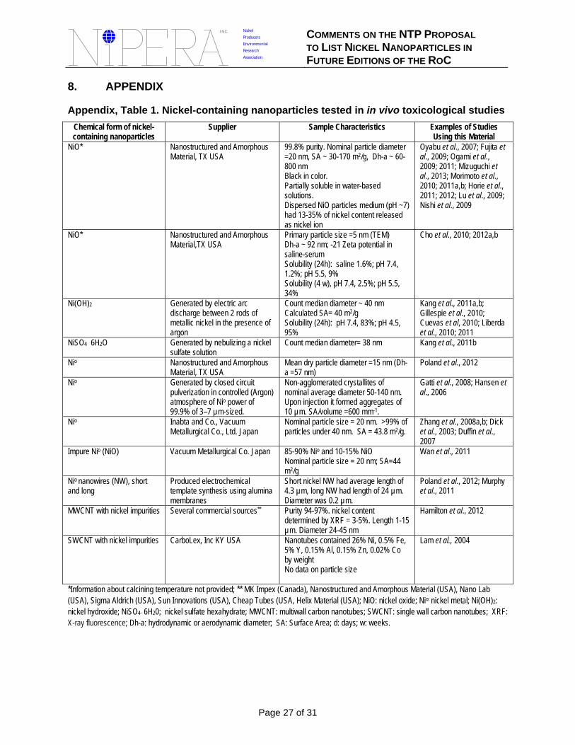
INC. Nickel
Producers COMMENTS ON THE NTP PROPOSAL Environmental Research TO LIST NICKEL NANOPARTICLES IN Association FUTURE EDITIONS OF THE ROC
8. APPENDIX
Appendix, Table 1. Nickel-containing nanoparticles tested in in vivo toxicological studies
Chemical form of nickel-containing nanoparticles
Supplier Sample Characteristics Examples of Studies Using this Material
NiO* Nanostructured and Amorphous Material, TX USA
99.8% purity. Nominal particle diameter =20 nm, SA ~ 30-170 m2/g, Dh-a ~ 60800 nm Black in color. Partially soluble in water-based solutions. Dispersed NiO particles medium (pH ~7) had 13-35% of nickel content released as nickel ion
Oyabu et al., 2007; Fujita et al., 2009; Ogami et al., 2009; 2011; Mizuguchi et al., 2013; Morimoto et al., 2010; 2011a,b; Horie et al., 2011; 2012; Lu et al., 2009; Nishi et al., 2009
NiO* Nanostructured and Amorphous Material,TX USA
Primary particle size =5 nm (TEM) Dh-a ~ 92 nm; -21 Zeta potential in saline-serum Solubility (24h): saline 1.6%; pH 7.4, 1.2%; pH 5.5, 9% Solubility (4 w), pH 7.4, 2.5%; pH 5.5, 34%
Cho et al., 2010; 2012a,b
Ni(OH)2 Generated by electric arc discharge between 2 rods of metallic nickel in the presence of argon
Count median diameter ~ 40 nm Calculated SA= 40 m2/g Solubility (24h): pH 7.4, 83%; pH 4.5, 95%
Kang et al., 2011a,b; Gillespie et al., 2010; Cuevas et al, 2010; Liberda et al., 2010; 2011
NiSO4 6H2O Generated by nebulizing a nickel sulfate solution
Count median diameter= 38 nm Kang et al., 2011b
Nio Nanostructured and Amorphous Material, TX USA
Mean dry particle diameter =15 nm (Dha =57 nm)
Poland et al., 2012
Nio Generated by closed circuit pulverization in controlled (Argon) atmosphere of Nio power of 99.9% of 3–7 µm-sized.
Non-agglomerated crystallites of nominal average diameter 50-140 nm. Upon injection it formed aggregates of 10 µm. SA/volume =600 mm-1.
Gatti et al., 2008; Hansen et al., 2006
Nio Inabta and Co., Vacuum Metallurgical Co., Ltd. Japan
Nominal particle size = 20 nm. >99% of particles under 40 nm. SA = 43.8 m2/g.
Zhang et al., 2008a,b; Dick et al., 2003; Duffin et al., 2007
Impure Nio (NiO) Vacuum Metallurgical Co. Japan 85-90% Nio and 10-15% NiO Nominal particle size = 20 nm; SA=44 m2/g
Wan et al., 2011
Nio nanowires (NW), short and long
Produced electrochemical template synthesis using alumina membranes
Short nickel NW had average length of 4.3 µm, long NW had length of 24 µm. Diameter was 0.2 µm.
Poland et al., 2012; Murphy et al., 2011
MWCNT with nickel impurities Several commercial sources** Purity 94-97%. nickel content determined by XRF = 3-5%. Length 1-15 µm. Diameter 24-45 nm
Hamilton et al., 2012
SWCNT with nickel impurities CarboLex, Inc KY USA Nanotubes contained 26% Ni, 0.5% Fe, 5% Y, 0.15% Al, 0.15% Zn, 0.02% Co by weight No data on particle size
Lam et al., 2004
*Information about calcining temperature not provided; ** MK Impex (Canada), Nanostructured and Amorphous Material (USA), Nano Lab (USA), Sigma Aldrich (USA), Sun Innovations (USA), Cheap Tubes (USA, Helix Material (USA); NiO: nickel oxide; Nio: nickel metal; Ni(OH)2: nickel hydroxide; NiSO4· 6H20; nickel sulfate hexahydrate; MWCNT: multiwall carbon nanotubes; SWCNT: single wall carbon nanotubes; XRF: X-ray fluorescence; Dh-a: hydrodynamic or aerodynamic diameter; SA: Surface Area; d: days; w: weeks.
Page 27 of 31
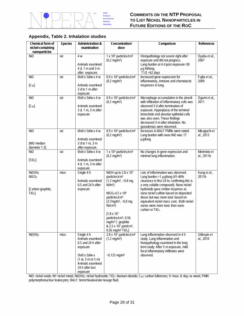
INC. Nickel
Producers COMMENTS ON THE NTP PROPOSAL Environmental Research TO LIST NICKEL NANOPARTICLES IN Association FUTURE EDITIONS OF THE ROC
Appendix, Table 2. Inhalation studies
Chemical form of nickel-containing
nanoparticles
Species Administration & examination
Concentration/ dose
Comparison References
NiO rat 4 w
Animals examined 4 d, 1 m and 3 m after exposure
1 x 10⁵ particles/cm3
(0.2 mg/m3) Histopathology not severe right after exposure and did not progress. Lung burden at 4 d post exposure=30 µg Ni/lung. T1/2 =62 days
Oyabu et al., 2007
NiO rat 6h/d x 5d/w x 4 w 0.9 x 10⁵ particles/cm3
(0.2 mg/m3) Increased gene expression for inflammatory, immune and chemotactic
Fujita et al., 2009
[C60] Animals examined 3 d to 1 m after exposure
responses in lung.
NiO rat 6h/d x 5d/w x 4 w 0.9 x 10⁵ particles/cm3
(0.2 mg/m3) Macrophage accumulation in the alveoli with infiltration of inflammatory cells was
Ogami et al., 2011
[C60] Animals examined 3 d, 1 m, 3 m after exposure
observed 3 d after termination of exposure. Hyperplasia of the terminal bronchiole and alveolar epithelial cells was also seen. These findings decreased 3 m after inhalation. No granulomas were observed.
NiO rat 6h/d x 5d/w x 4 w
Animals examined
0.9 x 10⁵ particles/cm3
(0.2 mg/m3) Increases in BALF PMNs were noted. Lung burden with nano NiO was 17 µg/lung.
Mizuguchi et al., 2013
[NiO median 3 d to 1 m, 3 m diameter 1.3] after exposure. NiO rat 6h/d x 5d/w x 4 w 1 x 10⁵ particles/cm3 No changes in gene expression and Morimoto et
(0.2 mg/m3) minimal lung inflammation. al., 2011b [TiO2] Animals examined
4 d, 1 m, 3 m after exposure
Ni(OH)2
NiSO4
[Carbon graphite, TiO2]
mice Single 4 h
Animals examined 0.5 and 24 h after exposure
NiOH up to 2.8 x 10⁷
particles/cm3
(1.2 mg/m3, ~0.8 mg Ni/m3)
NiSO4 4.5 x 10⁶
particles/cm3
(3.7mg/m3, ~0.8 mg Ni/cm3)
[1.8 x 10⁷
particles/cm3, 0.56 mg/m3 C graphite & 2.5 x 10⁷ part/cm3, 0.56 mg/m3 TiO2]
Lots of inflammation was observed. Lung burden =1 µg/lung (41-46% clearance in first 24 hs confirming this is a very soluble compound). Nano nickel hydroxide gave similar response as nano nickel sulfate based on deposited doses but was more toxic based on equivalent nickel mass conc. Both nickel nanos were more toxic than nano carbon or TiO2.
Kang et al., 2011b
Ni(OH)2 mice Single 4 h Animals examined 0.5 and 24 h after exposure
5h/d x 5d/w x (1 w, 3 m or 5 m) Animals examined 24 h after last exposure
2.8 x 10⁷ particles/cm3
(1.2 mg/m3)
~0.125 mg/m3
Lung inflammation observed in 4 h study. Lung inflammation and histopathology examined in the long term study. After 5 m exposure, mild focal inflammatory infiltrates were observed.
Gillespie et al., 2010
NiO: nickel oxide; Nio: nickel metal; Ni(OH)2: nickel hydroxide; TiO2: titanium dioxide; C60: carbon fullerenes; h: hour; d: day; w: week; PMN: polymorphonuclear leukocytes; BALF: bronchioalaveolar lavage fluid.
Page 28 of 31
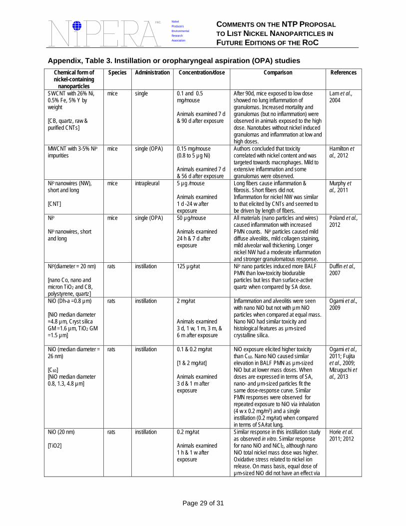
INC. Nickel
Producers COMMENTS ON THE NTP PROPOSAL Environmental Research TO LIST NICKEL NANOPARTICLES IN Association FUTURE EDITIONS OF THE ROC
Appendix, Table 3. Instillation or oropharyngeal aspiration (OPA) studies
Chemical form of nickel-containing
nanoparticles
Species Administration Concentration/dose Comparison References
SWCNT with 26% Ni, 0.5% Fe, 5% Y by weight
[CB, quartz, raw & purified CNTs]
mice single 0.1 and 0.5 mg/mouse
Animals examined 7 d & 90 d after exposure
After 90d, mice exposed to low dose showed no lung inflammation of granulomas. Increased mortality and granulomas (but no inflammation) were observed in animals exposed to the high dose. Nanotubes without nickel induced granulomas and inflammation at low and high doses.
Lam et al., 2004
MWCNT with 3-5% Nio mice single (OPA) 0.15 mg/mouse Authors concluded that toxicity Hamilton et impurities (0.8 to 5 µg Ni) correlated with nickel content and was
targeted towards macrophages. Mild to al., 2012
Animals examined 7 d extensive inflammation and some & 56 d after exposure granulomas were observed.
Nio nanowires (NW), short and long
[CNT]
mice intrapleural 5 µg /mouse
Animals examined 1 d -24 w after exposure
Long fibers cause inflammation & fibrosis. Short fibers did not. Inflammation for nickel NW was similar to that elicited by CNTs and seemed to be driven by length of fibers.
Murphy et al., 2011
Nio
Nio nanowires, short and long
mice single (OPA) 50 µg/mouse
Animals examined 24 h & 7 d after exposure
All materials (nano particles and wires) caused inflammation with increased PMN counts. Nio particles caused mild diffuse alveolitis, mild collagen staining, mild alveolar wall thickening. Longer nickel NW had a moderate inflammation and stronger granulomatous response.
Poland et al., 2012
Nio(diameter = 20 nm) rats instillation 125 µg/rat Nio nano particles induced more BALF PMN than low-toxicity biodurable
Duffin et al., 2007
[nano Co, nano and particles but less than surface-active micron TiO2 and CB, quartz when compared by SA dose. polystyrene, quartz] NiO (Dh-a =0.8 µm)
[NiO median diameter =4.8 µm, Cryst silica GM =1.6 µm, TiO2 GM =1.5 µm]
rats instillation 2 mg/rat
Animals examined 3 d, 1 w, 1 m, 3 m, & 6 m after exposure
Inflammation and alveolitis were seen with nano NiO but not with µm NiO particles when compared at equal mass. Nano NiO had similar toxicity and histological features as µm-sized crystalline silica.
Ogami et al., 2009
NiO (median diameter = 26 nm)
[C60] [NiO median diameter 0.8, 1.3, 4.8 µm]
rats instillation 0.1 & 0.2 mg/rat
[1 & 2 mg/rat]
Animals examined 3 d & 1 m after exposure
NiO exposure elicited higher toxicity than C60. Nano NiO caused similar elevation in BALF PMN as µm-sized NiO but at lower mass doses. When doses are expressed in terms of SA, nano- and µm-sized particles fit the same dose-response curve. Similar PMN responses were observed for repeated exposure to NiO via inhalation (4 w x 0.2 mg/m3) and a single instillation (0.2 mg/rat) when compared in terms of SA/rat lung.
Ogami et al., 2011; Fujita et al., 2009; Mizuguchi et al., 2013
NiO (20 nm) rats instillation 0.2 mg/rat Similar response in this instillation study as observed in vitro. Similar response
Horie et al. 2011; 2012
[TiO2] Animals examined 1 h & 1 w after exposure
for nano NiO and NiCl2, although nano NiO total nickel mass dose was higher. Oxidative stress related to nickel ion release. On mass basis, equal dose of µm-sized NiO did not have an effect via
Page 29 of 31
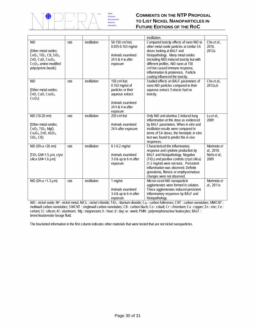
INC. Nickel
Producers COMMENTS ON THE NTP PROPOSAL Environmental Research TO LIST NICKEL NANOPARTICLES IN Association FUTURE EDITIONS OF THE ROC
instillation. NiO
[Other metal oxides: CeO2, TiO2, CB, SiO2, ZnO, CuO, Co3O4, Cr2O3, amine-modified polystyrene beads]
rats instillation 50-150 cm2/rat; 0.055-0.163 mg/rat
Animals examined 24 h & 4 w after exposure
Compared toxicity effects of nano NiO to other metal oxide particles at similar SA doses looking at BALF and histopathology. Many metal oxides (including NiO) induced toxicity but with different profiles. NiO nano at 150 cm2/rat caused immune response, inflammation & proteinosis. Particle coating influenced the toxicity.
Cho et al., 2010, 2012a
NiO
[Other metal oxides: ZnO, CuO, Co3O4, Cr2O3]
rats instillation 150 cm2/rat; 0.163 mg/rat of particles or their aqueous extract
Animals examined 24 h & 4 w after exposure
Studied effects on BALF parameters of nano NiO particles compared to their aqueous extract. Extracts had no toxicity.
Cho et al., 2012a,b
NiO (10-20 nm) rats instillation 250 cm2/rat Only NiO and alumina 2 induced lung inflammation at this dose as evidenced
Lu et al., 2009
[Other metal oxides: Animals examined by BALF parameters. When in vitro and CeO2, TiO2, MgO, 24 h after exposure instillation results were compared in Co3O4, ZnO, Al2O3, terms of SA doses, the hemolytic in vitro SiO2, CB] test was found to predict the in vivo
responses. NiO (Dh-a =26 nm) rats instillation 0.1-0.2 mg/rat Characterized the inflammatory
response and cytokine production by Morimoto et al., 2010;
[TiO2 GM=1.5 µm, cryst Animals examined BALF and histopathology. Negative Nishi et al., silica GM=1.6 µm] 3 d & up to 6 m after
exposure (TiO2) and positive controls (cryst silica) (1-2 mg/rat) were not toxic. Persistent inflammation was observed. Definite granuloma, fibrosis or emphysematous changes were not observed.
2009
NiO (Dh-a =1.3 µm) rats instillation 1 mg/rat
Animals examined 3 d & up to 6 m after exposure
Micron-sized NiO nanoparticle agglomerates were formed in solution. These agglomerates induced persistent inflammatory responses by BALF and histopathology.
Morimoto et al., 2011a
NiO : nickel oxide; Nio : nickel metal; NiCl2 : nickel chloride; TiO2 : titanium dioxide; C60 : carbon fullerenes; CNT : carbon nanotubes; MWCNT : multiwall carbon nanotubes; SWCNT : singlewall carbon nanotubes; CB : carbon black; Co : cobalt; Cr : chromium; Cu : copper; Zn : zinc; Ce : cerium; Si : silicon; Al : aluminum; Mg : magnesium; h : hour; d : day; w : week; PMN : polymorphonuclear leukocytes; BALF : bronchioalaveolar lavage fluid.
The bracketed information in the first column indicates other materials that were tested that are not nickel nanoparticles.
Page 30 of 31
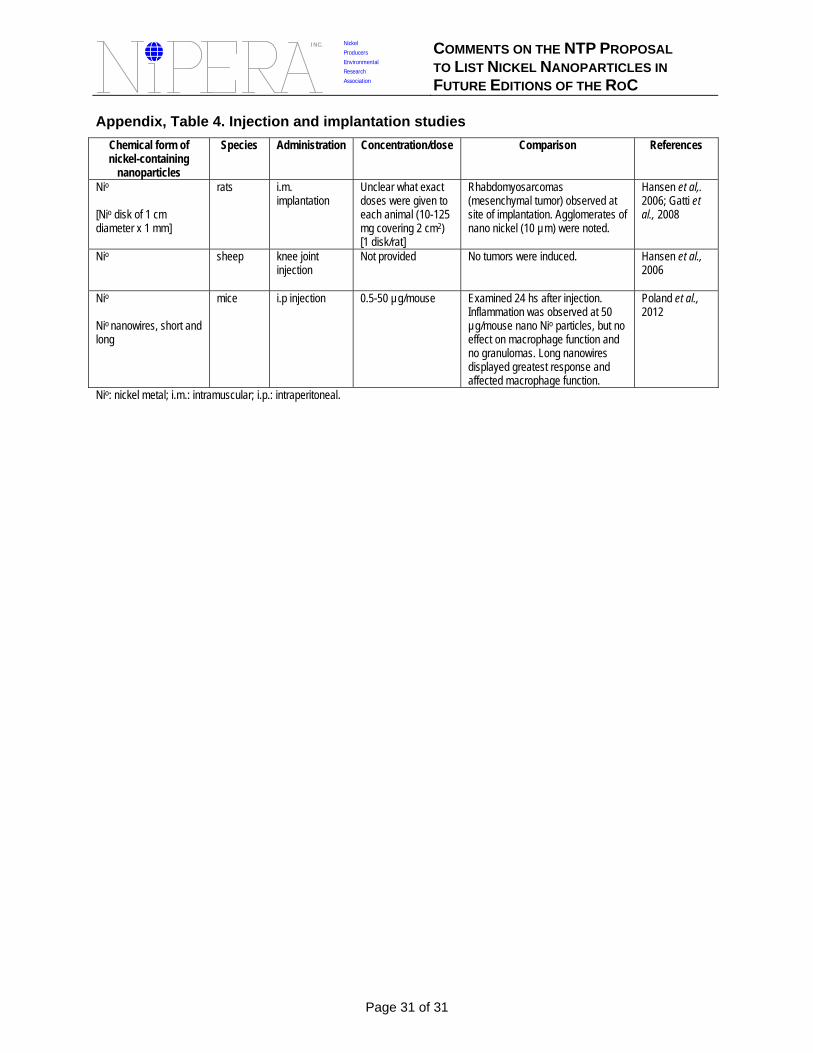
INC. Nickel
Producers COMMENTS ON THE NTP PROPOSAL Environmental Research TO LIST NICKEL NANOPARTICLES IN Association FUTURE EDITIONS OF THE ROC
Appendix, Table 4. Injection and implantation studies
Chemical form of nickel-containing
nanoparticles
Species Administration Concentration/dose Comparison References
Nio
[Nio disk of 1 cm diameter x 1 mm]
rats i.m. implantation
Unclear what exact doses were given to each animal (10-125 mg covering 2 cm2) [1 disk/rat]
Rhabdomyosarcomas (mesenchymal tumor) observed at site of implantation. Agglomerates of nano nickel (10 µm) were noted.
Hansen et al,. 2006; Gatti et al., 2008
Nio sheep knee joint injection
Not provided No tumors were induced. Hansen et al., 2006
Nio
Nio nanowires, short and long
mice i.p injection 0.5-50 µg/mouse Examined 24 hs after injection. Inflammation was observed at 50 µg/mouse nano Nio particles, but no effect on macrophage function and no granulomas. Long nanowires displayed greatest response and affected macrophage function.
Poland et al., 2012
Nio: nickel metal; i.m.: intramuscular; i.p.: intraperitoneal.
Page 31 of 31


