Page 1
8/6/2019 Nitric Oxide Cancer
http://slidepdf.com/reader/full/nitric-oxide-cancer 1/10
Cell Research (2002); 12(5-6):311-320http://www.cell-research.com
The role of nitric oxide in cancer
W EIMING XU1,*, LI ZHI LIU1, M ARILENA LOIZIDOU2, MOHAMED AHMED1, I AN G CHARLES1
1 Wolfson Institute for Biomedical Research, Cruciform Building, Gower Street, UCL, London, WC1 E 6AU, UK 2 Department of Surgery, Charles Bell House, 67-73 Riding House Street, UCL, London W1W 7EJ, UK
ABSTRACT
Nitric oxide (NO) is a pleiotropic regulator, critical to numerous biological processes, including vasodilatation,
neurotransmission and macrophage-mediated immunity. The family of nitric oxide synthases (NOS) com-
prises inducible NOS (iNOS), endothelial NOS (eNOS), and neuronal NOS (nNOS). Interestingly, various
studies have shown that all three isoforms can be involved in promoting or inhibiting the etiology of cancer.
NOS activity has been detected in tumour cells of various histogenetic origins and has been associated with
tumour grade, proliferation rate and expression of important signaling components associated with cancerdevelopment such as the oestrogen receptor. It appears that high levels of NOS expression (for example,
generated by activated macrophages) may be cytostatic or cytotoxic for tumor cells, whereas low level activ-
ity can have the opposite effect and promote tumour growth. Paradoxically therefore, NO (and related
reactive nitrogen species) may have both genotoxic and angiogenic properties. Increased NO-generation in
a cell may select mutant p53 cells and contribute to tumour angiogenesis by upregulating VEGF. In addition,
NO may modulate tumour DNA repair mechanisms by upregulating p53, poly(ADP-ribose) polymerase
(PARP) and the DNA-dependent protein kinase (DNA-PK). An understanding at the molecular level of the
role of NO in cancer will have profound therapeutic implications for the diagnosis and treatment of disease.
Key words: nitric oxide, oestrogen, cancer, p53, PARP, DNA-PKcs.
* Corresponding author: Dr. Weiming XU, Wolfson Institute
for Biomedical Research. University College London, Cruciform
Building, Gower Street, London WC1E 6AU.UK.
Fax: 44207813 2846 Tel: 442076796209
Email: [email protected] . Abbreviations: NO, nitric oxide; NOS, nitric oxide synthase;
iNOS, inducible nitric oxide synthase; eNOS, endothelial nitric
oxide synthase; nNOS, neuronal nitric oxide synthase; PARP,
poly(ADP-ribose) polymerase; DNA-PK, DNA-dependent pro-
tein kinase; DNA-PKcs, catalytic subunit of DNA-PK; BH4,
tetrahydrobiopterin; FAD, flavin adenosine dinucleotide; FMN,
flavin mononucleotide; sGC, soluble guanylate cyclase; VEGF,
vascular endothelial growth factor.
REVIEW
INTRODUCTION
Over the past decade or so, it has become evident
that the free radical gas nitric oxide (NO) acts as a
novel transcellular messenger molecule in many key
physiological and pathological processes[1]. NO plays
a central role in the cardiovascular system as the
endothelium - derived relaxing factor[2-5]. Within
the central nervous system, NO is a crucial compo-
nent of the signal transduction pathways used for
memory formation, sensory processing, and the
regulation of cerebral blood flow[6]. Interestingly,
as early as 1982, NO was implicated in the immuno-
defence network, as marked increase in urinary NO3
-
excretion was observed in human subjects with diar-
rhoea and fever[7], [8]. Further work showed that
the blood levels and urinary excretion of NO3
- in-
creased after exposure to Escherichia coli lipopolysac-
charide (LPS) in LPS-sensitive mice and that acti-
Page 2
8/6/2019 Nitric Oxide Cancer
http://slidepdf.com/reader/full/nitric-oxide-cancer 2/10
312
vated mouse peritoneal macrophages showed in-
creased NO2
- and NO3
- production in vitro[9]. The
mammalian immuno -defense network is involved
in tumour suppression, and macrophages are an im-
portant part of this process because of their abilityto destroy selectively a broad range of tumour types
upon specific activation. The role of NO in macroph-
age cytotoxicity was first described by Hibbs and
colleagues in 1987[10], and since that time numer-
ous studies have shown that cytokine activated ro-
dent macrophages can generate large concentrations
of NO by up-regulation of expression of the induc-
ible nitric oxide synthase gene (iNOS)[11]. The NO
generated by this process is capable of killing a range
of tumour cells of differing origin and grade[11-15].
Various direct and indirect mechanisms have beenproposed for the anti-tumour properties of NO.
Mechanisms include direct damage of DNA, inhibi-
tion of DNA synthesis and inhibition of the rate-
limiting enzyme ribonucleotide reductase. Reduced
activity of cis-aconitase and loss of a large fraction of
the iron pool, have also been suggested as possible
mechanisms. Importantly, NO-generation can efect
mitochondrial physiology leading to reduction of O2
consumption and damage to complexes I and II in
the mitochondrial electron transport chain, revers-
ible inhibition of complex IV activity and inductionof apoptosis[10-15].
Importantly, various studies have shown that all
three isoforms of NOS, (iNOS, eNOS and nNOS),
have been detected in tumour cells from a wide range
of isolates[16-18]. NOS activity has been observed
in human tumour cell lines and cells from tumour
biopsies. However, the precise function(s) of NO in
tumour biology remains unclear, and several lines
of research have indicated that NO may have dual
effects in cancer. In this review, we will present
some recent evidence on both the pro- and anti-tu-mour activities of NO and discuss the implications
of these data on the use of NO as a therapeutic agent
for the treatment of cancer.
Nitric oxide generation and its biologicaltargets
NO is a diatomic free radical molecule, and is a gas
at room temperature. Within mammalian cells a fam-
ily of NOS enzymes has been shown to be able to
The role of nitric oxide in cancer
generate NO, and all family members require a panel
of substrates and co-factors to be fully functional.
For example, the NO-generating reaction requires
L-arginine, NADPH and oxygen as substrates, and
tetrahydrobiopterin (BH4), thiol, flavin adenine di-nucleotide (FAD), and flavin mononucleotide (FMN)
as cofactors. In addition to NO, the NOS-catalysed
reaction produces citrulline and NADP as co-
products.
Three different isoforms of the NOS family have
been identified; endothelial NOS (eNOS), neuronal
NOS (nNOS) and inducible NOS (iNOS). The gene
symbol nomenclatures are: NOS1 for nNOS, NOS2
for iNOS and NOS3 for eNOS[6]. The nNOS and
eNOS isoforms are constitutively expressed in a va-
riety of cell types including the endothelium, platelets,and neurons. Typically, the constitutive NOS
isoforms can be activated as a result of calmodulin
(CaM) binding following a rise in intracellular
calcium. They may also be activated and/or inhib-
ited by phosphorylation via various protein kinases.
Unlike nNOS and eNOS, iNOS displays a high af-
finity for CaM, which is tightly bound within physi-
ological concentrations of calcium. As a consequence
of this, the activation of iNOS is not calcium-
dependent. However, the expression of iNOS can
be transcriptionally regulated by factors such ascytokines (e.g. interferon- (IFN- ), interleukin-1
(IL-1 ) and tumour necrosis factor- (TNF- ),
bacterial endotoxin (LPS) and oxidative stress (e.g.
under conditions encountered during hypoxia).
A good starting point to assess the function of
NO in mammalian physiology is to examine its
chemical properties. NO is a gas at room and body
temperature, making it highly diffusible within the
vasculature. As NO is a free radical, it is a highly
reactive molecule within biological systems, react-
ing with other free radicals, molecular oxygen andheavy metals. It has been suggested that the biologi-
cal effects of NO can be mediated by the products of
different NO metabolites. For example, NO can re-
act rapidly in the intracellular environment to form
nitrite and nitrate, S-nitroso-thiols or peroxynitrite.
These metabolites may play a key role in mediating
many of the key genotoxic effects, (such as DNA
damage), that are associated with the generation of
NO. Importantly, NO has been shown to bind
Page 3
8/6/2019 Nitric Oxide Cancer
http://slidepdf.com/reader/full/nitric-oxide-cancer 3/10
313
Wei Ming XU et al
rapidly, and with high affinity, to ferrous iron (Fe2+).
As a consequence of this, NO can bind easily to free
iron, iron within iron-sulphur centres, and iron
within haemoproteins (especially when the haem
contains a free ligand position). Many of the biologi-cal processes described for NO (involving smooth
muscle relaxation, neurotransmission, and inhibi-
tion of platelet aggregation and adhesion) results
from NO binding to the ferrous haem iron of the
enzyme soluble guanylate cyclase (sGC), which in
turn results in an increase in cGMP production. Due
its association with haem centres it is not surprising
that the binding of NO to haemoglobin is regarded
as a significant route by which NO can be broken
down in the body.
NO can cause DNA damage via the generation of peroxynitrite (ONOO-) and N2O3. Peroxynitrite can
oxidise and nitrate DNA and may potentially cause
single-strand DNA breaks through attack on the
sugar-phosphate backbone. N2O3 can nitrosate
amines to form N-nitrosamines, then alkylate DNA.
Nitrosation of primary amines, (e.g. in DNA bases)
leads to the formation of diazonium ions and subse-
quent deamination and DNA-crosslinks. The wide
range of differing biological effects arising from ex-
posure to NO is very much dependent upon many
factors, such as formation and metabolism of NO,the type of NOS enzymes that are present, the in-
teraction between NO utilising processes, and cru-
cially the concentration of NO that is present in the
given system.
Nitric oxide synthase expression intumours
The iNOS isoform has been mostly studied for its
role in immuno-mediated processes, for example,
iNOS knock-out mice have been generated and
shown to have increased susceptibility to infections
[18]. The first NOS isoform implicated in the mac-
rophage-mediated tumour killing process was also
iNOS, and as a consequence this isoform has been at
the center of attention for study of its expression incancer. In normal (non-tumourogenic) cells, iNOS
has been detected in macrophages and neutrophils,
as well as in hepatocytes, cardiac myocytes,
chondrocytes and many other cell types. Recently,
lack of iNOS in knock-out mice has been found to
promote intestinal tumorigenesis in the Apc(Min/+)
colon cancer mouse model, thereby substantiating
the role of iNOS within host defence mechanisms[19].
An initial study on iNOS expression in human
breast cancer suggested that iNOS activity was higher
in less differentiated tumours in a panel of 15 inva-sive breast carcinomas[17]. iNOS expression could
be detected predominantly in peritumoural and
intratumoural macrophages. NO biosynthesis was sig-
nificantly greater for grade III tumours as compared
with grade II in specimens from 10 breast cancer
tissues. Recently, three relatively large scale studies
[20-22], suggested that iNOS is not only expressed
in stromal cells and macrophages in the tumour, but
also in tumour cells themselves (Tab 1). Reveneau et
al reported NOS activity in 27 of 40 tumours studied
[20]. Vakkala et al showed that carcinomas with bothiNOS positive tumour and stromal cells had a higher
apoptotic index and a higher calculated microvessel
density index[21]. Loibl et al further demonstrated
that while none of the benign lesions were positive
for iNOS, 67% in situ carcinomas and 61% invasive
lesions showed iNOS tumour cell staining. eNOS ex-
pression was found in 33 invasive lesions (61%)[22].
Both iNOS expressing lesions and eNOS expressing
lesions showed strong co-expression (p=0.0008).
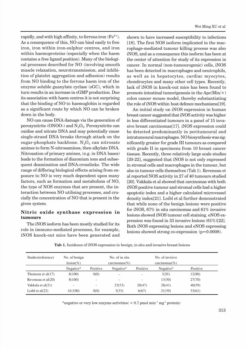
Studies(reference) No. of benign No. of in situ No. of invisive
lesion(%) carcinomas(%) carcinomas(%)
Negative* Positive Negative* Positive Negative* Positive
Thomsen et al(17) 8(100) 0(0) - - 3(20) 12(80)
Reveneau et al(20) 8(100) - - - 13(30) 27(70)
Vakkala et al(21) - - 23(53) 20(47) 28(41) 40(59)
Loibl et al(22) 41(100) 0(0) 3(33) 6(67) 21(39) 33(61)
*negative or very low enzyme activities( < 0.7 pmol min-1 mg -1 protein)
Tab 1. Incidence of iNOS expression in benign, in situ and invasive breast lesions
Page 4
8/6/2019 Nitric Oxide Cancer
http://slidepdf.com/reader/full/nitric-oxide-cancer 4/10
314
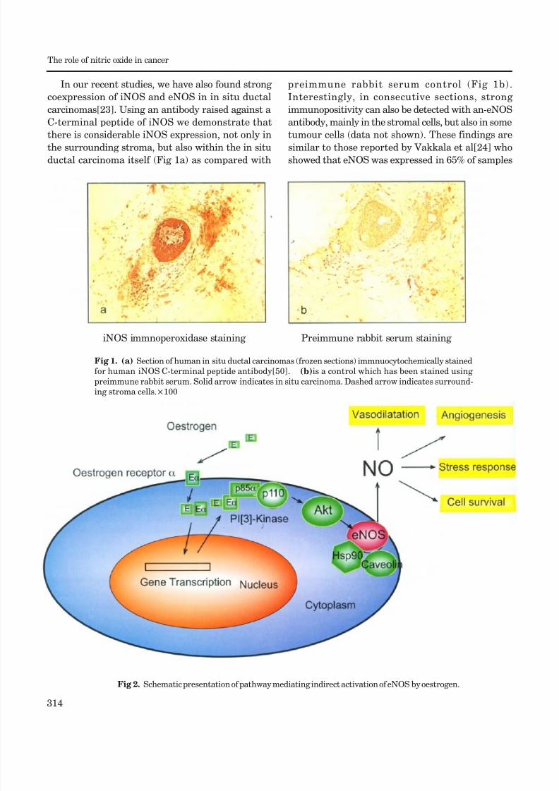
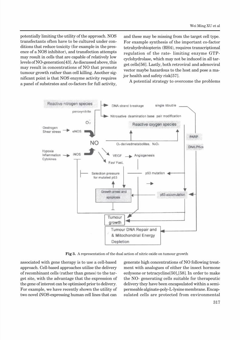
iNOS immnoperoxidase staining Preimmune rabbit serum staining
In our recent studies, we have also found strong
coexpression of iNOS and eNOS in in situ ductal
carcinomas[23]. Using an antibody raised against a
C-terminal peptide of iNOS we demonstrate that
there is considerable iNOS expression, not only inthe surrounding stroma, but also within the in situ
ductal carcinoma itself (Fig 1a) as compared with
preimmune rabbit serum control (Fig 1b).
Interestingly, in consecutive sections, strong
immunopositivity can also be detected with an-eNOS
antibody, mainly in the stromal cells, but also in some
tumour cells (data not shown). These findings aresimilar to those reported by Vakkala et al[24] who
showed that eNOS was expressed in 65% of samples
Fig 1. (a) Section of human in situ ductal carcinomas (frozen sections) immnuocytochemically stained
for human iNOS C-terminal peptide antibody[50]. (b)is a control which has been stained using
preimmune rabbit serum. Solid arrow indicates in situ carcinoma. Dashed arrow indicates surround-
ing stroma cells.×100
Fig 2. Schematic presentation of pathway mediating indirect activation of eNOS by oestrogen.
The role of nitric oxide in cancer
Page 5
8/6/2019 Nitric Oxide Cancer
http://slidepdf.com/reader/full/nitric-oxide-cancer 5/10
315
(80 cases in total) with immunopositivity seen both
in stromal structures and carcinoma cells[24].
Interestingly, Martin et al recently reported a sig-
nificant positive correlation between the percent-
age of tumour cells with eNOS expression and oestro-gen receptors[25]. We have also observed that some
eNOS positive in situ ductal carcinomas show posi-
tive staining with antibody to estrogen receptors (Xu
et al unpublished data). Recently several studies have
shown that the oestrogen/oestrogen receptor com-
plex binds to the p85a regulatory subunit of
phosphatidylinositol-3-OH kinase (PI-(3)K), which
leads to activation of protein kinase B/Akt [26], [27].
It is known that the catalytic activity of eNOS is aug-
mented by phosphorylation of a C-terminal serine
residue (Ser-1177 of human eNOS) through the PI-(3)K/Akt pathway[28],[29]. Therefore, it is possible
that oestrogen acting on the oestrogen receptor (ER σ )
located on the surface of cell membranes, could in-
directly activate the release of NO from membrane
bound eNOS (Fig 2). It is possible that the NO gen-
erated from this activation of eNOS may contribute
significantly to tumour cell survival under hypoxia
and other stress conditions. It should be noted that
there are complicated signalling network(s) in en-
dothelial cells capable of regulating eNOS activity.
For example, the Akt-mediated phosphorylation ac-tivity can be enhanced by binding to heat shock pro-
tein-90[30] and inhibited by binding to Caveolin-1
[31][Fig 2].
In addition to breast cancer, iNOS has also been
shown to be markedly expressed in approximately
60% of human adenomas and in 20-25% of colon
carcinomas, while expression was either low or ab-
sent in the surrounding normal tissues[32], [33]. In
human ovarian cancer, iNOS activity has been local-
ized in tumour cells and not found in normal tissue
[16]. Other tumours that have demonstrated iNOSgene expression are brain[1][34], head and neck[35],
esophagus[36], lung[37], prostate[38], bladder[39],
pancreatic[40], and Kaposi s sarcoma[41].
In the central nerve system, NO has a variety of
biological functions including vasorelaxation and
neurotransmision. Interestingly, nNOS has been
detected in some oligodendroglioma and neuroblas-
toma cell lines, althouth further studies are needed
to clarified the role of nNOS in tumour pathology
[34].
NO and tumour cell angiogenesis
While NO had been shown to have anti-tumour p
roperties[10], Jenkins et al[42][1995] first reporte
d the surprising finding that human carcinoma cells
transfected with a murine iNOS cDNA cassette (DLD-1 cells generating 20 pmol min-1 mg-1 NOS activ
ity) showed increased tumour growth, rather than d
ecreased growth. Using a nude mouse/xanograft mo
del it was shown that growth of these NO-generatin
g tumours was accompanied by increased neovascul
arization. These results were supported by Ambs et
al, who used recombinant iNOS expressing Calu-6 a
nd HT-29 human carcinoma cell lines containing m
utant p53[43] to look at tumour growth. The author
s demonstrated that an NO-mediated up-regulation
of VEGF corresponded with increased vascularisation in the xenograft tumours. Therefore it is possibl
e that NO generated by NOS (located either within t
he tumour or in the surrounding stroma) may prom
ote new blood vessel formation by up-regulating VE
GF. This neovasculaturization not only enhances th
e ability of the tumour to grow, but also increases it
s invasiveness and metastatic ability.
NO, p53, PARP and DNA-PKcs in DNA re-pair
As NO is a free radical, it is a highly reactive
molecule within biological systems, capable of inter-
action with other free radicals, molecular oxygen and
heavy metals. The biological effects of NO can be
mediated by the products of different NO
metabolites. For example, NO rapidly reacts intrac-
ellularly to form nitrite and nitrate, S-nitroso-thiols
or peroxynitrate, and these metabolites are believed
to play key roles in mediating many of the NO-asso-
ciated genotoxic effects. These effects include DNA
damage, which can be initiated by nitrosative
deamination, DNA strand breakage or DNA modifi-
cation[44].
One of the consequences of the NO- mediated
DNA damage is to trigger p53 accumulation, which
can induce apoptosis. This is a possible process by
which NO may induce death of tumour cells. An in-
crease in NOS activity (arising from increased tran-
scriptional activity, or from post-transcriptional/pro-
tein regulation activity) in tumour cells can conse-
quently cause the concentration of NO to be elevated
Wei Ming XU et al
Page 6
8/6/2019 Nitric Oxide Cancer
http://slidepdf.com/reader/full/nitric-oxide-cancer 6/10
316
such that it triggers p53-mediated growth arrest and
apoptosis[45],[46]. Interestingly, it has been dem-
onstrated that accumulation of p53 results ultimately
in down-regulation of iNOS expression by inhibi-
tion of iNOS promoter activity[47]. Thus a negativefeedback loop is formed between NO-generation and
p53 accumulation, that may constitute part of a
physiological mechanism, which responds to endog-
enously produced DNA damage due to NO. Overall,
this p53-mediated growth inhibition may be expected
to provide a strong selection pressure for mutant p53
expression in tumor cells.
In addition to p53, NO has also been shown to
activate poly (ADP-ribose) polymerase (PARP)[48]
and it has been proposed that this activation is due
to DNA damage. This damage may take the form of DNA strand breaks or nitrosative deamination of
DNA bases when NO is generated at high
concentrations. These high concentrations of NO
have been reported for NMDA-mediated neurotox-
icity as well as for tumouricidal and bactericidal ac-
tivation of cells[44]. Another important DNA repair
enzyme, DNA-dependent protein kinase (DNA-PK),
is also known to be essential for the maintenance of
the structural integrity of the genome. DNA-PK is a
serine/threonine protein kinase consisting of a large
catalytic subunit (DNA-PKcs) and a regulatory sub-unit (Ku). Recently, mammalian DNA-PKcs has been
shown to be an essential component of the DNA
double-strand repair pathway, as well as being cru-
cial for V(D)J recombination, involved in the gen-
eration of immunoglobulin and T-cell diversity. Scid
mice, which lack DNA-PKcs, show increased sus-
ceptibility to ionising radiation in addition to having
impaired V(D)J recombination and arrested T- and
B-cell development[49]. Interestingly, although
DNA-PK activity cannot be up-regulated by strong
doses of radiation, we found that NO can act a signal,increasing the activity of DNA-PK. Importantly, we
showed that this increase occurred by transcriptional
up-regulation of DNA-PKcs expression and occurred
under physiologically relevant ranges of NO concen-
trations[50]. Biologically, this NO-mediated increase
in enzymatically active DNA-PK not only protected
cells from the toxic effects of NO, but also provided
cross-protection against clinically important DNA-
damaging agents, such as X-ray radiation,
adriamycin, bleomycin and cisplatin[50].
The NO-mediated increase in DNA-PKcs path-
way not only plays an important role in tumour DNA
repair[51], but may also play an important role in
other tissue damage processes which involve NO-
mediated stress[52],[53]. For example, failing myocardium, (advanced heart failure due to idio-
pathic dilated cardiomyopathy) undergoes active
DNA repair, where DNA-PKcs expression is strongly
correlated with iNOS expression (r=0.53, p < 0.01)
[53]. Given the fact that one of the major substrates
of DNA-PKcs is p53[54] and DNA-PKcs itself is
subjected to ADP-ribosylation by PARP, it is pos-
sible that NO-mediated DNA damage and repair
could play a significant role in tumour development
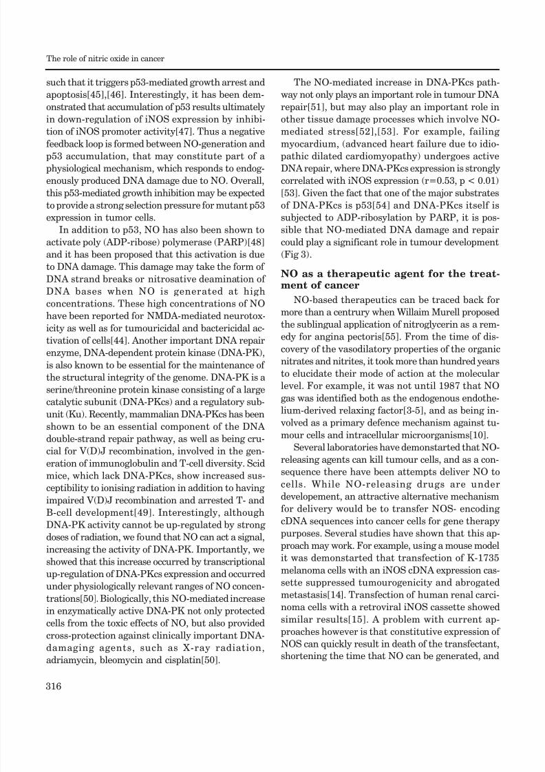
(Fig 3).
NO as a therapeutic agent for the treat-ment of cancer
NO-based therapeutics can be traced back for
more than a centrury when Willaim Murell proposed
the sublingual application of nitroglycerin as a rem-
edy for angina pectoris[55]. From the time of dis-
covery of the vasodilatory properties of the organic
nitrates and nitrites, it took more than hundred years
to elucidate their mode of action at the molecular
level. For example, it was not until 1987 that NO
gas was identified both as the endogenous endothe-
lium-derived relaxing factor[3-5], and as being in-
volved as a primary defence mechanism against tu-
mour cells and intracellular microorganisms[10].
Several laboratories have demonstarted that NO-
releasing agents can kill tumour cells, and as a con-
sequence there have been attempts deliver NO to
cells. While NO-releasing drugs are under
developement, an attractive alternative mechanism
for delivery would be to transfer NOS- encoding
cDNA sequences into cancer cells for gene therapy
purposes. Several studies have shown that this ap-
proach may work. For example, using a mouse model
it was demonstarted that transfection of K-1735
melanoma cells with an iNOS cDNA expression cas-
sette suppressed tumourogenicity and abrogated
metastasis[14]. Transfection of human renal carci-
noma cells with a retroviral iNOS cassette showed
similar results[15]. A problem with current ap-
proaches however is that constitutive expression of
NOS can quickly result in death of the transfectant,
shortening the time that NO can be generated, and
The role of nitric oxide in cancer
Page 7
8/6/2019 Nitric Oxide Cancer
http://slidepdf.com/reader/full/nitric-oxide-cancer 7/10
317
Fig 3. A representation of the dual action of nitric oxide on tumour growth
potentially limiting the utility of the approach. NOS
transfectants often have to be cultured under con-
ditions that reduce toxicity (for example in the pres-
ence of a NOS inhibitor), and transfection attempts
may result in cells that are capable of relatively low
levels of NO-generation[43]. As discussed above, this
may result in concentrations of NO that promote
tumour growth rather than cell killing. Another sig-
nificant point is that NOS enzyme activity requires
a panel of substrates and co-factors for full activity,
and these may be missing from the target cell type.
For example synthesis of the important co-factor
tetrahydrobiopterin (BH4), requires transcriptional
regulation of the rate- limiting enzyme GTP-
cyclohydrolase, which may not be induced in all tar-get cells[56]. Lastly, both retroviral and adenoviral
vector maybe hazardous to the host and pose a ma-
jor health and safety risk[57].
A potential strategy to overcome the problems
associated with gene therapy is to use a cell-based
approach. Cell-based approaches utilise the delivery
of recombinant cells (rather than genes) to the tar-
get site, with the advantage that the expression of
the gene of interest can be optimised prior to delivery.
For example, we have recently shown the utility of
two novel iNOS-expressing human cell lines that can
generate high concentrations of NO following treat-
ment with analogues of either the insect hormone
ecdysone or tetracycline[50],[58]. In order to make
the NO- generating cells suitable for therapeutic
delivery they have been encapsulated within a semi-
permeable alginate-poly-L-lysine membrane. Encap-
sulated cells are protected from environmental
Wei Ming XU et al
Page 8
8/6/2019 Nitric Oxide Cancer
http://slidepdf.com/reader/full/nitric-oxide-cancer 8/10
318
stresses encountered in the host (such as the host
immune response) and can be delivered to tumour
site(s) in a nude mouse model[58],[59]. Following
delivery, high concentrations of NO and reactive
nitrogen species can be generated by administra-tion of the appropriate inducer. This approach has
been very successful, and we have used it in a tu-
mour model showing 100% killing of SKOV-3
tumours and 54% killing of DLD-1 tumours[58].
Importantly, this strategy allowed the mechanism
of tumour killing to be determined as it was shown
that tumour killing was associated with concomitant
up-regulation of the Fas/FasL proteins. Overall we
believe that the cell-delivery approach addresses
some of the shortcomings of competing strategies and
has the potential to inhibit or kill many differenttypes of tumours from various histological origins[60].
CONCLUSION
The discovery of the generation of NO by mam-
malian tissues and the elucidation of some of its bio-
logical roles in cancer has thrown new light onto
many areas of tumour biology research. Although
initial findings suggested that the immune-cell gen-
erated NO is cytostatic or cytotoxic for tumour cells,
later findings have shown that NO can also possess
apparently contradictory activity leading to increasedtumour growth. NO can contribute to tumour angio-
genesis by upregulating VEGF and modulating tu-
mour DNA repair mechanism(s) by up-regulating
p53, PARP and DNA-PKcs. Overall, we can safely
say that NO is a Doubled-Edged Sword in cancer.
On the one hand, high concentrations of NO, (for
example, generated by activated macrophages) may
mediate cancer cell apoptosis and the inhibition of
cancer growth. On the other hand, at (relatively) low
concentrations of NO, (for example, at concentra-
tions measurable in many different types of clinicalcancer samples), tumour growth and proliferation is
promoted. The regulation of tumour growth by NO
represents an important new dimension in cancer
research. Further work into the precise mechanisms
of this process is required to help us develop new
therapeutic tools for the treatment of cancer.
ACKNOWLEDGMENTS
We would like to thank the Medical Research
Council (UK) for their support.
REFERENCES
[1] Moncada S, Palmer RM, Higgs EA. Nitric oxide: physiology,
pathophysiology, and pharmacology. Pharmacol Rev 1991;
43:109-42.
[2] Furchgott RF, Zawadzki JV. The obligatory role of endot-
helial cells in the relaxation of arterial smooth muscle by
acetylcholine. Nature 1980; 288:373-6.
[3] Ignarro LJ, Buga GM, Wood KS, Byrns RE, Chaudhuri G.
Endothelium-derived relaxing factor produced and re-
leased from the artery and vein is nitric oxide. Proc Natl
Acad Sci USA 1987; 84:9265-9.
[4] Katsuki S, Arnold W, Mittal C, Murad F. Stimulation of
guanylate cyclase by sodium nitroprusside, nitroglycerin
and nitric oxide in various tissue preparations and com-
parison to the effects of sodium and hydroxylamine. J
Cyclic Nucleotides Res 1977; 3:23-5.
[5] Palmer RM, Ferrige AG, Moncada S. Nitric oxide releaseaccounts for the biological activity of endothelium-de-
rived relaxing factor. Nature 1987; 327:524-6.
[6] Xu W, Liu L. Nitric Oxide: from a mysterious labile factor
to the molecule of the Nobel Prize. Recent progress in
nitric oxide research. Cell Research 1998; 8:251-8.
[7] Wagner DA, Young VR, Tannenbaum SR, Schultz DS, Deen
WM. Mammalian nitrate biochemistry: metabolism and
endogenous synthesis. IARC Sci Publ 1984; 57:247-53.
[8] Hegesh E, Shiloah J. Blood nitrates and infantile
methemoglobinemia. Clin Chim Acta 1982; 125:107-15.
[9] Stuehr DJ, Marletta MA. Mammalian nitrate biosynthesis:
mouse macrophages produce nitrite and nitrate in re-
sponse to Escherichia coli lipopolysaccharide. Proc Natl
Acad Sci USA 1985; 82:7738-42.
[10] Hibbs JB Jr, Taintor RR,Vavrin Z. Macrophage
cytotoxicity: role for L-arginine deiminase and imino
nitrogen oxidation to nitrite. Science 1987; 235:473-6.
[11] MacMicking J, Xie QW, Nathan C. Nitric oxide and mac-
rophage function. Annu Rev Immunol 1997; 15:323-50.
[12] Xu L, Xie K, Fidler IJ. Therapy of human ovarian cancer
by transfection with the murine interferon beta gene:
role of macrophage-inducible nitric oxide synthase. Hum
Gene Ther 1998; 9:2699-708.
[13] Garban HJ and Bonavida B. Nitric oxide sensitizes ova-
rian tumor cells to Fas-induced apoptosis. Gynecol Oncol
1999; 73:257-64.
[14] Xie K, Huang S, Dong Z. et al. Transfection with theinducible nitric oxide synthase gene suppresses tumori-
genicity and abrogates metastasis by K-1735 murine
melanoma cells. J Exp Med 1995; 181:1333-43.
[15] Juang SH, Xie K, Xu L, et al. Suppression of tumorige-
nicity and metastasis of human renal carcinoma cells by
infection with retroviral vectors harboring the murine
inducible nitric oxide synthase gene. Hum Gene Ther
1998; 9:845-85.
[16] Thomsen LL, Lawton FG, Knowles RG, Beesley JE,
Riveros-Moreno V, Moncada S. Nitric oxide synthase
The role of nitric oxide in cancer
Page 9
8/6/2019 Nitric Oxide Cancer
http://slidepdf.com/reader/full/nitric-oxide-cancer 9/10
Page 10
8/6/2019 Nitric Oxide Cancer
http://slidepdf.com/reader/full/nitric-oxide-cancer 10/10
320
oxide synthase expression in cancer-prone p53 knockout
mice. Proc Natl Acad Sci USA 1998; 95:8823-8.
[48] Zhang J, Dawson VL, Dawson TM, Snyder SH. Nitric
oxide activation of poly (ADP-ribose) synthetase in
neurotoxicity. Science 1994; 263:687-9.
[49] Smith GC and Jackson SP. The DNA-dependent proteinkinase. Genes Dev 1999; 13: 916-34.
[50] Xu W, Liu L, Smith GC, Charles IG. Nitric oxide
upregulates expression of DNA-PKcs to protect cells from
DNA-damaging anti-tumour agents. Nat Cell Biol 2000;
2:339- 45.
[51] Kolb JP. Pro- and anti-apoptotic role of nitric oxide, NO.
C R Acad Sci III 2001; 324:413-24.
[52] Culmsee C, Bondada S, Mattson MP. Hippocampal neu-
rons of mice deficient in DNA-dependent protein kinase
exhibit increased vulnerability to DNA damage, oxida-
tive stress and excitotoxicity. Brain Res Mol Brain Res
2001; 87:257-62.
[53] Bartunek J, Vanderheyden M, Knaapen M, Tack W, Kockx
M, Goethals M. Deoxyribonucleic acid damage/ repairproteins are elevated in the failing human myo-
cardium due to idiopathic dilated cardiomyopathy. J Am
Coll Cardiol 2002; 40:1097-103.
[54] Woo RA, McLure KG, Lees-Miller SP, Rancourt DE, Lee
PW. DNA-dependent protein kinase acts upstream of
p53 in response to DNA damage. Nature 1998; 394:700-
4.
[55] Murrell W. Nitroglycerin as a remedy for angina pectoris.
Lancet 1879; 1:113-5.[56] Tzeng E, Yoneyama T, Hatakeyama K, Shears LL, 2nd &
Billiar TR. Vascular inducible nitric oxide synthase gene
therapy: requirement for guanosine triphosphate
cyclohydrolase I. Surgery 1996; 120:315-21.
[57] Lehrman S. Virus treatment questioned after gene
therapy death. Nature. 1999; 401:517-8.
[58] Xu W, Liu L, Charles IG. Microencapsulated iNOS-ex-
pressing cells cause tumor suppression in mice. FASEB J
2002; 16:213-5.
[59] Read T. Sorensen DR, Mahesparan R, et al Local
endostatin treatment of gliomas administered by mi-
croencapsulated producer cells. Nat Biotechnol 2001; 19:
29-34.
[60] Hahne M, Rimoldi D, Schroter M, et al. Melanoma cellexpression of Fas (Apo-1/CD95) ligand: implications for
tumor immune escape. Science 1996; 274:1363-6.
The role of nitric oxide in cancer