Page 1
Nkx6.1 promoter
1
Transcriptional and Translational Regulation of -cell Differentiation Factor Nkx6.1
Hirotaka Watada*, Raghavendra G. Mirmira*§, Joey Leung*,
and Michael S. German*§¶
*Hormone Research Institute and §Department of Medicine, University of California
San Francisco, San Francisco, CA
¶Corresponding author: Hormone Research Institute, University of California San
Francisco, 513 Parnassus Avenue, San Francisco, CA 94143-0534
Tel: (415) 476-9262, Fax: (415) 731-3612
email: [email protected]
Running title; Nkx6.1 promoter
Copyright 2000 by The American Society for Biochemistry and Molecular Biology, Inc.
JBC Papers in Press. Published on August 9, 2000 as Manuscript M004981200 by guest on June 26, 2018
http://ww
w.jbc.org/
Dow
nloaded from
Page 2
Nkx6.1 promoter
2
SUMMARY
In the mature pancreas, the homeodomain transcription factor Nkx6.1 is uniquely
restricted to β-cells. Nkx6.1 also is expressed in developing β-cells and plays an essential
role in their differentiation. Among cell lines, both β- and α-cell lines express Nkx6.1
mRNA; but no protein can be detected in the α-cell lines, suggesting that post-
transcriptional regulation contributes to the restriction of Nkx6.1 to β-cells. To
investigate the regulator of Nkx6.1 expression, we outlined the structure of the mouse
Nkx6.1 gene and identified regions that directs cell-type-specific expression. The Nkx6.1
gene has a long 5’-untranslated region (5’UTR) downstream of a cluster of transcription
start sites. Nkx6.1 gene sequences from -5.6kb to +1.0kb have specific promoter activity
in β-cell lines but not in NIH3T3 cells. This activity is dependent on sequences located at
about –800bp and on the 5’UTR. Electrophoretic mobility shift assays demonstrate that
homeodomain transcription factors PDX1 and Nkx2.2 can bind to the sequence element
located at -800bp. In addition, dicistronic assays establish that the 5’UTR region
functions as a potent internal ribosomal entry site, providing cell-type specific
regulation of translation. These data demonstrate that complex regulation of both
Nkx6.1 transcription and translation provides the specificity of expression required
during pancreas development.
by guest on June 26, 2018http://w
ww
.jbc.org/D
ownloaded from
Page 3
Nkx6.1 promoter
3
INTRODUCTION
The development and differentiation of organs like the pancreas require the
coordinate activation of unique sets of transcription factors (1,2). Genetic studies in mice
have recently revealed the critical role of several pancreatic transcription factors in the
differentiation of the insulin-producing β-cells during pancreatic development (3-12).
Among the known pancreatic transcription factors, the homeodomain factor
Nkx6.1 is unique in its absolute restriction in the mature pancreas to the β-cells. In the
developing fetus, however, Nkx6.1 is initially expressed in almost all the epithelial cells
of the pancreatic buds. Starting around embryonic day 13 (E13), Nkx6.1 expression
becomes restricted to β-cells and β-cell precursors (13). Targeted disruption of the
Nkx6.1 gene causes a severe defect in β-cell differentiation in mice. The Nkx6.1 null
mutants have normal numbers of insulin-expressing cells through e12.5; but new β-cell
formation is blocked after e12.5. Hence, Nkx6.1 is a necessary component of the signals
triggering the major wave of β-cell differentiation and proliferation after e12.5 2. Nkx6.1
functions at one step in the hierarchy of transcription factors controlling pancreatic
development and differentiation. Although Nkx6.1 represses the transcription of target
genes (14), its downstream genetic targets have not been identified. Upstream of
Nkx6.1, two homeodomain transcription factors are known to control its pancreatic
expression: Nkx2.2 in the fetal pancreas after E12.5, and PDX1 in adult β-cells (9,15).
However, the mechanisms by which these factors control Nkx6.1 expression and the
potential roles of other factors are unknown.
Control of cell-type-specific gene expression frequently operates at the level of
gene transcription, but post-transcriptional mechanisms including controls at the level
of translation initiation may play essential roles as well. Generally, cap-dependent
by guest on June 26, 2018http://w
ww
.jbc.org/D
ownloaded from
Page 4
Nkx6.1 promoter
4
ribosomal scanning identifies translation start sites and initiates translation on the
majority of cellular mRNAs. This process is severely hampered on long 5’ untranslated
regions (5’UTR) containing multiple upstream reading frames and secondary structure
(16). Translation of such mRNAs may initiate through a cap-independent mechanism
utilizing an internal ribosomal entry site (IRES) in the 5’ UTR. Cellular mRNAs
containing IRESs can be a very specifically regulated, providing a post-transcriptional
mechanism to control their expression (17, 18). Using this mechanism, the translation of
a number of mammalian growth factor RNAs is specifically regulated during
differentiation or cell growth (19-22). Further, during Drosophila embryonic
development, the 5’UTRs of mRNAs encoding homeodomain transcription factors
Antp and Ubx are known to regulate protein expression in a spacio-temporal manner,
although there are no reports of the existence of IRESs in any mammalian
homeodomain transcription factors (23, 24).
To understand the mechanisms that regulate β-cell specific expression of Nkx6.1,
we outlined the structure of the mouse Nkx6.1 gene and identified a promoter that
directs cell-type-specific expression in β-cells. A promoter element found
approximately 800 bp upstream of the transcription initiation sites contains binding sites
for Nkx2.2 and PDX1 and functions as an important transcriptional enhancer. In
addition, a potent IRES in the 5’UTR further restricts Nkx6.1 expression to β-cells. These
findings establish that gene regulation through an IRES plays a similar role in
development of the mammalian pancreatic islet as in Drosophila development.
by guest on June 26, 2018http://w
ww
.jbc.org/D
ownloaded from
Page 5
Nkx6.1 promoter
5
EXPERIMENTAL PROCEDURES
Cell culture and transient transfections. βTC3 cells and αTC1.6 cells were grown in
Dulbecco’s Modified Eagle Medium (DMEM) supplemented with 2.5% fetal bovine
serum and 15% horse serum. HIT-T15 M2.22 cells and INR1 cells were grown in DME-
H16 medium supplemented with 10% fetal bovine serum. NIH3T3 cells were grown in
DMEM medium supplemented with 10% calf serum. Cos7 cells were grown in DMEM
medium with 10% fetal bovine serum with 4mM glutamine and INS-1 cells were grown
in RPMI 1640 medium supplemented with 10% fetal bovine serum with 50µM 2-
mercaptoethanol, 1mM pyruvic acid, 10mM HEPES.
For transient mammalian cell transfections, βTC3 cells, αTC1.6 cells and NIH3T3
cells were plated in six-well tissue culture plates 24 h before transfection. For the
standard reporter gene analysis, 1.8 µg of each luciferase reporter plasmid and 0.2 µg of
the CMVβ-Gal plasmid were cotransfected into the cells using Superfect® (Qiagen)
under conditions recommended by the manufacturer. Forty-eight hours after
transfection, cells were harvested and luciferase and β-galactosidase assays were
performed as described previously (14). Luciferase activity was corrected for
transfection efficiency by use of the cotransfected CMVβ-Gal plasmid. For evaluation
of PDX1 and Nkx2.2 effects on the reporter gene constructs, 50 ng of expression vector
(pBAT12-IPF1, pBAT12hNkx2.2, or expression vector without insert—pBAT12(14)) were
cotransfected into NIH3T3 cells with 2.0µg of each luciferase reporter plasmid. Cells
were harvested 48 hours later and assayed for luciferase activity. All reporter gene
analyses were performed on at least three occasions and data are expressed as mean ±
SEM.
by guest on June 26, 2018http://w
ww
.jbc.org/D
ownloaded from
Page 6
Nkx6.1 promoter
6
RNA isolation and Northern blot analyses. Total RNA from cell lines was isolated
using TRIzol® ( GibcoBRL) per the manufacture’s protocol. Northern blots were
performed by standard procedures using 10µg total RNA (25). A fragment of hamster
Nkx6.1 cDNA was used as a probe for Northern analysis and was prepared by
digesting pBAT12-Nkx6.1 (26) with KpnI and NotI and labeling the liberated fragment
with 32P-dCTP.
5’-Rapid Amplification of cDNA Ends (RACE). The 5’ end of mouse Nkx6.1 cDNA was
identified by 5’-RACE, using a modification of the protocol from the 5’-RACE System
for Rapid Amplification of cDNA Ends, Version 2.0 (GibcoBRL). For mouse cDNA, 2.5
pmol of specific primer HW8 (5’-GCG TTC GCT TTG ATG TAG GA-3’) was annealed to
1µg of total RNA from βTC3 cells. Reverse transcription was carried out using
SuperScript II reverse transcriptase (GibcoBRL). After first strand cDNA synthesis, the
original mRNA template was removed by treatment with RNase and homopolymeric
dCTP tails was then added to the 3’-end of the cDNA using terminal deoxynucleotidyl
transferase. Using this products as a template, we carried out 35 cycles of PCR using the
5’ RACE Abridged Anchor Primer (Gibco BRL) and HW9 (5’-CGC CTG GGG TAG CTT
CAA AG-3’) as primers. For the nested PCR, we used Abridged Universal Amplification
Primer (Gibco BRL) and HW11 (5’-GCG GAT CCG CCT CTG ATC TCG CTC GGA -3’)
as primers, and performed 35 cycles of PCR. The PCR products were subcloned into
pBluscript KS(+) and sequenced.
RNase Protection Assay. The fragment from nucleotide -159 to nucleotide +100 of the
Nkx6.1 gene was amplified by PCR and subcloned into pBluescript. Labeled antisense
by guest on June 26, 2018http://w
ww
.jbc.org/D
ownloaded from
Page 7
Nkx6.1 promoter
7
RNA probe was generated using this fragment as a template. RNase protection assays
were carried out using HybSpeed RPA® kit (Ambion) per manufacturer’s protocol.
Hybridization of the riboprobe to RNA was performed in a 10µl reaction containing
8X10⁴ cpm of probe and 10µg of total RNA from βTC3 cells and 40µg of yeast tRNA.
The control sample contained 50 µg of yeast tRNA alone.
Cloning of the mouse Nkx6.1 gene promoter. A λ DASH mouse genomic library was
screened for the Nkx6.1 gene using a mouse Nkx6.1 partial cDNA probe corresponding
to the coding reigoin of exon1. The λDASH clone encoding the longest 5’ region of the
Nkx6.1 gene was subcloned into the EcoRI site of pBluescript KS(+). This plasmid
contains an approximately 10kb fragment of Nkx6.1 gene (pBSNkx6.1-10kb). This clone
was characterized by restriction enzyme analysis and sequencing.
Reporter gene constructs and assay. To generate reporter plasmids, fragments of the 5’
region of the Nkx6.1 gene (obtained either by restriction digestion or PCR) were ligated
upstream of the luciferase gene in the plasmid pFOXLuc1 (14). Mutagenesis of the
reporter gene constructs was performed using the Quick Change mutagenesis kit
(Stratagene, CA). All constructs were confirmed by sequencing.
In vitro transcription and translation and elecrophoretic mobility shift assay (EMSA).
Nkx2.2 and PDX-1 proteins were produced in vitro using T7TNT Quick Coupled Lysate
System® (Promega). Single-stranded wild type oligonucleotide (5’-
GATCTAGCCCCTCATAAGTGATAATGATCTAGGGG-3’), corresponding to the
sequence between nucleotides -817 and -788 (B1), and (5’-
CGGAAGAGACGCACTTAAACTGCTTTTC-3’) corresponding to the sequence
by guest on June 26, 2018http://w
ww
.jbc.org/D
ownloaded from
Page 8
Nkx6.1 promoter
8
between –478 and -441 nucleotides were 5’ end-labeled with [γ-32P]-ATP using T4
polynucleotide kinase. The labeled oligonucleotide was column-purified and annealed
to an excess of complementary strand. EMSA buffers and electrophoresis conditions
were as previously described (27). One µl of the in vitro reaction mixture was used for
the EMSAs. The following oligonucleotides were used as competitors in EMSA reactions
(top strands shown):
M1: GATCTAGCCCCTCATAAGTGATGGTGATCTAGGGG
M2: GATCTAGCCCCTCATGGGTGATAATGATCTAGGGG
Western blotting analyses. Expression of Nkx6.1 in nuclear extracts was measured by
performing Western blot analysis using polyclonal anti-Nkx6.1 antibody (14). Western
blots were visualized by using the ECL Plus® system (Amersham).
Dicistronic plasmids and dicistronic assay. For generating the basic dicistronic
construct, the RSV promoter region driving the CAT gene was inserted upstream of the
luciferase gene in pFOXLuc1, to obtain pFoxRSV-CAT-Luc. The 5’UTR of Nkx6.1 was
inserted bi-directionally into the region between the CAT and luciferase genes, to
obtain pFoxRSV-CAT-5’UTR-Luc and pFoxRSV-CAT-5’UTR-R-Luc. Next, using PCR-
based site directed mutagenesis, the clonning sites and 5’UTR of the luciferase gene
were removed. The constructs were confirmed by sequencing. These dicistronic
reporter genes were cotransfected with pBluescript KS(+) into mammalian cells and 48
hours after the transfection cells were harvested and assayed for luciferase and CAT
activities, as described previously (28). Luciferase enzyme activity from each
transfection was normalized to the activity of CAT, and used as an index of IRES
activity.
by guest on June 26, 2018http://w
ww
.jbc.org/D
ownloaded from
Page 9
Nkx6.1 promoter
9
RESULTS
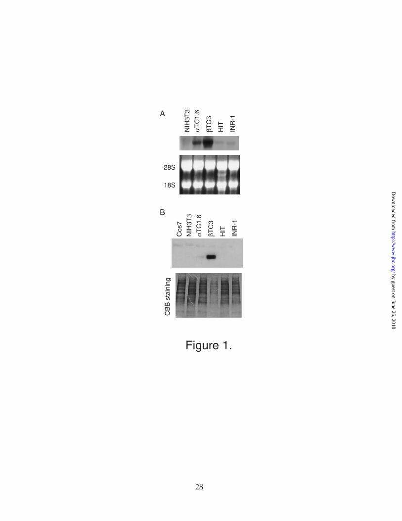
Expression of Nkx6.1 in cell lines. To identify cell-lines that express Nkx6.1, we
performed northern blot analysis with a hamster Nkx6.1 cDNA probe and western blot
analysis with antiserum directed against the carboxy-terminal end of the hamster
Nkx6.1 protein. Total RNA and nuclear extracts for these assays were prepared from β-
cell lines βTC3 and HIT-T15, and α-cell lines INR-1 and αTC1.6 and non-pancreatic cell
lines NIH3T3 and Cos7. As shown in Figure 1, Nkx6.1 mRNA is expressed in all four
pancreatic islet cell lines, but not in NIH3T3 cells. However, the Nkx6.1 protein can be
detected only in βTC3 cells. Although the expression level of Nkx6.1 mRNA is higher in
βTC3 cells than other cell line, this difference alone cannot explain the greater difference
in the expression of Nkx6.1 protein. These results suggest that the expression of Nkx6.1
is regulated both transcriptionally and post-transcriptionally.
Structure of the mouse Nkx6.1 gene. As an initial step towards characterizing the 5' end
of the Nkx6.1 gene, we cloned and sequenced exon1 and the 5’flanking region of the
mouse Nkx6.1 gene. 5’RACE performed on RNA from βTC3 cells identifies a cluster of
7 transcription start sites clustered at ~ 1 kb upstream from the translation start site
with no additional intervening introns (Figure 2b). RNase protection analysis with
βTC3 RNA confirms the position of four of these start sites.
The strongest band by RNase protection was defined as +1 bp, and other
nucleotides are numbered accordingly. The mouse genomic sequence lacks a TATAA
box upstream of the transcription start sites, but consensus CCAAT box sequences are
located at -220 bp and at –150bp (29) .
by guest on June 26, 2018http://w
ww
.jbc.org/D
ownloaded from
Page 10
Nkx6.1 promoter
10
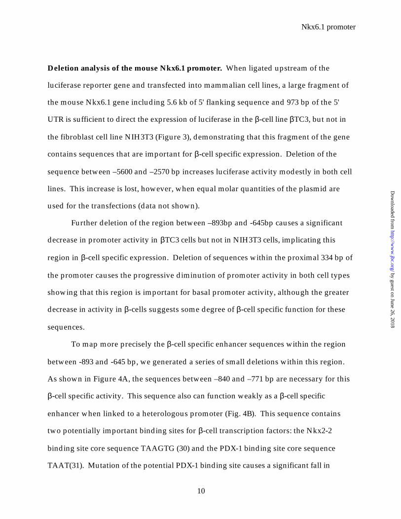
Deletion analysis of the mouse Nkx6.1 promoter. When ligated upstream of the
luciferase reporter gene and transfected into mammalian cell lines, a large fragment of
the mouse Nkx6.1 gene including 5.6 kb of 5' flanking sequence and 973 bp of the 5'
UTR is sufficient to direct the expression of luciferase in the β-cell line βTC3, but not in
the fibroblast cell line NIH3T3 (Figure 3), demonstrating that this fragment of the gene
contains sequences that are important for β-cell specific expression. Deletion of the
sequence between –5600 and –2570 bp increases luciferase activity modestly in both cell
lines. This increase is lost, however, when equal molar quantities of the plasmid are
used for the transfections (data not shown).
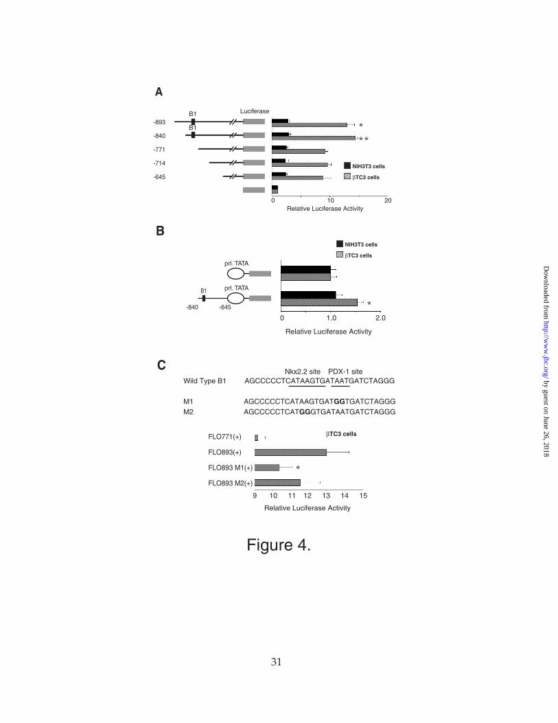
Further deletion of the region between –893bp and -645bp causes a significant
decrease in promoter activity in βTC3 cells but not in NIH3T3 cells, implicating this
region in β-cell specific expression. Deletion of sequences within the proximal 334 bp of
the promoter causes the progressive diminution of promoter activity in both cell types
showing that this region is important for basal promoter activity, although the greater
decrease in activity in β-cells suggests some degree of β-cell specific function for these
sequences.
To map more precisely the β-cell specific enhancer sequences within the region
between -893 and -645 bp, we generated a series of small deletions within this region.
As shown in Figure 4A, the sequences between –840 and –771 bp are necessary for this
β-cell specific activity. This sequence also can function weakly as a β-cell specific
enhancer when linked to a heterologous promoter (Fig. 4B). This sequence contains
two potentially important binding sites for β-cell transcription factors: the Nkx2-2
binding site core sequence TAAGTG (30) and the PDX-1 binding site core sequence
TAAT(31). Mutation of the potential PDX-1 binding site causes a significant fall in
by guest on June 26, 2018http://w
ww
.jbc.org/D
ownloaded from
Page 11
Nkx6.1 promoter
11
promoter activity, although mutation of the Nkx2-2 binding site causes a more modest
decrease in promoter activity (Fig. 4C).
PDX-1 and Nkx2-2 bind to the Nkx6.1 promoter. To address whether PDX-1 and Nkx2-
2 bind to these sites, we performed an electrophoretic mobility shift assay (EMSA) using
a double-stranded oligodeoxynucleotide corresponding to nucleotide –817 to –788 (B1)
as a probe. In vitro translated Nkx2-2 and PDX-1 can bind to this site (Figure 5A) and
they are competed by the unlabelled oligonucleotide. An unlabeled oligonucleotide
containing a mutation in the Nkx2-2 binding core (mutant M2) can still compete for
PDX-1 binding but not for Nkx2-2 binding. Interestingly, an unlabeled oligonucleotide
containing a mutation in the PDX-1 binding core (mutant M1) cannot compete for either
PDX-1 or Nkx2-2 binding.
When cotransfected into NIH3T3 cells, PDX-1 can activate the β-cell specific
enhancer linked to the rat prolactin promoter (Fig. 5B). Nkx2-2, however, cannot
activate the minienhancer by itself or in combination with PDX-1 (Fig. 5B), although it
can activate the intact Nkx6.1 promoter (data not shown). Neither factor affects the
expression of luciferase from the parent vector containing the prolactin alone. Recently
Sepulveda et al.(32) demonstated that the closely related cardiac homeodomain factor
Nkx2.5 cooperates with GATA-4, a zinc finger transcription factor, to activate the α-
actin promoter. Interestingly, for this interaction, a GATA4 binding site is not
necessary. To test the possibility that Nkx2.2 also co-operates with GATA factors in
pancreatic β-cells to activate the Nkx6.1 promoter, we co-transfected vectors expressing
either GATA4 or GATA6 along with the Nkx2.2 expression vector and a reporter
plasmid containing either the β-cell specific mini-enhancer linked to the rat prolactin
promoter or the intact Nkx6.1 promoter driving luciferase. However, neither GATA4
by guest on June 26, 2018http://w
ww
.jbc.org/D
ownloaded from
Page 12
Nkx6.1 promoter
12
nor GATA6 produced any additional activation of the Nkx6.1 promoter or
minienhancer (data not shown).
There are additional PDX-1 and Nkx2-2 binding sites in the Nkx6.1 promoter
outside of the β-cell specific enhancer region. There are multiple TAAT sequences that
fit the PDX-1 binding consensus within the proximal 900 bp of the promoter (see
boldfaced sequences in Fig. 2B) as well as several copies of the C/TAAG sequence that
forms the core of the Nkx2-2 binding sequence. One of these sites, located at -440 bp,
can function as a high affinity Nkx2-2 binding site (Fig. 5C).
It should be noted as well that there are other potential binding sites for β-cell
transcription factors in the Nkx6.1 promoter, including two copies of the HNF6 binding
site consensus sequence (33) (see Fig. 2B). The functional importance of these sites is
difficult to ascertain since they fall within a region of the proximal promoter that is also
important for expression in NIH3T3 cells.
Complex function of the Nkx6.1 gene 5’UTR. While its promoter plays a critical role in
expression of the Nkx6.1 gene, Fig. 3 demonstrates that sequences within the 5’ UTR
are at least as important. In βTC3 cells, the deletion of the 5’UTR causes a nearly
complete loss of luciferase expression from the Nkx6.1 promoter constructs. In NIH3T3
cells, however, removal of the 5’UTR increases luciferase activity.
When moved from its normal position downstream of the transcription start
site, the function of the 5'UTR changes. As shown in Figure 6, when positioned
downstream of the Nkx6.1 or herpes simplex virus thymidine kinase (TK) minimal
promoter, the 5’ UTR enhances the expression of luciferase in α and β cell lines but not
in the NIH3T3 cell line. In contrast, the 5'UTR produces no activity in α and β cells and
by guest on June 26, 2018http://w
ww
.jbc.org/D
ownloaded from
Page 13
Nkx6.1 promoter
13
significant repression in NIH3T3 cells when placed upstream of the Nkx6.1 or TK
promoters.
These results demonstrate that the 5’UTR can function as a position-independent
repressor in non-islet cells, and as an activator in islet cells when located in its normal
position downstream of the promoter. Cell-type specific function appears to be
dependent on an intact 5'UTR, since the 5'UTR loses all specificity when cut in half (Fig.
6).
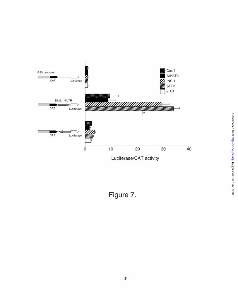
Identification of an internal ribosomal entry site (IRES) in the 5’ UTR. Several
features of the 5'UTR suggest that it may provide a poor template for protein synthesis
after cap-dependent scanning: it is long (973 bp), G/C rich (67.3%), and contains out of
frame ATG codons with reasonable Kozac consensus sequences. These limitations
could be overcome by an internal ribosomal entry site (IRES). In addition, the presence
of an IRES that functions in a cell-type specific manner could explain the functional
characteristics of the 5'UTR.
To test for this possibility, a dicistronic gene (pFoxRSV-CAT-Luc) was
constructed placing the CAT gene and the luciferase gene in series under the control of
RSV promoter. The 5’UTR of the Nkx6.1 was inserted between the two cistrons of this
plasmid (pFOX-CAT-5’UTR-Luc). In addition, the 5’UTR was inserted in an inverted
orientation (pFOX-CAT-5’UTR-R-Luc) as a nonspecific control. These plasmids were
transfected into two β-cell lines, βTC3 and INS1, the α-cell line αTC1.6, and two non-
islet cell lines NIH3T3 and COS7. CAT and luciferase activity were assayed 48 hours
after transfection. The ratio of luciferase activity to CAT activity provides a gauge of
IRES function. As shown in Figure 7, the 5'UTR can function as an IRES when placed in
its native orientation, and this activity is consistently greater in islet cell lines. Further, β-
by guest on June 26, 2018http://w
ww
.jbc.org/D
ownloaded from
Page 14
Nkx6.1 promoter
14
cell lines showed modestly higher activity than α-cell lines. These data demonstrate that
the IRES function of the 5’UTR contributes to the tissue specific expression of Nkx6.1.
by guest on June 26, 2018http://w
ww
.jbc.org/D
ownloaded from
Page 15
Nkx6.1 promoter
15
DISCUSSION
In the present study, we have characterized the Nkx6.1 promoter and mapped a
region involved in its cell-type specific expression. In addition, we found that the
expression of Nkx6.1 also is controlled at the post-transcriptional level, and an IRES in
the 5’ UTR plays an important role in directing its expression to islet cells.
Like many transcription factor genes, the 5’-flanking region of the mouse Nkx6.1
gene lacks a classic TATA box. The TATA box is typically located 30bp upstream of the
transcription initiation site and helps specify the transcription initiation site by directing
the binding of TFIID. Characteristic of genes that lack TATA boxes, the Nkx6.1 gene
has multiple transcription initiation sites as mapped by 5’ RACE and RNase protection
assay. Also characteristic of TATA-less genes, the transcription initiation sites lie just
downstream of two CCAAT boxes, at least one of which is functional in both islet and
non-islet cells.
Complex interactions among a number of transcription factors control the
temporal expression of genes during the development of the pancreas. Tight control
over the temporal and spacial expression of these factors is essential for proper
development of the endocrine cells. Nkx6.1 is expressed in at least three different cell
types during mouse pancreatic development: initial broad expression in the epithelial
cells that compose the dorsal and ventral buds, resticted expression after embryonic
day 13 in islet cell precursors, and finally in mature beta-cells. Studies of mice that lack
an intact Nkx2.2 gene demonstrate that Nkx2.2 is required for Nkx6.1 expression in the
pancreas after e13; and specific inactivation of the PDX-1 gene in insulin-expressing cells
demonstrates that PDX-1 is required for maintaining Nkx6.1 expression in
differentiated β-cells (9, 15). It can be concluded from these prior studies that Nkx6.1
by guest on June 26, 2018http://w
ww
.jbc.org/D
ownloaded from
Page 16
Nkx6.1 promoter
16
expression is regulated directly or indirectly by these two factors. The promoter studies
reported here support the conclusion that both factors drive Nkx6.1 expression directly,
by binding to the Nkx6.1 gene promoter.
Although there are several binding sites for Nkx2.2 and PDX-1 in the proximal
Nkx6.1 promoter, only the sites located at -800 are required for expression specifically
in the β-cell line. The more proximal sites may also contribute, but removal of more
proximal sequences affects expression in NIH3T3 cells as well. The presence of binding
sites for both factors in the β-cell specific enhancer located at -800 bp is intriguing given
the essential role of both factors in Nkx6.1 expression. Despite the juxtaposed binding
sites, however, Nkx2.2 does not activate the β-cell specific enhancer even in the
presence of co-expressed PDX-1.
We recently found that Nkx2.2 by itself cannot activate transcription even from a
construct with 7 tandem repeats of an ideal Nkx2.2 binding site. The NK2 domain just
downstream of the homeodomain in Nkx2.2 inhibits the activation domain, and some
modification the NK2 specific domain may be required to allow Nkx2.2 to activate
transcription (30). When Nkx2.2 is over-expressed as in the co-transfection experiments
reported here, the non-islet cells may lack specific modifiers of the NK2 domain, or the
capacity of the cells to modify the NK2 domain may be exceeded. Hence, we cannot
rule out the possibility that Nkx2.2 cooperates with PDX1 in regulating the Nkx6.1
promoter in the normal cellular context.
In addition, a different array of factors may control Nkx6.1 expression at
different points in development. The cells used for these experiments are probably
more representative of mature β-cells than undifferentiated pancreatic epithelial cells or
islet cell progenitors. During the differentiation of islet cells in the fetal mouse pancreas,
the islet cell progenitors transiently express the basic helix-loop-helix transcription
by guest on June 26, 2018http://w
ww
.jbc.org/D
ownloaded from
Page 17
Nkx6.1 promoter
17
factor neurogenin3 prior to further maturation and expression of PDX-1 (34). Some of
these early neurogenin3 expressing cells co-express Nkx6.1, but not PDX-1,
demonstrating that cells at this stage in differentiation do not require PDX-1 for Nkx6.1
expression (34). Other homeodomain proteins capable of binding to the TAAT sites, or
other factors such as HNF6 (35) may fill the PDX-1 role at this stage.
In addition to controls at the level of the promoter, expression from the Nkx6.1
gene is regulated by its long, complex 5’UTR, which has similarities to the 5'UTRs found
in some Drosophila homeobox genes. For example, ubx contains a 968 bp 5’UTR and 2
upstream ATGs, and antp contains a 1735 bp 5’UTR and 15 upstream ATGs. Both of
these 5'UTRs contain IRESs that promote developmentally regulated translation.
Similarly, the Nkx6.1 5'UTR functions as an IRES; and while it can function in all the cells
types tested, its activity is significantly higher in islet cell lines. These results
demonstrate that the IRES activity of the Nkx6.1 5'UTR shows cell type specificity. In
addition, the 5’UTR also inhibits transcription in the NIH3T3 cells, but not in islet cells, in
a position independent fashion. Taken together, the 5’UTR contributes significantly to
the cell-type specific expression of Nkx6.1.
The identified functions of the 5'UTR, however, cannot completely explain the
differences in expression of Nkx6.1 between α- and β-cell lines. No Nkx6.1 protein can
be detected in α-cell lines, despite the presence of Nkx6.1 mRNA, suggesting that post-
transcriptional regulation of Nkx6.1 expression contributes to its restriction from α-
cells. Although the IRES in the 5'UTR provides a mechanism for cell-type specific
translation of Nkx6.1 mRNA, the activity of the IRES is similar in αTC1 cells and β-TC3
cells. It is possible that in the context of the intact gene, the IRES may function in a
more tightly restricted fashion, or other posttranscriptional mechanisms must play
additional roles in the cell-type specific expression of Nkx6.1.
by guest on June 26, 2018http://w
ww
.jbc.org/D
ownloaded from
Page 18
Nkx6.1 promoter
18
It should be noted that further controls provide additional limits on the function
of Nkx6.1 once the protein is expressed. When it binds to target genes, Nkx6.1 is a
potent transcriptional repressor; but a sequence in the carboxyl-terminal end of the
molecule prevents DNA binding by the homeodomain (14). Presumably only when
this binding inhibition is relieved by interactions provided in the appropriate cellular
environment can Nkx6.1 then turn off target genes. Together, several layers of
regulation ensure that gene targeting by Nkx6.1 is tightly restricted temporally and
spatially.
by guest on June 26, 2018http://w
ww
.jbc.org/D
ownloaded from
Page 19
Nkx6.1 promoter
19
Acknowledgements We thank members of German laboratory for helpful
comments and criticisms and M. Sander for the mouse Nkx6.1 genomic clone. H.W is a
recipient of a Juvenile Diabetes Foundation International Postdoctral Fellowship and
RGM is a recipient of a Research Career Award (K08) from the National Institutes of
Health. This work was supported by National Institutes of Health grants DK31371 and
DK30255.
by guest on June 26, 2018http://w
ww
.jbc.org/D
ownloaded from
Page 20
Nkx6.1 promoter
20
FIGURE LEGENDS
Figure 1. The expression of Nkx6.1 in cell lines. (A) The northern blot shown is
probed with 32P-labelled hamster Nkx6.1 cDNA. Ten microgram of total RNA were
used from NIH3T3 cells (lane1), αTC1.6 cells (lane2), βTC3 cells (lane3), HIT T15 M2.2.2
cells (lane4), and INR-1 cells (lane5). (B) The western blot shown is probed with
antiserum raised in rabbit against hamster Nkx6.1. Five microgram of nuclear extract
were used from Cos7 cells (lane1), NIH3T3 cells (lane2), αTC1.6 cells (lane3), βTC3 cells
(lane4), HIT T15 M2.2.2 cells (lane5), and INR-1 cells (lane6).
Figure2. The mouse Nkx6.1 gene promoter. (A) The results of an RNase protection
assay are shown. Total RNA from βTC3 cells (10µg) was hybridized with a mouse
genomic Nkx6.1 antisense probe. The protected bands were indicated by arrows. (B)
2.4 kb of genomic DNA sequence upstream of the mouse Nkx6.1 coding region is
shown. Seven transcription start sites identified by 5’ RACE is shown in boldface, and 4
transcription sites identified by RNase protection are labeled with asterisks. The
translation start site is indicated by italics. Two potential CCAAT boxes and other
promoter-element are indicated in underlined-boldface. Several proximal TAAT
sequences are shown in boldface. The sequence of the mouse Nkx6.1 promoter region
and Exon1 are available in the Genbank database under accession number AF291666.
Figure3. Transcriptional activity of the Nkx6.1 promoter. Reporter plasmids were
constructed with the Nkx6.1 gene fragments indicated inserted upstream of the
luciferase gene, and then were cotransfected with a CMV promoter-driven β-
galactosidase expression plasmid into NIH3T3 cells (filled bars) of βTC3 cells (hatched
by guest on June 26, 2018http://w
ww
.jbc.org/D
ownloaded from
Page 21
Nkx6.1 promoter
21
bars). Relative luciferase activities are calculated with the activity of cells transfected
with the pFOXLuc1 plasmid alone set at 1. All data are shown as mean ± SEM.
Figure4. Identification of a -cell-specific enhancer element. (A) Reporter plasmids
were constructed with the truncated Nkx6.1 promoters indicated inserted upstream of
the luciferase gene and then were cotransfected with a CMV promoter-driven β-
galactosidase expression plasmid into NIH3T3 cells (filled bars) or βTC3 cells (hatched
bars). Relative luciferase activities are calculated with the activity of cells transfected
with the pFOXLuc1 plasmid alone set at 1. The asterisks indicates a P value <.01 for the
comparison of the activities of the -771 promoter with the -840 promoter and <.05 for
the comparison of the activities of the -771 promoter with the -893 promoter in βTC3
cells as calculated by the paired Student's T test. (B) A reporter plasmids was
constructed with the -840 to -645 bp fragment of the Nkx6.1 promoter inserted
upstream of the minimal rat prolactin promoter and the luciferase gene and then was
cotransfected with a CMV promoter-driven β-galactosidase expression plasmid into
NIH3T3 cells (filled bars) or βTC3 cells (hatched bars). Relative luciferase activities are
calculated with the activity of cells transfected with the pFOXLuc1 plasmid containing
only the prolactin promoter set at 1. The asterisk indicates a P value <.05 for the
comparison of the activities of the pFOXluc.prl.Nkx6.1(-840/-645) plasmid with the
pFOXluc.prl plasmid in βTC3 cells as calculated by the paired Student's T test. (C)
Reporter plasmids containing the -893 bp promoter fragment and complete 5’ UTR with
or without the mutations shown upstream of the luciferase gene were cotransfected
with a CMV promoter-driven β-galactosidase expression plasmid into βTC3 cells.
Relative luciferase activities are calculated with the activity of cells transfected with the
pFOXLuc1 plasmid alone set at 1. The asterisk indicates a P value <.05 for the
by guest on June 26, 2018http://w
ww
.jbc.org/D
ownloaded from
Page 22
Nkx6.1 promoter
22
comparison of the activities of the M1 mutant promoter with the wild-type promoter as
calculated by the paired Student's T test. All data are shown as mean ± SEM.
Figure5. PDX-1 and Nkx2.2 binding to the Nkx6.1 promoter. (A) An electrophoretic
mobility shift assay (EMSA) using in vitro translated Nkx2.2 and PDX-1 is shown. 32P-
labeled oligonucleotides encoding the B1 enhancer element (sequences are shown in
(Figure 4B)) were incubated with 1µl of each in vitro translated protein for 15min. at
room temperature, then subjected to electrophoresis on a 5% polyacrylamide gel.
Unlabeled competitor oligonucleotides (sequences are shown in (Figure 4B)) were
added at 20 and 200 fold molar excess. (B) A reporter plasmid containing 5 tandem
copies of the B1 enhancer element upstream of the prolactin minimal promoter driving
luciferase and pBAT12 expression plasmids expressing the Nkx2.2 and PDX-1 cDNAs
under the control of the CMV promoter were cotransfected into NIH3T3 cells. Relative
luciferase activities are calculated with the activity of cells transfected with the pBAT12
expression vector without cDNA insert set at 1. All data are shown as mean ± SEM. (C)
An EMSA using in vitro translated Nkx2.2 is shown. 32P-labeled oligonucleotides
encoding the B1 enhancer element (lanes 1-3) or the related sequence at –460 (lane 4, 5)
in the Nkx6.1 promoter (see Methods for sequence) were incubated with 1µl of the in
vitro translated protein for 15min. at room temperature, then subjected to
electrophoresis on a 5% polyacrylamide gel. The control lanes (2 and 4) contain in vitro
translated luciferase protein. N. S. indicates a non-specific protein-DNA complex
produced by proteins present in the rabbit reticulocyte lysate mix.
Figure6. Characterization of the 5’UTR region. (A) Reporter plasmids containing
Nkx6.1 5’UTR upstream or downstream of THE Nkx6.1 –344 minimal promoter region
by guest on June 26, 2018http://w
ww
.jbc.org/D
ownloaded from
Page 23
Nkx6.1 promoter
23
or the TK promoter driving the luciferase gene were cotransfected with a CMV
promoter-driven β-galactosidase expression plasmid into NIH3T3 cells (filled bar), βTC3
cells (hatched bar), and αTC1.6 cells (open bar). Relative luciferase activities are
calculated with the activity of cells transfected with the plasmid containing the promoter
alone without 5’UTR alone set at 1.
Figure 7. The 5’UTR of Nkx6.1 gene works as an IRES. Dicistronic reporter plasmid
containing the 5’ UTR of Nkx6.1 bidirectionally inserted between the CAT and luciferase
genes were transfected into the cell lines shown. CAT activity and Luciferase activity
were measured and the luciferase activity divided by CAT activity was used as an index
of IRES activity. All data are shown as mean ± SEM. The ratio of CAT/Luciferase
acrivity for the control dicistronic constructs without the 5’UTR is set at 1.0.
by guest on June 26, 2018http://w
ww
.jbc.org/D
ownloaded from
Page 24
Nkx6.1 promoter
24
REFERENCES
1. Sander, M. and German, M. S. (1997) J. Mol. Med. 75, 327-40
2. Edlund, H. (1998) Diabetes 47, 1817-23
3. Jonsson, J., Carlsson, L., Edlund, T. and Edlund, H. (1994) Nature 371, 606-609
4. Ahlgren, U., Pfaff, S. L., Jessell, T. M., Edlund, T. and Edlund, H. (1997) Nature 385,
257-60
5. St-Onge, L., Sosa-Pineda, B., Chowdhury, K., Mansouri, A. and Gruss, P. (1997)
Nature 387, 406-9
6. Sosa-Pineda, B., Chowdhury, K., Torres, M., Oliver, G. and Gruss, P. (1997) Nature
386, 399-402
7. Sander, M., Neubuser, A., Kalamaras, J., Ee, H. C., Martin, G. R. and German, M. S.
(1997) Genes Dev 11, 1662-73
8. Naya, F. J., Huang, H. P., Qiu, Y., Mutoh, H., DeMayo, F. J., Leiter, A. B. and Tsai, M.
J. (1997) Genes Dev 11, 2323-34
9. Sussel, L., Kalamaras, J., Hartigan-O'Connor, D. J., Meneses, J. J., Pedersen, R. A.,
Rubenstein, J. L. and German, M. S. (1998) Development 125, 2213-21
10. Gradwohl, G., Dierich, A., LeMeur, M. and Guillemot, F. (2000) Proc. Natl. Acad. Sci.
USA 97, 1607-1611
11. Li, H., Arber, S., Jessell, T. M. and Edlund, H. (1999) Nat Genet 23, 67-70
12. Harrison, K. A., Thaler, J., Pfaff, S. L., Gu, H. and Kehrl, J. H. (1999) Nat Genet 23, 71-
5
13. Oster, A., Jensen, J., Serup, P., Galante, P., Madsen, O. D. and Larsson, L. I. (1998) J
Histochem Cytochem 46, 707-15
14. Mirmira, R. G., Watada, H. and German, M. S. (2000) J. Biol. Chem. 275, 14743-14751
by guest on June 26, 2018http://w
ww
.jbc.org/D
ownloaded from
Page 25
Nkx6.1 promoter
25
15. Ahlgren, U., Jonsson, J., Jonsson, L., Simu, K. and Edlund, H. (1998) Genes Dev 12,
1763-8
16. Kozak, M. (1989) J Cell Biol 108, 229-41
17. Pelletier, J. and Sonenberg, N. (1988) Nature 334, 320-5
18. van der Velden, A. W. and Thomas, A. A. (1999) Int J Biochem Cell Biol 31, 87-106
19. Vagner, S., Gensac, M. C., Maret, A., Bayard, F., Amalric, F., Prats, H. and Prats, A.
C. (1995) Mol Cell Biol 15, 35-44
20. Teerink, H., Voorma, H. O. and Thomas, A. A. (1995) Biochim Biophys Acta 1264, 403-
8
21. Bernstein, J., Sella, O., Le, S. Y. and Elroy-Stein, O. (1997) J Biol Chem 272, 9356-62
22. Stein, I., Itin, A., Einat, P., Skaliter, R., Grossman, Z. and Keshet, E. (1998) Mol Cell
Biol 18, 3112-9
23. Oh, S. K., Scott, M. P. and Sarnow, P. (1992) Genes Dev 6, 1643-53
24. Ye, X., Fong, P., Iizuka, N., Choate, D. and Cavener, D. R. (1997) Mol Cell Biol 17,
1714-21
25. Sambrook, j., Fritsch, E. F. and Maniatis, T. Molecualr Cloning (Cold Spring Harbor
Laboratory Press, 1989).
26. Rudnick, A., Ling, T. Y., Odagiri, H., Rutter, W. J. and German, M. S. (1994) Proc Natl
Acad Sci U S A 91, 12203-7
27. German, M. S., Moss, L. G., Wang, J. and Rutter, W. J. (1992) Mol. Cell. Biol. 12, 1777-
1788
28. German, M. S., Wang, J., Chadwick, R. B. and Rutter, W. J. (1992) Genes & Dev. 6,
2165-2176
29. Mantovani, R. (1998) Nucleic Acids Res 26, 1135-43
by guest on June 26, 2018http://w
ww
.jbc.org/D
ownloaded from
Page 26
Nkx6.1 promoter
26
30. Watada, H., Mirmira, G. H., Kalamaras, J. and German, M. S. (2000) Proc Natl Acad
Sci U S A in press
31. Ohlsson, H., Karlsson, K. and Edlund, T. (1993) EMBO J. 12, 4251-4259
32. Sepulveda, J. L., Belaguli, N., Nigam, V., Chen, C. Y., Nemer, M. and Schwartz, R. J.
(1998) Mol Cell Biol 18, 3405-3415
33. Lannoy, V. J., Burglin, T. R., Rousseau, G. G. and Lemaigre, F. P. (1998) J Biol Chem
273, 13552-62
34. Schwitzgebel, V. M., Scheel, D. W., Conners, J. R., Kalamaras, J., Lee, J. E.,
Anderson, D. J., Sussel, L., Johnson, J. D. and German, M. S. (2000) Development 127,
3533-3542
35. Jacquemin, P., Durviaux, S. M., Jensen, J., Godfraind, C., Gradwohl, G., Guillemot, F.,
Madsen, O. D., Carmeliet, P., Dewerchin, M., Collen, D., Rousseau, G. G., and Lemaigre,
F. P. (2000) Mol Cell Biol 20, 4445-4454
by guest on June 26, 2018http://w
ww
.jbc.org/D
ownloaded from
Page 27
Nkx6.1 promoter
27
FOOTNOTES
1 The abbreviation used are: 5’ untranslated region; 5’UTR, internal ribosomal entry site;
IRES, Dulbecco’s Modified Eagle Medium; DMEM, Rapid Amplification of cDNA Ends;
RACE, Herpes simplex virus thymidine kinase; TK.
2 Sander M., Sussel L., Conners J., Kalamaras J., DelaCruz F., Schwitzgebel V., Hays-
Jordan A., and German, M.S. manuscript submitted.
by guest on June 26, 2018http://w
ww
.jbc.org/D
ownloaded from
Page 28
A
28S
18SN
IH3T
3
βTC
3
αTC
1.6
HIT
INR
-1
B
NIH
3T3
βTC
3
αTC
1.6
HIT
INR
-1
Cos
7
Figure 1.
28
CB
B s
tain
ing
by guest on June 26, 2018http://w
ww
.jbc.org/D
ownloaded from
Page 29
-1394 AAGCTTTGGAGAAGTAAGGG GACCGACCAGTTTAAGGCCT CCTGCTTGACATTCAGAGTC AAACTCCCTGGCGCTGCTCA GTTTTAATTCCTGGGTCATT TATCGCCTATTTCTGTTTTC TGAACCTTAAATTTGGACTC AATAATATGATGCAAATCTC TGCTGTGACACTCCCCCCCC CCCCCCACTGGTGTATCAAC TGCCCGATTTCTCAAGATCG ACCAAAGAGGTTTTTTCCTT GGTTTTGGTCAACCCTGAGC AGACCTTAAAGATCGGCCAG AGGGAGCAAAGCCCTTTTGA -1094 CCATCGCTCCCAATGCCAGC CTAGAAGTCGGTCGTCTCTA GTTTACTCAACTACCCCGAG TTGAGAGCTTGACCAGGCTT TCCAACAGTTACCTGTCTTC CCCCGAGGTATTCCTCTATC TAAAGTTGCCCTGTGAATTT TAGTGATCCTGCCTCATAAA TCCAACCAATAATAATAGAG GGAGGATTTTAAAAAATAAT TATCTCATTTCTGTTAGGTT TAGACACCACGCAGGAGATA AATATTCTCATTAAGCTGAT TTCATCCCCAGAGTACTGAG CCCCCTCATAAGTGATAATG -794 ATCTAGGGAGTGGGAGAGCG AAGACAAGAACGGAGAAAGA ACAGAAAAGAGCAGGAGACA GAAAGATGGTGAAGGGTGAC CCTTAGGCCTGCGAGGGGAT TTAAAAACATCTACGGGCTT AAGGAACAACAAATCAATTT ACACGGTTCTGGAAGAGCCC AGAGGGCCTTTAATTAATCC CTTCAAAAGAAGGAAGTCGG CCTGGGATGTGCCTCCTGCC TGCTCCATTAGCTCCCTTTT CGCAAGGGTCCAGACACCGT TGGAGGTGGGCGCTGCGCGC AAGCTGGTGGGGGAGGATGA -494 CGCGAGCTGGCGTGGGCGGA AGAGACGCACTTAAACTGCT TTTCCATAGAAGGGCTGGAT
TTTCATTATTCCTCTCTTTA AAAAGTAATGCCCTCTTCGT CCGTGCTCCCTCCTTCTCCT TTCCATTTTATTTTGCACAA TTAGTTGAGCCGGCCGCTGG CTCTAGACTGGAACCACTCT TTTCGCCAGGCCCCTCCCCT CTTGGCTCCGCCCAAGTGAA GCTGGGGCGGGGACTAGGAG GGCGCGTCCTTATGGCTCCC TAGTCTCAGCCAATCAAAAG CTGTGGCGCTCCCAGGTAGG
-194 CGTGTTCTAGGAGCGACGCC TTGCCCAAGCTGAGCGCTAT TGGAGGCGGTGTTTACGCCC
AGGACCCGGGCCCCGCTCCT CAGTCCCGCCCCGCCGAGCC GCCCCGGAATGACGTCCTCG AAAGTTCTCATTTTGGCCCC CCACCTCCCCTCCCTTGCGT CCCCCAGCTAAAGAGAGGCA GGGAGGGGTGCAAATATTTT ATTACCTTTGAGAGCTTCAT CCGAACTGTCAGGCCCAGAG GGAGAGAGGAGAGAAGGGAA GAGCCGCCGAGAGGGTCAGT TTGGCCAGAGGACAGGGCTT +106 GGAAGAGCCAAGCCTGGAAA GCCAAGGAGAAATGCCAGAG AGGCAAGAGAAAGGCGGAGA GTGAGAGGAAGAGAGGGCGC CAGGGGGTGGGGCGGGGGGT CTCCCGGCCGGCTTGCAGAA GCGGACTAACGATCGGAACC GGCCCGACAGACTTTTTCCA TACTTAGGTTTATTTTCTTT TATCCTTTTCGATCCGGCTA GTCCGGCGCCGCGTGGCTCG GAGGGGAAGCAGGCTCGTGG GCGCGCCAAGTCCCCAGCCG CCGGAGCGGTGCCACTGACT GCCCTTTCTGGCGCAGTAAC +406 GCCGGGCCCGAGGCAGTGGC GGGCGCGATACCCGCGCCGG CCGTTTGTGCTCTGTCCCCC GGGCGAGCGGTGAGTGCGAC TCAGCAGCGCGGCCGCCGGG AGAGCGGAGCGTCCGAGCGA GATCAGAGGCGCGCACCGGG CGGAACGCCGCCCGCTTTGA AGCTACCCCAGGCGAGCGAG CCGGCCCCCGCCCTCCTACA TCAAAGCGAACGCTCCGCGC CTCCCAACCTTGTTGCAAAC TCTCTGGGTCGGCTGCGGGG TACGTCTTGCTGATTTCCCG CGGGGGTGGAGAAGATGAGA +706 AGCAGAGCGCTCTGAGCCGG GAACGAGGGACCAGCGCCTG GGATCGAATCCCGGACTCCC GAAGCCGAGGAAGCGCTGAG CCCGCCCGCGCCCCCGCAGC CCTCGCCCCCGCCGCCTCCC GCGGGGCGTTTGGACATTTT TGCTGCGCAGCTCCCGGAGC CCGCGGCCGATCCACACTTC GCTTGCGCGCGCCCCCGGCA CCTCGGGTTCTCCCGAGCCC CGGCGGGGCCACCGACCTGC GTGGCTGCGGGTTCGGGTCT GGCTGTGGGATG
CCAAT box
Nkx2.2&PDX-1
Translation Start Site
* *
*
*
A B
yeas
t tR
NA
βTC
3
CCAAT box
HNF6 ?
Nkx2.2
HNF6 ?
Figure 2.
29
by guest on June 26, 2018http://w
ww
.jbc.org/D
ownloaded from
Page 30
-5600 +1-1000
-5600
-2570
-1580
-1390
-893
-645
-555
-334
-240
-207
-185
-149
-90
-1
-334
FLO5600(+)
FLO2570(+)
FLO1580(+)
FLO1390(+)
FLO893(+)
FLO645(+)
FLO555(+)
FLO334(+)
FLO240(+)
FLO207(+)
FLO185(+)
FLO149(+)
FLO90(+)
FLO1(+)
FLO334(-)
pFoxLuc1
Relative Luciferase Activity
0 10 18
+1000
-5600 FLO5600(-)
NIH3T3 cells
βTC3 cells
Figure 3.
30
by guest on June 26, 2018http://w
ww
.jbc.org/D
ownloaded from
Page 31
A
B
prl. TATA
prl. TATAB1
Relative Luciferase Activity
1.0 2.00
Wild Type B1 AGCCCCCTCATAAGTGATAATGATCTAGGG
M1 AGCCCCCTCATAAGTGATGGTGATCTAGGGM2 AGCCCCCTCATGGGTGATAATGATCTAGGG
FLO893(+)
FLO771(+)
FLO893 M1(+)
FLO893 M2(+)
Relative Luciferase Activity
9 10 11 12 13 14 15
Nkx2.2 site PDX-1 site
βTC3 cells
C
Luciferase
-893
-840
-771
-714
-645
Relative Luciferase Activity0 10 20
NIH3T3 cells
βTC3 cells
B1
B1
-840 -645
NIH3T3 cells
βTC3 cells
Figure 4.
***
*
*
31
by guest on June 26, 2018http://w
ww
.jbc.org/D
ownloaded from
Page 32
Nkx2.2 PDX-1
Co
mp
etit
or Wild type
M1M2
5XB1
Reporter gene construct
prl. TATA
Control
Nkx2.2
PDX-1
PDX-1+Nkx2.2
10 2
Relative Luciferase Activity
A
B
Luciferase
Nkx2.2
N.S.
PDX-1
1 2 3 4 5 6 7 8 9 10 1112 13 14 15
C
Nkx
2.2
Nkx
2.2
Co
ntr
ol
Co
ntr
ol
Nkx2.2
N.S.
1 2 3 4 5
NIH3T3 cells
Figure 5.
I.V.T.T.
-810 site-460 site
-810 site
32
pro
be
on
ly
by guest on June 26, 2018http://w
ww
.jbc.org/D
ownloaded from
Page 33
FLONk6
FLONk6-5'UTR
FLO5'UTR-Nk6
FLONk6(0-500)
FLONk6(500-1000)
FLO(0-500)Nk6
FLO(500-1000)Nk6
-344 Nkx6.1pro Luciferase
-344 +43
-344 +973
-344 +43
+9 +973-344 +500
-344 +43
-344 +43
+973+492
+9 +500
+973+492
-344 +43
+973
+9 +973+500
+973+492
+9 +500
+973+492
TK mini-promoter
FLOTK
FLOTK-5'UTR
FLO5'UTR-TK
FLOTK(0-500)
FLOTK(500-1000)
FLO(0-500)TK
FLO(500-1000)TK
Relative Luciferase Activity0 5
+9
TK
TK
TK
TK
TK
TK
TK
+9
NIH3T3
βTC3
αTC1
Figure 6.
33
by guest on June 26, 2018http://w
ww
.jbc.org/D
ownloaded from
Page 34
LuciferaseCAT
Nkx6.1 5'UTR
LuciferaseCAT
RSV promoter
LuciferaseCAT
Luciferase/CAT activity
Cos 7
NIH3T3
INS-1
βTC3
αTC1
0 10 20 30 40
Figure 7.
34
by guest on June 26, 2018http://w
ww
.jbc.org/D
ownloaded from
Page 35
Watada Hirotaka, Raghavendra G. Mirmira, Joey Leung and Michael S. GermanNkx6.1
Transcriptional and Translational Regulation of beta-cell Differentiation Factor
published online August 9, 2000J. Biol. Chem.
10.1074/jbc.M004981200Access the most updated version of this article at doi:
Alerts:
When a correction for this article is posted•
When this article is cited•
to choose from all of JBC's e-mail alertsClick here
by guest on June 26, 2018http://w
ww
.jbc.org/D
ownloaded from