Pfeuffer S et al. NMO Spectrum Disorders. Neurology International Open 2017; 1: E36–E47
Original Article
History and NomenclatureAs early as 1894, the French physician, Eugène Devic, described pa-tients with concomitant symptoms of optic neuritis and transverse myelitis, and designated it as “neuromyelitis optica” (NMO) [1]. For a long time, the NMO was not defined as a separate entity under chronic inflammatory CNS diseases, but as a sub-form of multiple sclerosis. This assessment was abandoned by the turn of the mil-lennium, when Wingerchuk and colleagues first published inde-pendent diagnostic criteria for the disease [2]. A major contribu-tion to this new understanding has been the discovery of antibod-ies in the serum of patients; these were initially referred to as NMO immunoglobulin G (IgG). The membrane protein aquaporin-4 (AQP4) was identified as the target structure of these antibodies [3, 4]. This discovery led to a revision of the diagnostic criteria with the implementation of antibody diagnostics as a biomarker of the disease in 2006. Since in about one-third of the patients with the clinical presentation of an NMO, such antibodies were not detect-able, a distinction was made between “seropositive” and “seron-egative” NMO [5].
Conversely, antibodies to AQP4 were detectable in patients who did not have the typical symptoms of NMO (optic neuritis and my-elitis, also referred to as “classic Devic’s syndrome”). Primarily for this reason, the concept of NMO-spectrum diseases (NMOSD) was
established and gradually expanded. With the last revision of the diagnostic criteria in 2015, this term was rendered uniform. Its use is now recommended for the description of all seropositive and se-ronegative CNS disorders with a typical distribution pattern and clinical course [6].
EpidemiologyBasically, the NMOSD is a much rarer entity of chronic inflamma-tory CNS disorders compared to multiple sclerosis (MS). The geo-graphical distribution of prevalence and incidence is markedly dif-ferent with a significantly higher incidence in Asian, African and South American patients compared to Caucasians.
In a relatively recent epidemiological study, a prevalence of 3.9/100 000 persons and an incidence of 0.7/1 000 000 person- years were determined in a Caucasian population in North America (Olmsted City, Minnesota). On the Antilles island of Martinique, the prevalence was 10/100 000 persons and the incidence of 7.3/1 000 000 person-years.
This is consistent with a Danish survey that reported a preva-lence of 4.4/100 000 inhabitants [7, 8] in the predominantly Cau-casian cohorts. Retrospective studies of (Caucasian) patients with chronic inflammatory, demyelinating CNS disorders support these
CorrespondenceProf. Heinz Wiendl, MDNeurology Clinic and Institute for translational NeurologyUniversity Hospital MünsterAlbert-Schweitzer-Campus 148149 Mü[email protected]
Abstr Act
Neuromyelitis optica spectrum disorders (NMOSD) represent a rare subset of chronic-inflammatory diseases of the central nervous system. Despite heterogeneities in disease activity, there is a higher degree of disability accumulation in NMOSD patients compared to MS patients. According to the revised diagnostic criteria, a recommendation was made to abandon the term NMO and to summarize these conditions as NMOSD. Clinical presentation of NMOSD patients in most cases is optic neuritis and transverse myelitis but nevertheless, NMOSD can affect most parts of the central nervous system (e. g. brainstem and hypothal-amus). Originally characterized as AQP4-antibody-dependent disease, it has recently been discussed whether conditions with presence of antibodies against myelin oligodendrocyte glycoprotein (MOG) belong to the family of NMOSD. Due to the severity of the disease with often devastating relapses, systematic therapy is necessary. Usually, immu-nosuppressants or monoclonal antibodies with anti-inflammatory prop-erties are used. Recently, four substances entered clinical testing for treatment of NMOSD.
Pfeuffer S et al. NMO Spectrum Disorders. Neurology International Open 2017; 1: E36–E47
numbers, NMOSD cases comprising approximately 1.5 % of all cases in the widest sense [9]. The findings from the Caribbean popula-tion underline the fact that, as in the whole of South America, NMOSD is much more common in the Caribbean than in Europe.
Surprisingly, in individual cohorts in Japan, the prevalence of NMOSD at 0.9/100 000 was unusually low [7]. However, it must be taken into account that in Japan often the diagnosis of “opticospi-nal MS” (OSMS) is made. Although OSMS is not conclusively as-signed to the NMOSD spectrum, patients with OSMS share numer-ous other similarities in addition to the typical distribution of the lesions with only occasional intracerebral demyelinating lesions. These include a comparable course with rapid accumulation of dis-ability; however, oligonoclonal bands [10] are only detected in ex-ceptional cases. However, the strongest argument for such an as-sociation is the prevalence of AQP4 antibodies in 50 % of OSMS pa-tients [11, 12].
Including OSMS patients, NMOSD represents approximately half of all cases of chronic inflammatory, demyelinating CNS disorders in Asian patients [13].
For these reasons, the current consensus criteria recommend that the term OSMS be used no longer, but that in these patients the diagnosis of an NMOSD (seronegative or seropositive) should be made.
The age at the initial manifestation of NMOSD is on average 37.8 years, which is approximately 10 years above the mean manifesta-tion age in MS patients. However, initial manifestations were also observed in significantly older patients.
With a ratio of approximately 9: 1 (f: m (female to male sex ratio)), NMOSD occurs significantly more frequently in females, es-pecially in its seropositive forms, than multiple sclerosis ( ratio f: m = 2–3: 1 [9, 14]. The preferential occurrence of NMSOD in women decreases in AQP4-seronegative patients and especially with those with detectable antibodies against myelin oligodendrocyte glyco-protein (MOG); MOG antibody-associated NMOSD forms occur even more frequently in men (0.6: 1 (m: f)) [15].
NMOSD patients also tend to develop other antibody-mediated (or at least antibody-associated) autoimmune diseases. Thyroid disorders (~14 % of patients), Sjögren's syndrome (~20 % of pa-tients) and myasthenia gravis (2 %) [16, 17] are the most common diseases in these patients. Overall, approximately one-third of NMOSD patients suffer from at least one further manifest autoim-mune phenomenon with marked preference for seropositive pa-tients (58.5 % for AQP4 + patients vs. 8.6 % for AQP4-patients) [14]. The tendency to form autoantibodies is illustrated by the fact that positive antinuclear antibodies without documented clinical man-ifestation are present in approximately half of all NMOSD patients [17]. There are case reports of manifest systemic lupus erythema-tosus [18].
Due to the high clinical relevance and the necessity for vigilance, it must be borne in mind that a bi-directional association exists be-tween Myasthenia gravis and NMOSD. Since NMOSD is most com-mon in myasthenia patients after thymus resection, this differen-tial diagnosis should be considered in the presence of the relevant clinical symptoms. It is unclear, however, whether presence of AQP4 antibodies without corresponding clinical symptoms in individual cases of myasthenia patients is a meaningful indication for screen-ing (if necessary, prior to indicated thymus resection) [19].
PathogenesisThe pathogenesis of NMOSD has not been conclusively clarified. By identifying the target structure Aquaporin-4, however, there has been a gain in the understanding of immunopathogenesis. In this article, only selected aspects, which can explain directly clinical findings, are recapitulated.
A key role in disease development is the autoimmune-mediat-ed destruction of astrocytes by the immune system [20]. The for-mation of autoantibodies against the water channel protein Aqua-porin-4 (AQP4) represents the first step of the immunopathogen-esis. This protein is expressed at astrocyte processes involved in the construction of the blood-brain barrier. Furthermore, AQP4 occurs more frequently in the gray matter of the spinal cord and the peri-aqueductal and periventricular zones [21].
The binding of AQP4 antibodies to the astrocyte processes of the hemophilia barrier leads to cell death of astrocytes, for which, among others, antibody-mediated complement activation and cy-totoxic T cells are responsible.
After disruption of the blood-brain barrier, inflammatory cells, in addition to lymphocytes, typically also neutrophils and eosino-philic granulocytes, migrate into the CNS [14]. The inflammatory response leads to secondary damage of neurons and oligodendro-cytes with corresponding atrophy and demyelination, possibly by glutamate overstimulation after increased release of this transmit-ter from dead astrocytes [22–24].
The death of astrocytes and neurons could be visualized in MRI of NMOSD patients. The results differed markedly from MS patients with detectable, primary damage to the oligodendrocytes [25].
In the case of NMOSD patients, the changes in the central nerv-ous system that have been detected so far are strictly limited to those of inflammatory origin. In the case of MS patients, however, structural abnormalities of the white substance not detected by MRI have also been described.
Possibly due to this, there is no generalized or localized (e. g., the thalamus) cerebral atrophy in NMOSD patients. This can explain the absence of a chronic-progressive course of the disease [26].
For the recruitment of the inflammatory cells in NMOSD, inter-leukin-6 (IL6) more frequently found in the CSF appears to be re-sponsible. IL6 is released, among others, by special subgroups of T lymphocytes (Th17 cells) [27, 28].
The involvement of Th17 cells is a possible explanation for the ineffectiveness of different MS therapeutics in NMOSD patients, as these cells use alternative strategies to invade the CNS compared to “conventional” T cells [29, 30].
However, typical markers of intrathecal antibody synthesis are absent in NMOSD, up to 90 % of the patients lack oligoclonal bands. An MRZ reaction can only be detected in isolated cases [31].
It is worth mentioning that AQP4 is also expressed in other or-gans, such as the placenta. This could be an explanation for the in-creased rate of miscarriages and pregnancy complications in NMOSD patients [32].
Manifestations of NMOSDA major improvement of the revised diagnosis criteria of 2015 is the recognition that although the optic nerve, the spinal cord and area postrema are the preferred sites of NMOSDs manifestation, in
E37
Pfeuffer S et al. NMO Spectrum Disorders. Neurology International Open 2017; 1: E36–E47
Original Article
principle they can affect all parts of the central nervous system. Ac-cordingly, three further manifestations were defined in addition to the three “classical” regions. These six categories are presented briefly below.
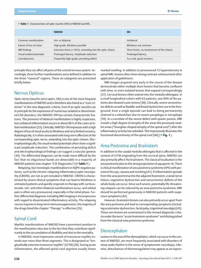
Nervus OpticusOptic nerve (neuritis nervi optici, ON) is one of the most frequent manifestations of NMOSD and is therefore also listed as a “core cri-terion” in the new diagnostic criteria. Even if an optic neuritis can in principle be the expression of numerous isolated or disseminat-ed CNS disorders, the NMOSD-ON has certain characteristic fea-tures. The presence of bilateral manifestation is highly suspicious, but unilateral inflammation occurs in about 80 % of the cases at in-itial manifestation [33]. Clinically, NMOSD-ON impresses with a high degree of loss of visual acuity to blindness and very limited recovery. Radiologically, it is often associated with long-term affection of the corresponding optic nerve, extending into the optic chiasm. Elec-trophysiologically, the visual evoked potentials often show a signif-icant amplitude reduction. The combination of persisting deficit and electrophysiological findings can suggest a vascular genesis [34]. The differential diagnosis can be made more difficult by the fact that no oligoclonal bands are detectable in a majority of NMOSD patients (see chapter “CSF Diagnostics”) (▶table 1 ).
Relapsing, but monotopic manifestations in AQP4-negative pa-tients, such as the chronic-relapsing inflammatory optic neuropa-thy (CRION), are not as yet included in NMOSD. CRION is charac-terized by severe clinical symptoms that can lead to blindness in untreated patients and quickly responds to therapy with corticos-teroids. Uni- and often bilateral manifestations occur, and orbital pain is often very pronounced, especially in the initial phase. Fur-ther differential diagnosis including MRI imaging is inconspicuous with regard to disseminated inflammatory activity. The relapsing course requires in long-term immunosuppression, the majority of the drugs listed the chapter “Therapy” is effective [35].
Spinal CordMyelitic manifestations of NMOSD have a prominent position in the manifestation sites due to the fact that they contribute signif-icantly to the accumulation of disability and also to the mortality.
In NMOSD, most impressive variant of transverse myelitis ex-tends over more than three segments. This is designated as “lon-gitudinally extensive transverse myelitis” (LETM) [36]. During acute inflammation, the affected spinal cord segment usually shows
marked swelling. In addition to pronounced T2 hyperintensity in spinal MRI, lesions often show strong contrast-enhancement after application of gadolinium.
MRI images acquired very early in the course of the disease demonstrate either multiple short lesions that become confluent with time, or even isolated lesions that expand correspondingly [37]. Cervical lesions often extend into the medulla oblongata. In a small longitudinal cohort with 63 patients, one-fifth of the pa-tients also showed cystic lesions [38]. Clinically, severe sensorimo-tor deficits as well as bladder and bowel dysfunction are in the fore-ground. Even a single episode can lead to being permanently chained to a wheelchair due to severe paraplegia or tetraplegia [39]. As a correlate of the severe defect with spastic paresis, MRI reveals a high degree of atrophy of the areas the previously swol-len areas (“hourglass-shaped atrophy of the spinal cord”) after the inflammatory activity has subsided. This impressively illustrates the functional discontinuity of the spinal cord [40] ( ▶Fig. 1 ).
Area Postrema and BrainstemIn addition to the caudal medulla oblongata that is affected in the context of LETM originating from the cervical spine, NMOSD can also primarily affect the brainstem. The classical localization is the area postrema (due to the strong expression of aquaporin-4). There is clinical manifestation of area postrema syndrome (APS) with per-sistent hiccups, nausea and vomiting [41]. If inflammation spreads from the area postrema into the adjacent brainstem, cranial nerve failure, vegetative dysfunction and sensorimotor deficits of the whole body can occur. Since such severe, potentially life-threaten-ing relapses can be induced by an area postrema syndrome, MRI should be performed generously in NMOSD patients with suspi-cious symptoms [42].
However, brainstem lesions can also primarily occur apart from the area postrema and lead to corresponding symptoms (includ-ing oculomotor dysfunction, facial palsy, trigeminal neuralgia) [43]. These rare lesions are summarized in the revised diagnostic crite-ria under the term “acute brainstem syndrome” and distinguished from the classical area postrema syndrome.
DiencephalonLesions in the area of the diencephalon, which can occur in the con-text of NMOSD, are most frequently associated with disorders of sleep-wake rhythm in the sense of symptomatic narcolepsy. Like-wise, disturbances of thermoregulation may appear as an expres-
▶table 1 Characteristics of optic neuritis (ON) in NMOSD and MS.
NMOsD Ms
Common manifestation Uni- or bilateral Unilateral
Extent of loss of vision High grade, blindness possible Blindness not common
MRI findings Extensive lesion ( > 50 %), extending into the optic chiasm Short lesion, no involvement of the chiasm
Convalescence Frequently high-grade, persisting deficit As a rule, good recovery
E38
Pfeuffer S et al. NMO Spectrum Disorders. Neurology International Open 2017; 1: E36–E47
sion of hypothalamic dysfunction or endocrinological syndromes with underlying pituitary insufficiency. MRI correlates of these dis-orders are signal alterations in the region of the hypophysis, hypo-thalamus or adjacent to the third ventricle [44, 45]. These symp-toms are summed up in the revised diagnosis criteria under the term “diencephalic syndrome”.
Cerebral HemispheresOne of the clearest changes in the diagnostic criteria of NMOSD is in relation to the occurrence of cerebral white matter lesions. In the previous revision of the NMO diagnostic criteria, such lesions were listed as “red flags”, if not as an exclusion criterion for diagnosis. The only exception was leukoencephalopathy of alternative origin, for instance, as a result of cerebral microangiopathy. Meanwhile, however, longitudinal studies have shown that up to two-thirds of NMOSD patients also develop inflammatory white matter lesions. There are some morphological differences compared to MS-typi-cal lesions. Thus, NMOSD-associated lesions are usually larger and in the acute phase more markedly edematous [46]. These lesions are often localized subcortical and periventricular, but the latter
are not oriented perpendicularly to the lateral ventricles (like the “Dawson’s fingers” in the MS), but align themselves parallel to this, affecting over 50 % of the roof of the ventricle. Subcortical lesions usually follow the course of corticospinal tracts. In contrast, NMOSD is unlikely if disseminated, juxtacortical lesions are present or the inferior temporal lobes are involved; these lesions should suggest a diagnosis of MS [47]. Isolated cases running a fulminant course with cerebral demyelination, cerebral edema with herniation and death have been described [48] ( ▶Fig. 2).
Diagnosis Based on the Revised Diagnos-tic CriteriaSeropositive NMOSDIn presence of AQP4 antibodies, a diagnosis of NMO-spectrum dis-order can and should be made if one of the six sites is affected. For example, in the case of optic neuritis and detection of AQP4 anti-bodies, a diagnosis of seropositive NMOSD should be strived with all its therapeutic consequences.
▶Fig. 1 MRI findings in NMOSD-associated ON and LETM of a single patient. a, b: Coronary T2 and T1Gd visualization of the optic nerve at the level of the optic chiasm with signs of blood brain barrier disruption and edematous swelling due to bilateral ON. c/d: Sagittal representation of the spinal cord in T2 b and T1Gd c. LETM extending over at least 9 segments with edematous swelling and barrier disorder. e/f: Axial representation correlated to c/d: at segment Th1. Typical is the concentric affection of the spinal cord, which also leads to the gray matter being affected. Clinically, the pa-tient presented with high-grade tetraplegia.
E39
Pfeuffer S et al. NMO Spectrum Disorders. Neurology International Open 2017; 1: E36–E47
Original Article
The criterion of temporal dissemination in time the diagnosis of NMOSD does not exist because of the existence of monophasic dis-ease coureses. This appears to be meaningful in the face of accu-mulation of severe disabilities during disease exacerbation and the associated necessity to start a disease-modifying therapy quickly.
It is important to note that cell-based detection methods should be used because they are superior to the ELISA-based method in specificity and sensitivity. False-positive antibody detection in co-horts with definitive diagnosis of MS was found in 1.3 % of cases in ELISA tests, whereas in similar cohorts, there were only 0.1 % false-positive results in studies using cell-based detection meth-ods [49].
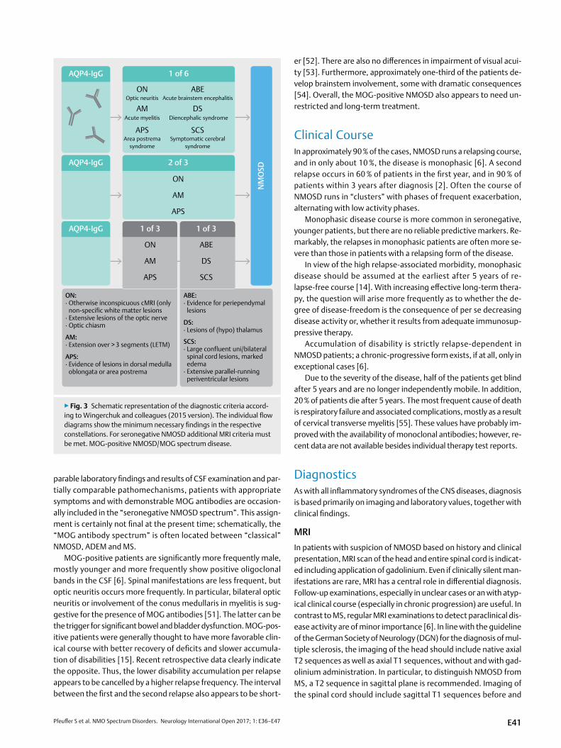
Seronegative NMOSDIn absence of antibodies against AQP4, more stringent criteria apply for classifying an inflammatory syndrome of the CNS as NMOSD. In contrast to the seropositive variant, there is dissemination to at least two sites necessary. At least one of which must correspond to a “clas-sical” site (spinal cord, optic nerve or area postrema). Furthermore, the additional requirements for MRI findings listed in ▶Fig. 3 apply. Special care is required to differentiate suspected NMOSD with optic
neuritis and cerebral demyelination areas. The diagnosis of NMOSD can only be made in this situation if the optic neuritis shows clear signs of NMOSD-ON. Due to therapeutic consequences of the as-signment of a case to either MS or NMOSD, diagnosis should be done in consultation with specialists or by a specialized center. As already mentioned, the diagnostic criteria do not include a criterion of dis-semination in time. Accordingly, NMOSD can not be diagnosed in seronegative patients with a monotopic manifestation but with a re-lapsing course (in ~10% of patients) [14].
MOG-positive NMOSD/MOG spectrum disorderIn recent years, autoantibodies to the myelin oligodendrocyte gly-coprotein (MOG) have been detected in about a quarter of the AQP antibody-negative patients with clinical suspicion of NMOSD [6]. In seropositive patients, however, they are virtually never demon-strable. In contrast to AQP4, MOG is not an astrocytic protein, but is expressed on the surface of oligodendrocytes and has been con-sidered as a possible autoantigen in multiple sclerosis. Antibodies against MOG have so far been reported mainly in children with acute demyelinating encephalomyelitis (ADEM), where they are more frequently detectable [50]. Due to the clinical course, com-
▶Fig. 2 MRI findings of intracerebral manifestations in NMOSD. a/b: axial and sagittal T2 imaging of the brain stem in patients with area postrema syndrome and multiple intracranial nerve deficits (including oculomotor dysfunction and dysphagia). The cervical spinal cord also shows a section of an LETM: section of the upper cervical region. c: Axial FLAIR representation of the dicephalon of a patient with newly diagnosed narcolepsy and evidence of AQP4 antibodies. d: Evidence of periventricular white matter lesions in patients with known NMOSD. In contrast to MS-typical lesions, here they run a parallel course along the ventricle.
E40
Pfeuffer S et al. NMO Spectrum Disorders. Neurology International Open 2017; 1: E36–E47
parable laboratory findings and results of CSF examination and par-tially comparable pathomechanisms, patients with appropriate symptoms and with demonstrable MOG antibodies are occasion-ally included in the “seronegative NMOSD spectrum”. This assign-ment is certainly not final at the present time; schematically, the “MOG antibody spectrum” is often located between “classical” NMOSD, ADEM and MS.
MOG-positive patients are significantly more frequently male, mostly younger and more frequently show positive oligoclonal bands in the CSF [6]. Spinal manifestations are less frequent, but optic neuritis occurs more frequently. In particular, bilateral optic neuritis or involvement of the conus medullaris in myelitis is sug-gestive for the presence of MOG antibodies [51]. The latter can be the trigger for significant bowel and bladder dysfunction. MOG-pos-itive patients were generally thought to have more favorable clin-ical course with better recovery of deficits and slower accumula-tion of disabilities [15]. Recent retrospective data clearly indicate the opposite. Thus, the lower disability accumulation per relapse appears to be cancelled by a higher relapse frequency. The interval between the first and the second relapse also appears to be short-
er [52]. There are also no differences in impairment of visual acui-ty [53]. Furthermore, approximately one-third of the patients de-velop brainstem involvement, some with dramatic consequences [54]. Overall, the MOG-positive NMOSD also appears to need un-restricted and long-term treatment.
Clinical CourseIn approximately 90 % of the cases, NMOSD runs a relapsing course, and in only about 10 %, the disease is monophasic [6]. A second relapse occurs in 60 % of patients in the first year, and in 90 % of patients within 3 years after diagnosis [2]. Often the course of NMOSD runs in “clusters” with phases of frequent exacerbation, alternating with low activity phases.
Monophasic disease course is more common in seronegative, younger patients, but there are no reliable predictive markers. Re-markably, the relapses in monophasic patients are often more se-vere than those in patients with a relapsing form of the disease.
In view of the high relapse-associated morbidity, monophasic disease should be assumed at the earliest after 5 years of re-lapse-free course [14]. With increasing effective long-term thera-py, the question will arise more frequently as to whether the de-gree of disease-freedom is the consequence of per se decreasing disease activity or, whether it results from adequate immunosup-pressive therapy.
Accumulation of disability is strictly relapse-dependent in NMOSD patients; a chronic-progressive form exists, if at all, only in exceptional cases [6].
Due to the severity of the disease, half of the patients get blind after 5 years and are no longer independently mobile. In addition, 20 % of patients die after 5 years. The most frequent cause of death is respiratory failure and associated complications, mostly as a result of cervical transverse myelitis [55]. These values have probably im-proved with the availability of monoclonal antibodies; however, re-cent data are not available besides individual therapy test reports.
DiagnosticsAs with all inflammatory syndromes of the CNS diseases, diagnosis is based primarily on imaging and laboratory values, together with clinical findings.
MRIIn patients with suspicion of NMOSD based on history and clinical presentation, MRI scan of the head and entire spinal cord is indicat-ed including application of gadolinium. Even if clinically silent man-ifestations are rare, MRI has a central role in differential diagnosis. Follow-up examinations, especially in unclear cases or an with atyp-ical clinical course (especially in chronic progression) are useful. In contrast to MS, regular MRI examinations to detect paraclinical dis-ease activity are of minor importance [6]. In line with the guideline of the German Society of Neurology (DGN) for the diagnosis of mul-tiple sclerosis, the imaging of the head should include native axial T2 sequences as well as axial T1 sequences, without and with gad-olinium administration. In particular, to distinguish NMOSD from MS, a T2 sequence in sagittal plane is recommended. Imaging of the spinal cord should include sagittal T1 sequences before and
▶Fig. 3 Schematic representation of the diagnostic criteria accord-ing to Wingerchuk and colleagues (2015 version). The individual flow diagrams show the minimum necessary findings in the respective constellations. For seronegative NMOSD additional MRI criteria must be met. MOG-positive NMOSD/MOG spectrum disease.
E41
Pfeuffer S et al. NMO Spectrum Disorders. Neurology International Open 2017; 1: E36–E47
Original Article
after gadolinium application as well as a sagittal T2 sequence. Axial sequences in axial T1 and T2 sequences weighting complete the basic program. In the case of clinical suspicion of an affection of certain specific structures (optic nerve, brain stem, etc.), appropri-ate thin-sliced imaging of these structures is recommended [6].
CSF diagnosisLumbar puncture should be performed for the purpose of differen-tial diagnosis. Typically, NMOSD patients exhibit lymphogranulo-cytic pleocytosis with the presence of neutro- and eosinophilic granulocytes. The cell count can increase significantly in acute at-tack, values of more than 50 cells/μL have been regularly observed. Oligoclonal bands are only detectable in 10–20 % of the patients and should give rise to a more thorough diagnostic process. A poly-specific antiviral immune response (“MRZ reaction”) is detectable only in isolated cases [56]. This is in contrast to MS, where oligo-clonal bands are detectable in over 90 % of cases [23]. Numerous other observations have been published which have a high speci-ficity for NMOSD, but are still of no relevance in clinical practice.
These include, among others, an increase in the levels of inter-leukin-6 and detection of glial fibrillary acidic protein as a correlate of astrocytic damage [57]. Extensive measurements of the cytokine levels in the CSF or the determination of AQP4 or MOG antibodies in the CSF have no confirmed value [58].
Diagnosis of AQP4 and MOG Antibodies in SerumDue to the impressive specificity of a positive AQP4 antibody find-ing, a test should always be carried out in case of suspicion. Three important findings are to be taken into account in the test: firstly, the strength of a finding depends to a considerable extent on the related diagnostic process. Secondly, AQP4 titers may drop after therapy or in phases of low disease activity or seroconversion may even occur in a patient. Thirdly, in patients initially seronegative, AQP4 antibodies may be detected over time [59].
The first available tests detected AQP4 antibodies using ELISA technology. Since no native but denatured AQP4 was used in this test method, the sensitivity was only 60 %. Cell-based methods, in which serum is incubated with AQP4-expressing cells and subse-quently evaluated by means of flow cytometry or microscopy, reach values of up to 77 %. Accordingly, only the latter methods should be applied [49].
In seronegative patients with suspicion of NMOSD, a diagnosis should be based on the presence of MOG antibodies. Here too, cell-based test methods should be used [50].
In the case of seronegative patients with suspicion of NMOSD, the test should be repeated with regard to AQP4 and MOG anti-bodies, and blood should be drawn before the start of therapy. In individual cases, the corresponding antibodies were detectable in the CSF in patients with inconspicuous serum findings. According-ly, in seronegative patients with high clinical suspicion, such CSF diagnosis may be useful [60].
Differential diagnosisThe most important differential diagnosis of NMO-spectrum dis-orders is multiple sclerosis. A confirmed diagnostic classification is essential due to major differences in therapy. It is therefore not sur-prising that most of the “red flags” explicitly named in the diagnos-tic criteria for NMOSD of 2015 are simultaneously reliable charac-teristics of MS.
MRI findings of periventricular lesions, lesions in the inferior temporal lobe, “Dawson fingers” and juxtacortical lesions, espe-cially involving the U fibers, more likely suggest multiple sclerosis.
The cranial MRI of NMOSD patients is usually inconspicuous, es-pecially when patients with symptomatic cerebral syndrome are excluded [47]. Even if due to the localization there is no suspicion of MS, cases with relevant lesions in the cerebral cranial MRI require particularly critical evaluation.
The detection of oligoclonal bands in the CSF is more likely suggestive of multiple sclerosis, whereas the demonstration of granulocytes in CSF suggests NMOSD. ▶table 2 lists “red flags” in the differential diagnosis of the NMOSD.
In addition to multiple sclerosis, other systemic and CNS auto-immune diseases, vasculitides, malignomas as well as hereditary and infectious CNS diseases should be taken into account in differ-ential diagnosis. Attention is specially drawn to neurosarcoidosis since it can manifest itself with NMOSD-typical symptoms such as optic neuritis and LETM, and presents great difficulties in differen-tial diagnosis [61].
In general, LETM as a common manifestation cannot be classi-fied solely on the basis of MRI findings. Corresponding cases have been documented by Trebst and colleagues [36].
The diagnosis is sometimes made more difficult by the detec-tion of antinuclear antibodies in NMOSD patients [62]. Further-more, several autoimmune diseases can also be present concomi-tantly. For instance, there are reports of patients with concomitant systemic lupus erythematosus and NMOSD [63]. ▶table 3 gives a selected overview of differential diagnoses.
▶table 2 “Red flags” in NMOSD, modified according to [6].
Course of the disease– Chronic-progressive course, relapse-independent deterioration– Progressive worsening of suspicious symptoms over several weeks– Spontaneous remission of neurological deficits after a short time
Clinical and laboratory findings– Detection of oligoclonal bands in the CSF– Serologic testing of serum/CSF (e. g., HIV, syphilis, borreliosis)– Detection of solid or hematological tumor diseases– Proof of generalized or hilar lymphadenopathy
MRI findings– relevant lesion burden in cranial MRI, in particular with– Detection of MS-typical lesions (“Dawson’s fingers”)– Detection of cortical lesions– Detection of lesions of the inferior temporal lobes– Proof of persistent gadolinium-enhancement– Detection of persistent short-range lesions in the spinal cord
E42
Pfeuffer S et al. NMO Spectrum Disorders. Neurology International Open 2017; 1: E36–E47
TherapyTreatment of acute relapsesAs with multiple sclerosis, there are essentially four procedures available for the treatment of acute relapses of NMOSD: corticos-teroids, plasmapheresis, immunoadsorption and intravenous im-munoglobulins.
High-dose administration of intravenous corticosteroids (1 g/d methylprednisolone over 5 days) is currently the most frequently used therapy for acute attacks [64, 65]. In the absence of improve-ment after therapy with 1 g/d methylprednisolone, a further cycle of 2 g/d can be added on for a further 5 days. Ulcer and thrombosis prophylaxis are essential.
Alternatively, plasmapheresis or immunoadsorption can be car-ried out. Both methods have shown comparable efficacy. The se-lection can accordingly be oriented, besides availability, to the side effects/to the side effects profile profile (necessity of having to sub-stitute foreign blood components after plasmapheresis, stronger affection of the coagulation function after plasmapheresis). The effect of plasmapheresis or immunoadsorption is not categorical-ly different in seropositive or seronegative patients [66].
In a recent retrospective study of the NEMOS study group, it was found that there was complete remission in only 21.6 % of the pa-tients and partial remission in 72.4 % of the patients after all ther-apeutic procedures had been exhausted. It is astonishing that al-ready 19.1 % of the patients achieved complete remission after the first of up to five therapy courses. Plasma exchange methods have led to improved symptom control especially in patients with acute myelitis. The positive effect of plasma exchange decreased with the increase in previously undergone therapy procedures. These data were not significant when comparing all kinds of relapses but other studies support the estimation that plasma exchange is superior [65, 67]. Individual studies also showed a cumulative effect of com-bined steroid pulse and apheresis treatment (for example plasma exchange and infusion of corticosteroids on alternating days) [68].
Intravenous immunoglobulins (IVIG) are currently only used in exceptional cases for the treatment of relapses, as for instance in children. Currently there are no larger studies, but patients are being recruited for a study on the treatment of acute transverse myelitis with IVIG [69]. Different case series have documented the positive effects of IVIG so that its use as second-line therapy can be justified (0.4 g/kg for five consecutive days) [70]. On the one hand, the therapy should be carried out only after plasma exchange has been completed because efficacy is not clearly established and there is the risk of leaching. Individual studies in pediatric patients showed a positive effect of IVIG in maintenance therapy (dose: 0.4 g/kg bw every 4 weeks) [71] ( ▶table 4).
Disease-modifying therapyContrasting the 15 approved medications for the treatment of MS, there is no approved drug for the treatment of NMOSD. This is mainly due to the absence of relevant randomized, placebo-con-trolled trials. In addition to the rarity of the disease, other factors contribute to this situation. On the one hand, approval authorities usually do not recognize comparative studies between several un-authorized medications. On the other hand, there are significant ethical concerns about placebo-controlled studies in the face of threat of severe impairment after a single relapse. For the first time, different drugs are tested in controlled clinical trials. Different
▶table 4 Overview of available acute therapies for relapses in NMOSD.
Medication Dosage and interval
Corticosteroids [63] 1 g Methylprednisolone/d over 5 days, if needed escalation with 2 g Methylprednisolone/d (latter over a maximum of 5 days)
Plasmapheresis [64] 5–7 cycles every other day (exchange of 1.1 to 1.5 plasma volumes per session)
Immunadsorption [64] 5–6 cycles on consecutive days; tryptophan or protein A-columns (adsorption of 2 to 2.5 plasma volumes per session)
Intravenous immunoglobulins [69, 70] 0.4 g/kg/d over 3–5 days, maintenance dose: 0.4 g/kg once a month
▶table 3 Overview of selected differential diagnoses of the NMOSD.
Pfeuffer S et al. NMO Spectrum Disorders. Neurology International Open 2017; 1: E36–E47
Original Article
methods, for instance, asymmetric randomization in favor of the study medication, definition of the first relapse as clinical end point or narrow time limitation of the study medication, were used for risk minimization [72].
With the exception of mitoxantrone, rituximab and azathio-prine, most drugs known from MS therapy are either ineffective (e. g., glatiramer acetate) or even counterproductive (e. g., interferon-beta, fingolimod, natalizumab, alemtuzumab). Under no circumstances should they be used in the therapy of NMOSD. Instead, classical immunosuppressants and various monoclonal an-tibodies are prefered in treatment of NMOSD [73].
As a rule, according to how well a drug is clinically established, three categories of drugs can be defined: (1) “established” drugs with longer experience in off-label use, (2) medication successfully used in individual cases, (3) drugs currently being tested in studies.
Even though a wide range of drugs has already been used in the therapy of NMOSD, the greatest amount of experience is available in relation to azathioprine, mycophenolate mofetil and rituximab. Accordingly, these drugs are regarded as first choice drugs. In some cases, the recommendation is made to use azathioprine and mycophenolate mofetil for “mild” forms and rituximab for “highly active” forms of the disease. However, in view of the risk of mortal-ity, such considerations should be left to a specialized center.
For azathioprine, several studies with approximately 100 pa-tients each showed significant reduction in the relapse rate and dis-ability accumulation [74, 75]. The high dropout rate of around 60 % after two years, usually due to side effects, as for example elevated liver enzymes, appears to limit its use. Due to the delayed onset of drug effect, overlapping therapy with oral prednisone appears to be useful after steroid pulse [76].
Mycophenolate mofetil (MMF) was effective in several smaller studies with up to 59 patients [77]. Randomized prospective stud-ies, which allow a definite statement compared to azathioprine, are not available. However, the fact that therapy dropouts were rarer during the observation period than in the case of azathioprine speaks in its favor.
There are also positive data in support of the use of anti-CD20 an-tibody rituximab in NMOSD patients. Numerous studies showed a sig-
nificant reduction in the rate of relapses and accumulation of disabil-ity. Long-term data of up to 67 months are available for this drug [78, 79]. Overall, its efficacy is superior to azathioprine and MMF, which is why its use in “severe disease course” is recommended [80]. Because a priori the risk of rapid disability accumulation is already high after a single relapse event in NMOSD, we tend to use primarily rituximab, particularly in seropositive patients. We believe that this approach is supported by the lack of reliable prognostic markers.
In particular, with regard to rituximab, there are two treatment schemes both of which are equally effective: Scheme (1) was orig-inally designed for the treatment of lymphoma. Despite the high-er amount of drug used, it lacks superiority compared to scheme (2). As costs of this off-label use have to be negotiated with the health insurance, scheme (1) has largely been abandoned in Ger-many.
Maintenance therapies should be performed when there is a re-newed increase of CD19+ B cells or CD27 + memory B cells in the peripheral blood, but at the latest after 6 months in each case. Var-ious biomarkers, such as the FCGR3A polymorphism, associated with a poorer response to therapy, are not currently established in the clinical routine [79].
Tocilizumab is the second successfully used monoclonal anti-body. Tocilizumab binds to the IL6 receptor, without activating it and thus blocks the proinflammatory effect of the cytokine IL6. In 2013, first positive data were published [81] and in 2015, the drug was found to be effective in a small study of 8 rituximab-refracto-ry 8 patients [82]. Usually 8 mg/kg body weight is infused every 4 weeks ( ▶table 5).
Eculizumab, which intervenes in the complement cascade by binding to the protein C5, is an option as well; data, however, are sparse but positive. In 2013, an open-label study with 14 patients observed over a period of 48 weeks reported disease stabilization [83]. Under treatment with eculizumab, there was a significantly increased susceptibility to meningococcal meningitis. According-ly, in addition to high clinical vigilance, the administration of a pro-tective vaccination and/or an antibiotic prophylaxis according to summary of product characteristics (SMPC) is indicated.
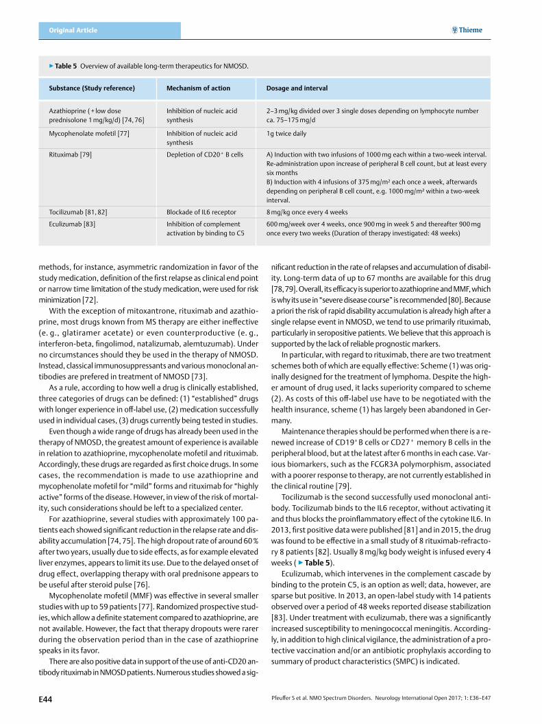
▶table 5 Overview of available long-term therapeutics for NMOSD.
substance (study reference) Mechanism of action Dosage and interval
2–3 mg/kg divided over 3 single doses depending on lymphocyte number ca. 75–175 mg/d
Mycophenolate mofetil [77] Inhibition of nucleic acid synthesis
1g twice daily
Rituximab [79] Depletion of CD20 + B cells A) Induction with two infusions of 1000 mg each within a two-week interval. Re-administration upon increase of peripheral B cell count, but at least every six monthsB) Induction with 4 infusions of 375 mg/m² each once a week, afterwards depending on peripheral B cell count, e.g. 1000 mg/m² within a two-week interval.
Tocilizumab [81, 82] Blockade of IL6 receptor 8 mg/kg once every 4 weeks
Eculizumab [83] Inhibition of complement activation by binding to C5
600 mg/week over 4 weeks, once 900 mg in week 5 and thereafter 900 mg once every two weeks (Duration of therapy investigated: 48 weeks)
E44
Pfeuffer S et al. NMO Spectrum Disorders. Neurology International Open 2017; 1: E36–E47
Current therapies of NMOSD and PerspectivesAlthough, as stated at the beginning of the previous section, clin-ical studies on the implementation of therapies in NMOSD remain difficult, several large-scale Phase III trials are recruiting patients.
As a consequence of the positive but highly preliminary results of an open-label study of eculizumab, the manufacturer initiated a phase III study, which is expected to deliver first data by the end of 2016. However, only AQP4-seropositive patients were included. Eculizumab was added to an existing immunosuppression – with the exception of rituximab.
In the N-MOMENTUM study, patients are randomized 3: 1 to MEDI-551, a B cell-depleting antibody directed against CD19. The therapy duration is limited to 197 days, followed by the option to participate in an extension study [72].
Two studies were started to evaluate the IL6 receptor antibody SA-237, which is a derivative of tocilizumab and has an approxi-mately fourfold longer half-life. In the first phase III trial, 70 patients are scheduled to receive either SA-237 as monotherapy or place-bo. The parallel-running Sakura-SKY study randomizes 90 patients to SA-237 or placebo under continuation of the existing immuno-suppressive therapy. The results of the studies on MEDI-551 and SA-237 are not expected to be available until 2019 at the earliest.
Other therapeutics are in early stages of development, such as the monoclonal antibody aquaporumab, which binds to AQP4 and prevents the interaction of the protein with endogenous AQP4 antibodies and the resulting cell damage [84]. However, no clinical trials have yet been registered with this antibody. Ublituximab is another B-cell depleting antibody in Phase I trial (NCT02276963) ( ▶table 6).
Conclusion for PracticeBy revising the diagnostic criteria, the term neuromyelitis optica (NMO) is being increasingly replaced by NMO-spectrum diseases (NMOSD). In the future, the new term will cover a broader range of clinical presentations with presumably comparable immunopatho-genesis. The “MOG Antibody Spectrum”, a new entity crystallized out of diseases, has its own biomarker and partly already included in the NMO spectrum. However, monotopic presentations such as
isolated, relapsing transverse myelitis or chronic recurrent inflam-matory optic neuritis are not (yet) included in this spectrum. For the first time, controlled therapies have been initiated for NMOSD patients, but results are still pending.
The initial diagnosis should include spinal and cranial MRI images as well as CSF and laboratory diagnostics. Special attention should be paid to cell-based antibody diagnosis. Following acute therapy where necessary, NMOSD patients should be referred to a specialized center for further diagnosis and implementation of long-term immune therapy concepts. Taking up contact to na-tional or international study groups (e.g., german NMO study group (www.nemos-net.de)) is recommended.
Conflict of interest
The authors declare that they were not guided by any economic in-terests in the preparation of this article. Steffen Pfeuffer declares that he has received travel reimbursements by Sanofi Genzyme and lecture honoraria by Sanofi Genzyme and Biogen. Christine Strip-pel declares no conflicts of interests. Heinz Wiendl received fees for work in scientific committees of the companies Bayer Health-care, Biogen GmbH Germany and Biogen Idec, Sanofi-Genzyme, Merck-Serono, Novartis, Roche and Teva. He received lecture fees and reimbursement of travel costs from Bayer VitalGmbH, der Bayer Schering AG, der BiogenGmbH Deutschland und Biogen Idec, CSL Behring, EMD Serono, Fresenius Medical Care, Genzyme, Merck-Serono, Omniamed,Novartis, Sanofi-Aventis und Teva, and consultant fees from Biogen GmbH Deutschland und Biogen Idec, Merck-Serono, Novartis, Omniamed, Roche und Sanofi-Genzyme. His scientific work is supported by Bayer HealthCare, Bayer Health-Care, Biogen GmbH Germany and Biogen Idec, Merck-Serono, No-vartis, Sanofi-Genzyme, Sanofi US and Teva, and the German Fed-eral Ministr y of Education and Research (BMBF) ), the Else-Kröner-Fresenius Foundation, the Fresenius Foundation, the Hertie Foundation, the North Rhine-Westphalian Ministry of Edu-cation and Research, the Interdisciplinary Center for Clinical Research (IZKF) Münster and the RE Children’s Foundation.
▶table 6 Overview of active phase III therapy studies on NMOSD, with study data registered up to 09/2016.
study Drug studied Mechanism of action benchmark data Identification number (clinicaltrials.gov)
“PREVENT” Eculizumab/Placebo Inhibition of complementactivation by binding to C5
Completion planned 12/2016 (132 Patients), only AQP4 +
NCT01892345
„N-MOmentum“ MEDI-551/Placebo Depletion of CD19+ B cells Completion planned 04/2020 (212 Patients), AQP4 + / −
Pfeuffer S et al. NMO Spectrum Disorders. Neurology International Open 2017; 1: E36–E47
Original Article
References
[1] Jarius S, Wildemann B. The history of neuromyelitis optica. J Neuroinflammation 2013; 10: 8
[2] Wingerchuk DM, Hogancamp WF, O‘Brien PC et al. The clinical course of neuromyelitis optica (Devic’s syndrome). Neurology 1999; 53: 1107–1114
[3] Lennon VA, Wingerchuk DM, Kryzer TJ et al. A serum autoantibody marker of neuromyelitis optica: Distinction from multiple sclerosis. Lancet 2004; 364: 2106–2112
[4] Lennon VA, Kryzer TJ, Pittock SJ et al. IgG marker of optic-spinal multiple sclerosis binds to the aquaporin-4 water channel. J Exp Med 2005; 202: 473–477
[5] Wingerchuk DM, Lennon VA, Pittock SJ et al. Revised diagnostic criteria for neuromyelitis optica. Neurology 2006; 66: 1485–1489
[6] Wingerchuk DM, Banwell B, Bennett JL et al. International consensus diagnostic criteria for neuromyelitis optica spectrum disorders. Neurology 2015; 85: 177–189
[7] Flanagan EP, Cabre P, Weinshenker BG St et al. Epidemiology of aquaporin-4 autoimmunity and neuromyelitis optica spectrum. Ann Neurol 2016; doi: 10.1002/ana.24617
[8] Asgari N, Lillevang ST, Skejoe HP et al. A population-based study of neuromyelitis optica in Caucasians. Neurology 2011; 76: 1589–1595
[9] Bizzoco E, Lolli F, Repice AM et al. Prevalence of neuromyelitis optica spectrum disorder and phenotype distribution. J Neurol 2009; 256: 1891–1898
[10] Kira J, Kanai T, Nishimura Y et al. Western versus Asian types of multiple sclerosis: Immunogenetically and clinically distinct disorders. Ann Neurol 1996; 40: 569–574
[11] Tanaka K, Tani T, Tanaka M et al. Anti-aquaporin 4 antibody in selected Japanese multiple sclerosis patients with long spinal cord lesions. Mult Scler 2007; 13: 850–855
[12] Tanaka M, Tanaka K, Komori M et al. Anti-aquaporin 4 antibody in Japanese multiple sclerosis: The presence of optic spinal multiple sclerosis without long spinal cord lesions and anti-aquaporin 4 antibody. J Neurol Neurosurg Psychiatry 2007; 78: 990–992
[13] Kira J. Multiple sclerosis in the Japanese population. Lancet Neurol 2003; 2: 117–127
[14] Jarius S, Ruprecht K, Wildemann B et al. Contrasting disease patterns in seropositive and seronegative neuromyelitis optica: A multicentre study of 175 patients. J Neuroinflammation 2012; 9: 14
[15] Sato DK, Callegaro D, Lana-Peixoto MA et al. Distinction between MOG antibody-positive and AQP4 antibody-positive NMO spectrum disorders. Neurology 2014; 82: 474–481
[16] McKeon A, Lennon VA, Jacob A et al. Coexistence of myasthenia gravis and serological markers of neurological autoimmunity in neuromyeli-tis optica. Muscle Nerve 2009; 39: 87–90
[17] Nagaishi A, Takagi M, Umemura A et al. Clinical features of neuromy-elitis optica in a large Japanese cohort: Comparison between phenotypes. J Neurol Neurosurg Psychiatry 2011; 82: 1360–1364
[18] Wingerchuk DM, Weinshenker BG. The emerging relationship between neuromyelitis optica and systemic rheumatologic autoimmune disease. Mult Scler 2012; 18: 5–10
[19] Spillane J, Christofi G, Sidle KC et al. Myasthenia gravis and neuromyeli-tis opica: A causal link. Mult Scler Relat Disord 2013; 2: 233–237
[20] Wingerchuk DM, Lennon VA, Lucchinetti CF et al. The spectrum of neuromyelitis optica. Lancet Neurol 2007; 6: 805–815
[21] Pittock SJ, Weinshenker BG, Lucchinetti CF et al. Neuromyelitis optica brain lesions localized at sites of high aquaporin 4 expression. Arch Neurol 2006; 63: 964–968
[22] Jarius S, Wildemann B. AQP4 antibodies in neuromyelitis optica: diagnostic and pathogenetic relevance. Nat Rev Neurol 2010; 6: 383–392
[23] Marignier R, Nicolle A, Watrin C et al. Oligodendrocytes are damaged by neuromyelitis optica immunoglobulin G via astrocyte injury. Brain 2010; 133: 2578–2591
[24] Hinson SR, Roemer SF, Lucchinetti CF et al. Aquaporin-4-binding autoantibodies in patients with neuromyelitis optica impair glutamate transport by down-regulating EAAT2. J Exp Med 2008; 205: 2473–2481
[25] Ciccarelli O, Thomas DL, De Vita E et al. Low myo-inositol indicating astrocytic damage in a case series of neuromyelitis optica. Ann Neurol 2013; 74: 301–305
[26] Matthews L, Kolind S, Brazier A et al. Imaging surrogates of disease activity in neuromyelitis optica allow distinction from multiple sclerosis. PLoS One 2015; 10: e0137715
[27] Uzawa A, Mori M, Arai K et al. Cytokine and chemokine profiles in neuromyelitis optica: Significance of interleukin-6. Mult Scler 2010; 16: 1443–1452
[28] Jarius S, Paul F, Franciotta D et al. Cerebrospinal fluid findings in aquaporin-4 antibody positive neuromyelitis optica: results from 211 lumbar punctures. J Neurol Sci 2011; 306: 82–90
[29] Schneider-Hohendorf T, Rossaint J, Mohan H et al. VLA-4 blockade promotes differential routes into human CNS involving PSGL-1 rolling of T cells and MCAM-adhesion of TH17 cells. J Exp Med 2014; 211: 1833–1846
[30] Barnett MH, Prineas JW, Buckland ME et al. Massive astrocyte destruction in neuromyelitis optica despite natalizumab therapy. Mult Scler 2012; 18: 108–112
[31] Jarius S, Aboul-Enein F, Waters P et al. Antibody to aquaporin-4 in the long-term course of neuromyelitis optica. Brain 2008; 131: 3072–3080
[32] Nour MM, Nakashima I, Coutinho E et al. Pregnancy outcomes in aquaporin-4-positive neuromyelitis optica spectrum disorder. Neurology 2016; 86: 79–87
[33] Piccolo L, Woodhall M, Tackley G et al. Isolated new onset ‘atypical’ optic neuritis in the NMO clinic: serum antibodies, prognoses and diagnoses at follow-up. J Neurol 2016; 263: 370–379
[34] Kim HJ, Paul F, Lana-Peixoto MA et al. MRI characteristics of neuromy-elitis optica spectrum disorder: An international update. Neurology 2015; 84: 1165–1173
[35] Petzold A, Plant GT. Chronic relapsing inflammatory optic neuropathy: A systematic review of 122 cases reported. J Neurol. 2014; 261: 17–26
[36] Trebst C, Raab P, Voss EV et al. Longitudinal extensive transverse myelitis – it’s not all neuromyelitis optica. Nat Rev Neurol 2011; 7: 688–698
[37] Flanagan EP, Weinshenker BG, Krecke KN et al. Short myelitis lesions in aquaporin-4-IgG-positive neuromyelitis optica spectrum disorders. JAMA Neurol 2015; 72: 81–87
[38] Kim SH, Huh SY, Hyun JW et al. A longitudinal brain magnetic resonance imaging study of neuromyelitis optica spectrum disorder. PLoS One 2014; 9: e108320
[39] Weinshenker BG, Wingerchuk DM, Vukusic S et al. Neuromyelitis optica IgG predicts relapse after longitudinally extensive transverse myelitis. Ann Neurol 2006; 59: 566–569
[40] Jiao Y, Fryer JP, Lennon VA et al. Aquaporin 4 IgG serostatus and outcome in recurrent longitudinally extensive transverse myelitis. JAMA Neurol 2014; 71: 48–54
[41] Popescu BF, Lennon VA, Parisi JE et al. Neuromyelitis optica unique area postrema lesions: Nausea, vomiting, and pathogenic implications. Neurology 2011; 76: 1229–1237
[42] Takahashi T, Miyazawa I, Misu T et al. Intractable hiccup and nausea in neuromyelitis optica with anti-aquaporin-4 antibody: A herald of acute exacerbations. J Neurol Neurosurg Psychiatry 2008; 79: 1075–1078
Pfeuffer S et al. NMO Spectrum Disorders. Neurology International Open 2017; 1: E36–E47
[44] Kume K, Deguchi K, Ikeda K et al. Neuromyelitis optica spectrum disorder presenting with repeated hypersomnia due to involvement of the hypothalamus and hypothalamus-amygdala linkage. Mult Scler 2015; 21: 960–962
[45] Poppe AY, Lapierre Y, Melancon D et al. Neuromyelitis optica with hypothalamic involvement. Mult Scler 2005; 11: 617–621
[46] Pittock SJ, Lennon VA, Krecke K et al. Brain abnormalities in neuromy-elitis optica. Arch Neurol 2006; 63: 390–396
[47] Matthews L, Marasco R, Jenkinson M et al. Distinction of seropositive NMO spectrum disorder and MS brain lesion distribution. Neurology 2013; 80: 1330–1337
[49] Waters PJ, Pittock SJ, Bennett JL et al. Evaluation of aquaporin-4 antibody assays. Clin Exp Neuroimmunol 2014; 5: 290–303
[50] Kitley J, Woodhall M, Waters P et al. Myelin-oligodendrocyte glycoprotein antibodies in adults with a neuromyelitis optica phenotype. Neurology 2012; 79: 1273–1277
[51] Kitley J, Waters P, Woodhall M et al. Neuromyelitis optica spectrum disorders with aquaporin-4 and myelin-oligodendrocyte glycoprotein antibodies: A comparative study. JAMA Neurol 2014; 71: 276–283
[52] Jarius S, Ruprecht K, Kleiter I et al. MOG-IgG in NMO and related disorders: a multicenter study of 50 patients. Part 2: Epidemiology, clinical presentation, radiological and laboratory features, treatment responses, and long-term outcome. J Neuroinflammation 2016; 13: 280
[53] Pache F, Zimmermann H, Mikolajczak J et al. MOG-IgG in NMO and related disorders: A multicenter study of 50 patients. Part 4: Afferent visual system damage after optic neuritis in MOG-IgG-seropositive versus AQP4-IgG-seropositive patients. J Neuroinflammation 2016; 13: 282
[54] Jarius S, Kleiter I, Ruprecht K et al. MOG-IgG in NMO and related disorders: A multicenter study of 50 patients. Part 3: Brainstem involvement – frequency, presentation and outcome. J Neuroinflam-mation 2016; 13: 281
[55] Wingerchuk DM, Weinshenker BG. Neuromyelitis optica: Clinical predictors of a relapsing course and survival. Neurology 2003; 60: 848–853
[56] Jarius S, Franciotta D, Bergamaschi R et al. Polyspecific, antiviral immune response distinguishes multiple sclerosis and neuromyelitis optica. J Neurol Neurosurg Psychiatry 2008; 79: 1134–1136
[57] Takano R, Misu T, Takahashi T et al. Astrocytic damage is far more severe than demyelination in NMO: A clinical CSF biomarker study. Neurology 2010; 75: 208–216
[58] Chang KH, Ro LS, Lyu RK et al. Biomarkers for neuromyelitis optica. Clin Chim Acta 2015; 440: 64–71
[59] Jiao Y, Fryer JP, Lennon VA et al. Updated estimate of AQP4-IgG serostatus and disability outcome in neuromyelitis optica. Neurology 2013; 81: 1197–1204
[60] Sato DK, Callegaro D, de Haidar Jorge FM et al. Cerebrospinal fluid aquaporin-4 antibody levels in neuromyelitis optica attacks. Ann Neurol 2014; 76: 305–309
[61] Flanagan EP, Kaufmann TJ, Krecke KN et al. Discriminating long myelitis of neuromyelitis optica from sarcoidosis. Ann Neurol. 2016; 79: 437–447
[62] Pittock SJ, Lennon VA, de Seze J et al. Neuromyelitis optica and non organ-specific autoimmunity. Arch Neurol 2008; 65: 78–83
[63] Adawi M, Bisharat B, Bowirrat A. Systemic Lupus Erythematosus (SLE) complicated by Neuromyelitis optica (NMO – Devic’s Disease): Clinic-pathological report and review of the literature. Clin Med Insights Case Rep 2014; 7: 41–47
[65] Kleiter I, Gahlen A, Borisow N et al. Neuromyelitis optica: Evaluation of 871 attacks and 1153 treatment courses. Ann Neurol 2016; 79: 206–216
[66] Bonnan M, Valentino R, Olindo S et al. Plasma exchange in severe spinal attacks associated with neuromyelitis optica spectrum disorder. Mult Scler 2009; 15: 487–492
[67] Watanabe S, Nakashima I, Misu T et al. Therapeutic efficacy of plasma exchange in NMO-IgG-positive patients with neuromyelitis optica. Mult Scler 2007; 13: 128–132
[68] Abboud H, Petrak A, Mealy M et al. Treatment of acute relapses in neuromyelitis optica: Steroids alone versus steroids plus plasma exchange. Mult Scler 2016; 22: 185–192
[69] Absoud M, Gadian J, Hellier J et al. Protocol for a multicentre randomiSed controlled TRial of IntraVEnous immunoglobulin versus standard therapy for the treatment of transverse myelitis in adults and children (STRIVE). BMJ Open 2015; 5: e008312
[70] Elsone L, Panicker J, Mutch K et al. Role of intravenous immunoglobu-lin in the treatment of acute relapses of neuromyelitis optica: Experience in 10 patients. Mult Scler. 2014; 20: 501–504
[71] Viswanathan S, Wong AH, Quek AM et al. Intravenous immunoglobulin may reduce relapse frequency in neuromyelitis optica. J Neuroimmu-nol 2015; 282: 92–96
[72] Cree BA, Bennett JL, Sheehan M et al. Placebo-controlled study in neuromyelitis optica-Ethical and design considerations. Mult Scler 2016; 22: 862–872
[73] Kleiter I, Gold R. Present and future therapies in neuromyelitis optica spectrum disorders. Neurotherapeutics 2016; 13: 70–83
[74] Elsone L, Kitley J, Luppe S et al. Long-term efficacy, tolerability and retention rate of azathioprine in 103 aquaporin-4 antibody-positive neuromyelitis optica spectrum disorder patients: A multicentre retrospective observational study from the UK. Mult Scler 2014; 20: 1533–1540
[75] Costanzi C, Matiello M, Lucchinetti CF et al. Azathioprine: Tolerability, efficacy, and predictors of benefit in neuromyelitis optica. Neurology 2011; 77: 659–666
[76] Mandler RN, Ahmed W, Dencoff JE. Devic’s neuromyelitis optica: A prospective study of seven patients treated with prednisone and azathioprine. Neurology 1998; 51: 1219–1220
[77] Huh SY, Kim SH, Hyun JW et al. Mycophenolate mofetil in the treatment of neuromyelitis optica spectrum disorder. JAMA Neurol 2014; 71: 1372–1378
[78] Kim SH, Huh SY, Lee SJ et al. A 5-year follow-up of rituximab treatment in patients with neuromyelitis optica spectrum disorder. JAMA Neurol 2013; 70: 1110–1117
[79] Kim SH, Jeong IH, Hyun JW et al. Treatment outcomes with rituximab in 100 patients with neuromyelitis optica: Influence of FCGR3A polymorphisms on the therapeutic response to rituximab. JAMA Neurol 2015; 72: 989–995
[80] Trebst C, Jarius S, Berthele A et al. Update on the diagnosis and treatment of neuromyelitis optica: Recommendations of the Neuromyelitis Optica Study Group (NEMOS). J Neurol 2014; 261: 1–16
[81] Ayzenberg I, Kleiter I, Schroder A et al. Interleukin 6 receptor blockade in patients with neuromyelitis optica nonresponsive to anti-CD20 therapy. JAMA Neurol 2013; 70: 394–397
[82] Ringelstein M, Ayzenberg I, Harmel J et al. Long-term therapy with interleukin 6 receptor blockade in highly active neuromyelitis optica spectrum disorder. JAMA Neurol 2015; 72: 756–763
[83] Pittock SJ, Lennon VA, McKeon A et al. Eculizumab in AQP4-IgG-posi-tive relapsing neuromyelitis optica spectrum disorders: An open-label pilot study. Lancet Neurol 2013; 12: 554–562