74
Chapter 19 NMR Spectroscopy

Chapter 19
NMR Spectroscopy
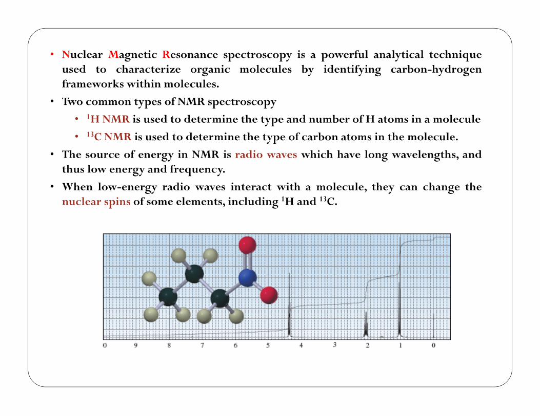
• Nuclear Magnetic Resonance spectroscopy is a powerful analytical techniqueused to characterize organic molecules by identifying carbon-hydrogenframeworks within molecules.
• Two common types of NMR spectroscopy
• 1H NMR is used to determine the type and number of H atoms in a molecule
• 13C NMR is used to determine the type of carbon atoms in the molecule.
• The source of energy in NMR is radio waves which have long wavelengths, andthus low energy and frequency.
• When low-energy radio waves interact with a molecule, they can change thenuclear spins of some elements, including 1H and 13C.
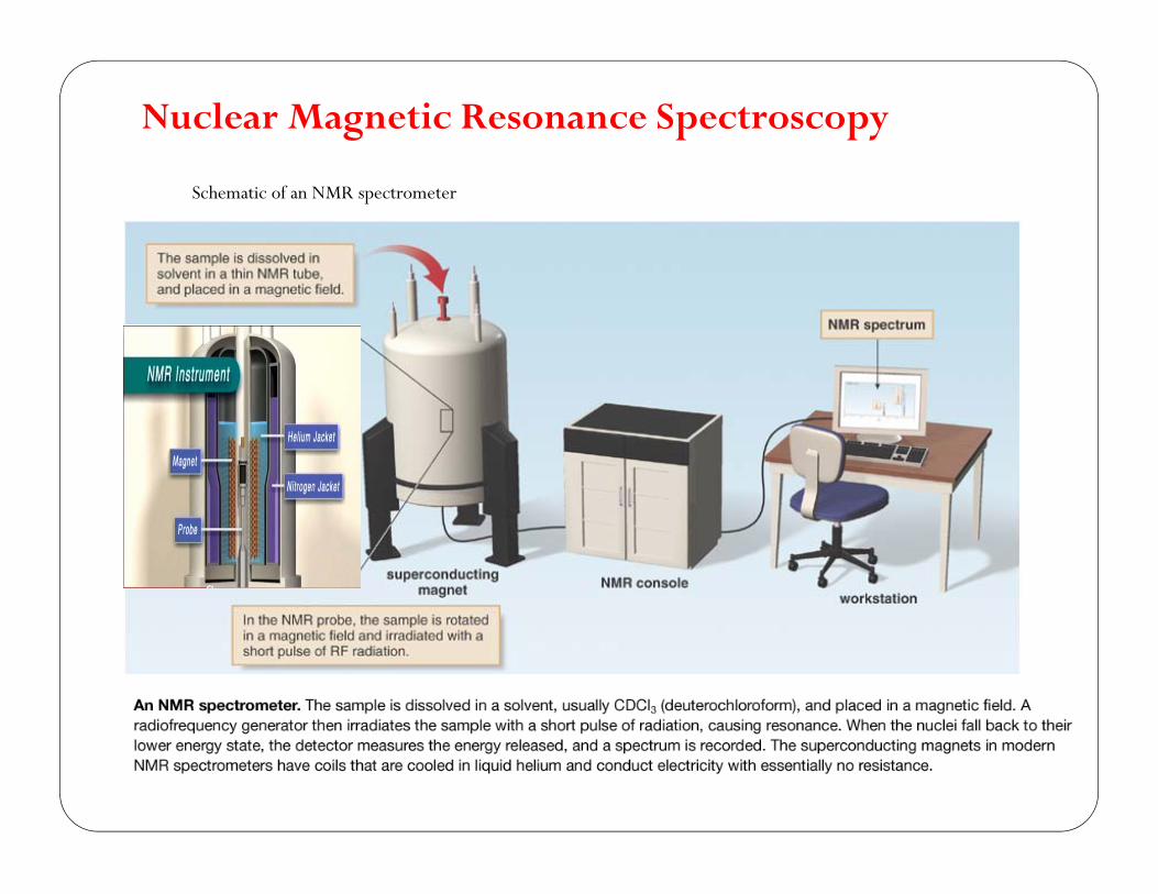
Nuclear Magnetic Resonance Spectroscopy
Schematic of an NMR spectrometer
3

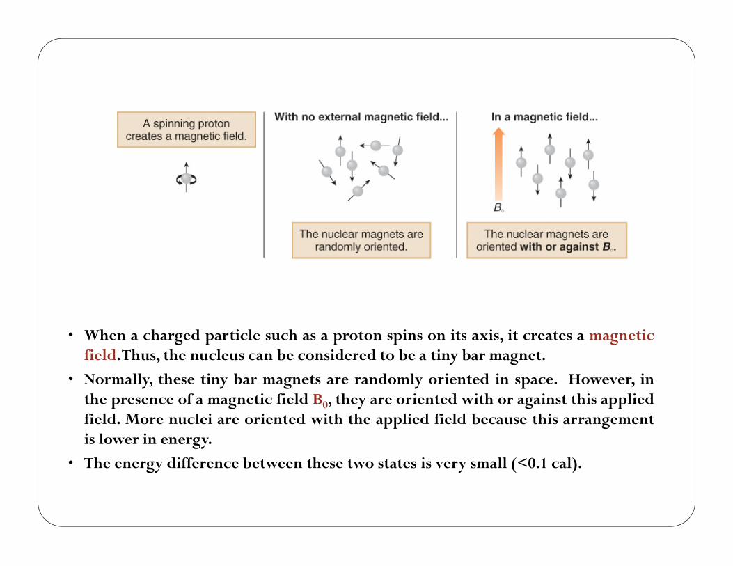
• When a charged particle such as a proton spins on its axis, it creates a magneticfield.Thus, the nucleus can be considered to be a tiny bar magnet.
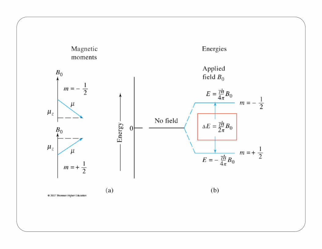
• Normally, these tiny bar magnets are randomly oriented in space. However, inthe presence of a magnetic field B0, they are oriented with or against this appliedfield. More nuclei are oriented with the applied field because this arrangementis lower in energy.
• The energy difference between these two states is very small (<0.1 cal).
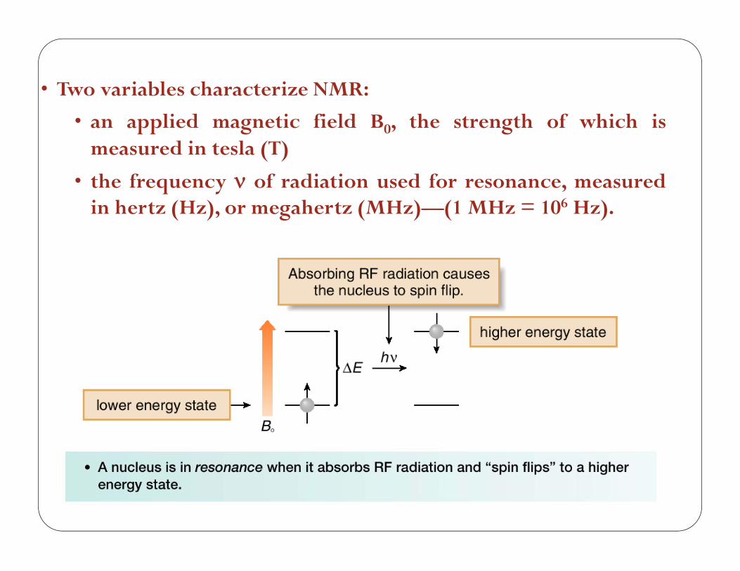
• Two variables characterize NMR:
• an applied magnetic field B0, the strength of which ismeasured in tesla (T)
• the frequency of radiation used for resonance, measuredin hertz (Hz), or megahertz (MHz)—(1 MHz = 106 Hz).
6
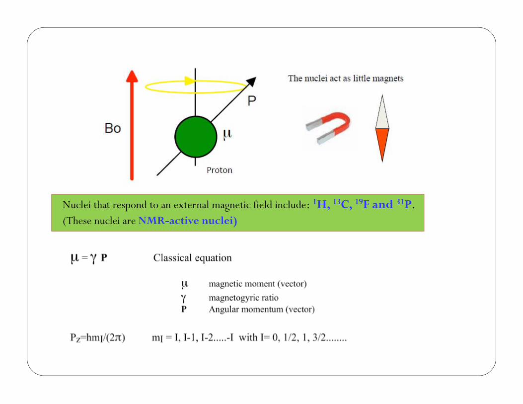
Nuclei that respond to an external magnetic field include: 1H, 13C, 19F and 31P.(These nuclei are NMR-active nuclei)


Example: Many proton NMR instruments employ a magnet that provides a field strength of 4.69T. At what frequency would the hydrogen nucleus absorb in such field?
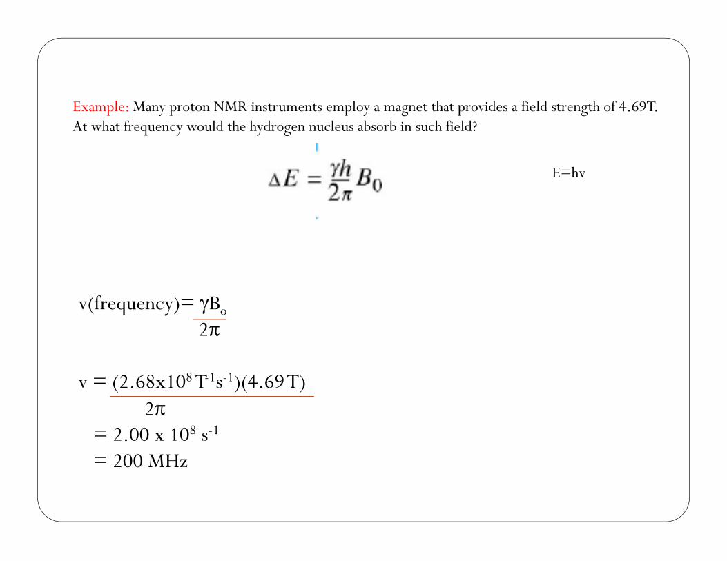
Example: Many proton NMR instruments employ a magnet that provides a field strength of 4.69T. At what frequency would the hydrogen nucleus absorb in such field?
E=hv
v(frequency)= Bo
2
v = (2.68x10⁸ T-1s-1)(4.69 T)2
= 2.00 x 10⁸ s-1
= 200 MHz
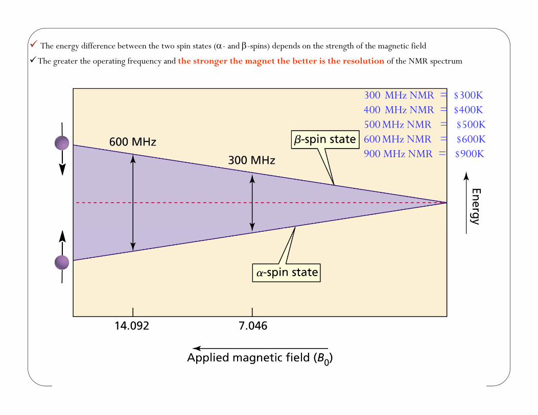
The energy difference between the two spin states (- and -spins) depends on the strength of the magnetic field
The greater the operating frequency and the stronger the magnet the better is the resolution of the NMR spectrum
300 MHz NMR = $300K400 MHz NMR = $400K500 MHz NMR = $500K600 MHz NMR = $600K900 MHz NMR = $900K
12

Magnetic resonance imaging (MRI)Nobel Laureate: Peter Mansfield
Routine method within medical diagnostics
Worldwide, more than 60 million
investigations with MRI are performed/year
MRI has replaced several invasive modes
of examination and thereby reduced the
risk and discomfort for many patients.
Examination with MRI is especially valuable for detailed imaging of the brain and the spinal cord.
http://nobelprize.org/nobel_prizes/medicine/laureates/2003/press.html13

MRI
Magnetic resonance imaging, noninvasive
“Nuclear” is omitted because of public’s fear that it would be radioactive.
Only protons in one plane can be in resonance at one time.
Computer puts together “slices” to get 3D.
Tumors readily detected.
14
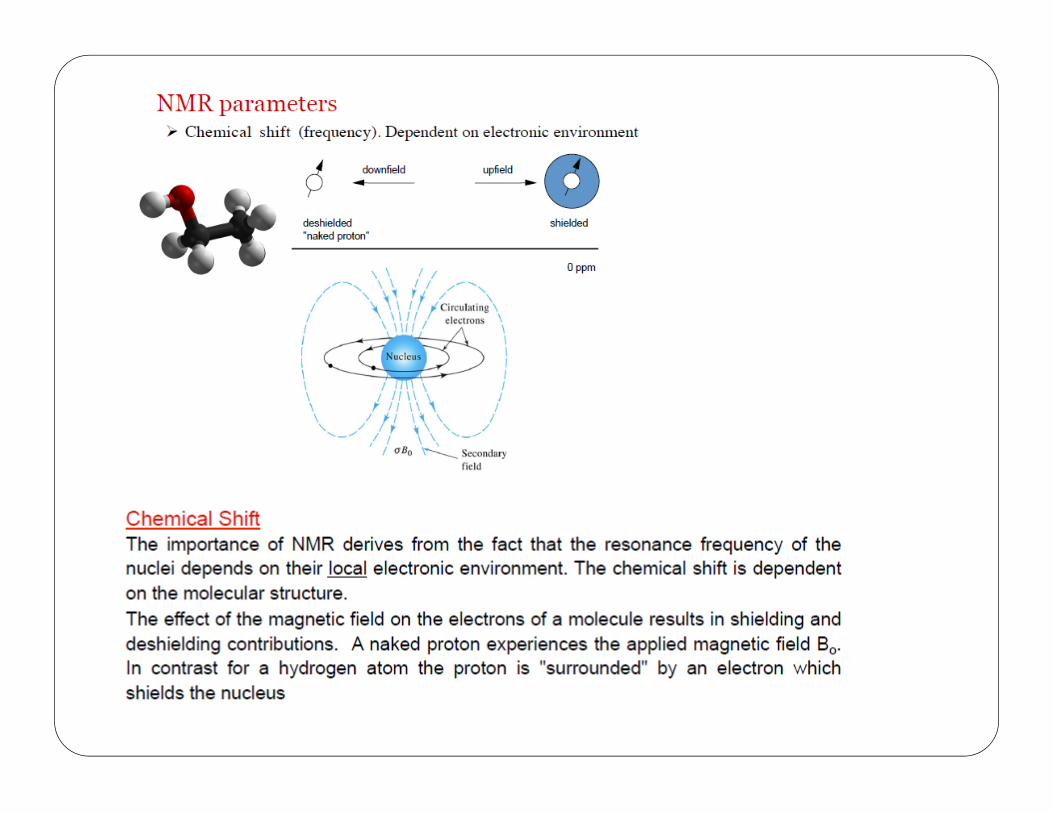

The common scale for chemical shifts =
=distance downfield from TMS (Hz)
operating frequency of the spectrometer (MHz)
Measured in parts per million (ppm).Same value for 60, 100, or 300 MHz machine.Called the delta scale.


The chemical shift is independent of the operating frequency of the spectrometer
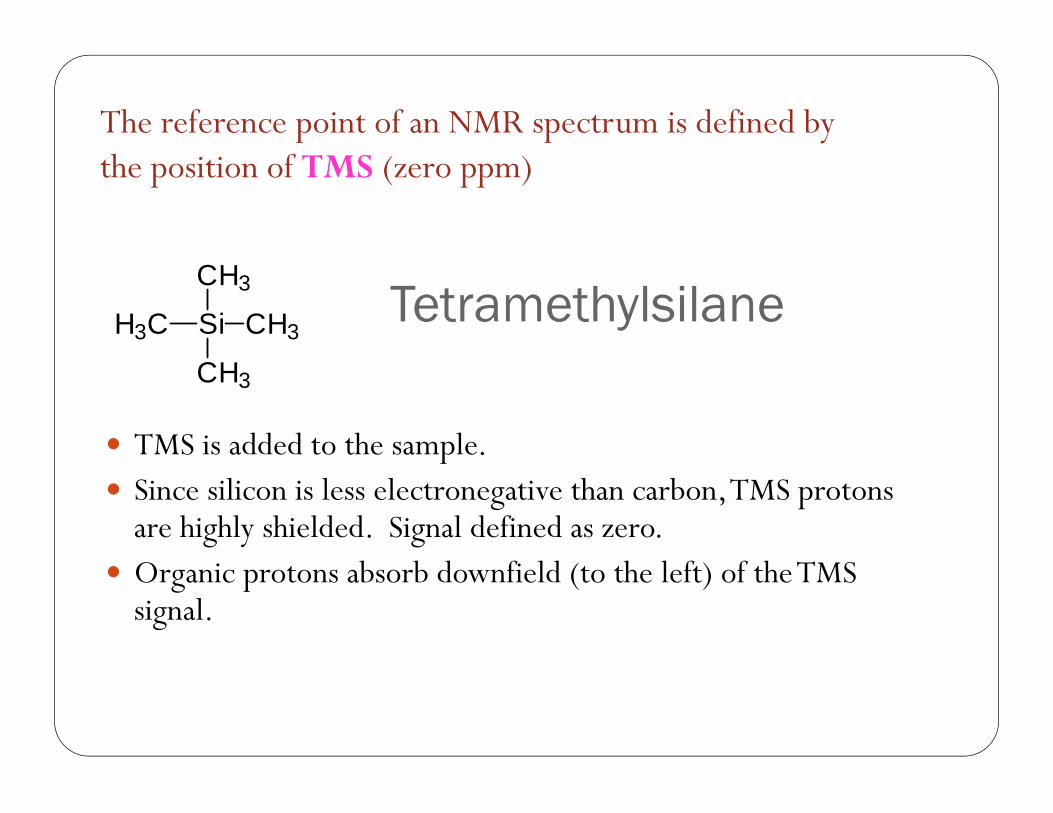
Tetramethylsilane
TMS is added to the sample. Since silicon is less electronegative than carbon, TMS protons
are highly shielded. Signal defined as zero. Organic protons absorb downfield (to the left) of the TMS
signal.
Si
CH3
CH3
CH3
H3C
21
The reference point of an NMR spectrum is defined bythe position of TMS (zero ppm)
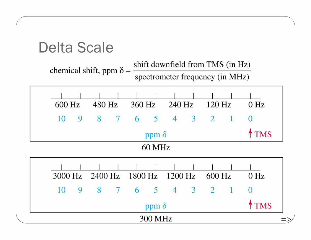
Delta Scale
=>22
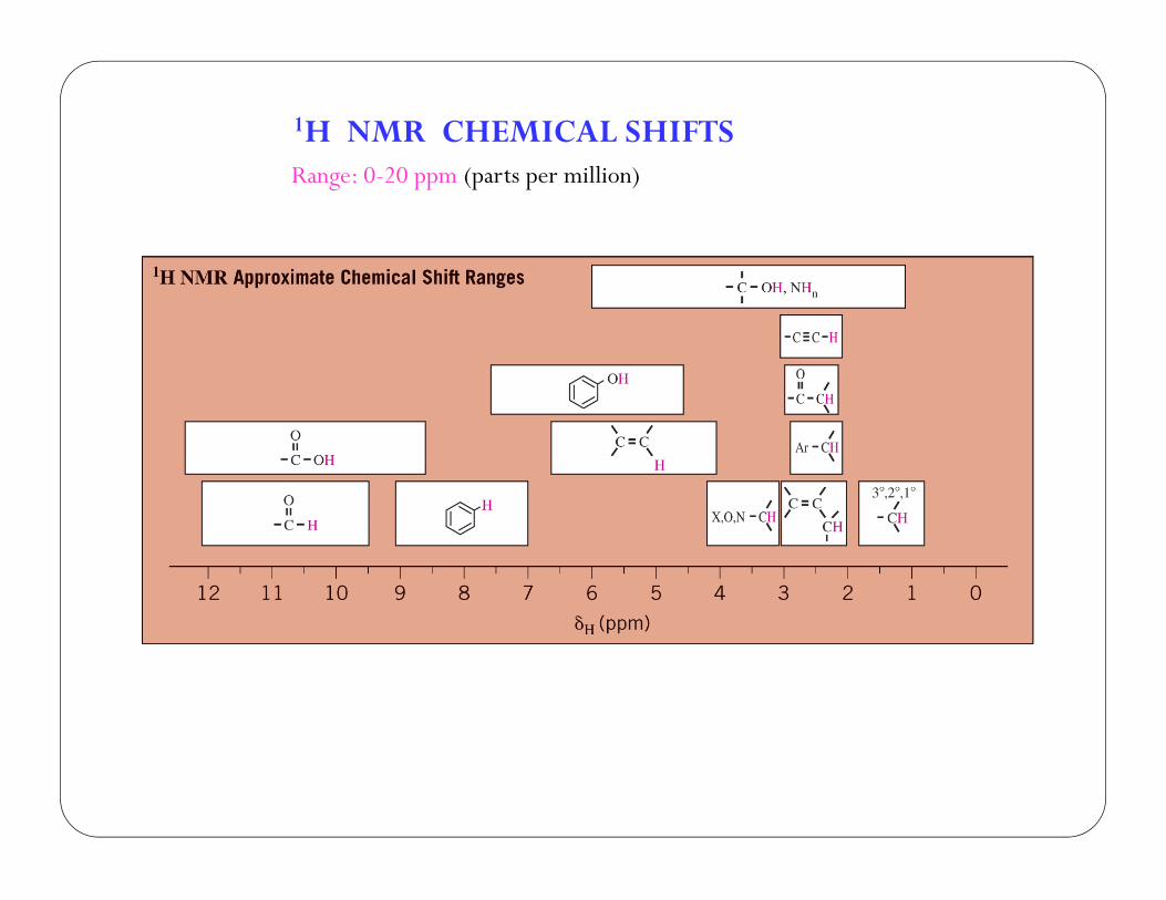
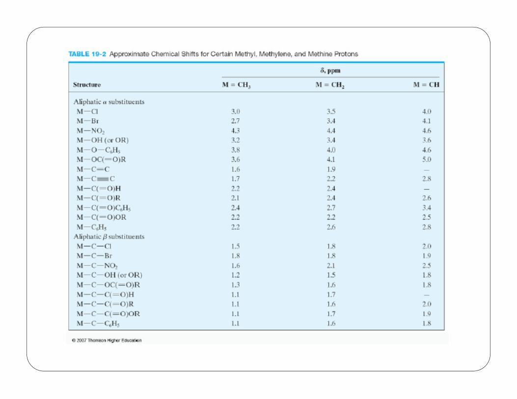
1H NMR CHEMICAL SHIFTSRange: 0-20 ppm (parts per million)
25
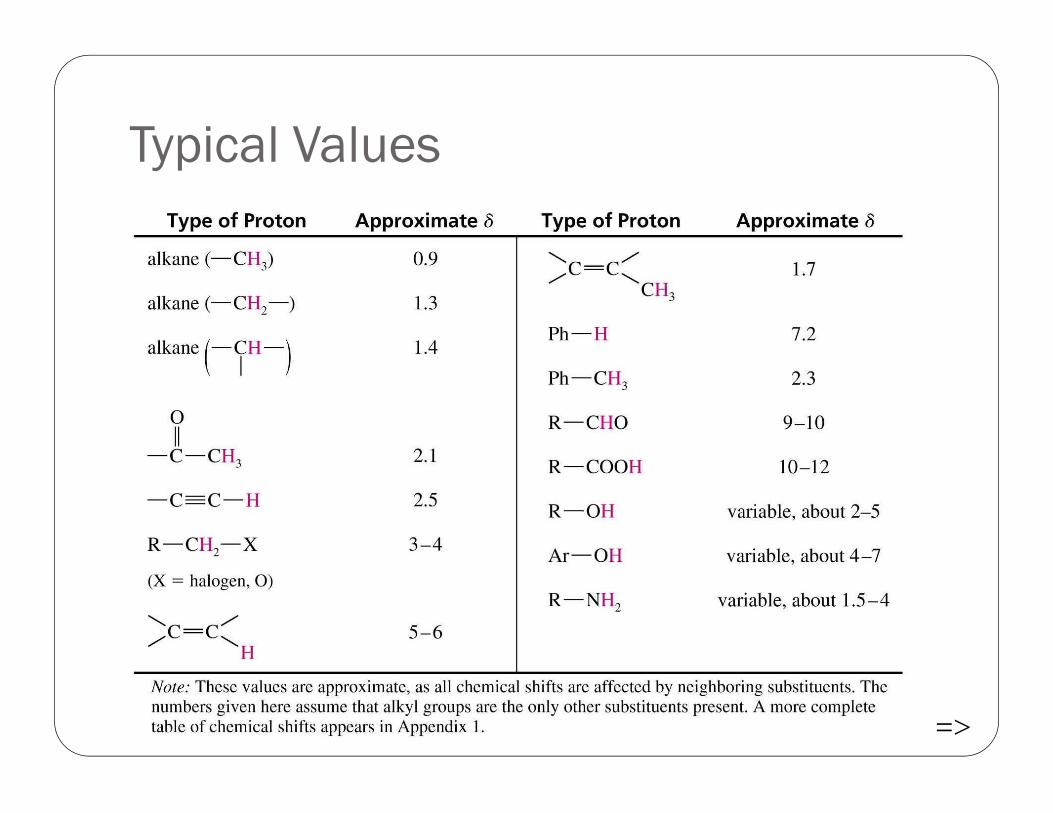
Typical Values
=>26
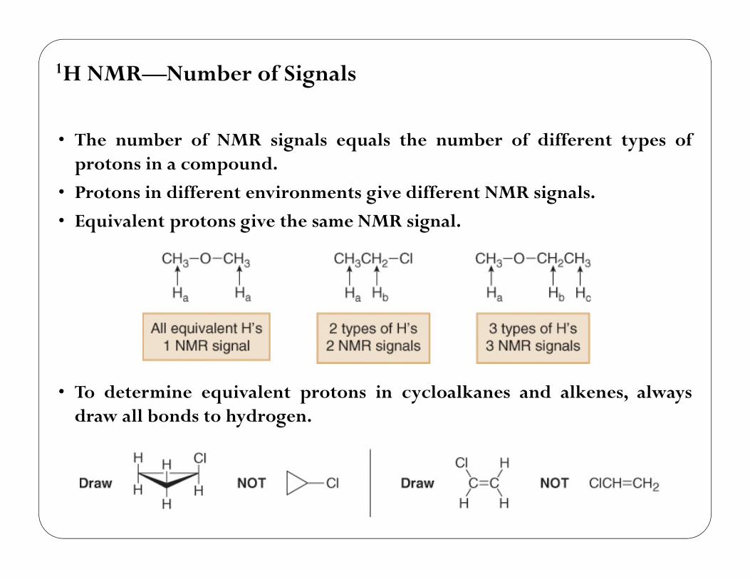
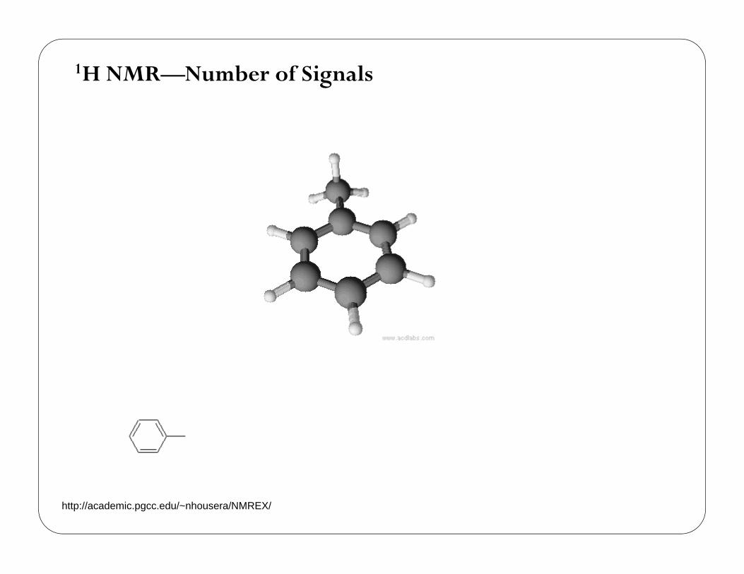
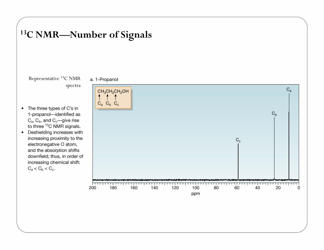
• The number of NMR signals equals the number of different types ofprotons in a compound.
• Protons in different environments give different NMR signals.
• Equivalent protons give the same NMR signal.
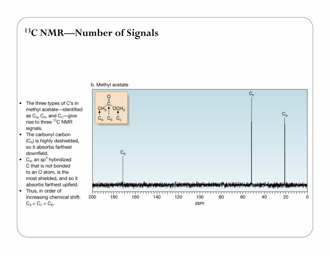
1H NMR—Number of Signals
• To determine equivalent protons in cycloalkanes and alkenes, alwaysdraw all bonds to hydrogen.
27
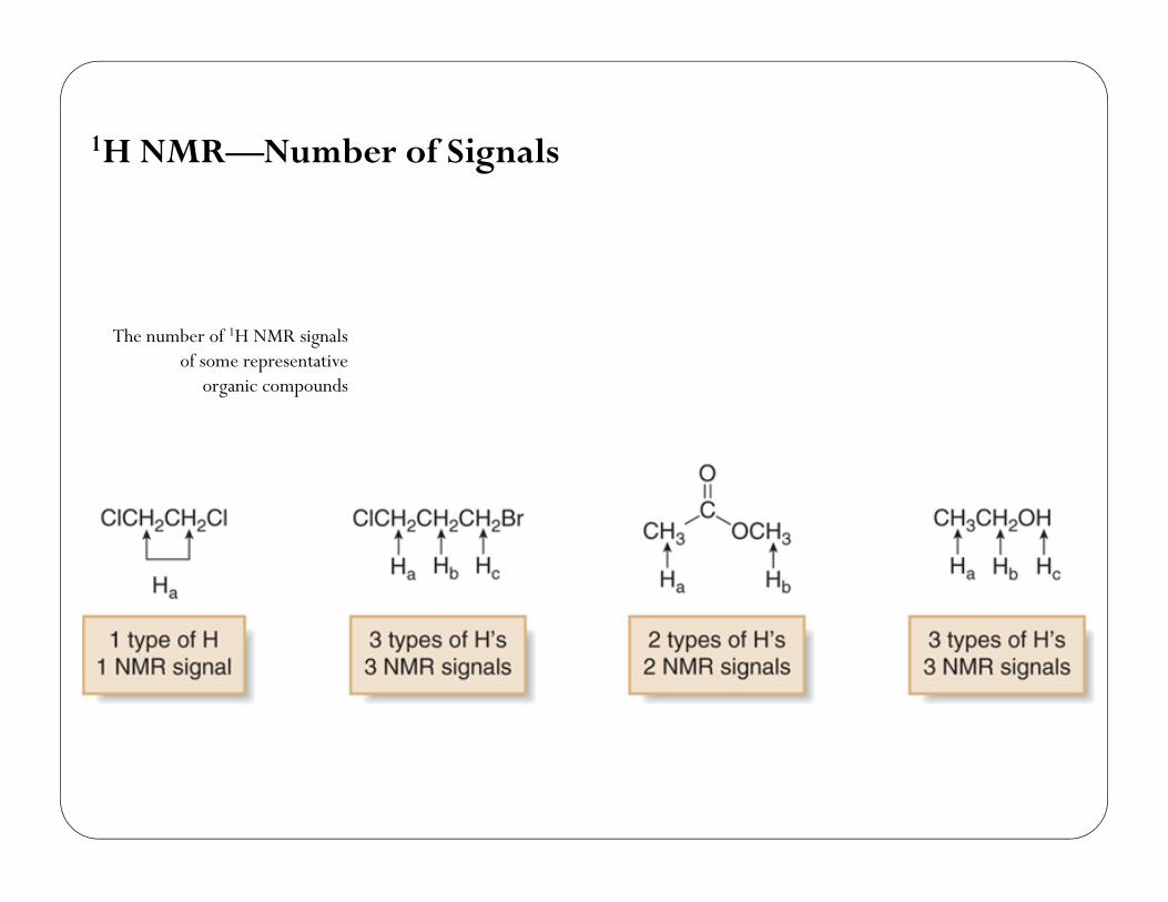
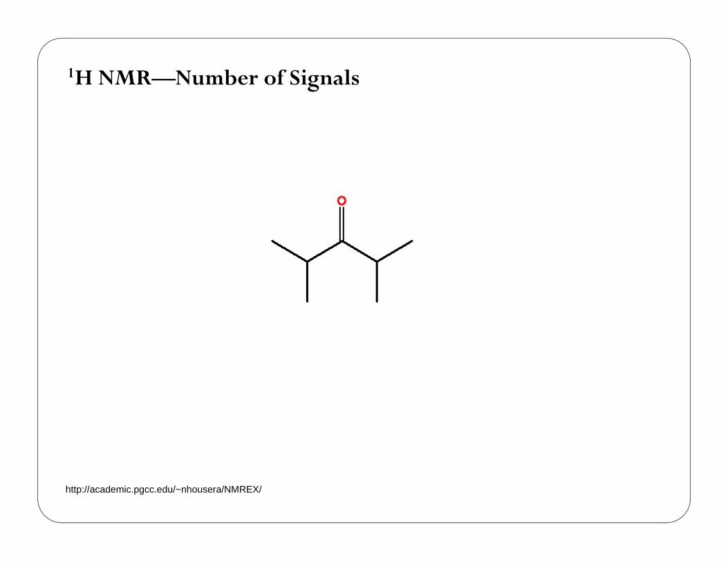
1H NMR—Number of Signals
The number of 1H NMR signalsof some representative
organic compounds
28
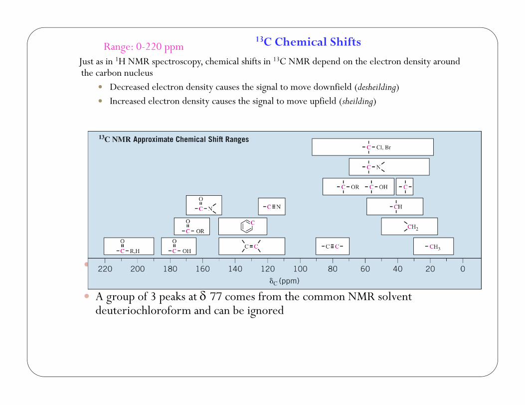
Range: 0-220 ppmJust as in 1H NMR spectroscopy, chemical shifts in 13C NMR depend on the electron density around the carbon nucleus
Decreased electron density causes the signal to move downfield (desheilding) Increased electron density causes the signal to move upfield (sheilding)
Because of the wide range of chemical shifts, it is rare to have two 13C peaks coincidentally overlap
A group of 3 peaks at 77 comes from the common NMR solvent deuteriochloroform and can be ignored
13C Chemical Shifts
29
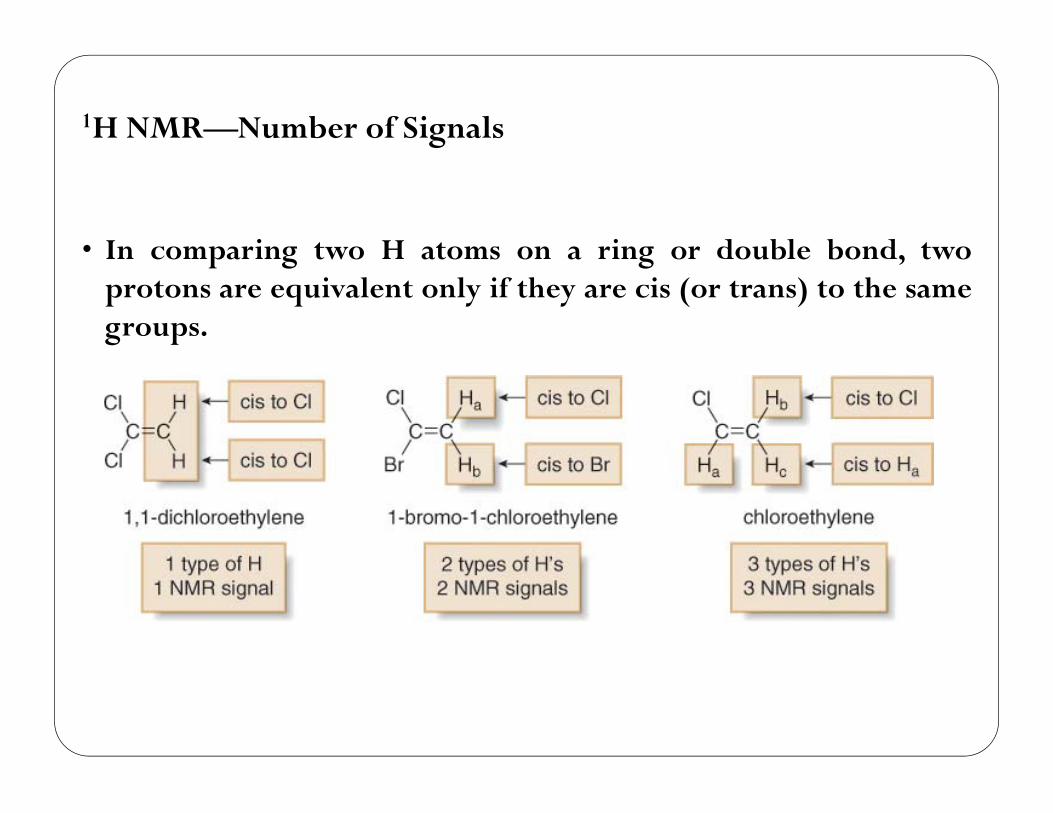
• In comparing two H atoms on a ring or double bond, twoprotons are equivalent only if they are cis (or trans) to the samegroups.
1H NMR—Number of Signals
30
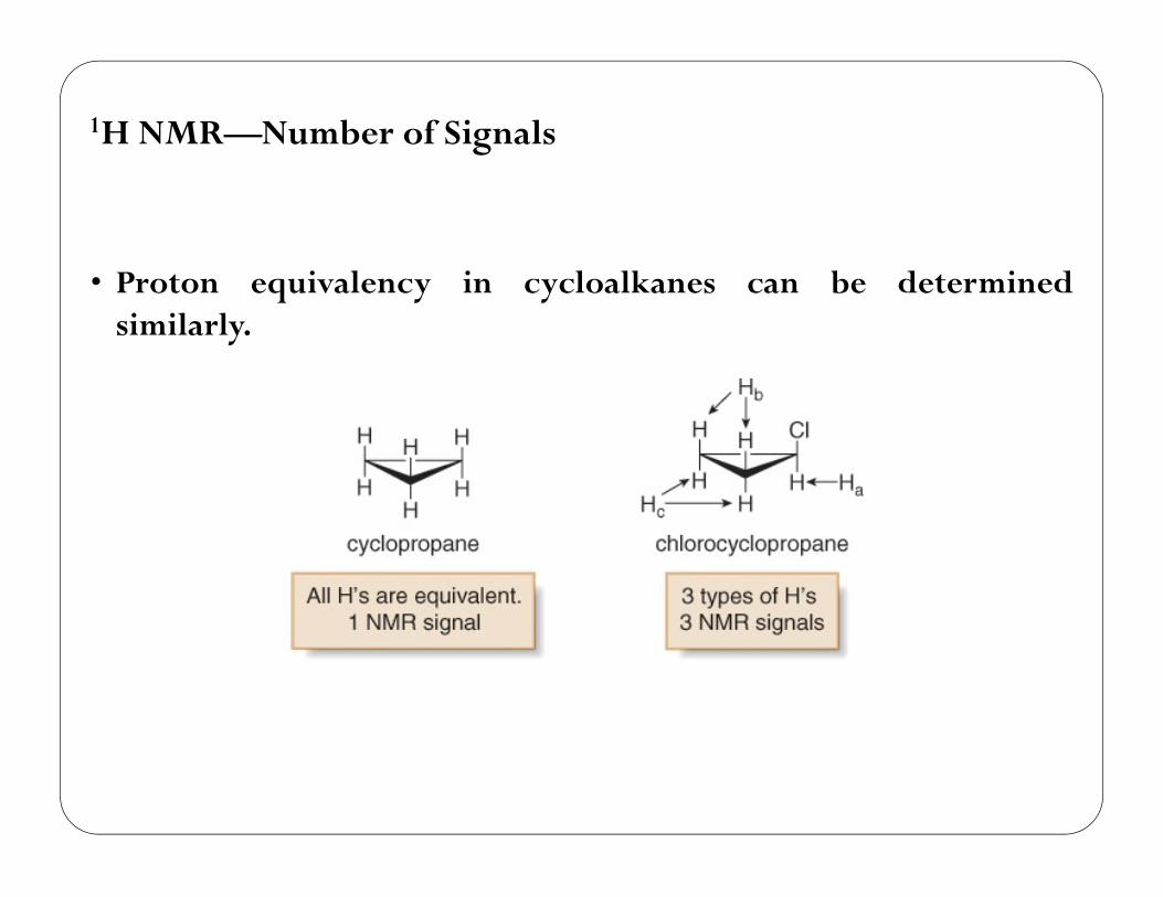
• Proton equivalency in cycloalkanes can be determinedsimilarly.
1H NMR—Number of Signals
31

CH3CH2CH2-NO2
1H NMR—Number of Signals
http://academic.pgcc.edu/~nhousera/NMREX/
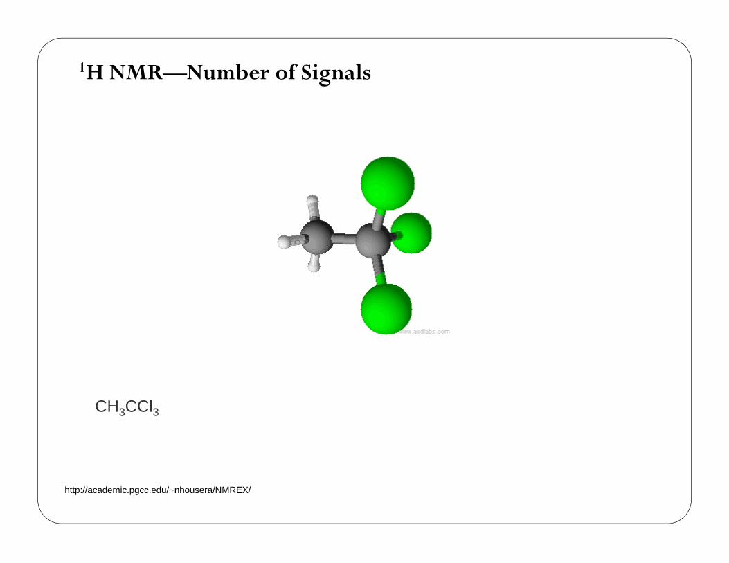
CH3CCl3
1H NMR—Number of Signals
http://academic.pgcc.edu/~nhousera/NMREX/
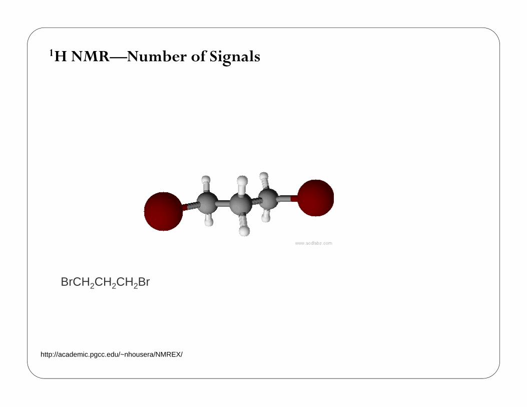
BrCH2CH2CH2Br
1H NMR—Number of Signals
http://academic.pgcc.edu/~nhousera/NMREX/
1H NMR—Number of Signals
http://academic.pgcc.edu/~nhousera/NMREX/
1H NMR—Number of Signals
http://academic.pgcc.edu/~nhousera/NMREX/
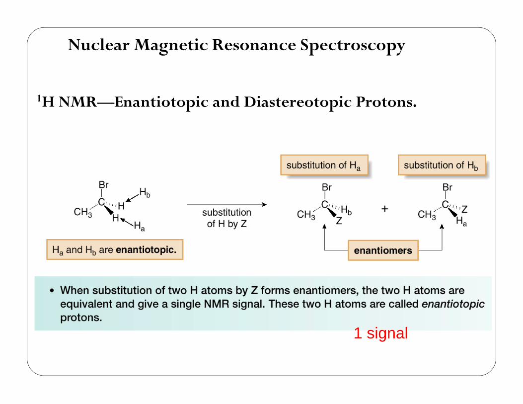
Nuclear Magnetic Resonance Spectroscopy
1H NMR—Enantiotopic and Diastereotopic Protons.
37
1 signal
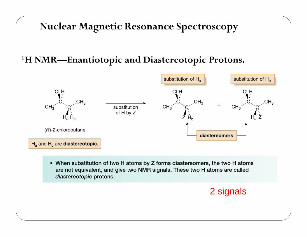
Nuclear Magnetic Resonance Spectroscopy
1H NMR—Enantiotopic and Diastereotopic Protons.
38
2 signals
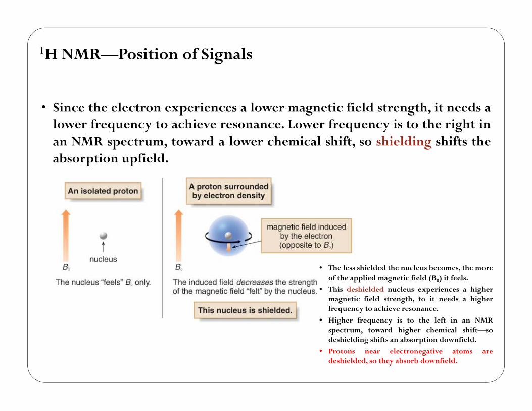
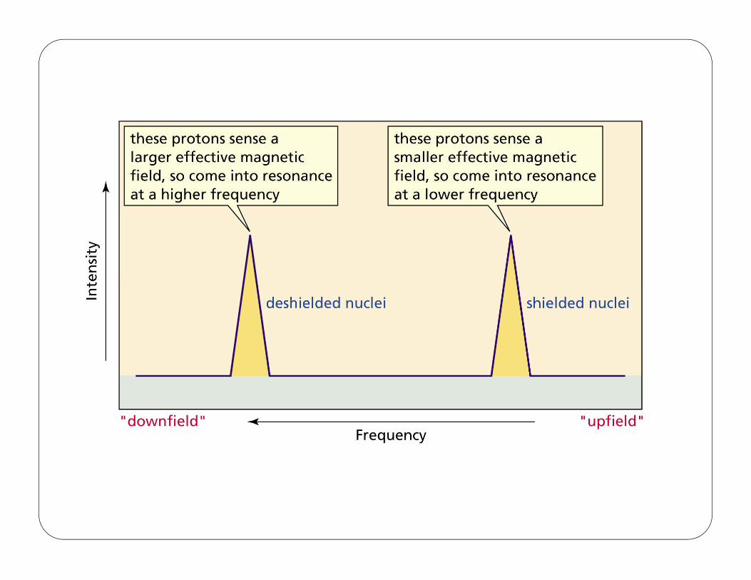
• Since the electron experiences a lower magnetic field strength, it needs alower frequency to achieve resonance. Lower frequency is to the right inan NMR spectrum, toward a lower chemical shift, so shielding shifts theabsorption upfield.
1H NMR—Position of Signals
39
• The less shielded the nucleus becomes, the moreof the applied magnetic field (B0) it feels.
• This deshielded nucleus experiences a highermagnetic field strength, to it needs a higherfrequency to achieve resonance.
• Higher frequency is to the left in an NMRspectrum, toward higher chemical shift—sodeshielding shifts an absorption downfield.
• Protons near electronegative atoms aredeshielded, so they absorb downfield.
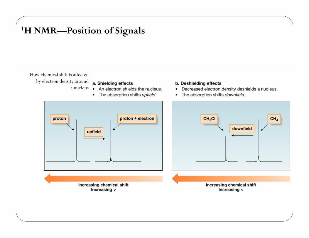
1H NMR—Position of Signals
How chemical shift is affectedby electron density around
a nucleus
40
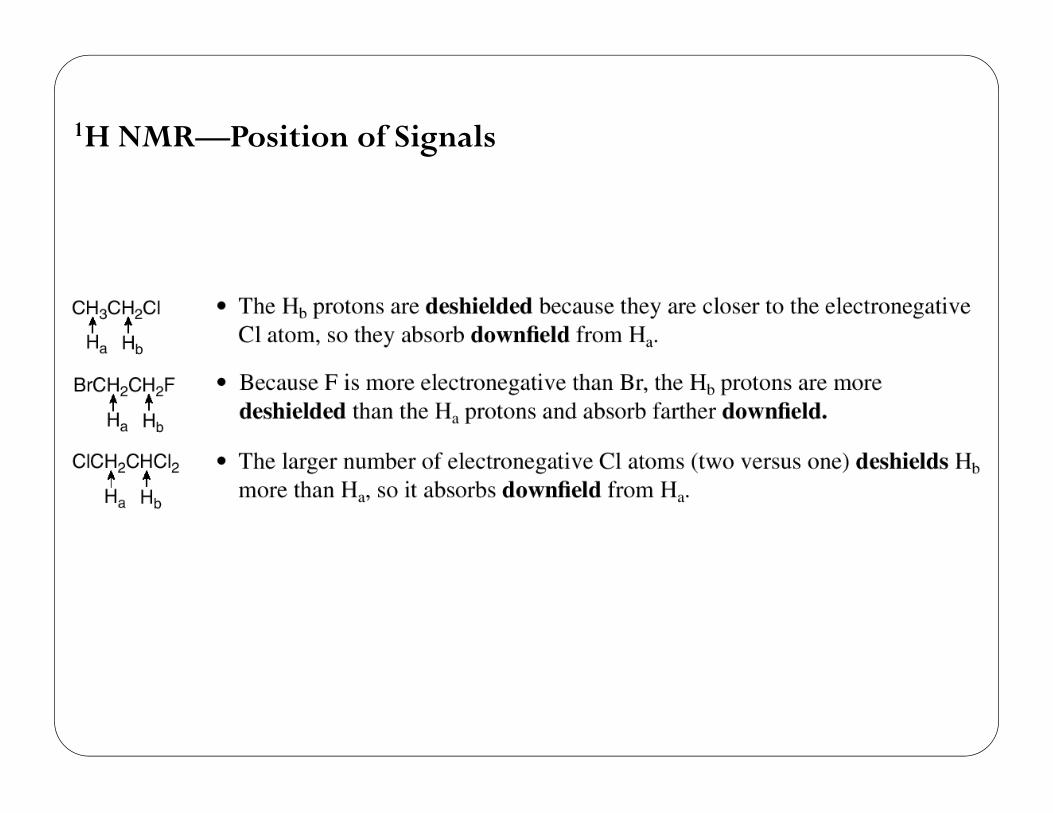
1H NMR—Position of Signals
41
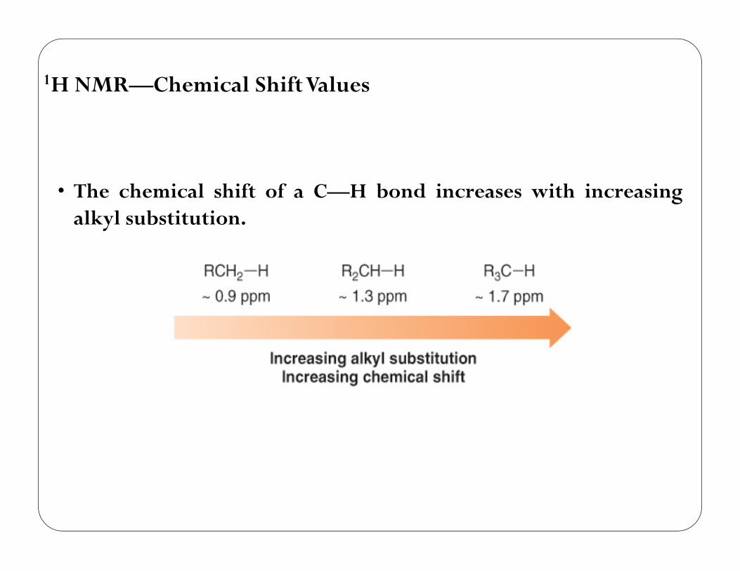
• The chemical shift of a C—H bond increases with increasingalkyl substitution.
1H NMR—Chemical ShiftValues
42
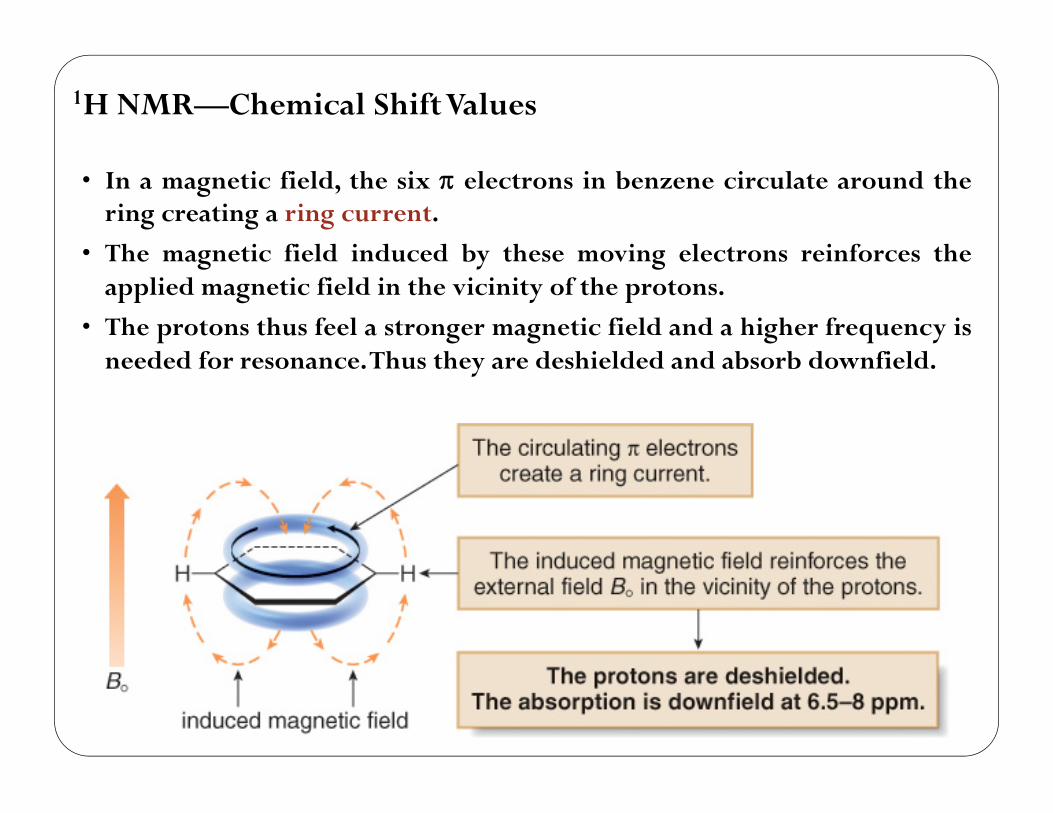
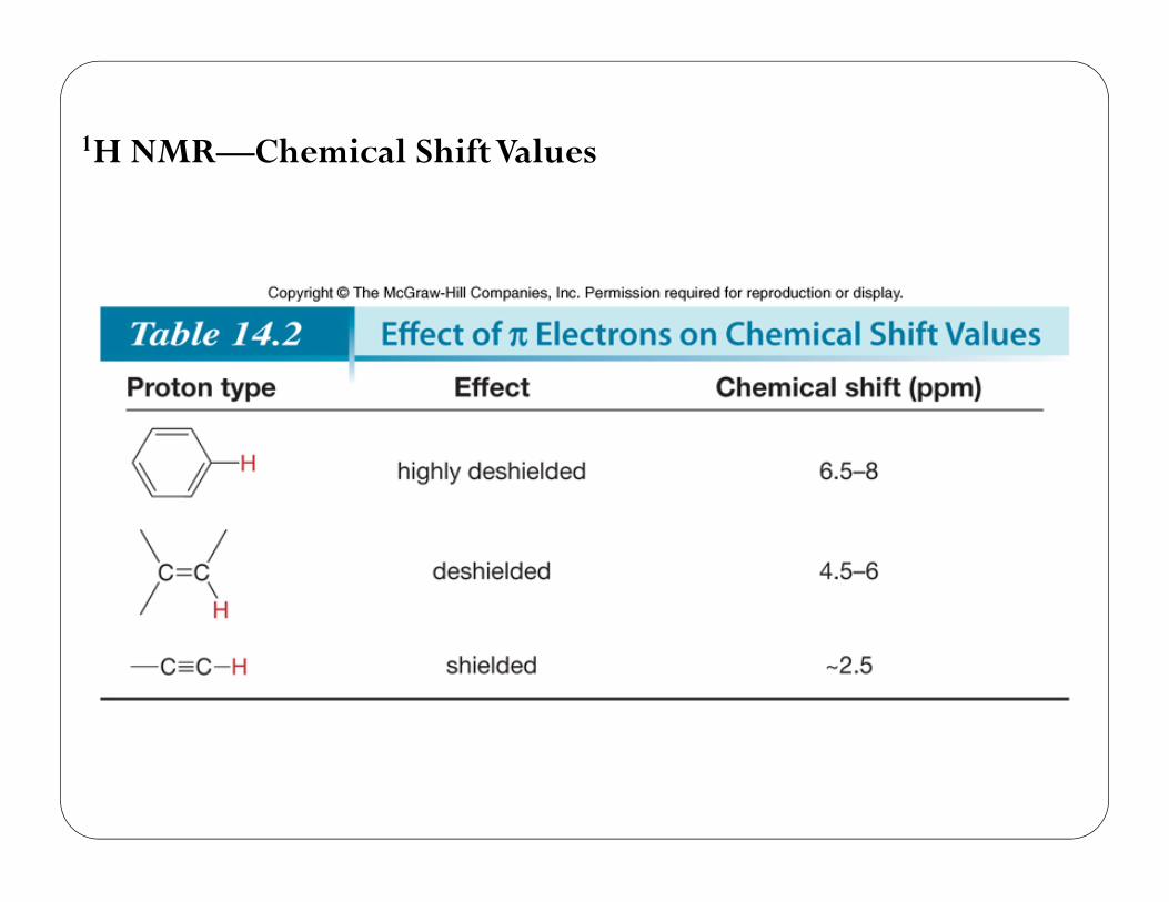
• In a magnetic field, the six electrons in benzene circulate around thering creating a ring current.
• The magnetic field induced by these moving electrons reinforces theapplied magnetic field in the vicinity of the protons.
• The protons thus feel a stronger magnetic field and a higher frequency isneeded for resonance.Thus they are deshielded and absorb downfield.
1H NMR—Chemical ShiftValues
43
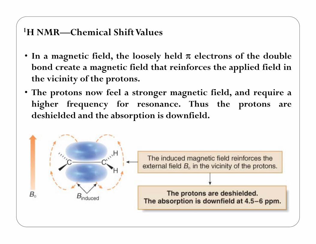
• In a magnetic field, the loosely held electrons of the doublebond create a magnetic field that reinforces the applied field inthe vicinity of the protons.
• The protons now feel a stronger magnetic field, and require ahigher frequency for resonance. Thus the protons aredeshielded and the absorption is downfield.
1H NMR—Chemical ShiftValues
44
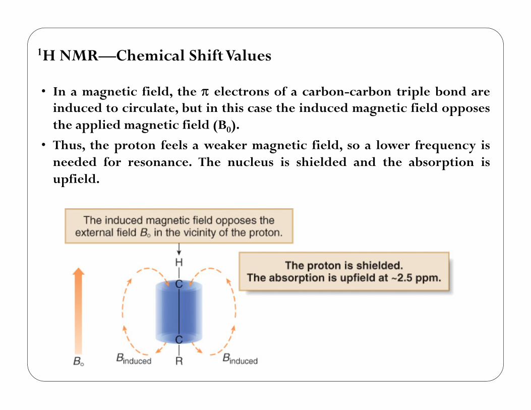
• In a magnetic field, the electrons of a carbon-carbon triple bond areinduced to circulate, but in this case the induced magnetic field opposesthe applied magnetic field (B0).
• Thus, the proton feels a weaker magnetic field, so a lower frequency isneeded for resonance. The nucleus is shielded and the absorption isupfield.
1H NMR—Chemical ShiftValues
45
1H NMR—Chemical Shift Values
46
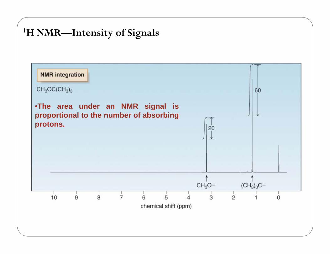
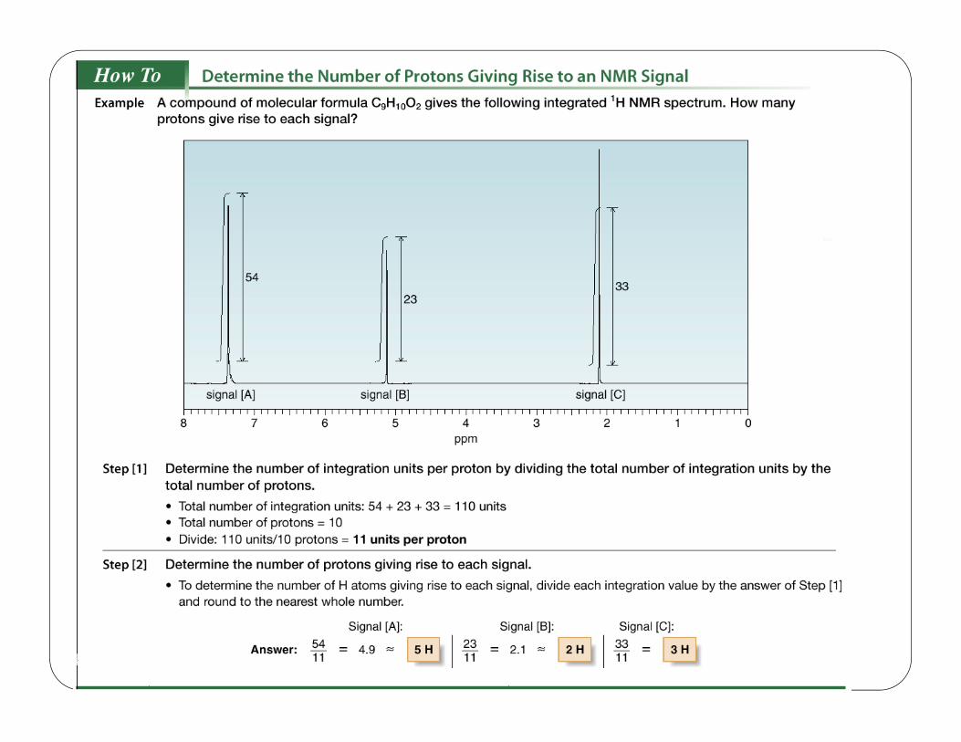
1H NMR—Intensity of Signals
47
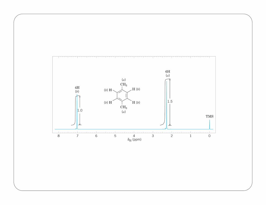
•The area under an NMR signal isproportional to the number of absorbingprotons.
48
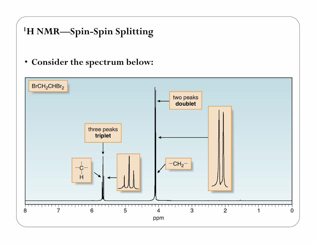
• Consider the spectrum below:
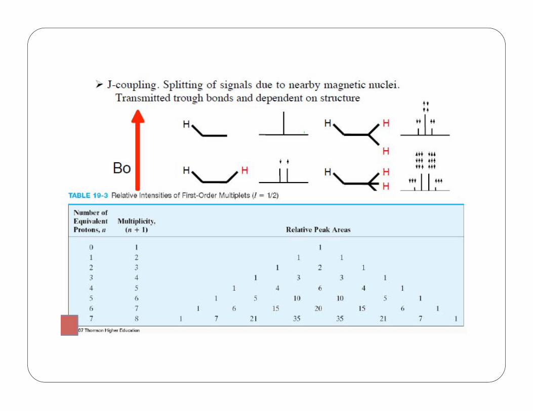
1H NMR—Spin-Spin Splitting
49
1H NMR—Spin-Spin Splitting
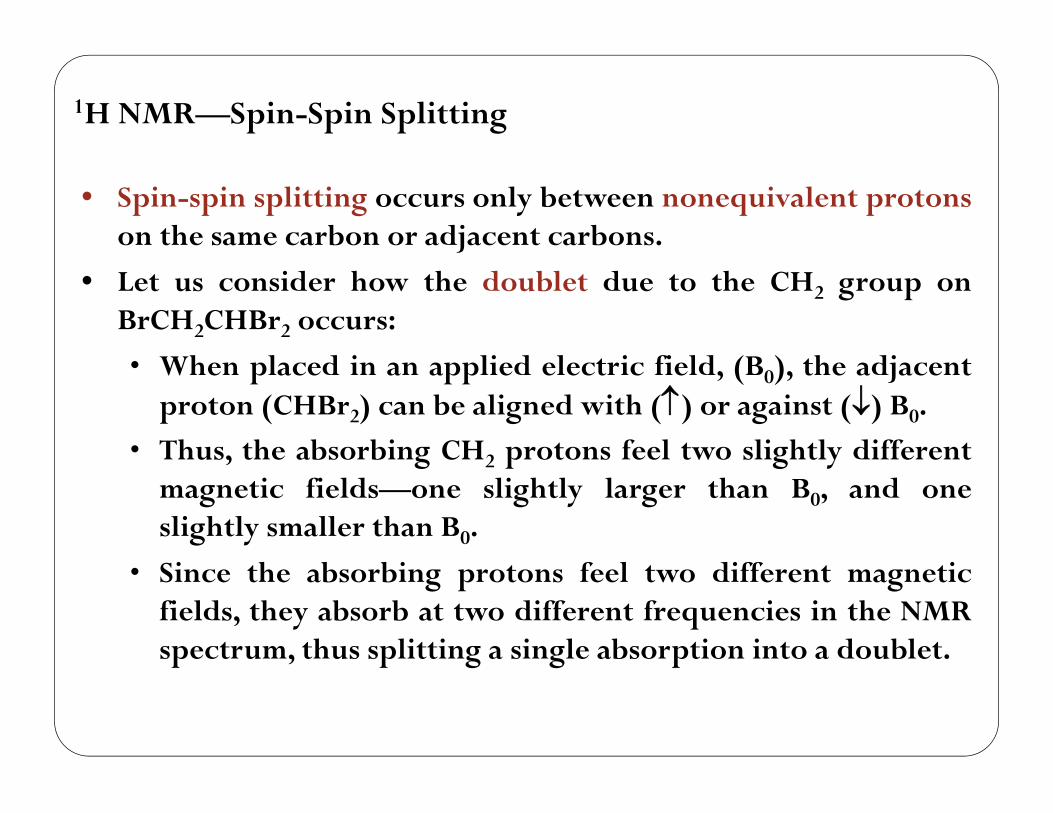
• Spin-spin splitting occurs only between nonequivalent protonson the same carbon or adjacent carbons.
• Let us consider how the doublet due to the CH2 group onBrCH2CHBr2 occurs:
• When placed in an applied electric field, (B0), the adjacentproton (CHBr2) can be aligned with () or against () B0.
• Thus, the absorbing CH2 protons feel two slightly differentmagnetic fields—one slightly larger than B0, and oneslightly smaller than B0.
• Since the absorbing protons feel two different magneticfields, they absorb at two different frequencies in the NMRspectrum, thus splitting a single absorption into a doublet.
50
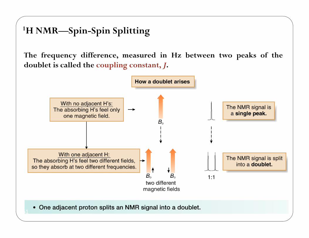
1H NMR—Spin-Spin Splitting
The frequency difference, measured in Hz between two peaks of thedoublet is called the coupling constant, J.
51
1H NMR—Spin-Spin Splitting
52
1H NMR—Spin-Spin Splitting
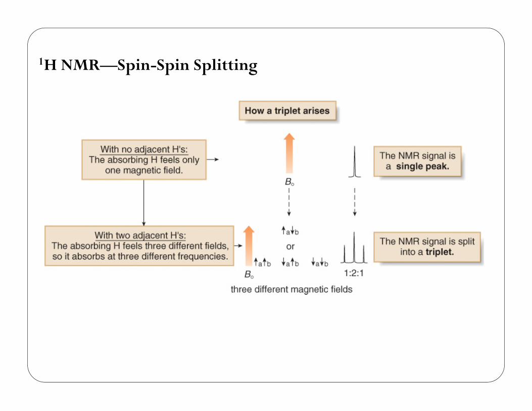
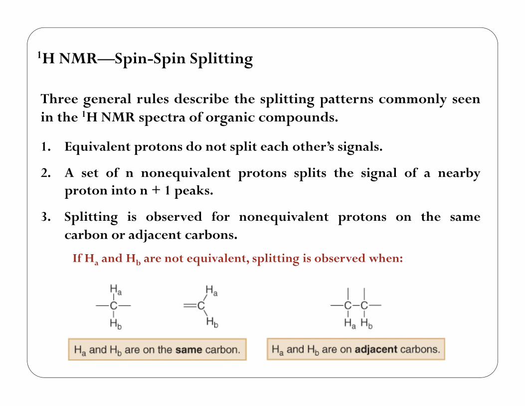
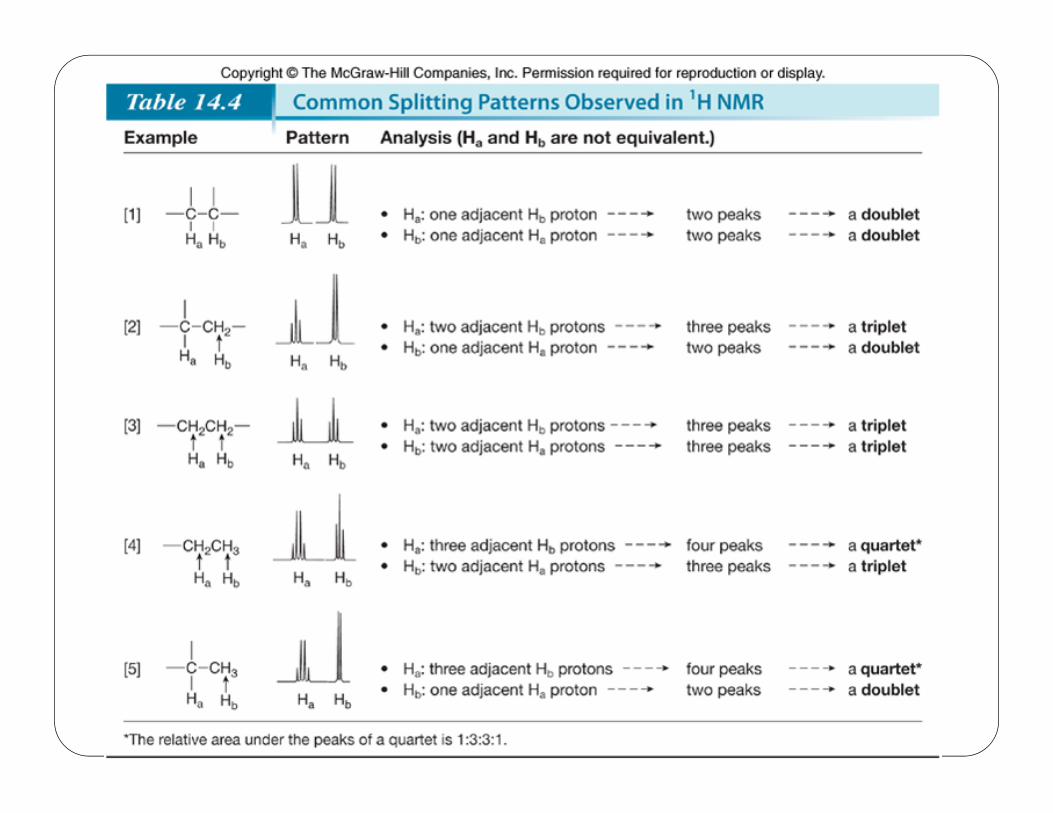
Three general rules describe the splitting patterns commonly seenin the 1H NMR spectra of organic compounds.
1. Equivalent protons do not split each other’s signals.
2. A set of n nonequivalent protons splits the signal of a nearbyproton into n + 1 peaks.
3. Splitting is observed for nonequivalent protons on the samecarbon or adjacent carbons.
If Ha and Hb are not equivalent, splitting is observed when:
53
1H NMR—Spin-Spin Splitting
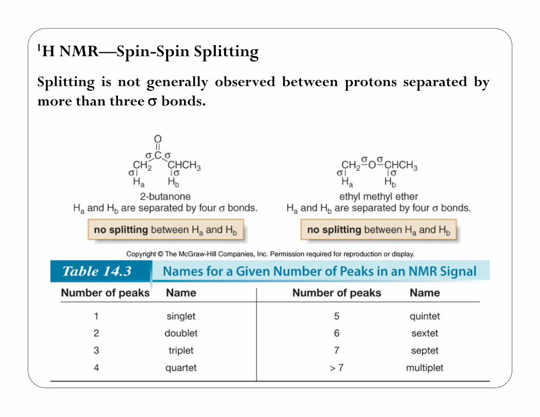
Splitting is not generally observed between protons separated bymore than three bonds.
54
55
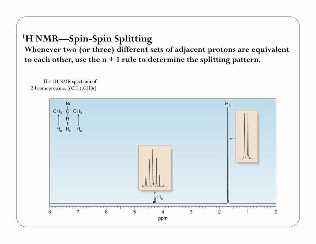
1H NMR—Spin-Spin SplittingWhenever two (or three) different sets of adjacent protons are equivalentto each other, use the n + 1 rule to determine the splitting pattern.
The 1H NMR spectrum of2-bromopropane, [(CH3)2CHBr]
56
1H NMR—Spin-Spin Splitting
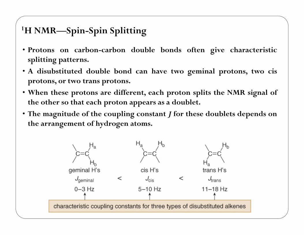
• Protons on carbon-carbon double bonds often give characteristicsplitting patterns.
• A disubstituted double bond can have two geminal protons, two cisprotons, or two trans protons.
• When these protons are different, each proton splits the NMR signal ofthe other so that each proton appears as a doublet.
• The magnitude of the coupling constant J for these doublets depends onthe arrangement of hydrogen atoms.
57
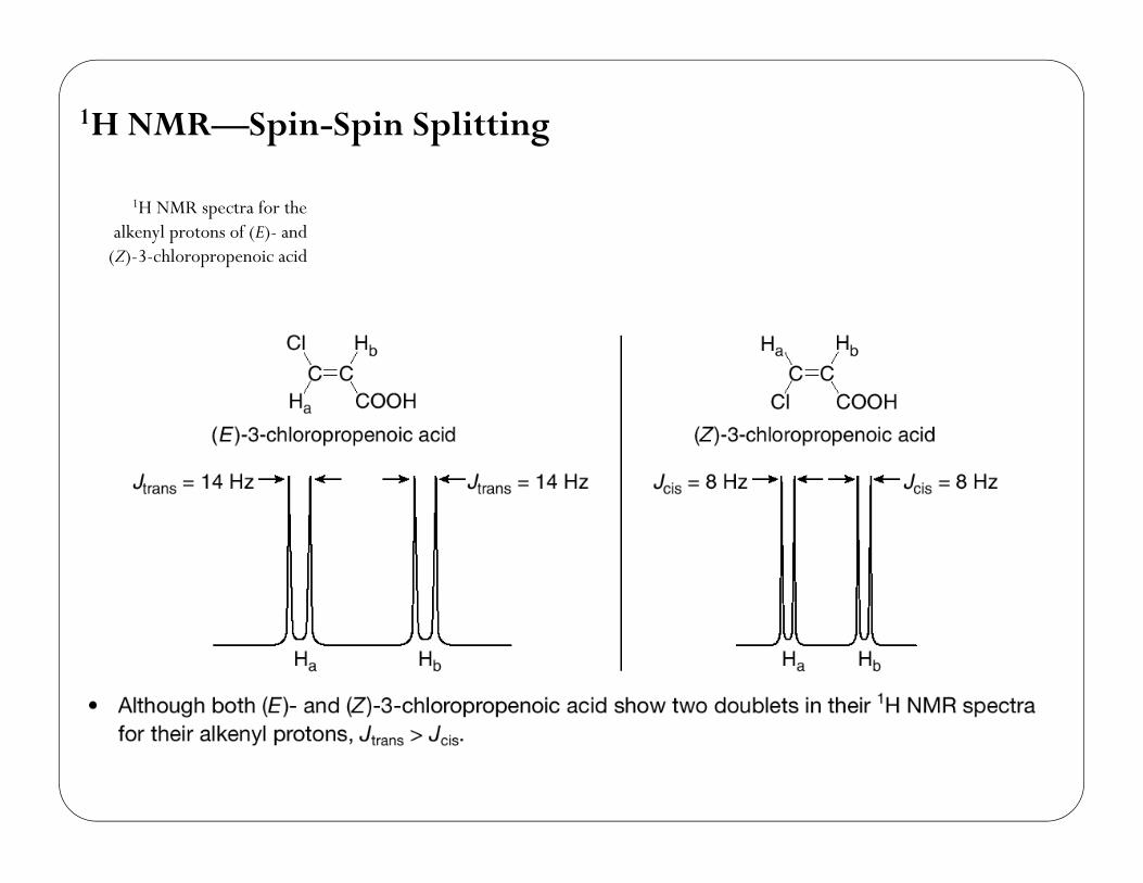
1H NMR—Spin-Spin Splitting
1H NMR spectra for thealkenyl protons of (E)- and
(Z)-3-chloropropenoic acid
58
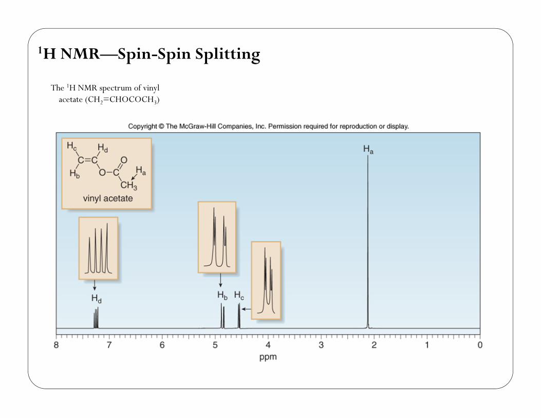
1H NMR—Spin-Spin Splitting
The 1H NMR spectrum of vinylacetate (CH2=CHOCOCH3)
59
1H NMR—Spin-Spin Splitting
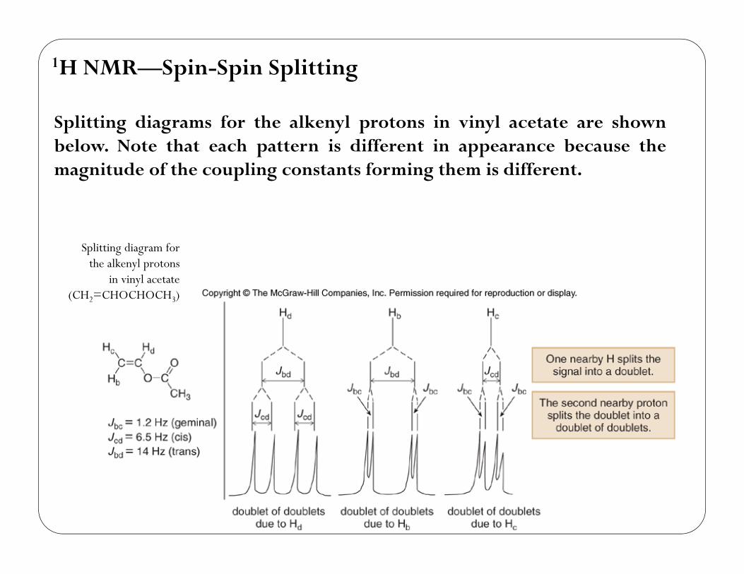
Splitting diagrams for the alkenyl protons in vinyl acetate are shownbelow. Note that each pattern is different in appearance because themagnitude of the coupling constants forming them is different.
Splitting diagram forthe alkenyl protons
in vinyl acetate(CH2=CHOCHOCH3)
60
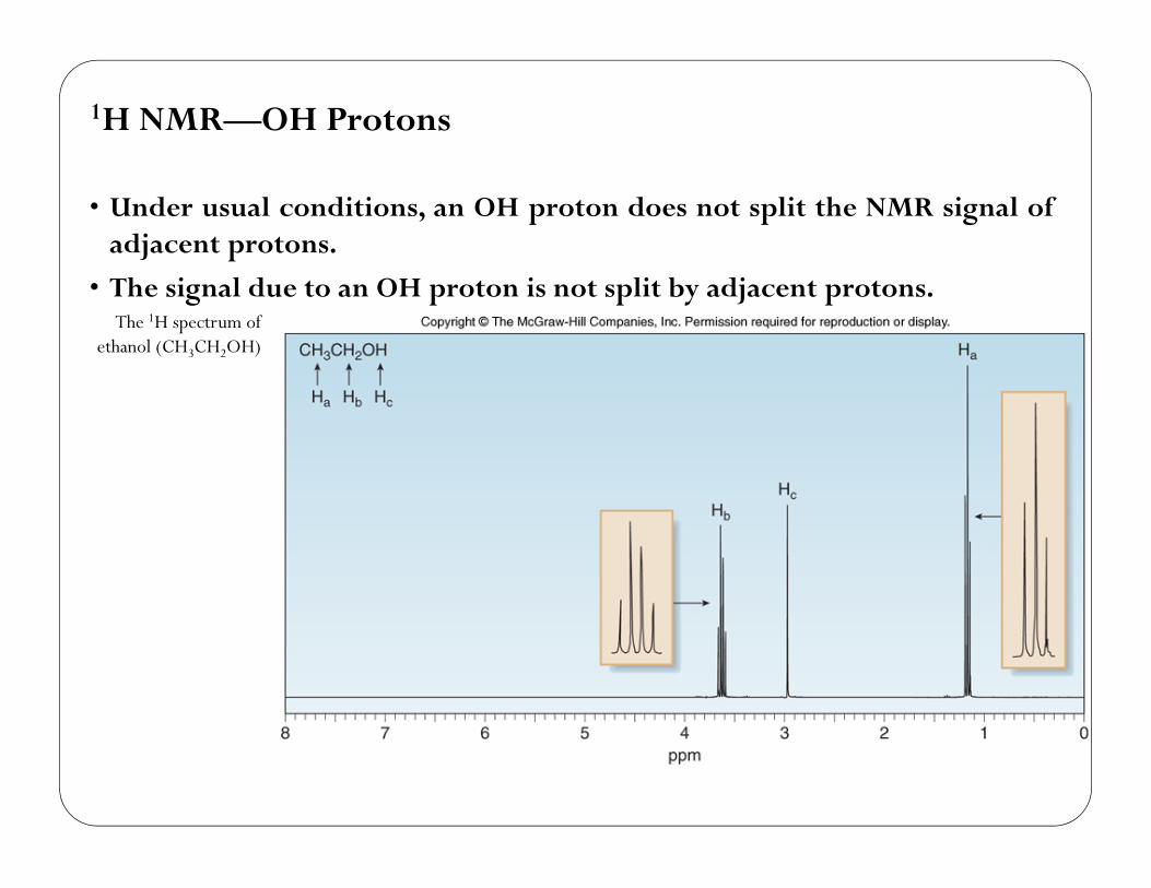
1H NMR—OH Protons
• Under usual conditions, an OH proton does not split the NMR signal ofadjacent protons.
• The signal due to an OH proton is not split by adjacent protons.The 1H spectrum of
ethanol (CH3CH2OH)
61
1H NMR—OH Protons
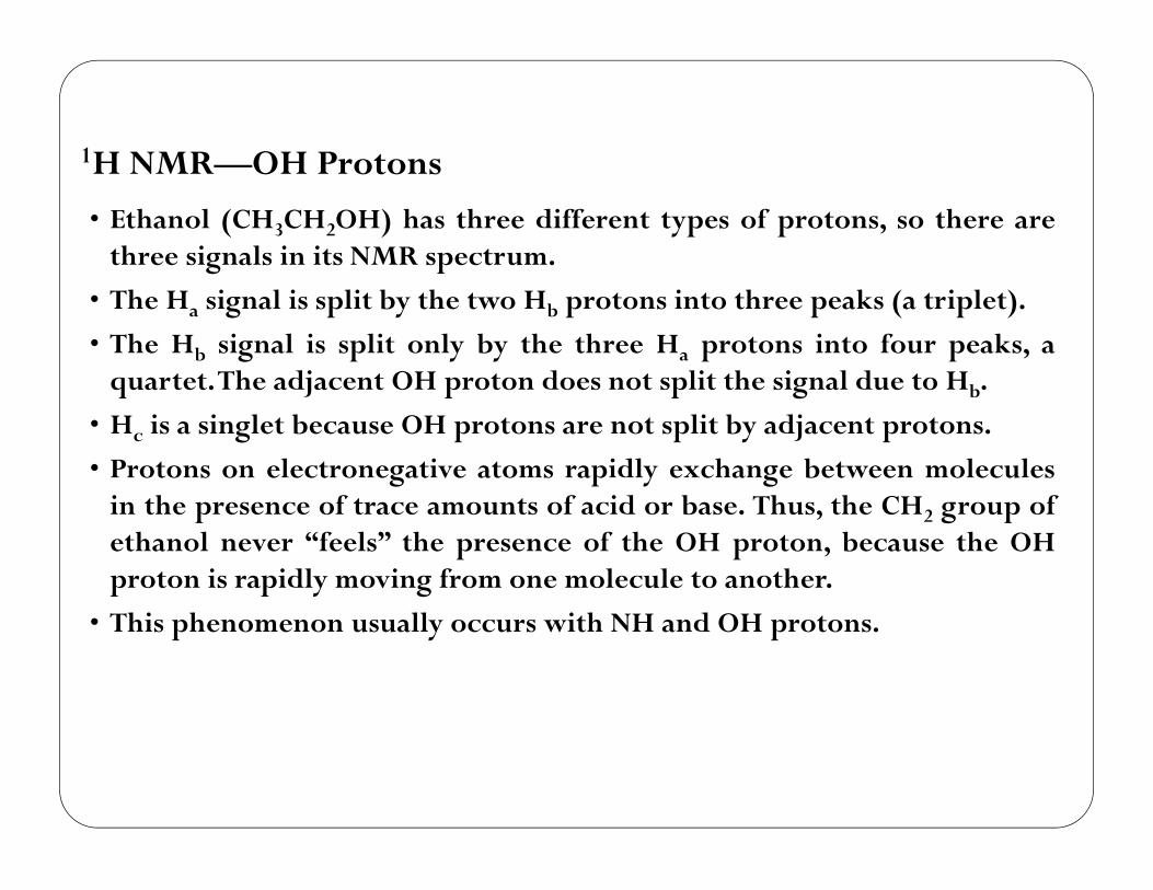
• Ethanol (CH3CH2OH) has three different types of protons, so there arethree signals in its NMR spectrum.
• The Ha signal is split by the two Hb protons into three peaks (a triplet).
• The Hb signal is split only by the three Ha protons into four peaks, aquartet.The adjacent OH proton does not split the signal due to Hb.
• Hc is a singlet because OH protons are not split by adjacent protons.
• Protons on electronegative atoms rapidly exchange between moleculesin the presence of trace amounts of acid or base. Thus, the CH2 group ofethanol never “feels” the presence of the OH proton, because the OHproton is rapidly moving from one molecule to another.
• This phenomenon usually occurs with NH and OH protons.
62
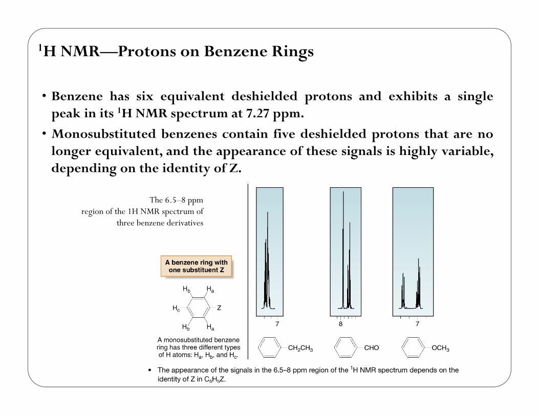
1H NMR—Protons on Benzene Rings
• Benzene has six equivalent deshielded protons and exhibits a singlepeak in its 1H NMR spectrum at 7.27 ppm.
• Monosubstituted benzenes contain five deshielded protons that are nolonger equivalent, and the appearance of these signals is highly variable,depending on the identity of Z.
The 6.5–8 ppmregion of the 1H NMR spectrum of
three benzene derivatives
63
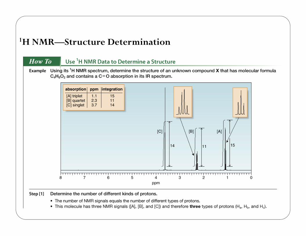
1H NMR—Structure Determination
64
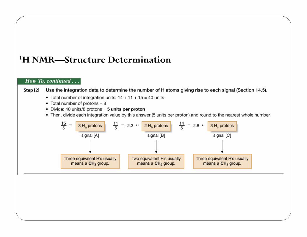
1H NMR—Structure Determination
65
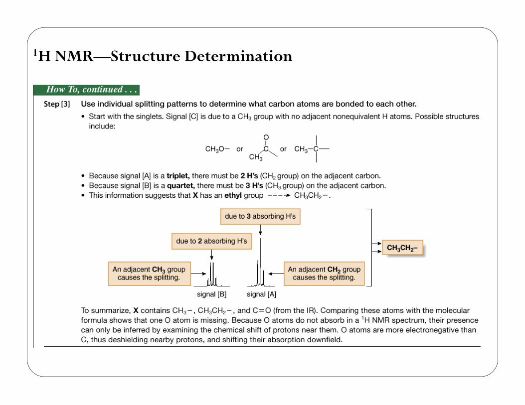
1H NMR—Structure Determination
66
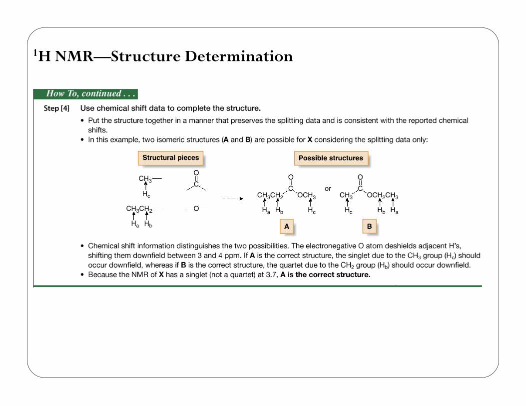
1H NMR—Structure Determination
67
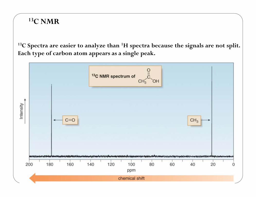
13C NMR
13C Spectra are easier to analyze than 1H spectra because the signals are not split.Each type of carbon atom appears as a single peak.
68
13C NMR
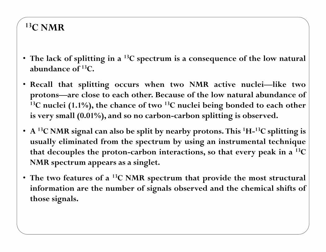
• The lack of splitting in a 13C spectrum is a consequence of the low naturalabundance of 13C.
• Recall that splitting occurs when two NMR active nuclei—like twoprotons—are close to each other. Because of the low natural abundance of13C nuclei (1.1%), the chance of two 13C nuclei being bonded to each otheris very small (0.01%), and so no carbon-carbon splitting is observed.
• A 13C NMR signal can also be split by nearby protons. This 1H-13C splitting isusually eliminated from the spectrum by using an instrumental techniquethat decouples the proton-carbon interactions, so that every peak in a 13CNMR spectrum appears as a singlet.
• The two features of a 13C NMR spectrum that provide the most structuralinformation are the number of signals observed and the chemical shifts ofthose signals.
69
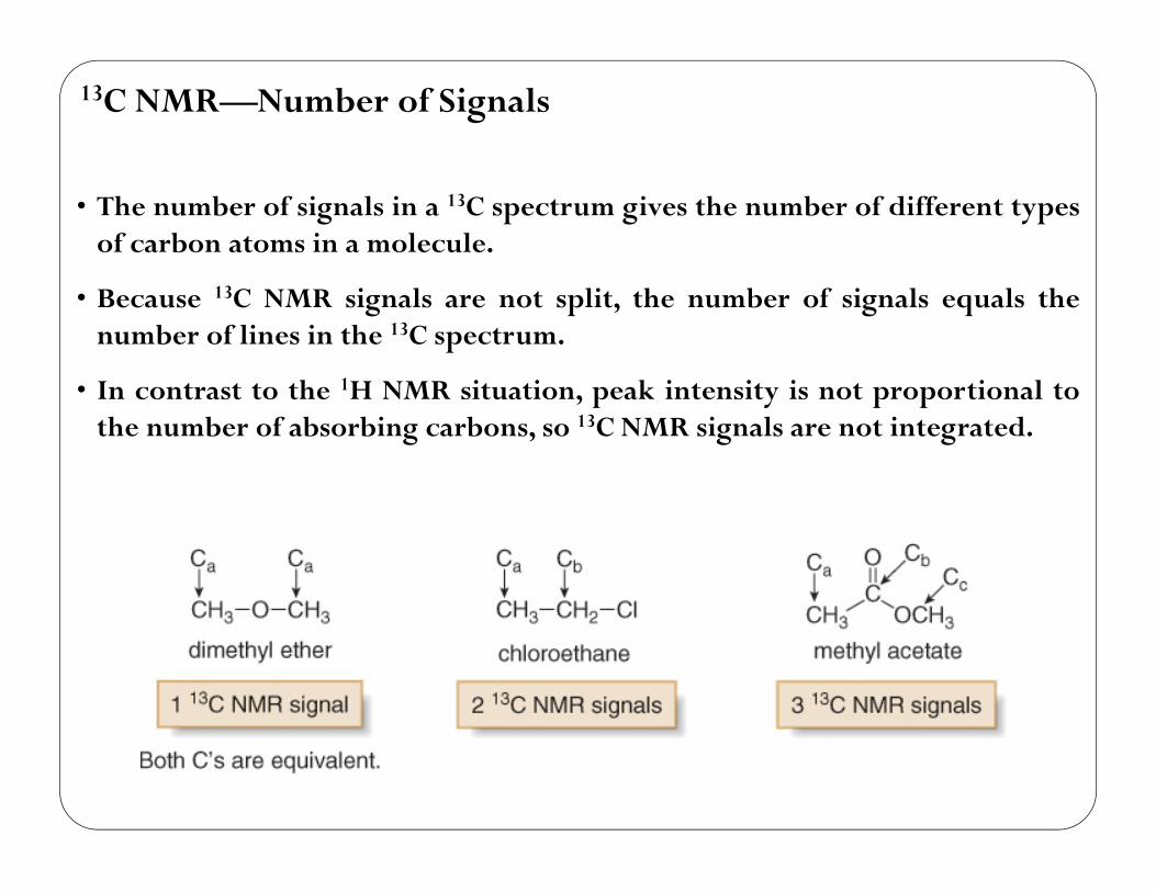
13C NMR—Number of Signals
• The number of signals in a 13C spectrum gives the number of different typesof carbon atoms in a molecule.
• Because 13C NMR signals are not split, the number of signals equals thenumber of lines in the 13C spectrum.
• In contrast to the 1H NMR situation, peak intensity is not proportional tothe number of absorbing carbons, so 13C NMR signals are not integrated.
70

13C NMR—Position of Signals
• In contrast to the small range of chemical shifts in 1H NMR (1-10 ppmusually), 13C NMR absorptions occur over a much broader range (0-220ppm).
• The chemical shifts of carbon atoms in 13C NMR depend on the same effectsas the chemical shifts of protons in 1H NMR.
71
13C NMR—Number of Signals
Representative 13C NMRspectra
72
13C NMR—Number of Signals
73

http://academic.pgcc.edu/~nhousera/NMREX/
TRY PROBLEMS


