INTRODUCTION Cervical margin relocation (CMR) technique has been proposed as a non-invasive pretreatment for the restoration of deep Class II cavities with proximal cervical margins extending below cemento-enamel junction (CEJ) 1,2) . This concept suggests application of a layer of composite in the deepest parts of the proximal areas in order to reposition the cervical margin supragingivally, which would facilitate rubber dam isolation, impression taking and subsequent adhesive cementation of the indirect restoration. Some of the issues related to CMR technique that concern both the researchers and the clinicians are adhesion to deep cervical dentine, quality and durability of the seal at the subgingival margins, as well as the periodontal tissue response to the performed treatment 3) . Scientific evidence on these topics obtained through clinical studies is scarce. So far only one randomized clinical study exists, which investigated the influence of the CMR pretreatment on the periodontal health of posterior teeth restored with indirect restorations 4) . The results from that study indicated higher incidence of inflammation of periodontal tissue around teeth that underwent CMR pretreatment prior to definitive restoration compared to teeth without CMR at 12-month follow up. All the other available data on CMR technique is provided by in vitro studies. Several clinical protocols and case reports suggested the use of various adhesive systems, composite materials and application techniques for the CMR 5-8) . In order to evaluate the marginal quality in vitro, different methods have been used. The majority of studies used scanning electron microscopy (SEM) to observe the margins and to assess the presence of gaps and irregularities 9-15) . Couple of other studies tested the microtensile bond strength of the restoration to the floor of the proximal cavity 16) or the sealing ability of the subgingival proximal margin by performing the microleakage test 17) . The above- mentioned studies aimed to compare the performance of teeth with and without cervically relocated gingival margins or teeth where CMR was done with different adhesives and composites, applied in one or more layers, and inconsistent and inconclusive results have been reported. Although each one of these studies provided certain information about the quality and the integrity of the margin, the question that might be posed is which of the methods provide more useful and clinically relevant evidence and even more interestingly, whether or not different in vitro tests performed on the same samples would give consistent results. For example, would higher presence of gaps at the margins as observed by SEM also imply higher microleakage scores, and vice versa. Therefore, the aim of this study was to evaluate the quality of the margins created by the CMR technique using different materials and to analyze the consistency of the results obtained by two in vitro methods commonly used for such evaluations. The null hypotheses tested were that: (1) there is no significant relationship between the results of the marginal quality obtained from microleakage test and SEM analysis, (2) CMR technique would not affect the microleakage at the cervical margins, and (3) the marginal quality would not be influenced by the type of adhesive system and No correlation between two methodological approaches applied to evaluate cervical margin relocation Jelena JULOSKI 1,2 , Serhat KÖKEN 1 and Marco FERRARI 1 1 Department of Medical Biotechnologies, University of Siena, Viale Mario Bracci, 16, 53100 Siena, Italy 2 Clinic for Pediatric and Preventive Dentistry, School of Dental Medicine, University of Belgrade, Doktora Subotica Starijeg 11, 11000 Belgrade, Serbia Corresponding author, Jelena JULOSKI; E-mail: [email protected]The study evaluated the quality of gingival margins created by cervical margin relocation (CMR) technique using different materials and assessed the consistency of the results obtained by two in vitro methods: microleakage test and scanning electron microscopy (SEM). Mesio-occlusal-distal cavities with subgingival proximal margins were prepared. Mesial margins were elevated supragingivally with total-etch adhesive and flowable composite (Group 1) or with universal adhesive and bulk-fill flowable composite (Group 2). Distal margins were not elevated. Teeth were restored with CAD/CAM overlays. Marginal quality was evaluated by microleakage test and SEM observation of epoxy resin replicas. Statistical analyses showed no significant correlations between microleakage scores and percentage of marginal integrity observed under SEM at CMR margins, lower microleakage scores at margins without CMR compared to CMR margins, lower microleakage scores in Group 2 than in Group 1 and no difference in SEM integrity between groups at CMR margins. Keywords: Cervical margin relocation, Adhesion, Composite, Marginal quality, Marginal seal Color figures can be viewed in the online issue, which is avail- able at J-STAGE. Received Dec 9, 2018: Accepted Aug 27, 2019 doi:10.4012/dmj.2018-410 JOI JST.JSTAGE/dmj/2018-410 Dental Materials Journal 2020; 39(4): 624–632
Transcript
IntroductIon
Cervical margin relocation (CMR) technique has been proposed as a non-invasive pretreatment for the restoration of deep Class II cavities with proximal cervical margins extending below cemento-enamel junction (CEJ)1,2). This concept suggests application of a layer of composite in the deepest parts of the proximal areas in order to reposition the cervical margin supragingivally, which would facilitate rubber dam isolation, impression taking and subsequent adhesive cementation of the indirect restoration.
Some of the issues related to CMR technique that concern both the researchers and the clinicians are adhesion to deep cervical dentine, quality and durability of the seal at the subgingival margins, as well as the periodontal tissue response to the performed treatment3). Scientific evidence on these topics obtained through clinical studies is scarce. So far only one randomized clinical study exists, which investigated the influence of the CMR pretreatment on the periodontal health of posterior teeth restored with indirect restorations4). The results from that study indicated higher incidence of inflammation of periodontal tissue around teeth that underwent CMR pretreatment prior to definitive restoration compared to teeth without CMR at 12-month follow up. All the other available data on CMR technique is provided by in vitro studies.
Several clinical protocols and case reports suggested the use of various adhesive systems, composite materials and application techniques for the CMR5-8). In order to
evaluate the marginal quality in vitro, different methods have been used. The majority of studies used scanning electron microscopy (SEM) to observe the margins and to assess the presence of gaps and irregularities9-15). Couple of other studies tested the microtensile bond strength of the restoration to the floor of the proximal cavity16) or the sealing ability of the subgingival proximal margin by performing the microleakage test17). The above-mentioned studies aimed to compare the performance of teeth with and without cervically relocated gingival margins or teeth where CMR was done with different adhesives and composites, applied in one or more layers, and inconsistent and inconclusive results have been reported. Although each one of these studies provided certain information about the quality and the integrity of the margin, the question that might be posed is which of the methods provide more useful and clinically relevant evidence and even more interestingly, whether or not different in vitro tests performed on the same samples would give consistent results. For example, would higher presence of gaps at the margins as observed by SEM also imply higher microleakage scores, and vice versa.
Therefore, the aim of this study was to evaluate the quality of the margins created by the CMR technique using different materials and to analyze the consistency of the results obtained by two in vitro methods commonly used for such evaluations. The null hypotheses tested were that: (1) there is no significant relationship between the results of the marginal quality obtained from microleakage test and SEM analysis, (2) CMR technique would not affect the microleakage at the cervical margins, and (3) the marginal quality would not be influenced by the type of adhesive system and
No correlation between two methodological approaches applied to evaluate cervical margin relocationJelena JuLoSKI1,2, Serhat KÖKEn1 and Marco FErrArI1
1 Department of Medical Biotechnologies, University of Siena, Viale Mario Bracci, 16, 53100 Siena, Italy2 Clinic for Pediatric and Preventive Dentistry, School of Dental Medicine, University of Belgrade, Doktora Subotica Starijeg 11, 11000 Belgrade,
SerbiaCorresponding author, Jelena JULOSKI; E-mail: [email protected]
The study evaluated the quality of gingival margins created by cervical margin relocation (CMR) technique using different materials and assessed the consistency of the results obtained by two in vitro methods: microleakage test and scanning electron microscopy (SEM). Mesio-occlusal-distal cavities with subgingival proximal margins were prepared. Mesial margins were elevated supragingivally with total-etch adhesive and flowable composite (Group 1) or with universal adhesive and bulk-fill flowable composite (Group 2). Distal margins were not elevated. Teeth were restored with CAD/CAM overlays. Marginal quality was evaluated by microleakage test and SEM observation of epoxy resin replicas. Statistical analyses showed no significant correlations between microleakage scores and percentage of marginal integrity observed under SEM at CMR margins, lower microleakage scores at margins without CMR compared to CMR margins, lower microleakage scores in Group 2 than in Group 1 and no difference in SEM integrity between groups at CMR margins.
Keywords: Cervical margin relocation, Adhesion, Composite, Marginal quality, Marginal seal
Color figures can be viewed in the online issue, which is avail-able at J-STAGE.Received Dec 9, 2018: Accepted Aug 27, 2019doi:10.4012/dmj.2018-410 JOI JST.JSTAGE/dmj/2018-410
Dental Materials Journal 2020; 39(4): 624–632
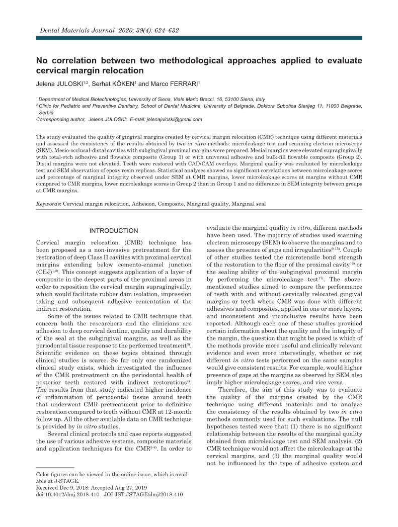
Fig. 1 Schematic representation of the cavity design.
restorative material used for relocation of the cervical margin.
MAtErIALS And MEtHodS
Teeth preparationFourteen intact, healthy, similar sized human molars without any visible cracks, cavities or restorations were selected for the study after informed consent of the patients was obtained. Teeth were mechanically cleaned with hand scalers, brushed with a pumice and were stored in 0.1% thymol solution for no longer than three months. Standardized mesio-occlusal-distal (MOD) cavity preparations were created using water-cooled diamond burs from the set for ceramic inlays and partial crowns preparation (Komet Burs Expert Set 4562/4562ST, Komet, Lemgo, Germany) mounted on a high-speed handpiece. The remaining axial walls had 2 mm of thickness, and they were reduced for a cuspal coverage. Proximally, box-shaped preparations were made, 2 mm in the mesio-distal and 5 mm in bucco-lingual direction. The inner angles of the cavities were rounded, and the margins were not beveled. Proximal margins in both, mesial and distal sides, were located in dentin, approximately 1 mm below the CEJ. Distal proximal margins were not elevated in any of the samples. In all the samples the cervical margins on the mesial sides were relocated above CEJ with flowable composite (Fig. 1). To achieve CMR of the mesial proximal margins and immediate dentin sealing (IDS) appropriate adhesives were used, according to the assigned experimental groups. The adhesive layer, as well as each 1-mm increment of the flowable composite, was light cured for 20 s from the occlusal direction using the light curing unit with a curing output of 1,200 mw/cm2 (BA Optima 10, B.A. International, Northampton, UK). The adaptation of the flowable composites was performed with flowability of the material itself and with a ball ended hand instruments and a microbrush. Special care was taken not to layer the composite more than 2 mm in thickness. Kerr 2181 Adapt® SuperCap® (Kerr, Orange, CA, USA) matrices in steel (0.038, 5.0 mm
high) were used to create the CMR. The circumferential matrix was carefully adjusted to eliminate the risk of overhanging of the material over the margins and 2 mm space is marked on the inner side of the matrix to avoid overfilling of the proximal box. Water-cooled diamond burs (Komet Burs Expert Set 4562/4562ST, Komet) on high-speed handpiece were used to give final shape of each cavity after CMR was performed.
ImpressionExtraoral scanner GC Aadva Lab Scanner (GC, Tokyo, Japan) was used for making digital impressions of the prepared teeth. Scanned files were sent to Milling Center (GC Europe, Leuven, Belgium) to create the overlays out of hybrid ceramic computer-aided design/computer-aided manufacturing (CAD/CAM) blocks (Cerasmart, GC). The prepared teeth without any temporary restorations were kept in fresh water for two weeks at room temperature until the overlays were luted. The fit of the overlays was examined under the digital microscope (Nikon Shuttle Pix, Nikon, Tokyo, Japan) and the digital photographs were taken under 10× magnification.
Luting procedureBefore luting, teeth were cleaned and preparation surfaces were gently dried. The overlays were sandblasted at approximately 300 kPa pressure with 50-μm aluminum oxide particles. Inner sandblasted surface of the overlays was silanized: in Group 1 Silane primer (Kerr) was applied and in Group 2 Monobond Plus (Ivoclar Vivadent, Schaan, Lichtenstein). Luting procedures were performed with an appropriate resin cement, according to the assigned experimental group. The resin cements were mixed with its special mixing tip, initial mixture was discarded on a clean paper. The latter mixture was applied on restoration’s inner surface and preparation surface. The overlays were pressed firmly on teeth and the excess luting material was cleaned with a microbrush. The restoration margins were covered with a water-based glycerine gel (Airblock™, Dentsply DeTrey, Konstanz, Germany). Each axial wall was light cured for 60 s and finally occlusal surface was cured 60 s (BA Optima 10). Margins were gently finished with flexible disks (SofLex Pop-on, 3M ESPE, St. Paul, MN, USA).
GroupsTeeth were randomly assigned to two equal groups of 7 specimens each, according to the adhesive materials employed, as follows (Fig. 2):
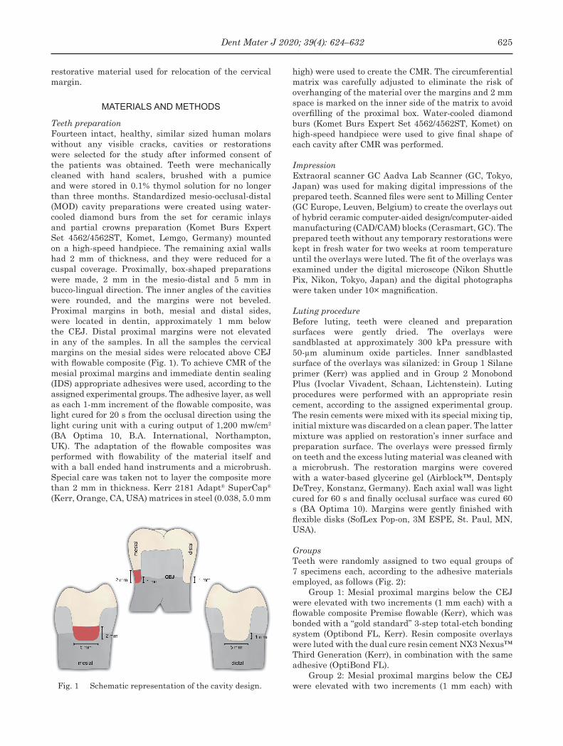
Group 1: Mesial proximal margins below the CEJ were elevated with two increments (1 mm each) with a flowable composite Premise flowable (Kerr), which was bonded with a “gold standard” 3-step total-etch bonding system (Optibond FL, Kerr). Resin composite overlays were luted with the dual cure resin cement NX3 Nexus™ Third Generation (Kerr), in combination with the same adhesive (OptiBond FL).
Group 2: Mesial proximal margins below the CEJ were elevated with two increments (1 mm each) with
625Dent Mater J 2020; 39(4): 624–632
Fig. 2 Schematic representation of the experimental groups tested in the study.
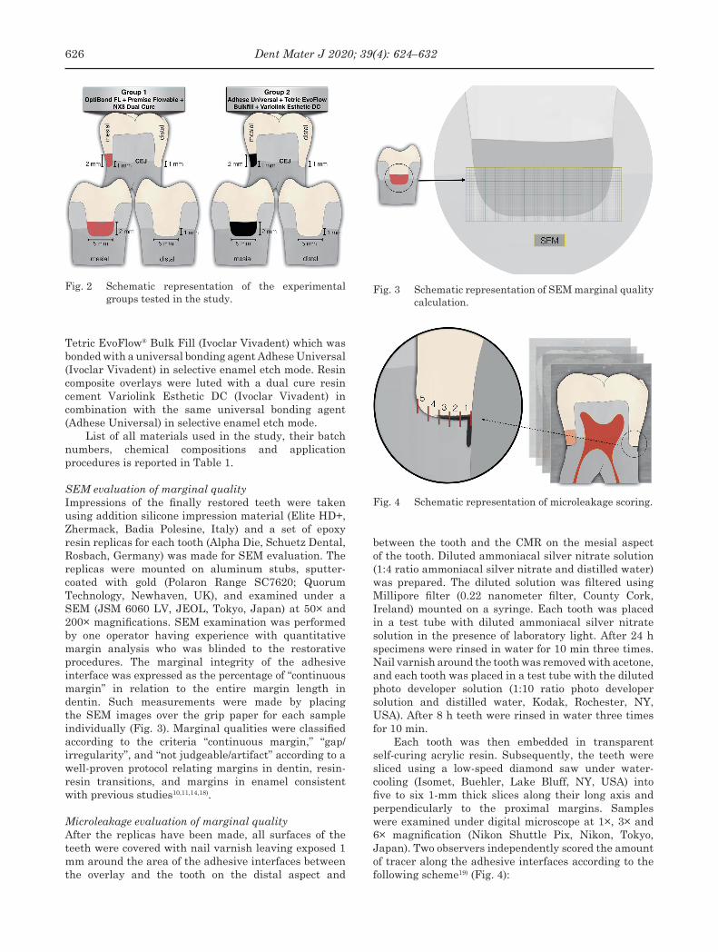
Fig. 3 Schematic representation of SEM marginal quality calculation.
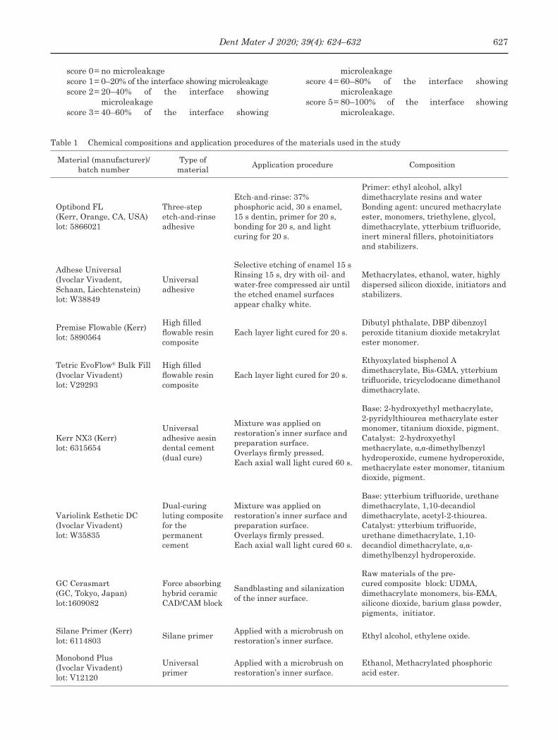
Fig. 4 Schematic representation of microleakage scoring.
Tetric EvoFlow® Bulk Fill (Ivoclar Vivadent) which was bonded with a universal bonding agent Adhese Universal (Ivoclar Vivadent) in selective enamel etch mode. Resin composite overlays were luted with a dual cure resin cement Variolink Esthetic DC (Ivoclar Vivadent) in combination with the same universal bonding agent (Adhese Universal) in selective enamel etch mode.
List of all materials used in the study, their batch numbers, chemical compositions and application procedures is reported in Table 1.
SEM evaluation of marginal qualityImpressions of the finally restored teeth were taken using addition silicone impression material (Elite HD+, Zhermack, Badia Polesine, Italy) and a set of epoxy resin replicas for each tooth (Alpha Die, Schuetz Dental, Rosbach, Germany) was made for SEM evaluation. The replicas were mounted on aluminum stubs, sputter-coated with gold (Polaron Range SC7620; Quorum Technology, Newhaven, UK), and examined under a SEM (JSM 6060 LV, JEOL, Tokyo, Japan) at 50× and 200× magnifications. SEM examination was performed by one operator having experience with quantitative margin analysis who was blinded to the restorative procedures. The marginal integrity of the adhesive interface was expressed as the percentage of “continuous margin” in relation to the entire margin length in dentin. Such measurements were made by placing the SEM images over the grip paper for each sample individually (Fig. 3). Marginal qualities were classified according to the criteria “continuous margin,” “gap/ irregularity”, and “not judgeable/artifact” according to a well-proven protocol relating margins in dentin, resin-resin transitions, and margins in enamel consistent with previous studies10,11,14,18).
Microleakage evaluation of marginal qualityAfter the replicas have been made, all surfaces of the teeth were covered with nail varnish leaving exposed 1 mm around the area of the adhesive interfaces between the overlay and the tooth on the distal aspect and
between the tooth and the CMR on the mesial aspect of the tooth. Diluted ammoniacal silver nitrate solution (1:4 ratio ammoniacal silver nitrate and distilled water) was prepared. The diluted solution was filtered using Millipore filter (0.22 nanometer filter, County Cork, Ireland) mounted on a syringe. Each tooth was placed in a test tube with diluted ammoniacal silver nitrate solution in the presence of laboratory light. After 24 h specimens were rinsed in water for 10 min three times. Nail varnish around the tooth was removed with acetone, and each tooth was placed in a test tube with the diluted photo developer solution (1:10 ratio photo developer solution and distilled water, Kodak, Rochester, NY, USA). After 8 h teeth were rinsed in water three times for 10 min.
Each tooth was then embedded in transparent self-curing acrylic resin. Subsequently, the teeth were sliced using a low-speed diamond saw under water-cooling (Isomet, Buehler, Lake Bluff, NY, USA) into five to six 1-mm thick slices along their long axis and perpendicularly to the proximal margins. Samples were examined under digital microscope at 1×, 3× and 6× magnification (Nikon Shuttle Pix, Nikon, Tokyo, Japan). Two observers independently scored the amount of tracer along the adhesive interfaces according to the following scheme19) (Fig. 4):
626 Dent Mater J 2020; 39(4): 624–632
Table 1 Chemical compositions and application procedures of the materials used in the study
Material (manufacturer)/batch number
Type of material
Application procedure Composition
Optibond FL (Kerr, Orange, CA, USA) lot: 5866021
Three-stepetch-and-rinse adhesive
Etch-and-rinse: 37% phosphoric acid, 30 s enamel, 15 s dentin, primer for 20 s, bonding for 20 s, and light curing for 20 s.
Primer: ethyl alcohol, alkyl dimethacrylate resins and water Bonding agent: uncured methacrylate ester, monomers, triethylene, glycol, dimethacrylate, ytterbium trifluoride, inert mineral fillers, photoinitiators and stabilizers.
Sandblasting and silanization of the inner surface.
Raw materials of the pre-cured composite block: UDMA, dimethacrylate monomers, bis-EMA, silicone dioxide, barium glass powder, pigments, initiator.
Silane Primer (Kerr) lot: 6114803
Silane primerApplied with a microbrush on restoration’s inner surface.
Ethyl alcohol, ethylene oxide.
Monobond Plus (Ivoclar Vivadent) lot: V12120
Universal primer
Applied with a microbrush on restoration’s inner surface.
Ethanol, Methacrylated phosphoric acid ester.
score 0= no microleakagescore 1= 0–20% of the interface showing microleakagescore 2= 20–40% of the interface showing
microleakagescore 3= 40–60% of the interface showing
microleakagescore 4= 60–80% of the interface showing
microleakagescore 5= 80–100% of the interface showing
microleakage.
627Dent Mater J 2020; 39(4): 624–632
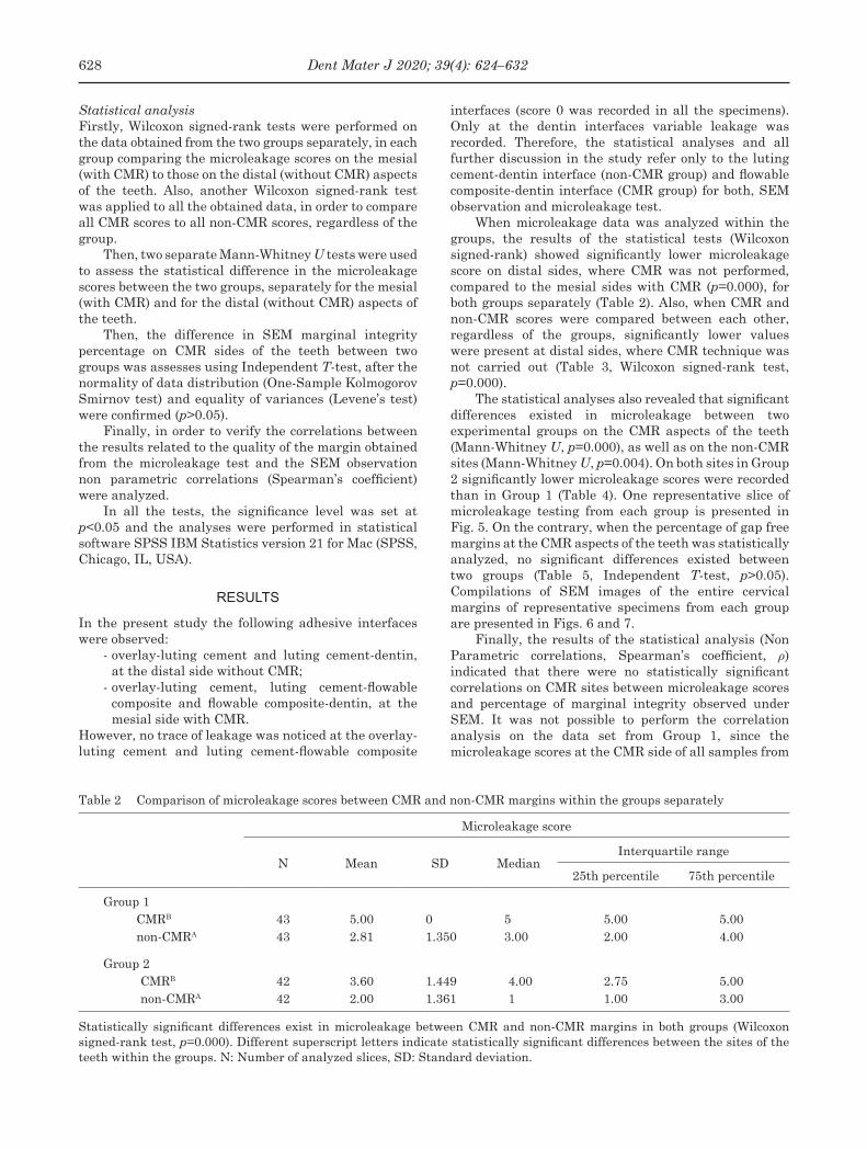
Table 2 Comparison of microleakage scores between CMR and non-CMR margins within the groups separately
Microleakage score
N Mean SD MedianInterquartile range
25th percentile 75th percentile
Group 1CMRB
non-CMRA
4343
5.002.81
01.350
53.00
5.002.00
5.004.00
Group 2CMRB
non-CMRA
4242
3.602.00
1.4491.361
4.001
2.751.00
5.003.00
Statistically significant differences exist in microleakage between CMR and non-CMR margins in both groups (Wilcoxon signed-rank test, p=0.000). Different superscript letters indicate statistically significant differences between the sites of the teeth within the groups. N: Number of analyzed slices, SD: Standard deviation.
Statistical analysisFirstly, Wilcoxon signed-rank tests were performed on the data obtained from the two groups separately, in each group comparing the microleakage scores on the mesial (with CMR) to those on the distal (without CMR) aspects of the teeth. Also, another Wilcoxon signed-rank test was applied to all the obtained data, in order to compare all CMR scores to all non-CMR scores, regardless of the group.
Then, two separate Mann-Whitney U tests were used to assess the statistical difference in the microleakage scores between the two groups, separately for the mesial (with CMR) and for the distal (without CMR) aspects of the teeth.
Then, the difference in SEM marginal integrity percentage on CMR sides of the teeth between two groups was assesses using Independent T-test, after the normality of data distribution (One-Sample Kolmogorov Smirnov test) and equality of variances (Levene’s test) were confirmed (p>0.05).
Finally, in order to verify the correlations between the results related to the quality of the margin obtained from the microleakage test and the SEM observation non parametric correlations (Spearman’s coefficient) were analyzed.
In all the tests, the significance level was set at p<0.05 and the analyses were performed in statistical software SPSS IBM Statistics version 21 for Mac (SPSS, Chicago, IL, USA).
rESuLtS
In the present study the following adhesive interfaces were observed:
- overlay-luting cement and luting cement-dentin, at the distal side without CMR;
- overlay-luting cement, luting cement-flowable composite and flowable composite-dentin, at the mesial side with CMR.
However, no trace of leakage was noticed at the overlay-luting cement and luting cement-flowable composite
interfaces (score 0 was recorded in all the specimens). Only at the dentin interfaces variable leakage was recorded. Therefore, the statistical analyses and all further discussion in the study refer only to the luting cement-dentin interface (non-CMR group) and flowable composite-dentin interface (CMR group) for both, SEM observation and microleakage test.
When microleakage data was analyzed within the groups, the results of the statistical tests (Wilcoxon signed-rank) showed significantly lower microleakage score on distal sides, where CMR was not performed, compared to the mesial sides with CMR (p=0.000), for both groups separately (Table 2). Also, when CMR and non-CMR scores were compared between each other, regardless of the groups, significantly lower values were present at distal sides, where CMR technique was not carried out (Table 3, Wilcoxon signed-rank test, p=0.000).
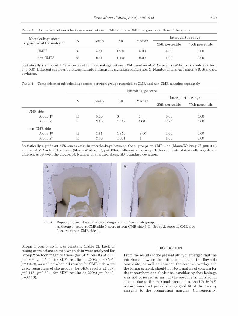
The statistical analyses also revealed that significant differences existed in microleakage between two experimental groups on the CMR aspects of the teeth (Mann-Whitney U, p=0.000), as well as on the non-CMR sites (Mann-Whitney U, p=0.004). On both sites in Group 2 significantly lower microleakage scores were recorded than in Group 1 (Table 4). One representative slice of microleakage testing from each group is presented in Fig. 5. On the contrary, when the percentage of gap free margins at the CMR aspects of the teeth was statistically analyzed, no significant differences existed between two groups (Table 5, Independent T-test, p>0.05). Compilations of SEM images of the entire cervical margins of representative specimens from each group are presented in Figs. 6 and 7.
Finally, the results of the statistical analysis (Non Parametric correlations, Spearman’s coefficient, ρ) indicated that there were no statistically significant correlations on CMR sites between microleakage scores and percentage of marginal integrity observed under SEM. It was not possible to perform the correlation analysis on the data set from Group 1, since the microleakage scores at the CMR side of all samples from
628 Dent Mater J 2020; 39(4): 624–632
Table 3 Comparison of microleakage scores between CMR and non-CMR margins regardless of the group
Microleakage scoreregardless of the material
N Mean SD MedianInterquartile range
25th percentile 75th percentile
CMRB 85 4.31 1.235 5.00 4.00 5.00
non-CMRA 84 2.41 1.408 2.00 1.00 3.00
Statistically significant differences exist in microleakage between CMR and non-CMR margins (Wilcoxon signed-rank test, p=0.000). Different superscript letters indicate statistically significant difference. N: Number of analyzed slices, SD: Standard deviation.
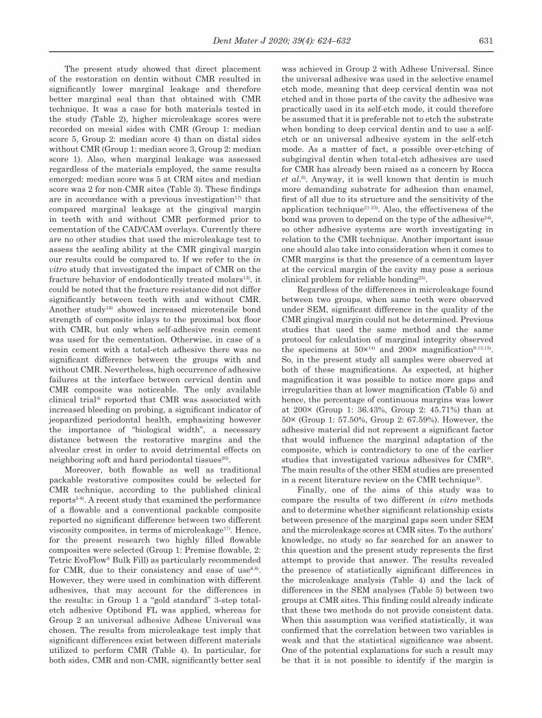
Table 4 Comparison of microleakage scores between groups recorded at CMR and non-CMR margins separately
Microleakage score
N Mean SD MedianInterquartile range
25th percentile 75th percentile
CMR sideGroup 1B
Group 2A
4342
5.003.60
01.449
54.00
5.002.75
5.005.00
non-CMR sideGroup 1B
Group 2A
4342
2.812.00
1.3501.361
3.001
2.001.00
4.003.00
Statistically significant differences exist in microleakage between the 2 groups on CMR side (Mann-Whitney U, p=0.000) and non-CMR side of the teeth (Mann-Whitney U, p=0.004). Different superscript letters indicate statistically significant differences between the groups. N: Number of analyzed slices, SD: Standard deviation.
Fig. 5 Representative slices of microleakage testing from each group. A; Group 1: score at CMR side 5, score at non-CMR side 3. B; Group 2: score at CMR side
2, score at non-CMR side 1.
A B
Group 1 was 5, so it was constant (Table 2). Lack of strong correlations existed when data were analyzed for Group 2 on both magnifications (for SEM results at 50×: ρ=0.306, p=0.504; for SEM results at 200×: ρ=−0.505, p=0.248), as well as when all results for CMR side were used, regardless of the groups (for SEM results at 50×: ρ=0.115, p=0.694; for SEM results at 200×: ρ=−0.443, p=0.113).
dIScuSSIon
From the results of the present study it emerged that the interfaces between the luting cement and the flowable composite, as well as between the ceramic overlay and the luting cement, should not be a matter of concern for the researchers and clinicians, considering that leakage was not observed in any of the specimens. This could also be due to the maximal precision of the CAD/CAM restorations that provided very good fit of the overlay margins to the preparation margins. Consequently,
629Dent Mater J 2020; 39(4): 624–632
Table 5 Comparison of marginal integrity of CMR sides between two groups observed on SEM images under 50× and 200× magnifications
Percentage of gap free margins (%)
N Mean Standard Deviation Standard Error p value
50× magnificationGroup 1A
Group 2A
77
57.50%67.59%
18.7017.99
7.076.80
0.324
200× magnificationGroup 1A
Group 2A
77
36.43%45.71%
14.887.77
5.622.93
0.169
Same superscript letters indicate lack of statistically significant differences between the groups (Independent T-test, p>0.05). N: Number of analyzed teeth.
Fig. 6 Compilation of SEM images of the entire cervical margin of a specimen from Group 1.
Fig. 7 Compilation of SEM images of the entire cervical margin of a specimen from Group 2.
a good seal without any imperfections or gaps at the interface could be achieved, which did not permit any marginal leakage. It may be assumed that restorations created in a traditional way would have given different results. However, this study confirmed that the only troublesome interface for the adhesion was dentin, which became the matter of further interest of this investigation.
The results indicate that there was not statistically significant relationship between the results obtained from two in vitro methods used for evaluation of the quality of the margins. Consequently, the first null
hypothesis was accepted. Furthermore, the CMR technique impaired the sealing at the cervical margins, as significant differences in microleakage scores were recorded at sides of the teeth with and without CMR, so the second null hypothesis had to be rejected. And finally, the marginal microleakage was significantly influenced by the type of adhesive system and restorative material used for relocation of the cervical margin, whereas the percentage of the gap-free margin assessed on the SEM images did not differ between the different materials. Therefore, the third null hypothesis was only partially rejected.
630 Dent Mater J 2020; 39(4): 624–632
The present study showed that direct placement of the restoration on dentin without CMR resulted in significantly lower marginal leakage and therefore better marginal seal than that obtained with CMR technique. It was a case for both materials tested in the study (Table 2), higher microleakage scores were recorded on mesial sides with CMR (Group 1: median score 5, Group 2: median score 4) than on distal sides without CMR (Group 1: median score 3, Group 2: median score 1). Also, when marginal leakage was assessed regardless of the materials employed, the same results emerged: median score was 5 at CRM sites and median score was 2 for non-CMR sites (Table 3). These findings are in accordance with a previous investigation17) that compared marginal leakage at the gingival margin in teeth with and without CMR performed prior to cementation of the CAD/CAM overlays. Currently there are no other studies that used the microleakage test to assess the sealing ability at the CMR gingival margin our results could be compared to. If we refer to the in vitro study that investigated the impact of CMR on the fracture behavior of endodontically treated molars13), it could be noted that the fracture resistance did not differ significantly between teeth with and without CMR. Another study16) showed increased microtensile bond strength of composite inlays to the proximal box floor with CMR, but only when self-adhesive resin cement was used for the cementation. Otherwise, in case of a resin cement with a total-etch adhesive there was no significant difference between the groups with and without CMR. Nevertheless, high occurrence of adhesive failures at the interface between cervical dentin and CMR composite was noticeable. The only available clinical trial4) reported that CMR was associated with increased bleeding on probing, a significant indicator of jeopardized periodontal health, emphasizing however the importance of “biological width”, a necessary distance between the restorative margins and the alveolar crest in order to avoid detrimental effects on neighboring soft and hard periodontal tissues20).
Moreover, both flowable as well as traditional packable restorative composites could be selected for CMR technique, according to the published clinical reports5-8). A recent study that examined the performance of a flowable and a conventional packable composite reported no significant difference between two different viscosity composites, in terms of microleakage17). Hence, for the present research two highly filled flowable composites were selected (Group 1: Premise flowable, 2: Tetric EvoFlow® Bulk Fill) as particularly recommended for CMR, due to their consistency and ease of use6,8). However, they were used in combination with different adhesives, that may account for the differences in the results: in Group 1 a “gold standard” 3-step total-etch adhesive Optibond FL was applied, whereas for Group 2 an universal adhesive Adhese Universal was chosen. The results from microleakage test imply that significant differences exist between different materials utilized to perform CMR (Table 4). In particular, for both sides, CMR and non-CMR, significantly better seal
was achieved in Group 2 with Adhese Universal. Since the universal adhesive was used in the selective enamel etch mode, meaning that deep cervical dentin was not etched and in those parts of the cavity the adhesive was practically used in its self-etch mode, it could therefore be assumed that it is preferable not to etch the substrate when bonding to deep cervical dentin and to use a self-etch or an universal adhesive system in the self-etch mode. As a matter of fact, a possible over-etching of subgingival dentin when total-etch adhesives are used for CMR has already been raised as a concern by Rocca et al.6). Anyway, it is well known that dentin is much more demanding substrate for adhesion than enamel, first of all due to its structure and the sensitivity of the application technique21-23). Also, the effectiveness of the bond was proven to depend on the type of the adhesive24), so other adhesive systems are worth investigating in relation to the CMR technique. Another important issue one should also take into consideration when it comes to CMR margins is that the presence of a cementum layer at the cervical margin of the cavity may pose a serious clinical problem for reliable bonding25).
Regardless of the differences in microleakage found between two groups, when same teeth were observed under SEM, significant difference in the quality of the CMR gingival margin could not be determined. Previous studies that used the same method and the same protocol for calculation of marginal integrity observed the specimens at 50×14) and 200× magnification9-13,15). So, in the present study all samples were observed at both of these magnifications. As expected, at higher magnification it was possible to notice more gaps and irregularities than at lower magnification (Table 5) and hence, the percentage of continuous margins was lower at 200× (Group 1: 36.43%, Group 2: 45.71%) than at 50× (Group 1: 57.50%, Group 2: 67.59%). However, the adhesive material did not represent a significant factor that would influence the marginal adaptation of the composite, which is contradictory to one of the earlier studies that investigated various adhesives for CMR9). The main results of the other SEM studies are presented in a recent literature review on the CMR technique3).
Finally, one of the aims of this study was to compare the results of two different in vitro methods and to determine whether significant relationship exists between presence of the marginal gaps seen under SEM and the microleakage scores at CMR sites. To the authors’ knowledge, no study so far searched for an answer to this question and the present study represents the first attempt to provide that answer. The results revealed the presence of statistically significant differences in the microleakage analysis (Table 4) and the lack of differences in the SEM analyses (Table 5) between two groups at CMR sites. This finding could already indicate that these two methods do not provide consistent data. When this assumption was verified statistically, it was confirmed that the correlation between two variables is weak and that the statistical significance was absent. One of the potential explanations for such a result may be that it is not possible to identify if the margin is
631Dent Mater J 2020; 39(4): 624–632
sealed efficiently by observing it under SEM. Probably much higher magnifications or diverse techniques are necessary to reveal the adhesive interfaces that are not efficiently sealed. One study has proposed microcomputed tomography as a non-destructive technique for inspection of the tooth-restoration interfaces and interfacial leakage26). However, it would be valuable to investigate more about the most appropriate in vitro method to appraise the quality of the CMR adhesive margins. One limitation of the present study is a small sample size, which was used in order to obtain the initial data. Further research should verify the results of this study using larger number of the specimens and clarify which of the in vitro tests provide more reliable data and which one could give more accurate predictions for clinical success. Undisputedly, only well-designed clinical trials could provide strong scientific evidence that would allow for definitive clinical recommendations.
concLuSIonS
Based on the results obtained under conditions of the present study, several conclusions could be drawn:
1. CMR seems to provide less adequate seal of the margin than the one achieved by cementing the restoration directly to dentin without CMR.
2. The sealing ability of the marginal interface depends on the adhesive materials used for performing CMR.
3. Differences in the quality of the marginal adaptation between two different materials used for CMR could not be detected by SEM observations.
4. SEM examination of the marginal adaptation does not allow for the predictions of the functional sealing of the margins.
rEFErEncES
1) Dietschi D, Spreafico R. Current clinical concepts for adhesive cementation of tooth-colored posterior restorations. Pract Periodontics Aesthet Dent 1998; 10: 47-54; quiz 56.
2) Magne P, Spreafico R. Deep margin elevation: a paradigm shift. Am J Esthet Dent 2012; 2: 86-96.
3) Juloski J, Köken S, Ferrari M. Cervical margin relocation in indirect adhesive restorations: A literature review. J Prosthodont Res 2018; 62: 273-280.
4) Ferrari M, Köken S, Grandini S, Ferrari Cagidiaco E, Joda T, Discepoli N. Influence of cervical margin relocation (CMR) on periodontal health: 12-month results of a controlled trial. J Dent 2018; 69: 70-76.
5) Veneziani M. Adhesive restorations in the posterior area with subgingival cervical margins: new classification and differentiated treatment approach. Eur J Esthet Dent 2010; 5: 50-76.
6) Rocca GT, Rizcalla N, Krejci I, Dietschi D. Evidence-based concepts and procedures for bonded inlays and onlays. Part II. Guidelines for cavity preparation and restoration fabrication. Int J Esthet Dent 2015; 10: 392-413.
7) Kielbassa AM, Philipp F. Restoring proximal cavities of molars using the proximal box elevation technique: systematic review and report of a case. Quintessence Int 2015; 46: 751-764.
8) Dietschi D, Spreafico R. Evidence-based concepts and procedures for bonded inlays and onlays. Part I. Historical perspectives and clinical rationale for a biosubstitutive approach. Int J Esthet Dent 2015; 10: 210-227.
9) Lefever D, Gregor L, Bortolotto T, Krejci I. Supragingival relocation of subgingivally located margins for adhesive inlays/onlays with different materials. J Adhes Dent 2012; 14: 561-567.
10) Roggendorf MJ, Krämer N, Dippold C, Vosen VE, Naumann M, Jablonski-Momeni A, et al. Effect of proximal box elevation with resin composite on marginal quality of resin composite inlays in vitro. J Dent 2012; 40: 1068-1073.
11) Frankenberger R, Hehn J, Hajtó J, Krämer N, Naumann M, Koch A, et al. Effect of proximal box elevation with resin composite on marginal quality of ceramic inlays in vitro. Clin Oral Investig 2013; 17: 177-183.
12) Zaruba M, Göhring TN, Wegehaupt FJ, Attin T. Influence of a proximal margin elevation technique on marginal adaptation of ceramic inlays. Acta Odontol Scand 2013; 71: 317-324.
13) Ilgenstein I, Zitzmann NU, Bühler J, Wegehaupt FJ, Attin T, Weiger R, et al. Influence of proximal box elevation on the marginal quality and fracture behavior of root-filled molars restored with CAD/CAM ceramic or composite onlays. Clin Oral Investig 2015; 19: 1021-1028.
14) Spreafico R, Marchesi G, Turco G, Frassetto A, Di Lenarda R, Mazzoni A, et al. Evaluation of the in vitro effects of cervical marginal relocation using composite resins on the marginal quality of CAD/CAM crowns. J Adhes Dent 2016; 18: 355-362.
15) Müller V, Friedl KH, Friedl K, Hahnel S, Handel G, Lang R. Influence of proximal box elevation technique on marginal integrity of adhesively luted Cerec inlays. Clin Oral Investig 2017; 21: 607-612.
16) Da Silva Goncalves D, Cura M, Ceballos L, Fuentes MV. Influence of proximal box elevation on bond strength of composite inlays. Clin Oral Investig 2017; 21: 247-254.
17) Köken S, Juloski J, Sorrentino R, Grandini S, Ferrari M. Marginal sealing of relocated cervical margins of mesio-occluso-distal overlays. J Oral Sci 2018; 60: 460-468.
18) Frankenberger R, Tay FR. Self-etch vs etch-and-rinse adhesives: effect of thermo-mechanical fatigue loading on marginal quality of bonded resin composite restorations. Dent Mater 2005; 21: 397-412.
19) Saboia VP, Nato F, Mazzoni A, Orsini G, Putignano A, Giannini M, et al. Adhesion of a two-step etch-and-rinse adhesive on collagen-depleted dentin. J Adhes Dent 2008; 10: 419-422.
20) Ingber JS, Rose LF, Coslet JG. The “biologic width”: a concept in periodontics and restorative dentistry. Alpha Omegan 1977; 70: 62-65.
21) Perdigão J. Dentin bonding as a function of dentin structure. Dent Clin North Am 2002; 46: 277-301.
22) Van Meerbeek B, Van Landuyt K, De Munck J, Hashimoto M, Peumans M, Lambrechts P, et al. Technique-sensitivity of contemporary adhesives. Dent Mater J 2005; 24: 1-13.
23) Cardoso MV, de Almeida Neves A, Mine A, Coutinho E, Van Landuyt K, De Munck J, et al. Current aspects on bonding effectiveness and stability in adhesive dentistry. Aust Dent J 2011; 56 Suppl 1: 31-44.
24) Kugel G, Ferrari M. The science of bonding: from first to sixth generation. J Am Dent Assoc 2000; 131 Suppl: S20-25.
25) Ferrari M, Cagidiaco MC, Davidson CL. Resistance of cementum in Class II and V cavities to penetration by an adhesive system. Dent Mater 1997; 13: 157-162.
26) Rengo C, Goracci C, Ametrano G, Chieffi N, Spagnuolo G, Rengo S, et al. Marginal leakage of class V composite restorations assessed using microcomputed tomography and scanning electron microscope. Oper Dent 2015; 40: 440-448.