This article was downloaded by: [University of California Santa Cruz] On: 08 March 2013, At: 20:49 Publisher: Taylor & Francis Informa Ltd Registered in England and Wales Registered Number: 1072954 Registered office: Mortimer House, 37-41 Mortimer Street, London W1T 3JH, UK Journal of Biomaterials Science, Polymer Edition Publication details, including instructions for authors and subscription information: http://www.tandfonline.com/loi/tbsp20 Non-fouling properties of polysaccharide-coated surfaces Marco Morra a & Clara Cassineli b a Nobil Bio Ricerche, Str. S. Rocco 32, 14018 Villafranca d'Asti, Italy b Nobil Bio Ricerche, Str. S. Rocco 32, 14018 Villafranca d'Asti, Italy Version of record first published: 02 Apr 2012. To cite this article: Marco Morra & Clara Cassineli (1999): Non-fouling properties of polysaccharide-coated surfaces, Journal of Biomaterials Science, Polymer Edition, 10:10, 1107-1124 To link to this article: http://dx.doi.org/10.1163/156856299X00711 PLEASE SCROLL DOWN FOR ARTICLE Full terms and conditions of use: http://www.tandfonline.com/page/terms-and- conditions This article may be used for research, teaching, and private study purposes. Any substantial or systematic reproduction, redistribution, reselling, loan, sub- licensing, systematic supply, or distribution in any form to anyone is expressly forbidden. The publisher does not give any warranty express or implied or make any representation that the contents will be complete or accurate or up to date. The accuracy of any instructions, formulae, and drug doses should be independently verified with primary sources. The publisher shall not be liable for any loss, actions, claims, proceedings, demand, or costs or damages whatsoever or howsoever caused arising directly or indirectly in connection with or arising out of the use of this material.
Transcript
This article was downloaded by: [University of California Santa Cruz]On: 08 March 2013, At: 20:49Publisher: Taylor & FrancisInforma Ltd Registered in England and Wales Registered Number: 1072954Registered office: Mortimer House, 37-41 Mortimer Street, London W1T 3JH, UK
Journal of Biomaterials Science,Polymer EditionPublication details, including instructions for authorsand subscription information:http://www.tandfonline.com/loi/tbsp20
Non-fouling properties ofpolysaccharide-coated surfacesMarco Morra a & Clara Cassineli ba Nobil Bio Ricerche, Str. S. Rocco 32, 14018 Villafrancad'Asti, Italyb Nobil Bio Ricerche, Str. S. Rocco 32, 14018 Villafrancad'Asti, ItalyVersion of record first published: 02 Apr 2012.
To cite this article: Marco Morra & Clara Cassineli (1999): Non-fouling properties ofpolysaccharide-coated surfaces, Journal of Biomaterials Science, Polymer Edition, 10:10,1107-1124
To link to this article: http://dx.doi.org/10.1163/156856299X00711
PLEASE SCROLL DOWN FOR ARTICLE
Full terms and conditions of use: http://www.tandfonline.com/page/terms-and-conditions
This article may be used for research, teaching, and private study purposes.Any substantial or systematic reproduction, redistribution, reselling, loan, sub-licensing, systematic supply, or distribution in any form to anyone is expresslyforbidden.
The publisher does not give any warranty express or implied or make anyrepresentation that the contents will be complete or accurate or up to date. Theaccuracy of any instructions, formulae, and drug doses should be independentlyverified with primary sources. The publisher shall not be liable for any loss,actions, claims, proceedings, demand, or costs or damages whatsoever orhowsoever caused arising directly or indirectly in connection with or arising outof the use of this material.
Non-fouling properties of polysaccharide-coated surfaces
MARCO MORRA * and CLARA CASSINELI
Nobil Bio Ricerche, Str. S. Rocco 32, 14018 Villafranca d'Asti, Italy
Received 19 January 1999; accepted 18 May 1999
Abstract-Hyaluronic acid and alginic acid, examples of hydrophilic polysaccharides whose biologi- cal and technological properties are deeply related to strong interaction with water, have been coupled to substrate surfaces. These coatings can prevent mammalian cells adhesion and greatly reduce bac- terial cells adhesion in vitro and in several in vivo applications. The anti-adhesive properties of these surfaces are discussed in terms of the surface fractional coverage by the polysaccharide, as evaluated
by XPS analysis and water contact angle. The implications of chemical-molecular considerations to the properties of these coatings are discussed from an analytical and mechanistic point of view.
Surface coatings based on naturally-occurring polysaccharides are presently used
and/or investigated in several different fields of biomaterials science. Heparin
coatings for blood-contacting devices are probably the best known example [1-3]. The rationale for trying to immobilize polysaccharides to surfaces is to exploit the
intrinsic properties of these natural molecules to impart a specific characteristic
to the surface. Referring again to coatings based on heparin, according to the
prevailing views, the goal is to exploit its capability to interact specifically with
antithrombin [ 1, 2]. When it comes to fouling resistance, or the ability to resist adhesion of biological
moieties in the aqueous environments, polysaccharides present interesting possibil- ities. Several of their main properties in nature have directly to do with 'insulation'
of particles or molecules from the surroundings. For instance, they have a capacity to stabilize cellular suspensions and to facilitate bulk transport in muci and gels. Several polysaccharides are used industrially to the extent of the millions of met-
ric tons in food technology because of their unique power to stabilize suspensions
*To whom correspondence should be addressed. E-mail: [email protected]
Dow
nloa
ded
by [
Uni
vers
ity o
f C
alif
orni
a Sa
nta
Cru
z] a
t 20:
49 0
8 M
arch
201
3
1108
under all ionic conditions [4, 5]. As nicely underlined by Rau and Parsegian, the
key to the understanding of these properties seems to be in understanding the phys- ical interactions of these molecules with each other as well as with their aqueous environment 141. By direct measurement by the osmotic stress method, Rau and
Parsegian were able to measure forces between Xanthan and other polysaccharides molecules. One of their conclusions is quoted here as the starting point of this work:
'The same hygroscopic power that stabilizes suspensions under all ionic conditions
is seen as a hydration force insensitive to the kind and concentration of salt in the
suspending medium' [4]. The working hypothesis stemming from these results is that, by clever molecular
engineering, nature makes use of hydration to prevent non-specilic interactions
in aqueous media. The polysaccharide way to fouling resistance is an attempt to translate the peculiar molecular behavior shown in homogeneous phases or
suspensions to materials surfaces. The first step of this attempt is the choice of
the proper polysaccharide molecule to be linked to the surface. According to
the general principles described above, the candidate should be looked for among those polysaccharides explicitly engineered by nature to maximize interaction with
water. Additionally, it should bear chemical moieties that allow a straightforward chemical coupling to the surface (this consideration is particularly important when
the complex shape and geometry of most medical devices is taken into account). The aim of this paper is to present several results of the work going on in our
laboratory on the use of polysaccharides to modify medical devices, and for fouling resistance in particular. Most of this work is based on two water-scooping molecular
machines: alginic acid and hyaluronic acid. Alginate (AA) is a natural polymer that
exists in many species of seaweed, where its main role is to regulate the water
content [6]. Its technological applications are founded on interaction with water:
it is commonly used in anti-freeze formulation (the strong interaction with water
lowers the freezing point). In the biomedical field it is mostly used in wound-
dressing materials, where its interaction with water is at the basis of the 'moist-
healing' mechanism [6]. AA is composed of two sterically different repeating units,
( 4) a-L-guluronate (G unit) and ( 4) fl-D-mannuronate (M unit). The
relative abundance of G and M blocks affects the properties and structure of alginate macromolecular chains and their interaction with water and dissolved cations. A
commercially available alginate mostly containing mannuronate units, or 'high M'
alginate, was used in these studies. In a MM chain the saccharide units are joined
by the fl( 1 ,4) glycosidic bond, resulting in a straight-chain macromolecule.
Hyaluronan (HA) is a macromolecule whose basic disaccharide unit is composed of N-acetyl-D-glucosamine and o-glucuronic acid. Scott pointed out about 10 years
ago that HA is a molecule of paradoxes and contrasts [7]. On the one hand, it
is a simple homopolytner, doing very simple mechanical jobs related to its strong interaction with water, such as space filler or lubricant. On the other hand, it takes
part in cell surface phenomena of great specificity and can engage in very specific interactions with a number of proteins [7, 8]. It has been claimed that the diminished
Dow
nloa
ded
by [
Uni
vers
ity o
f C
alif
orni
a Sa
nta
Cru
z] a
t 20:
49 0
8 M
arch
201
3
1109
capacity to interact with other molecules, a necessary requisite for the role HA
plays in aqueous channels in the pericellular matrix, is a direct consequence of the
secondary structure of HA in aqueous solution [7]. Clearly, it is tempting to try to
exploit these properties to impart fouling resistance to medical devices surfaces.
This work will describe some aspects of fouling-resistant polysaccharide coatings. The two main issues that will be described are: (i) the molecular basis for
fouling resistance; and (ii) the characterization of polysaccharide coated surfaces
by physico-chemical means (X-ray photoelectron spectroscopy (XPS) and contact
angle measurement). Surface analysis data will be used to elaborate on results of
in vitro testing of bacterial and cell adhesion. It is important to remind that coatings based on the principles and the materials described in this paper are under trial and
have already produced excellent results in in vivo testing concerning biomedical
devices such as intraocular lenses, the prevention of post-surgical adhesion, and the reduction of bacterial adhesion and colonization to urologic catheters and biliary stents [9].
EXPERIMENTAL
Materials
Surface modification was performed on different sets of samples. For cell adhesion
experiments and XPS analysis, 5-cm diameter bacteriological-grade polystyrene (PS) Petri dishes were used (Corning). Bacterial adhesion testing and contact angle measurement were performed on surface-modified microscope cover glass slides.
High molecular weight polyethyleneimine (PEI, average Mw * 25 000, 50%
solution in water) and 'medium viscosity', water soluble Na salt of AA (product no. A-2158, Mw = 80 000-120 000), from Macroc.ystis pyrifera were from
Sigma-Aldrich. Sodium hyaluronate (Mw * 200000) was from Fidia Advanced
Biopolymers.
Surface modification
Surface modification of PS was obtained by subjecting bacteriological-grade PS Petri dishes to an air plasma treatment in a stainless steel, capacitively coupled
parallel-plate reactor, with the samples located on the water-cooled grounded electrode. The reactor volume is about 3 dm3, and the distance between the
electrodes 10 cm. Air flow rate, controlled by a MKS mass flow controller, was 20 seem (standard cubic centimeters per minute), the pressure inside the
chamber before the onset of the discharge was 2 Pa. The power discharge was
30 W and the treatment time 30 s. The plasma treated PS will be coded OPS below. After treatment, 2 ml of a 0.5% (w/v) aqueous solution of PEI were
put into the treated Petri dishes, and adsorption was performed for 2 h. After
prolonged rinsing in doubly distilled water, samples were dried under a laminar flow hood. PEI-coated PS samples will be coded PEIPS below. Then, 5 ml of
Dow
nloa
ded
by [
Uni
vers
ity o
f C
alif
orni
a Sa
nta
Cru
z] a
t 20:
49 0
8 M
arch
201
3
1110
an aqueous polysaccharide solution were placed into the Petri dishes. Chemical
coupling between surface amino groups and carboxyl groups of the polysaccharides chain was performed by carbodiimide condensation [10]. To obtain different
surface densities of polysaccharide for XPS and cell adhesion experiments, different
concentrations of polysaccharide were used: a 0.5% solution was used to obtain
cell-resistant coatings, while a 0.01 (w/v) solution was employed to obtain a
defective coating (a more precise description of these terms will be given in the
following sections). These samples will be coded O.OIAA, O.O1HA, and so on.
After overnight adsorption, the samples were extensively rinsed and stored
overnight in doubly distilled water. Before cell adhesion, testing and surface
characterization samples were dried under a laminar flow hood.
The surface modification of cover glass slides was obtained in the same way,
except that a hydrocarbon layer was deposited on both sides of glass from ethylene
plasma, using the same reactor described above. The hydrocarbon surface obtained
in this way was then oxidized by air plasma, and HA and AA were coupled using the same procedure adopted for PS modification.
Surface characterization
XPS analysis was performed with a Perkin Elmer PHI 5500 ESCA system. The
instrument is equipped with a monochromatic X-ray source (Al Ka anode) operating at 14 kV and 250 W. The diameter of the analyzed spot is approximately 400 itm, the base pressure 10-s Pa. The angle between the electron analyzer and the
sample surface was maintained at 68 deg. Curve fitting and quantification of
elements were accomplished using the software and sensitivity factors supplied by the manufacturer. In high-resolution spectra, all binding energies were referenced
by setting the CH_r peak maximum in the resolved C 1 s spectra to 285 eV.
Contact angle measurement
The measurement of the contact angle of doubly distilled water on the surface-
modified glass samples was performed by the Wilhelmy plate method, using a Cahn
DCA 312microbalance. The stage velocity was 40 f-lm s-1.
Cell-adhesion experiments
The resistance of polysaccharide-coated surfaces to cell adhesion was evaluated us-
ing the continuous mouse fibroblasts cell line L-929. This is a robust cell line that
readily adheres to most substrates and is a good test for resistance to cell adhesion.
The cell behavior on the modified surfaces was monitored by direct observation
in an inverted microscope and adhesion evaluated by cell counting. Experimental
procedures were the following: Experimental cell culture medium (BIOCHROM
KG, Berlin) consisted of Minimum Eagle's Medium without L-glutamine, 10% fetal
bovine serum, streptomycin (100 itg I- 1), penicillin 100 U ml- 1), and 2 mmol I- 1
Dow
nloa
ded
by [
Uni
vers
ity o
f C
alif
orni
a Sa
nta
Cru
z] a
t 20:
49 0
8 M
arch
201
3
1111
L-glutamine in a 250-ml plastic culture flask (CorningTM). Cells were cultured at
37°C in a humidified incubator equilibrated with 5% C02. Fibroblasts were har-
vested prior to confluence by means of a sterile trypsin-EDTA solution (0.5 gl-l
trypsin, 0.2 EDTA in normal phosphate buffered saline (PBS), pH 7.4), resus-
pended in the experimental cell culture medium, and di luted to 1 x 10⁵ cells ml- 1.
For experiments, 5 ml of the cell suspension were seeded into the surface-modified
PS Petri dishes. For cell counting (see below), experiments were performed in trip- licate. The cell behavior on the modified surfaces was monitored by direct observa-
tion in an inverted microscope and by cell counting. The number of cells adhered to the test surfaces was counted after 4 h, to evaluate
the short-term adhesion, and after 3 days (at this time, cells on the control, plasma- treated PS, had reached confluence). Cell behavior on the polysaccharide-coated surfaces was monitored further for 3 more days.
The number of cells on the surface at the selected time was evaluated by harvesting the cells by trypsin treatment and counting the cell number in a hemocytometer. As
described above, three surface-modified Petri dishes for each experimental point were used and five measurements were performed on each Petri dish. Data are
expressed as mean and standard deviation of the fifteen values obtained.
Bacterial adhesion experiments
The strain Staphylococcus epidermidis RP62A (ATCC 35984) used in this study is
a slime and capsular polysaccharide producer. It was routinely maintained both on
tryptic soy agar (TSA, Sigma) plates and transferred to new plates monthly and as
frozen suspension in liquid culture medium supplemented with 10% glycerol. Bac-
terial suspension was obtained by inoculating 100 ml tryptic soy broth (TSB, Sigma) and incubating overnight at 37°C. After incubating overnight, the suspension was
washed three times with PBS (Sigma), and finally resuspended in PBS. PBS used
for this experiment has the following composition: 0.2 g KCI, 0.2 g 1-' KHzP04, 8.0 NaCI, 1.15 Na2HP04, pH at RT = 7.3 + 0.3, osmolality (mOsm kg-I H20) = 290 ±5%. The bacterial suspension, according to previous experiments, was spectrophotometrically adjusted to the optical density required to obtain 5 x 10g
colony forming units (cfu) ml-1.
For adhesion experiments, 5 ml of bacterial suspension were poured in 5-cm di-
ameter bacteriological grade Petri dishes, containing a total of four replicates for
each sample. The bacterial suspension was incubated with the samples for 2 h at
37°C. After incubation time, samples were carefully rinsed with PBS in order to
remove non-adherent bacterial cells, then fixed in a 5% glutaraldheyde-PBS so-
lution and dehydrated using increasing concentrations of ethanol in water-ethanol
solutions up to 100% ethanol. The final dehydration step was performed with hexa-
methyldisilazane (HMDS, Aldrich). Dehydrated samples were gold sputter-coated (AGAR Auto Sputter Coater) and observed with a LEO 420 scanning electron mi-
croscope (SEM). Adherent bacteria on each replicate were counted in ten different
fields (each field = 3174 ¡Lm2) and normalized to the number of bacteria per cm2.
Dow
nloa
ded
by [
Uni
vers
ity o
f C
alif
orni
a Sa
nta
Cru
z] a
t 20:
49 0
8 M
arch
201
3
1112
The strain E.scherichia coli used in this study was isolated from a patient bearing a biliary stent. It was routinely maintained both on TSA plates and as frozen
suspension in TSB supplemented with 10% glycerol. Bacterial suspension was
obtained by inoculating 100 ml TSB and incubating overnight at 37°C, after which
the suspension was washed three times with PBS and finally resuspended in PBS
with the above described composition. Bacterial suspension, according to previous
experiments, was spectrophotometrically adjusted to the optical density required to
obtain 5 x 10s cfu itil- 1. The same experimental procedure described above was
used for bacterial adhesion and evaluation of the results.
Stati,sticczl allalysis
Contact angle, cell and bacterial adhesion data were subjected to analysis of
variance and post hoc LSD (least significant difference) test to evaluate whether
differences between data were statistically significant.
RESULTS
Surface composition
The surface composition, as detected by XPS analysis, of the polysaccharide-coated PS Petri dishes is shown in Table 1. The elemental modifications observed after
plasma oxidation and PEI adsorption are quite straightforward. Polysaccharides
coupling causes an increase of the O/C and O/N ratios, as intuitively expected from the high O/C ratio typical of the parent macromolecules, and this increase
is directly related to the polysaccharide concentration in the coupling solution. A
less immediate task is to elaborate, quantitatively, on the relationship between the
raw XPS data and the surface coverage by the polysaccharide in the AA and HA
samples. This aspect has been recently tackled by a simple overlayer model that
takes into account that the overall surface composition data actually stem from
the convoluted signal from the underlayer, the PEI intermediate layer, and the
Table 1. Surface composition, as detected from XPS analysis, of control and AA or HA coated PS
Note: Ca, Mg, Na, below I %., were detected on all samples but PS and OPS.
Dow
nloa
ded
by [
Uni
vers
ity o
f C
alif
orni
a Sa
nta
Cru
z] a
t 20:
49 0
8 M
arch
201
3
1113
polysaccharide topmost layer [I I ]. Even if a model based on sharp boundaries is a very crude approximation of a much more complicated reality, it has been shown that predictions based on this model are in good quantitative agreement with experimental data and allow a value for the surface fractional coverage y to
be calculated. In Table 1, besides experimental surface composition, the y-value calculated from the comparison between experimental and theoretical composition, as shown in ref. [ I I ], is reported. The yield of the coupling reaction at a given AA
concentration is different from that reported in ref. [II], as shown by the different
value of the surface composition. This is due to the fact that data in [11] refer to
ionically linked AA, while in the present case it is covalently coupled. Nevertheless, the same increase of fractional coverage within the polysaccharide concentration in
the coupling solution is detected, until a fractional coverage close to I is reached.
Contact angle measurements
Whilelmy plate experiments were performed on 0.5AA- and 0.5HA-coated polysac- charide glass slides. A plasma-cleaned glass slide was used as thc control. Results
are shown in Table 2. All surfaces are very hydrophilic, as evaluated by this mea-
surement. As far as the polysaccharide-coated surfaces are concerned, AA yields an advancing angle slightly but significantly lower than that of HA. A couple of
cycles were performed without observing evidence of delamination of the plasma-
deposited underlayer and/or polysaccharide overlayer. As to the glass sample, the
time from plasma-cleaning of the glass sample to contact angle measurement was
about 2 h, enough for detecting a finite contact angle on the glass surface.
Cell adhesion experiments
The results of cell adhesion experiments are shown in Table 3. A numerical
comparison is presented for the 4 h and the 3 day results. After 3 days, cells
reached confluence on OPS. Figure 1 a-c shows images obtained by the phase contrast inverted microscope (100 x) after 3 days. In particular, Fig. 1 a shows
a confluent monolayer of cells on OPS. A marked decrease of the cell density and
a more rounded morphology is observed on 0.01AA, as shown in Fig. lb. Finally, in the case of 0.5AA (Fig. lc), no cells adhere to the surface (the badly focused,
Table 2.
Advancing water contact angles measured by the
Wilhelmy plate method on control and AA or HA coated glass
Note: the receding angle was zero in every case.
Dow
nloa
ded
by [
Uni
vers
ity o
f C
alif
orni
a Sa
nta
Cru
z] a
t 20:
49 0
8 M
arch
201
3
1114
Figure 1. Phase contrast microscope images ( 1 00 x ) showing results of cell adhesion after 3 days to:
(a) OPS; (b) 0.01 AA; and (c) 0.5AA. Cells were toluidine blue-stained just before taking photographs.
Dow
nloa
ded
by [
Uni
vers
ity o
f C
alif
orni
a Sa
nta
Cru
z] a
t 20:
49 0
8 M
arch
201
3
1115
Figure 1. (Continued).
Table 3. Results of cell (L-929 fibroblalsts) adhesion experiments, expressed as the number of cells cm-2 x 10⁴
a-d Differences between values characterized by different letters are statistically significant (p > 0.05).
rounded shapes are actually suspended cells). The cell behavior was monitored
qualitatively up to 6 days on AA and HA samples, and a comment is reported in the
table. It is important to add that at the end of the 6-day period, cell viability was
greater than 95%, as evaluated by the Tripan blue exclusion test.
With regard to numerical values, the data in Table 3 clearly show, from one
side, the cell-resistant effect imparted to the surfaces by the polysaccharide coating and from the other side, the need for a quantitative control of the polysaccharide
Dow
nloa
ded
by [
Uni
vers
ity o
f C
alif
orni
a Sa
nta
Cru
z] a
t 20:
49 0
8 M
arch
201
3
1116
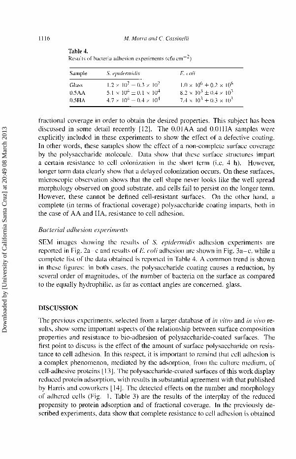
Table 4. Results of bacteria adhesion experiments (cfu cm-2)
fractional coverage in order to obtain the desired properties. This subject has been discussed in some detail recently [12]. The O.OIAA and O.O1HA samples were
explicitly included in these experiments to show the effect of a defective coating. In other words, these samples show the effect of a non-complete surface coverage
by the polysaccharide molecule. Data show that these surface structures impart a certain resistance to cell colonization in the short term (i.e. 4 h). However,
longer term data clearly show that a delayed colonization occurs. On these surfaces,
microscopic observation shows that the cell shape never looks like the well spread morphology observed on good substrate, and cells fail to persist on the longer term.
However, these cannot be defined cell-resistant surfaces. On the other hand, a
complete (in terms of fractional coverage) polysaccharide coating imparts, both in
the case of AA and HA, resistance to cell adhesion.
Bacterial adhesior2 experiments
SEM images showing the results of S. epidermidis adhesion experiments are
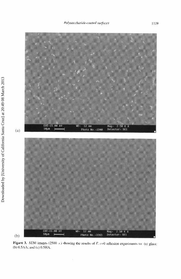
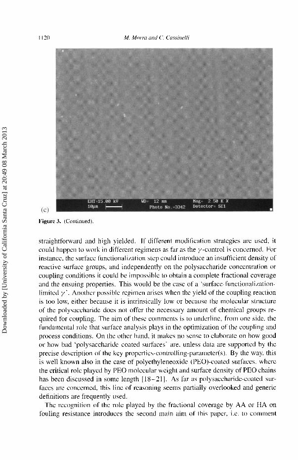
reported in Fig. 2a-c and results of E. coli adhesion are shown in Fig. 3a-c, while a
complete list of the data obtained is reported in Table 4. A common trend is shown in these figures: in both cases, the polysaccharide coating causes a reduction, by several order of magnitudes, of the number of bacteria on the surface as compared to the equally hydrophilic, as far as contact angles are concerned, glass.
DISCUSSION
The previous experiments, selected from a larger database of in vitro and in vivo re-
sults, show some important aspects of the relationship between surface composition
properties and resistance to bio-adhesion of polysaccharide-coated surfaces. The
first point to discuss is the effect of the amount of surface polysaccharide on resis- tance to cell adhesion. In this respect, it is important to remind that cell adhesion is a complex phenomenon, mediated by the adsorption, from the culture medium, of cell-adhesive proteins [ 1 3] . The polysaccharide-coated surfaces of this work display reduced protein adsorption, with results in substantial agreement with that published by Harris and coworkers [ 14]. The detected effects on the number and morphology of adhered cells (Fig. 1, Table 3) are the results of the interplay of the reduced
propensity to protein adsorption and of fractional coverage. In the previously de- scribed experiments, data show that complete resistance to cell adhesion is obtained
Dow
nloa
ded
by [
Uni
vers
ity o
f C
alif
orni
a Sa
nta
Cru
z] a
t 20:
49 0
8 M
arch
201
3
1117
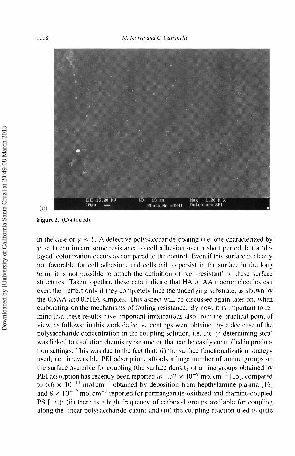
Figure 2. SEM images (1000 x) showing the results of S. epidermidis adhesion experiments to:
(a) glass; (b) 0.5AA; and (c) 0.5HA.
Dow
nloa
ded
by [
Uni
vers
ity o
f C
alif
orni
a Sa
nta
Cru
z] a
t 20:
49 0
8 M
arch
201
3
1118
Figure 2. (Continued).
in the case of y * 1. A defective polysaccharide coating (i.e. one characterized by
y < 1) can impart some resistance to cell adhesion over a short period, but a 'de-
layed' colonization occurs as compared to the control. Even if this surface is clearly not favorable for cell adhesion, and cells fail to persist in the surface in the long term, it is not possible to attach the definition of 'cell-resistant' to these surface
structures. Taken together, these data indicate that HA or AA macromolecules can
exert their effect only if they completely hide the underlying substrate, as shown by the 0.5AA and 0.5HA samples. This aspect will be discussed again later on, when
elaborating on the mechanisms of fouling resistance. By now, it is important to re-
mind that these results have important implications also from the practical point of
view, as follows: in this work defective coatings were obtained by a decrease of the
polysaccharide concentration in the coupling solution, i.e. the 'y-determining step' was linked to a solution chemistry parameter, that can be easily controlled in produc- tion settings. This was due to the fact that: (i) the surface functionalization strategy used, i.e. irreversible PEI adsorption, affords a huge number of amino groups on
the surface available for coupling (the surface density of amino groups obtained by PEI adsorption has recently been reported as 1.32 x mol CM-2 [ 15J, compared to 6.6 x mol cm obtained by deposition from hepthylamine plasma [16] and 8 x mol cm-2 reported for permanganate-oxidized and diamine-coupled PS [17]); (ii) there is a high frequency of carboxyl groups available for coupling
along the linear polysaccharide chain; and (iii) the coupling reaction used is quite
Dow
nloa
ded
by [
Uni
vers
ity o
f C
alif
orni
a Sa
nta
Cru
z] a
t 20:
49 0
8 M
arch
201
3
1119
Figure 3. SEM images (2500 x) showing the results of E. coli adhesion experiments to: (a) glass; (b) 0.5AA; and (c) 0.5HA.
Dow
nloa
ded
by [
Uni
vers
ity o
f C
alif
orni
a Sa
nta
Cru
z] a
t 20:
49 0
8 M
arch
201
3
1120
Figure 3. (Continued).
straightforward and high yielded. If different modification strategies are used, it
could happen to work in different regimens as far as the y-control is concerned. For
instance, the surface functionalization step could introduce an insufficient density of
reactive surface groups, and independently on the polysaccharide concentration or
coupling conditions it could be impossible to obtain a complete fractional coverage and the ensuing properties. This would be the case of a 'surface-functionalization-
limited y'. Another possible regimen arises when the yield of the coupling reaction
is too low, either because it is intrinsically low or because the molecular structure
of the polysaccharide does not offer the necessary amount of chemical groups re-
quired for coupling. The aim of these comments is to underline, from one side, the
fundamental role that surface analysis plays in the optimization of the coupling and
process conditions. On the other hand, it makes no sense to elaborate on how good or how bad 'polysaccharide coated surfaces' are, unless data are supported by the
precise description of the key properties-controlling-parameter(s). By the way, this
is well known also in the case of polyethyleneoxide (PEO)-coated surfaces, where
the critical role played by PEO molecular weight and surface density of PEO chains
has been discussed in some length [ 1 8-2 1 ]. As far as polysaccharide-coated sur-
faces are concerned, this line of reasoning seems partially overlooked and generic definitions are frequently used.
The recognition of the role played by the fractional coverage by AA or HA on
fouling resistance introduces the second main aim of this paper, i.e. to comment
Dow
nloa
ded
by [
Uni
vers
ity o
f C
alif
orni
a Sa
nta
Cru
z] a
t 20:
49 0
8 M
arch
201
3
1121
on the mechanism(s) responsible for the observed behavior. When thinking of
(bio)particles-surfaces repulsion, the colloidal chemical heritage of surface chem-
istry immediately invokes the long-range charge to charge electrostatic repulsion described by the DLVO theory. In the present case, it is easy to show that repulsion between the negatively charged cells or bacteria and the substrate does not play a
significant role: first of all, PSO and glass are also negatively charged, and this does
not prevent cell and bacterial adhesion. Then, due to the coupling reaction used and
the high density of amino groups introduced by PEI, the anionic sites on HA and
AA are at least partially charge-neutralized by bonding. Most important, given the
ionic strength of the solutions used for cell and bacterial adhesion experiments, the
range of electrostatic interaction in the present experiments is definitely low and the
relevant Debye length less than 1 nm [22].
According to another bulky heritage, steric repulsion and general 'mobility' effects are frequently invoked to account for fouling resistance. This widespread use mostly stems from the success of theories based on these ideas to explain the properties of PEO-coated surfaces [ 18, 19]. In the present case it is hard to
account for these results in terms of freely fluctuating chains, especially if one
takes into account the requirements for the building up of an actual brush-like
regimen, as recently discussed by Szleifer [19]: after all, this is a polyanion layer over a polycation underlayer, and it is rather difficult to think of freely fluctuating
negatively charged chains over a high density of positive charge. Then, there is
the multi-point covalent attachment, which is nothing like a brush-like regimen. The interfacial reaction used in this work for immobilization of HA and AA, and
the high density of amino and carboxyl groups as described before, suggest that
the polysaccharide will adopt a 'side on' configuration on the substrate surface,
according to the definition of Harris and coworkers [ 14]. In our opinion the approach towards the understanding of the properties of
polysaccharide-coated surfaces must necessarily pass through considerations on the
role of the molecular structure and its effects on the surrounding environment. In
this respect, results of force measurement are a valuable source of information [4, 5]. Direct force measurements between certain polysaccharide molecules show peculiar features as compared to forces measured between other hydrophilic materials.
Hydration forces seem a general and dominating feature of all hydrophilic surfaces
at close examination 141. But measurements such as those performed between
Xanthan molecules, already quoted in the beginning of this paper [ 4 ], indicate the
unique nature of the polysaccharide-water interaction. Unlike hydration forces
measured between mica, or even between other biopolymers such as DNA, forces
between the stiff helices of Xanthan are not sensititive to the kind of ionic species and their concentration in the bathing environment. This means, according to
Rau and Parsegian, that there is either no ion binding or no secondary hydration due to ions bound to the polysaccharide helices. Even more interesting and more
unique, this happens even if the Xanthan molecule is a polyelectrolyte that contains
charged carboxyl groups in its repeating unit. The latter have a marked effect on
Dow
nloa
ded
by [
Uni
vers
ity o
f C
alif
orni
a Sa
nta
Cru
z] a
t 20:
49 0
8 M
arch
201
3
1122
the strength of hydration forces: in fact, the removal of charged pyruvate groups,
exposing neutral (but 'hydrophilic', in a general sense) hydroxyl groups reduces the
magnitude of hydration forces by a factor of 3 [ 4, 5 ] . Quoting again from Rau and
Parsegian: 'Altough forces at close approach appear dominated by water structuring and not by electrostatic double-layer interactions between charged surfaces, the
power of charged surface groups to organize water is clearly apparent in the force
curves' [4]. It is not surprising that in a different biomolecular field, the excellent
work of Prof. D. Hurry has elucidated the role of water-structuring chemical
groups such as carboxylate in the control of the properties of the biomolecules
that are at the basis of the mechanism of motion in living organisms [25]. Both
classes of biomolecule, however apart, share a common engineering principle: to
exploit the opportunities offered by the water structure and orientation-dependent
properties [26]. The HA and AA macromolecules used in the present work are specifically
engineered to interact with water, giving rise to hydration forces, and these
properties are widely exploited in nature and industry. Direct force measurements
performed on related molecules strongly underline the specificity and dependence on molecular details of the water-polysaccharide molecule. The mechanism
at the basis of hydration forces, i.e. hydrogen bonding between electron-donor
electron-acceptor surface sites and water molecules and the ensuing effect on water
structure [26], are specific chemical interactions, characterized by bond length and angles, albeit existing in a dynamic structure whose typical lifetime is of the
order of picoseconds [27]. Based on these arguments, the pivotal role played
by 'surface crowding' of AA or HA molecules, shown by the dependence of
the anti-adhesive properties on fractional coverage, can be simply understood in
terms of the need to create an effective hydration layer that shields the underlying substrate. Along the same line, the strong emphasis on the dependence of the
properties of `polysaccharide-coated surfaces' on strictly chemical parameters (as a consequence of the 'specificity' of the water-polysaccharide interaction and as
opposed to chemically 'blind' approaches that consider hydrophilic molecules only as chains fluctuating in a good solvent), underlines the fundamental role played by the structure of the polysaccharide molecule and the details of the coupling reaction
on coating performances.
CONCLUSIONS
In conclusion, in this paper several aspects related to the fouling-resistance of
surfaces coated by hydrophilic polysaccharides have been discussed. AA and HA, as prototypal molecular machines explicitly engineered to maximize interaction
with water, have been selected as coating materials, and coated surfaces have been
tested in in vitro mammalian and bacterial cells adhesion experiments.
Experimental data show that the fouling-resistant properties of these surfaces are
related to the fractional coverage by the polysaccharide, and that the the simple
Dow
nloa
ded
by [
Uni
vers
ity o
f C
alif
orni
a Sa
nta
Cru
z] a
t 20:
49 0
8 M
arch
201
3
1123
water contact angle, or related parameters that suggest a univocal relationship between the water contact angle and bioadhesive response, is not enough to predict the bio-adhesive response.
Borrowing evidence from general technological applications of these polysaccha- rides and results from direct force measurements between related molecules in solu-
tion, it is suggested that the mechanism on which the adhesion-resistant properties of these surfaces is based is much more strictly and directly related to molecular
details and specific water-coated surface chemical interactions than generally as-
sumed. This aspect should always be considered in the development of effective
We thank Dr. Claudio Della Volpe (University of Trento, Italy) for the Wilhelmy
plate measurements performed on these samples.
REFERENCES
I. O. Larm, R. Larsson and P. Olsson, Biomat. Med. Dev. Art. Org. 11, 161 (1983). 2. J. Sanchez, G. Elgue, J. Riesenfeld and P. Olsson, J. Biomed. Mater Res. 29, 655 (1995). 3. R. H. West, A. J. Paul, S. Hibbert, P. Cahalan, L. Cahalan, M. Verhoeven, M. Hendriks and
B. Fouache, J. Mat. Sci. Mata Med. 6, 63 (1995). 4. D. C. Rau and V. A. Parsegian, Science 249, 1278 ( 1990). 5. P. M. Claesson, in: Biopolymers at Interfaces, M. Malmsten (Ed.), p. 281. Surfactant Science
Series, Marcel Dekker, New York (1998). 6. Y. Qin and G. K. Gilding, Med. Dev. Tech. 7, 32 (1996). 7. J. E. Scott, in: The Biology of Hyaluronan. D. Evered and J. Whelan (Eds), p. 6. Wiley,
Chichester ( 1989). 8. N. H. Choi-Miura, T. Tobe, J. Sumiya. Y. Nakano, Y. Sano, T. Mazda and M. Tomita, J. Biochem.
119, 1157 (1996). 9. Pavesio. D. Renier, C. Cassinelli and M. Morra. Med. Dev. Tech. 8, 20 (1997).
10. M. Bodanzsky, Peptide Chemistry. Springer-Verlag, Berlin (1988). 11. M. Morra and C. Cassinelli, Surf Interface Anal. 26, 742 (1998). 12. M. Morra and C. Cassinelli, Langmuir (in press). 13. T. A. Horbett and L. A. Klumb, in: Interfacial Phenomena and Bioproducts, J. L. Brash and
P. W. Wojciechowski (Eds), p. 351. Marcel Dekker, New York (1996). 14. E. Osterberg, K. Bergstrom, K. Holmberg, T. P. Schuman, J. A. Riggs, N. L. Burns, J. M. Van
Alstine and J. M. Harris, J. Biomed. Mater Res. 29, 741 (1995). 15. J. J. Chance and W. C. Purdy, Langmuir 13, 4487 (1997). 16. R. C. Chatelier, X. Xie, T. R. Gerenbach and H. J. Griesser, Langmuir 11,4122 (1995). 17. N. Zammatteo, C. Girardeaux, D. Delforge, J. J. Pireaux and J. Remacle, Analyt. Biochem. 236,
85(1996). 18. S. Nagaoka, Y. Mori, H. Tanzawa, Y. Kikuchi, F. Inagaki, Y. Yokota and Y. Noishiki, ASAIO.J.
10, 76 (1987). 19. I. Szleifer, Physica A 244, 370 (1997). 20. S. J. Jeon, J. H. Lee, J. D. Andrade and P. G. de Gennes, J. Colloid Interface Sci. 142, 149 (1991 ). 21. S.J. Jeon and J. D. Andrade, J. Colloid Interface Sci. 142, 159 (1991 ).
Dow
nloa
ded
by [
Uni
vers
ity o
f C
alif
orni
a Sa
nta
Cru
z] a
t 20:
49 0
8 M
arch
201
3
1124
22. F. Garbassi, M. Morra and E. Occhiello, Polymer Surfaces: from Physics to Technology, Ch. 1.
Wiley, Chichester (1998). 23. S. D. Johnson, J. M., Anderson and R. E. Marchant, J. Biomed. Mater. Res. 26. 915 (1992 ). 24. D. R. Absolom, F. F. Lamberti, Z. Policova, W. Zingg, C. J. Van Oss and A. W. Neumann. Appl.
Environ. Microbiol. 46, 90 (1983). 25. D. W. Hurry, Angew. Chem. Int. Ed. Engl. 32, 819 (1993). 26. N. A. M. Besseling. Langmuir 13, 2113 (1997). 27. E. A. Vogler, Adv. Coll. Interface Sci. 74, 69 (1998).