J. Anat. (1973), 115, 1, pp. 1-22 With 15 figures Printed in Great Britain Normal development of the jaws and teeth in pigs, and the dela and malocclusion produced by calorie deficiencies C. H. TONGE AND R. A. McCANCE University of Newcastle-upon-Tyne Dental School, Newcastle-upon-Tyne and Sidney Sussex College, Cambridge (Accepted 30 January 1973) INTRODUCTION The occlusion of the teeth, at maturity, depends upon the integrated growth of the dental, skeletal and soft tissues of both jaws. Genes determine the maximum growth of which each individual and organ is capable, but all growth is liable to environmental limitations. Therefore, in considering the causes of dental irregularities and malocclusions, genetic direction, growth potential, the developmental time scale, and dietary variations must all be considered. Lundstrom (1951), for instance, put forward evidence that the size of the jaw and the size of the teeth were under separate genetic direction, so that malocclusions and irregularities might result from the fusion of two ethnic groups. By comparing the permanent dentitions of Anglo-Saxons with their modern British representatives Lavelle (1968) has shown that the incisors, premolars and canines have become smaller during the last 12-14 centuries, and so have the dimensions of the jaw; the size of the molars, on the other hand, has increased. Jeffreys (1969) claimed that in Wistar rats environmental influences on the pattern of growth were small compared to those operating on the rate of growth, but that tooth size was subject to environmental influence, and that the degree of 'catch up' growth varied according to the time and extent of the period of retard- ation. Tonge & McCance (1965) depressed the overall growth of pigs by giving them a calorie-deficient diet until they were a year old. This retarded the development of the jaw more than that of the teeth and resulted in overcrowding, displacement and malocclusion. The rehabilitation of similarly undernourished pigs (McCance, Owens & Tonge, 1968) led to considerable 'catch up' growth but abnormal siting, im- paction and malalignment of the teeth remained and, with them, faulty occlusion. An alteration in the environment at the appropriate age had, therefore, upset the harmonious development of the jaws and teeth, and left permanent stigmata behind it. Further work on this subject seemed highly desirable, notably (1) to work out the time course of normal dental development in 'Large White' pigs, and (2) to set against this the delays and abnormalities caused by undernutrition and to study quantitatively the immediate and subsequent effects. There are two ways of considering the effects of undernutrition on the jaws and teeth. The first is the way in which normal dental development is often set out, showing in a table the chronological time from conception at which each tooth can I ANA II5
Transcript
J. Anat. (1973), 115, 1, pp. 1-22With 15 figuresPrinted in Great Britain
Normal development of the jaws and teeth in pigs, and the delaand malocclusion produced by calorie deficiencies
C. H. TONGE AND R. A. McCANCE
University of Newcastle-upon-Tyne Dental School, Newcastle-upon-Tyne andSidney Sussex College, Cambridge
(Accepted 30 January 1973)
INTRODUCTION
The occlusion of the teeth, at maturity, depends upon the integrated growth of thedental, skeletal and soft tissues of both jaws. Genes determine the maximumgrowth of which each individual and organ is capable, but all growth is liable toenvironmental limitations.
Therefore, in considering the causes of dental irregularities and malocclusions,genetic direction, growth potential, the developmental time scale, and dietaryvariations must all be considered. Lundstrom (1951), for instance, put forwardevidence that the size of the jaw and the size of the teeth were under separate geneticdirection, so that malocclusions and irregularities might result from the fusion oftwo ethnic groups. By comparing the permanent dentitions of Anglo-Saxons withtheir modern British representatives Lavelle (1968) has shown that the incisors,premolars and canines have become smaller during the last 12-14 centuries, and sohave the dimensions of the jaw; the size of the molars, on the other hand, hasincreased. Jeffreys (1969) claimed that in Wistar rats environmental influences on thepattern of growth were small compared to those operating on the rate of growth,but that tooth size was subject to environmental influence, and that the degree of'catch up' growth varied according to the time and extent of the period of retard-ation.Tonge & McCance (1965) depressed the overall growth of pigs by giving them a
calorie-deficient diet until they were a year old. This retarded the development of thejaw more than that of the teeth and resulted in overcrowding, displacement andmalocclusion. The rehabilitation of similarly undernourished pigs (McCance, Owens& Tonge, 1968) led to considerable 'catch up' growth but abnormal siting, im-paction and malalignment of the teeth remained and, with them, faulty occlusion.An alteration in the environment at the appropriate age had, therefore, upset theharmonious development of the jaws and teeth, and left permanent stigmata behindit. Further work on this subject seemed highly desirable, notably (1) to work out thetime course of normal dental development in 'Large White' pigs, and (2) to setagainst this the delays and abnormalities caused by undernutrition and to studyquantitatively the immediate and subsequent effects.There are two ways of considering the effects of undernutrition on the jaws and
teeth. The first is the way in which normal dental development is often set out,showing in a table the chronological time from conception at which each tooth can
I ANA II5
C. H. TONGE AND R. A. McCANCE
Table 1. The numbers of animals killed at each age, their characteristic weightat this time and their treatment during life
Experimental animalsNormal animals always well-fed calorie-deficient for 12 months, then well-fed
Ageat death, ---A
all animals Number killed Characteristic Months Number killed Characteristic(months) at each age weights kg rehabilitated at each age weights kg
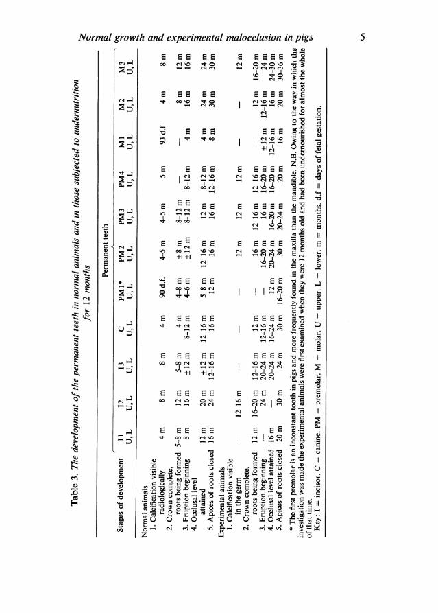
first be detected and the course of its calcification and eruption. This is satisfactoryenough for normal development, when everything else may be assumed to be goingaccording to the genetic plan, and has been employed in Tables 2 and 3. The effectsof undernutrition, however, cannot be represented satisfactorily by this methodalone, for undernutrition produces relative and absolute changes in the spatialarrangements of the teeth in the jaws. The only way to present this data is bydiagrams and this has accordingly been done in Figs. 1-15.
MATERIALS AND METHODS
Seventy normal 'Large White' pigs ranging in age from the 80th day of gestationto the 5th year of life were used to establish the growth rate of normal pigs in thecolony. Thirty-seven of these were used for the study of normal dental develop-ment (Table 1). Forty other 'Large White' pigs were subjected to severe caloricrestriction from early in life till they were a year old. Thirteen of these were thenkilled, and twenty-two others were rehabilitated by being given an excellent diet adlib., and killed for examination at appropriate intervals afterwards. Thus the numberof undernourished animals used to establish their growth curves also exceeded thoseused for the dental work (see Table 1).The pigs were reared on the same food and very much on the lines described by
McCance (1960) and Cabak, Gresham & McCance (1962). These diets provideplenty of protein for young growing pigs and a sufficiency of vitamins and minerals.Minor modifications, however, were made in the light of experience, and the stan-dardized procedure in the present investigation was as follows. (1) The piglets weretaken from the sow when they weighed 3-2 to 3 4 kg and were about 10 days old.(2) They were weighed every day. (3) Their food intake was measured accuratelyevery day. The amount required was usually about 45 g of the 'pellets' and 45 gof the meal mixture. (4) The piglets were allowed to gain about 50 g each week byvarying the food intake a little according to their health and progress. The number
2
Normal growth and experimental malocclusion in pigsof animals killed as controls for the undernourished ones and the characteristicweights of all of them at the various age intervals are shown in Table 1. As alreadystated, the numbers of animals weighed exceeded the numbers used for the dentalwork. When the animals, normal or abnormal, were killed, the head was removedand the jaws were dissected out, cleaned, photographed, and measured. The detailsof their articulation and occlusion were then studied by placing the head of themandibular condyle in the glenoid fossa and examining the patterns of attritionfound on the teeth. The upper and lower jaws were then bisected longitudinally,and X-ray photographs were obtained by standardized methods. Abnormalitiesfound in the experimental group were compared with the findings in the normalgroup. Tracings were made from the radiographs, and by reference to the jawsthemselves and their occlusal relationships these tracings were then superimposedin 'centric' occlusion. A further overall tracing was made in this position.The teeth were classified according to the formula given by Peyer (1968), and not
as they were classified by Bodegom (1969). The latter has investigated in some detailthe dental development of the miniature pig during life till the animals were a littleover a year old. Unfortunately, his approach and techniques were so different fromours, that it is very difficult to compare the two sets of results.
Normal animalsThe early stages
Birth takes place at about 1 5 days gestation in 'Large White' pigs, and the courseof development of the deciduous teeth is given in Table 2. Minor differences betweentimings in the upper and lower jaws were noted but are not included in the table.Histologically, calcification was observed in all the deciduous teeth except the thirdmolar by the 80th day of gestation. The first permanent premolars had calcifiedmost of their crowns by the end of gestation, but there was no radiologicalevidence of calcification in any of the other permanent teeth at this time.
Four weeks after birthFig. 1 shows a tracing of the jaws of an animal 4 weeks old in occlusion. Further
information about such jaws was given by Tonge & McCance (1965). There wereagain minor differences between the two jaws which are not noted in Tables 2 and 3.There was only contact between two teeth on each side in the molar segment, butthese animals were still being wholly breast-fed, and inspection showed that, if theeruption of the other teeth went as it should, an interdigitating pattern of the cuspswould be established. There was adequate space for the eruption and proper occlu-sion of the deciduous dentition, and the evidence suggested that this would also beso for the permanent teeth, for histological examination demonstrated the presenceof the germs of the uncalcified ones in the expected positions.
Four months after birthFig. 2 shows the dental development of a normal animal 4 months old, and the
stages reached by the various teeth are given in Tables 2 and 3. The deciduous oneswere well advanced and occluded normally. The first premolar had erupted in the
I-2
3
4
Cs
F-
H. TONGE
E -0
>e cUtCI
AND R. A.C.
,-; C. "; 4 W; .6 0-0.4
Normal growth and experimental malocclusion in pigs 5
oo CA C 't o
_-00 IC IRt<.l
c
e ° Q~~~~~~~~~~~ Cd
E E E E E E .-en It tr 00 +14 'o 0 =
+ 0
E-S E E E ~~EE E- - ~~~~~~~~C: CZstWI C14 'IC 0 00 4
E E E E E E E 'Xor.tn CA 'I1O C-
0 o0
W:E E E E E E *= 0 E+I +I C1 cq en E 4 0,
E E E E E E E E 1t C1 11o 'I r4 \. to
co~~~~~~~~g cqCd
N N£ £ t t t Y e 2tn +I +I CAoo
E E E E E E E E . o
E E E E E E EEc
bo 4=v
°°°geD r2 c.E ¢, e ° N 2 E*_ C * _u-ce w ,_Ceu
e o°° v Q +Q E > E o ° - <<Ecd ba O )U UWO
bo .= O 6
ZS
cq
*t
*E3
4-
c.C'4 4
.icim
,..q *.
. .510.4
en 04- .s
C
(U
E0
ce
laC*.V0
(D.4
ux ._
E cg*U 0
Ed UCd az;
C. H. TONGE AND R. A. McCANCE
---Icm
Fig. 1. Normal jaws 4 weeks after birth. In all the figures the outlines of the deciduous teeth areunshaded and those of the permanent teeth shaded.
i L 1I 1
Fig. 2. Normal jaws 4 months after birth.
maxilla, but the roots were not completely calcified, and the corresponding tooth(Table 3) was not visible in the mandible. The first permanent molar was justemerging in its correct position and alignment in both jaws. The cusps of the secondpermanent molars were beginning to calcify in both jaws. In the upper one the crownlay in a good position to emerge as the maxilla grew in the posterior direction andin the lower one a space was beginning to form for its eruption between the firstpermanent molar and the base of the coronoid process of the mandible.
Eight months after birthThe position reached by each tooth is shown spatially in Fig. 3, and all the
individual stages of development in Tables 2 and 3. The deciduous dentition occludednormally. The permanent canines had not yet erupted, and were lying more hori-zontally than would be expected at eruption, but there was adequate room for thisto take place. The mental foramen was situated below and between the third andfourth premolars in the mandible. The crowns of the second and third molars inboth jaws were calcifying and lying in good positions to erupt, and the permanentdentition promised to reach normal centric occlusion.
6
Normal growth and experimental malocclusion in pigs
I I I I
cm
Fig. 3. Normal jaws 8 months after birth.
I I 1 1-
cm
Fig. 4. Normal jaws 12 months after birth.
Twelve months after birthFig. 4 shows the dental development of one of the five normal animals killed at
this age. The jaws had grown considerably in the last 4 months, and the deciduousincisors were being replaced by the permanent ones. The lower canine had erupted,but only part of the upper one. Diastemata were opening up in each jaw between
7
C. H. TONGE AND R. A. McCANCE
i I I I IJ L 1 Jcm
Fig. 5. Normal jaws 16 months after birth.
the incisor segment and the canines, and also between the latter and the premolars.There was a first premolar in every maxilla examined at this age, but this tooth wasfound in only one of the mandibles. The deciduous molars were giving place to thesecond, third and fourth premolars, and in all the specimens the first and secondpermanent molars had erupted and established good occlusion, while the thirdmolars were still lying, partially calcified, in their crypts in good mesiodistal align-ment. The occlusion was therefore good in the premolar-molar region and the teethin both arches lay in a straight line mesiodistally. The contact area between the firstand second upper molars lay beneath the zygomatic process of the maxilla.
Sixteen months after birthIn Fig. 5 the state of the jaws and teeth in one of the five animals killed when they
were 16 months old is illustrated, and should be studied in association with Tables 2and 3. In one of these animals the maxillary canine had a forked root. Several otherswere found in pigs during this investigation but it is certainly unusual in teeth withcontinuous growth. There was generally plenty of room for all the teeth to occludenormally, but in two of the specimens there were slight signs of premolar over-crowding. This may have resulted from the forked root of the canine in one of them.
8
Normal growth and experimental malocclusion in pigs
I Ut--1 I I I I
cm
Fig. 6. Normal jaws 20 months after birth.
I I I I
cm
Fig. 7. Normal sow's jaws 24 months after birth. Note the bifurcation of the root of the maxillarycanine, probably genetic in origin.
9
C. H. TONGE AND R. A. McCANCE
I.l._ L.. 1_cm
Fig. 8. Normal hog's jaws (right side) 30 months after birth. Note again the bifurcationof the root of the upper canine.
Twenty months after birthBy this age the permanent dentition, with the exception of the distal parts of the
crowns of the 3rd molars, had fully erupted and was in normal occlusion. As shownin Fig. 6, some remnants of the second deciduous incisors and first molars mightremain, but there was no sign of overcrowding in any of the jaws studied, and thediastemata were roomy.
Later stagesAt 24 months the occlusion and alignment of the teeth was always good and the
diastemata in the anterior segments were satisfactory. The second molars, fullyerupted, were by now lying beneath the zygomatic process of the maxilla. The distalparts of the third molars had only partially erupted. In one of the jaws examined atthis age and shown in Fig. 7 the maxillary canine had a forked root, and anothersuch tooth was found in the maxilla of a pig aged 30 months (Fig. 8). By this timeall the permanent dentition had fully erupted, and there was always some attritionof the first molar teeth in both jaws. The roots of the premolars and first and secondmolars were complete, but not always those of the third (Table 2).
lo
Normal growth and experimental malocclusion in pigs
LI I 1 1 I I I
cm
Fig. 9. The state of the jaws after 12 months of calorie deficiency.
1 I L I
cm
Fig. 10. The jaws on the right-hand side of an animal after calorie deficiency for12 months and rehabilitation for 4 months.
At 36 months of age occlusion, alignment and diastemata were satisfactory, butattrition of all the molars was becoming conspicuous. By this age the third maxillarymolar was lying in its permanent position below the zygomatic process.
Effects of undernutrition for the first twelve months of lifeFig. 9 is representative of the condition of the jaws of animals which had been
undernourished till the age of one year, and weighed about 5-5 kg. It should becompared with Fig. 1, which shows the jaws of a normal animal of about the sameweight, but much younger, and with Fig. 4, which shows the jaws of a normal animalof the same age, but much larger (see Table 1). As the scales on the three figuresshow, the jaws of the undernourished pig were larger than those of the younger pig,which had the same body weight, but much smaller than those of the normal pig ofthe same age.The dental development of these pigs requires examination in detail. The deciduous
incisors and canines were still present, although the crowns of most of their per-
I1I
C. H. TONGE AND R. A. McCANCE
manent successors had begun to calcify. In the majority the first upper premolarhad erupted, and its roots lay close to the permanent canine, which was justdeveloping. There was no corresponding tooth in the lower jaw. The three deciduousmolars in each arch were still present, and showing signs of heavy attrition. Therewas radiological evidence of the crypts for the succeeding premolars having formed,and in the lower jaw some of these teeth had begun to calcify at the extremities oftheir cusps. The first lower permanent molars had completely erupted, but the upperones only partially. The second permanent molars had not, but their crowns wereapproaching complete calcification. These teeth, therefore, were in a much moreforward state than the weight of the animal or the size of the jaw would have ledone to expect. Compared with those shown in Fig. 4, however, they were not farenough forwards in the jaws. Worse still was the condition of the third molars, forthe lower ones, which in this specimen were just beginning to calcify, were lyinghigh up in the ramus of the mandible, and the upper ones were only separated fromthe pterygo-palatine fossa by a thin plate of bone. Further details of the chrono-logical delays in dental development at this age brought about by the undernutritionare given in Tables 2 and 3, together with the effects of rehabilitation and catch upgrowth.The diastemata between the incisors and the canines, and between the latter and
the deciduous molars, were very much smaller than those in normal pigs of the sameage or at the same stage of dental development.
In radiographs, lines of arrested growth were usually conspicuous on the posteriorborder of the ramus of the mandible. The radio-opacity of the jaws was slight com-pared with that of normal animals of the same age, and the angle at menton wasmore obtuse (Figs. 1 to 4; Tonge & McCance, 1965).
In occlusion, the lower first deciduous molar was unopposed and the whole of themolar segment in the lower jaw seemed to be too far forward relative to the upperone. All this evidence of overcrowding can be explained by the fact that the dentaldevelopment of these pigs corresponded to that of a normal animal about 4 monthsold, whereas the jaws had not yet attained the same size (Figure 2; Owens, 1968).
The effects of rehabilitation for:Four months
Fig. 10 shows the state of the jaws and teeth after 4 months rehabilitation, whenthe animals weighed about 75 kg, and it should be compared with the jaws shownin Figs. 2 and 3. Small diastemata were only beginning to appear between theincisors and the canines, and between the latter and the first premolars. There wasgross overcrowding in both jaws, and, on X-ray examination, the third molar in theright maxilla of the specimen illustrated (Fig. 10) appeared to be absent; this wasconfirmed by dissection. In the specimen illustrated both the third mandibular molarsand the left maxillary one appeared smaller than those ofnormal animals, for they layin a bucco-lingual plane at right angles to the normal one. Owing to this abnormalsiting of the teeth, their occlusion was bad. The maxillary incisors occluded with theinternal aspect of the mandibular ones, but the teeth were not in contact. Thecanines did not occlude, the lower one in fact being anterior to the third incisor. The
12
Normal growth and experimental malocclusion in pigs
l1 1 1 _L1
cm
Fig. 11. The effect of calorie deficiency for 12 months followed by rehabilitation for 8 months.
maxillary first and second premolars articulated with the deciduous third molar andthe first permanent molar on the left side but not on the right. The second maxillarymolars were not in occlusion with any teeth in the lower jaw.The teeth of the other animal killed at this stage of rehabilitation were in an equally
disorganized state. The first and third permanent incisors had erupted, but the secondmaxillary one was lying horizontally, in a position which would have made eruptionnearly impossible. The second upper premolar was wedged between the apex of theroot of the canine, which had erupted, and the distal border of the first premolar.The second permanent molar in the upper jaw was equally wedged, and the calcifyingcrown of the third was lying with its occlusal surface directed vertically upwardstowards the pterygoid region. In the mandible of this animal the mental foramenwas situated between the third and fourth premolars, and the third molar presentin both jaws was calcifying in a crypt at the base of the coronoid process.
Eight monthsAfter eight months rehabilitation the mandibular teeth of one of the two animals
examined were not so crowded as the ones shown in Fig. 11, but radiographs showedthat the third permanent molars had not erupted, and that they were lying bucco-lingually, with their crowns rotated distally to the normal plane. There was, more-over, still gross overcrowding and tooth rotation in the premolar region of the maxilla.The teeth of the other animal, illustrated in Fig. 11, were most irregular, with over-crowding of the premolars in both jaws and impaction of the second permanentmandibular molars. The third molars in the jaws were less developed and equallydisplaced. The occlusion was poor and stability depended upon the occlusal contactof the molar area. Thus the teeth were misplaced, very crowded, and still develop-
13
C. H. TONGE AND R. A. McCANCE
cm
Fig. 12. The left side of the jaws after calorie deficiency for 12 months and rehabilitation for12 months. Note the angle of growth of the upper third incisor and canine and the twinning ofthe third maxillary molar.
mentally backward compared with those of the normal animal illustrated in Fig. 6,and the premolars and molars were occupying less space in a smaller jaw.
Twelve monthsAfter rehabilitation for 12 months, the animals were now two years old and all
the erupted and unerupted teeth were permanent ones except the second permanentincisors, which had not yet replaced their deciduous predecessors. In Fig. 12 theupper third incisor and canine appear not to have erupted. This was because theangle at which these teeth were growing was such that their lowest edges lay abovethe level of the bone of the hard palate. The four upper premolars were partiallyrotated, with the third one overlapping the second distally. There was no first lowerpremolar, and though the other three were in good arch alignment, their occlusalrelationships with the upper teeth were not normal, for the high centre cusps wereoccluding with those of the upper teeth instead of interdigitating with them. More-over, the upper first and lower second premolar did not appear to be in occlusion,although, since their roots were not yet complete, occlusion might still have occurred.
In the molar region, the first upper molar was in post normal relationship withits lower counterpart. The second molar was partially erupted, but impacted betweenthe first molar and the more mesial of two unerupted third left molars. The secondmolar showed signs of decay, and its roots were not visible. The two third molars,found only on the left side, were impacted, the more mesial presenting its root sur-face to the oral cavity and lying in a rotated position. The distal one was in morecorrect antero-posterior alignment, but was impacted mesially against its twin.Both teeth had their crowns calcified. This was one of only two animals in whichtwo third maxillary molars were found. The other had been undernourished for oneyear and rehabilitated for 18 months (Fig. 14). The lower first and second molars
14
Normal growth and experimental malocclusion in pigs
[ 1 1 I -
cm
Fig. 13. Jaws showing a comparatively good recovery after calorie deficiencyfor 12 months and rehabilitation for 18 months.
had erupted in good alignment with the premolars, but the third one was rotatedthrough 900 and impacted, its mesial surface facing lingually.
Spaces were present in the anterior segments, indicating very near normality inthe incisor area, but the lower canine had drifted mesially and did not make contactwith the upper one. In normal jaws, the canines were always in contact either inocclusion or during the movement of the jaws. The overcrowding therefore had beenovercome to a great extent, except in the third molar area.
Eighteen monthsAfter 18 months rehabilitation, when the pigs were 30 months old, the two avail-
able specimens showed rather different effects of rehabilitation. In one (Fig. 13), therehabilitation had resulted in a fair recovery with the exception of (a) the persistenceof a rather greater anterior proclination of the mandible in the incisor region, (b) ashorter maxilla as compared with the normal control at the age of 30 months(Fig. 8), (c) some overcrowding and malocclusion in the molar area of the upperjaw, and small anterior diastemata.
In the other specimen (Fig. 14), the degree of overcrowding before rehabilitationhad been such that, during rehabilitation, considerable tooth displacement hadremained. The relative relocation of the teeth, accompanied by wear on occlusalsurfaces and loss of teeth, had left serious overcrowding and a malocclusion associ-ated with inadequate cusp fissure relationships between the teeth of the two jaws.In the left maxilla (not shown), the first premolar was worn down to its roots, thefourth premolar had been lost, the second molar had its mesial cusps impactedagainst the distal area of the first molar, and the third molar was not fully erupted
15
C. H. TONGE AND R. A. McCANCE
-- L 1 1 1 1 X 1
cm
Fig. 14. Jaws on the right hand side after calorie deficiency for 12 months and rehabilitation for18 months. Note the multiple permanent abnormalities that remained, among them twin thirdmolars in the maxilla.
distally. This tooth had attenuated roots, and it was unlikely to develop further. Theright maxilla, which is the one illustrated in Fig. 14, was even worse, although therewas rather less attrition of the first premolar. The fourth premolar, however, wasfirmly wedged between the third premolar and first molar teeth. There were alsotwin third molars. Overall, the mandible had done better than the maxilla. On theleft side, the first premolar was impacted against the canine; on the right side (Fig. 14)the first premolar was in a more normal position, but on both sides the third molarshad rotated buccolingually, with resulting impaction and malocclusion.
Thirty-six monthsIn all the animals examined after being rehabilitated for two years there were still
considerable but variable degrees of malocclusion. The more common forms were:(1) reductions in the mesiodistal lengths of the upper and lower third molars.(2) Rotation and/or impaction of the third molars. (3) Tilting and/or rotation of thepremolars. (4) Variable antero-posterior siting of the canine. (5) Absence of the thirdlower or upper incisor (this was never observed in normal adult pigs). There wereother less noteworthy differences between the two sides of the same jaw and betweenthe two jaws.
In the tracing shown in Fig. 15, the most notable feature in the anterior segmentsis that the first lower premolar, which is not invariably present, had been transposedmesial to the canine. The transposition had been bilateral in this pig, as also in
16
Normal growth and experimental malocclusion in pigs
I , II I I
cm
Fig. 15. The jaws of a pig after a calorie deficiency for 12 months followed by rehabilitation for24 months. Note the transposition of the first lower premolar and the canine and the absence ofany visible third molars
others, but this was not always so. The lower canines were in contact with the palatalmucosa at the gingival margin of the upper ones and had produced crater-likedepressions in the bone. The second, third and fourth lower premolars were tilteddistally and the fourth was rotated through approximately 900. Just mesial to thesecond was a retained fragment of the root of the first deciduous molar. The threelower molars had all erupted and completed their roots, but they were overcrowded.The third molar was characteristically foreshortened mesiodistally and rotated a littlelingually. In the upper arch all four premolars were present, but the first three showedsome degree of tilting. The crowns of the second molars were both compressed, andthe pattern of their cusps differed from that observed in normal adult pigs. Onlythe first and second permanent molars were present, and there was no evidence ofthe third one ever having developed or been present.
This was the only animal in which both third molars were absent although therewere crypts and space for them. In one animal at this stage the first and second lowermolars were absent, but there were retained root fragments indicating that the teethhad formed. The occlusion of all the undernourished animals, even after full re-habilitation, was always worse than that of any of the normal animals.
DISCUSSION
The word occlusion means more than the static relationship of teeth in appositionto one another. While, therefore, the present work may seem only to have compared
2 ANA II5
17
C. H. TONGE AND R. A. McCANCE
this static picture in two series of animals which had been differently reared, centricocclusion can be normal only if the growth and development of the jaws and teethhave also been normal - both separately and as an integrated unit. An attempt hastherefore been made, first, to work out the steps of development that lead to normaocclusion in normal pigs, and secondly to find out why malocclusion develops inundernourished pigs. The first was necessary because the criteria of normality havenot yet been so well established in the pig as they have been in man, but fortunatelythe task was simplified by the finding that, after making allowances for the differencesin morphology and numbers of the teeth, many of the accepted criteria of normalityin man were found to be applicable to pigs. Thus the upper teeth occluded with thelower ones so that (1) their buccal surfaces were in apposition, (2) their cusps fittedinto the fissures of, or between, opposing teeth, and thus presented an orderlypattern of interdigitation. Particular attention on this point was paid to the firstpermanent molars, since in man the relationship of these teeth to each other is an
important index of normality, and it is agreed that the angular ridge of the mesio-buccal cusps of the upper tooth should bite into the buccal groove of the lower one.
It is not possible to give such a fine definition in the pig but Figs. 2, 3, 4, 6 and 7show that the relationship of these two teeth in pigs bears a close resemblance totheir relationship in man. Figs. 5 and 8, however, show that marginally less goodalignments must be considered as falling within the range of normality. Attrition inthe pig is much more conspicuous than in man, and loss of the cusps on the crowns
of the third deciduous molar and the first permanent molar becomes pronounced,since these teeth are mainly responsible for maintaining the stability of the dentitionduring mastication as the deciduous teeth are replaced by the permanent set.
In occlusion (3) the distal surfaces of both the deciduous and permanent thirdmolars lay in the same or parallel vertical planes (Fig. 3); (4) apart from the firstupper premolar and the last deciduous and permanent molars, each upper toothwas opposed by its counterpart in the lower jaw or the tooth immediately distal toit; (5) the lower canines always lay mesial to the upper ones in both dentitions(Figs. 2 and 5).
In the animals which had been undernourished till they were a year old, dentaldevelopment was delayed, but not to the same extent as the growth of the jaw. Fig. 9shows clearly that there was considerable overcrowding of the standing and un-
erupted teeth, and malocclusion. The natural diastemata were obliterated at thattime in both jaws, the developing molar teeth were displaced backwards, and theshape of the jaws was altered anteriorly to make room for the teeth (McCance,Owens & Tonge, 1968). Rehabilitation did not make good these defects because,although moderately normal diastemata appeared anteriorly, the jaws never attainedtheir normal size. The teeth, on the other hand, did, certainly in the anterior seg-
ments. The roots of the molar teeth tended to be small but the crowns did not,except sometimes those of the third and occasionally those of the second molars.This made a difference to the weights of the molars, particularly in the mandible(Owens, 1968), but not to the impaction and overcrowding.The experimental creation of malocclusion by varying the plane of nutrition can
be explained as a particular example of one of the great general principles of bio-logical growth, and the effect of undernutrition upon it. It was well recognized by
18
Normal growth and experimental malocclusion in pigs 19anatomists in the first half of this century (Donaldson, 1908; Jackson, 1909; Lowrey,1911; Jackson & Lowrey, 1912) that the various parts of the body seldom grew atthe same rate at the same time, and that some had completed their developmentalmost before others had begun. The normal adult is the outcome of this highlyintegrated system, which Widdowson (1970) described as the harmony of growth,and which is, as it were, programmed from conception both in time and space.Growth can proceed as it should only in a favourable environment, and one of the
essentials in this environment is a good plane of nutrition. Calorie deficiency delaysall the processes of development but it does not delay them all to the same extent.It delays the time from conception at which the various parts should develop lessthan anything else, and at the right time (or a little late) these parts begin to develop.If there are not enough nutrients available for all parts of the body to grow to capacityat the same time, those parts scheduled for rapid development at that time continueto develop, even if more slowly than they should, and sometimes actually at theexpense of the others. There is, moreover, a further point. There comes a time in thechronological life of every animal when further growth, let us say in height, becomesimpossible. This is well known, but it has not been so well recognized until recentlythat this applies also to important parts of the body, such as the brain. Unless,therefore, an organ, or the body as a whole, can complete its development within aspecified chronological time from conception it may never be able to do so. Itfollows from all this that undernutrition upsets the biological programme, leads todiscordant growth and sometimes to incomplete development (McCance, 1962).The growth of the teeth is closely linked to chronological age, more so than that
of the bones, and, even when undernutrition has been as severe and prolonged asin the present experiments, the teeth develop before the jaws have grown large enoughto accommodate them. Moreover, they continue to do so regardless of the develop-ment of the jaws, and this results in the state of affairs illustrated in Figs. 9, 10 and11. The position is not fully corrected by rehabilitation because (I) during rehabili-tation the jaws do not become large enough to accommodate the teeth; (2) duringthe period of undernutrition the siting of the teeth had already become permanentlydisorganized; (3) some of the molar teeth, and particularly their roots, do not allgrow to their full size, possibly for the same reason as the jaws and possibly for purelylocal ones. For example, their abnormal siting may not allow enough space in whichto do so, and may also diminish the necessary supply of nutrients (McCance, 1968;Owens, 1968).The general principles of growth and development apply to man as much as they
do to pigs, but to what extent undernutrition can be held responsible for mal-occlusion, or indeed maldevelopment, in man is at present uncertain (Brash, McKeag& Scott, 1956), except for the possible significance of linear hypoplasia (Jelliffe &Jelliffe, 1971; Sweeney & Saffir, 1971). Garn, Lewis & Kerewsky (1965) attributed90% of the variations in the size of the teeth, the timing of their calcification, theirmovement, and their attainment of occlusal level to genetic factors. Garn, Lewis &Blizzard (1965), however, showed that in 19 human growth failures the teeth averaged91 % of their chronological expectancy, and skeletal development only 68%. Incretins the figures were 93% and 38% respectively. Garn, Lewis & Blizzard (1965)showed, furthermore, that if hormonal reasons made skeletal growth precocious it
2-2
C. H. TONGE AND R. A. McCANCE
became more advanced than that of the teeth, which remained closely linked to theindividual's chronological age.
Infantile undernutrition has been shown to lead to a degree of dental developmentgreater than that to be expected from a child's size, but malocclusion has rarely ifever been shown to follow (Trowell, Davies & Dean, 1964). This is understandableowing to (1) the periods of undernutrition, from which children have usuallysuffered, being so short relative to their long period of growth, (2) failure to maintaincontact with the children afterwards. Careful observation may well bring cases tolight.Gamn & Russell (1971), whilst admitting that the bearing of nutrition on dental
development in man is an unsolved problem, and one which has not been muchstudied, suggested that in making nutritional surveys the inclusion or exclusion ofdata on tooth formation and eruption should depend upon the known response ofthe teeth to nutritional deficiency or excess. This implies limiting by preselection therange of the observations to be made. In the present state of our knowledge aboutsuch matters, however, it does not seem to us that such a procedure is wise or justi-fiable.
Tables 2, 3 and 4 showed that a severe but carefully controlled calorie deficiencyled to the experimental animals taking 12 months to attain a dental status compar-able to that of a normal pig at 4-8 months. Rehabilitation on an excellent diet didnot enable them to make good this initial delay and by the time they were aged30 months they were only at a stage of dental development which would have beennormal in an animal about 6 months younger. There was, moreover, a degree ofmalocclusion, impaction and attrition quite in excess of anything so far observed innormal pigs, because, even after the most successful rehabilitation, the diastematabetween the incisors and canines, and between the latter and the premolar/molarelements, were always smaller than they should have been.Among the variations in the teeth found in the normal and experimental animals,
the absence of the first mandibular premolar and the presence of double rootedcanines are almost certainly genetic in origin. The two third molar teeth in the leftmaxilla in an animal (Fig. 12) rehabilitated for 12 months, and the two on the rightside in an animal rehabilitated for eighteen months (Fig. 14), were probably experi-mental in origin. This explanation is also thought to apply to the absence of anythird upper molars in another pig (Fig. 15) which had been rehabilitated for twoyears. At any rate no previous record has been found of absent third molars in pigs.Undernutrition certainly explains the fact that the third molars were often too smalleven after full rehabilitation. At the age when the calorie restrictions were imposedthe third molars were at a very early bud stage of development. This in itself will goa long way towards explaining their small size, and even the failure of some of thethird molars to develop at all. Abnormal pressure or movement of a tooth germmight result in its bisection and duplicate development, for Glasstone (1952) grewtwo teeth in vitro from halved tooth germs, and Kollar & Baird (1970) showed thatthe dominant factor concerned with the morphology of a tooth was the inductivecapacity of the dental papilla. In the experimental pigs the third molar tooth germsmust have split in the long axis of the tooth, leaving each of the two portions ofodontogenic epithelium in contact with some of the mesenchyme ofthe dental papilla.
20
Normal growth and experimental malocclusion in pigsSince the third molars were never found to be absent or duplicated in the normalanimals an explanation along these lines seems the only one possible at present.
CONCLUSIONS
The normal development of the teeth and jaws has been studied in 'Large White'pigs. Many of the accepted standards of normal occlusion in man can be appliedto these animals but it is not possible to define the criteria so exactly, and attritionbecomes much more conspicuous as pigs mature.
Severe undernutrition at the appropriate period of development has been shownto produce:
(1) a delay in the formation and eruption of the whole dentition, but(2) a greater delay in the development and growth of the jaws, consequently(3) overcrowding of the developing and standing teeth and particularly of the
permanent molars, hence(4) malocclusion between the opposing teeth and malalignment of the whole
dentition(5) partial or complete elimination of the diastemata proper to the jaw of the pig.
Rehabilitation led to:(1) rapid growth and development of both the jaws and the dental complex but(2) persistent malocclusion and disorder among all the permanent teeth and fre-
quently small, misshapen or foreshortened third molars,(3) small roots in the molars,(4) occasional failure of a tooth to form, sometimes the transposition of two teeth
or the duplication of third molars.These effects have not hitherto been demonstrated experimentally.
It is a pleasure to acknowledge the contribution made to this work by P. D. A.Owens, particularly while he was at Queen's University, Belfast. We owe much alsoto the technical skill of Mr D. Reid, and more recently to the support Mr K. White-law has given him. The secretarial work has been carried out by Mrs L. Goodwinand one of us (C. H. Tonge) acknowledges the support of the Medical ResearchCouncil in the earlier phases of the work.The animals have all been cared for morning, noon and night, seven days a week,
by Terry Cowen. The work would have been utterly impossible without him.
REFERENCES
BODEGOM, J. C. (1969). Thesis Experiments on Tooth Eruption in Miniature Pigs, pp. 1-78. Nijmegen:Drukkerij Gebr. Janssen N.V.
BRASH, J. C., McKEAG, H. T. A. & Scorr, J. H. (1956). The Aetiology ofIrregularity and Malocclusion ofthe Teeth, 2nd ed. London: Dental Board of U.K.
CABAK, V., GRESHAM, G. A. & MCCANCE, R. A. (1962). Severe undernutrition in growing and adultanimals: 10 The skin and hair of pigs. British Journal ofNutrition 16, 635-640.
DONALDSON, H. H. (1908). A comparison of the albino rat with man in respect to the growth of the brainand of the spinal cord. Journal of Comparative Neurology 18, 345-389.
GARN, S. M., LEWIs, A. B. & BLIZZARD, R. M. (1965). Endocrine factors in dental development. Journalof Dental Research 44, Suppl. 243-258.
21
C. H. TONGE AND R. A. McCANCE
GARN, S. M., LEWlS, A. B. & KEREWSKY, R. S. (1965). Genetic, nutritional and maturational correlatesof dental development. Journal of Dental Research 44, Suppl. 228-242.
GARN, S. M. & RUSSELL, A. L. (1971). Effect of nutritional extremes on dental development. AmericanJournal of Clinical Nutrition 24, 285-286.
GLASSTONE, S. (1952). The development of halved tooth germs: a study of experimental embryology.Journal ofAnatomy 86, 12-15.
JACKSON, C. M. (1909). On the prenatal growth of the human body and the relative growth of its variousparts. American Journal ofAnatomy 9, 119-167.
JACKSON, C. M. & LOWREY, L. G. (1912). On the relative growth of the component parts (head, trunkand extremities) and systems (skin, skeleton, musculature and viscera) of the albino rat. AnatomicalRecord 6, 449-472.
JEFFREYS, J. F. (1969). Thesis Growth Pattern and Environment pp. 1-108. Nijmegen: Drukkerij Gebr.Janssen N.V.
JELLIFFE, D. B. & JELLIFFE, E. F. P. (1971). Linear hypoplasia of deciduous teeth in malnourished children.American Journal of Clinical Nutrition 24, 893.
KOLLAR, E. J. & BAIRD, G. R. (1970). Tissue interactions in embryonic mouse tooth germs. 1. Reorganis-ation of the dental epithelium during tooth-germ reconstruction. Journal of Embryology and Experi-mental Morphology 24, 159-171.
LAVELLE, C. L. B. (1968). Anglo-Saxon and modern British teeth. Journal of Dental Research 47, 811-815.LOWREY, L. G. (1911). Prenatal growth of the pig. American Journal ofAnatomy 12, 107-138.LUNDSTROM, A. (1951). The etiology of crowding of the teeth (based on studies of twins and on mor-
phological investigations) and its bearing on orthodontic treatment (expansion or extraction). Trans-actions of the European Orthodontic Society, pp. 176-191.
MCCANCE, R. A. (1960). Severe undernutrition in growing and adult animals: 1. Production and generaleffects. British Journal of Nutrition 14, 59-73.
MCCANCE, R. A. (1962). Food, growth and time. Lancet 2, 621-626, 671-676.MCCANCE, R. A. (1968). See Calorie Deficiencies and Protein Deficiencies. Proceedings of Glaxo Confer-
ence. London: Churchill.MCCANCE, R. A., OWENS, P. D. A. & TONGE, C. H. (1968). Severe undernutrition in growing and adult
animals. 18. The effects of rehabilitation on the teeth and jaws of pigs. British Journal of Nutrition 22,357-368.
OWENS, P. D. A. (1968). The effect of undernutrition and rehabilitation on the jaws and teeth in pigs.See Calorie Deficiencies and Protein Deficiencies. Proceedings of Glaxo Conference. London: Churchill.
PEYER, B. (1968). Comparative Odontology. Chicago: Chicago University Press.SWEENEY, E. A., SAFFIR, A. J. & DE LEON, R. (1971). Linear hypoplasia of deciduous incisor teeth in mal-
nourished children. American Journal of Clinical Nutrition 24, 29-31.TONGE, C. H. & MCCANCE, R. A. (1965). Severe undernutrition in growing and adult animals. 15. The
mouth, jaws and teeth of pigs. British Journal of Nutrition 19, 361-372.TROWELL, H. C., DAVIES, J. N. P. & DEAN, R. F. A. (1964). Kwashiorkor. London: Arnold.WIDDOWSON, E. M. (1970). The harmony of growth. Lancet 1, 901-905.


![Clinical Study Implant Restoration of Edentulous Jaws with ...downloads.hindawi.com/journals/ijd/2013/683423.pdfintroduced in the early s requires two steps [ ]andthe use of a removable](https://static.documents.pub/doc/80x56/5f426c0e8f7ad0577e2e163f/clinical-study-implant-restoration-of-edentulous-jaws-with-introduced-in-the.jpg)


