Page 1
©AIUM
Normal Ob Gyne Ultrasound:
Only the Basics
Jennifer Lim-Dunham, MD
Dept of Radiology
Loyola University Stritch
School of Medicine
and
American Institute for
Ultrasound in Medicine AIUM
Page 2
©AIUM
Overview
Pelvic sonography is the imaging
modality of choice for evaluating the
female pelvis.
US uses NO ionizing radiation (which
can cause cancer and birth defects in
fetus)
Page 3
©AIUM
GOALS&OBJECTIVES
• Be familiar with how US images are
obtained, US image orientation, US
terminology, how sound waves travel
• Be familiar with appearance of normal
uterine and ovarian anatomy
• Be familiar with first and second trimester
pregnancy normal appearance and
measurements used for dating
• This is NOT intended to cover all Ob Gyne
pathology
Page 4
©AIUM
US terminology
Isoechoic- Same brightness as surrounding
soft tissue structures
Hyperechoic- Brighter than surrounding soft
tissue, “whiter”
Hypoechoic- Darker than surrounding soft
tissue, “blacker”
Anechoic- Completely black, no echoes.
This is what fluid looks like.
Page 5
©AIUM
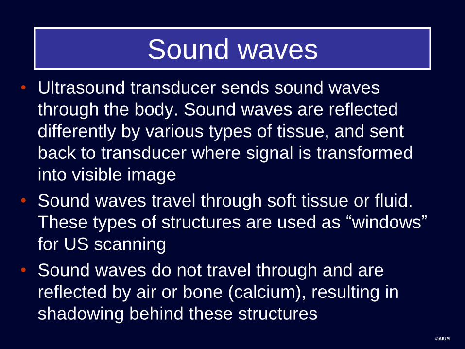
Sound waves
• Ultrasound transducer sends sound waves
through the body. Sound waves are reflected
differently by various types of tissue, and sent
back to transducer where signal is transformed
into visible image
• Sound waves travel through soft tissue or fluid.
These types of structures are used as “windows”
for US scanning
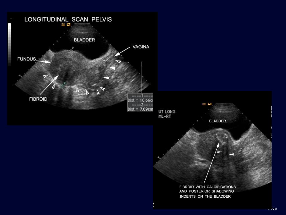
• Sound waves do not travel through and are
reflected by air or bone (calcium), resulting in
shadowing behind these structures
Page 7
©AIUM
Technique
• The standard pelvic examination
• Composed of the traditional
transabdominal approach (TAS)
• Combined with transvaginal
sonography (TVS)
• Frequently using Doppler
sonography
Page 8
©AIUM
Technique
• Transabdominal sonography uses
a distended bladder as window to
pelvic structures for a global view.
Page 9
©AIUM
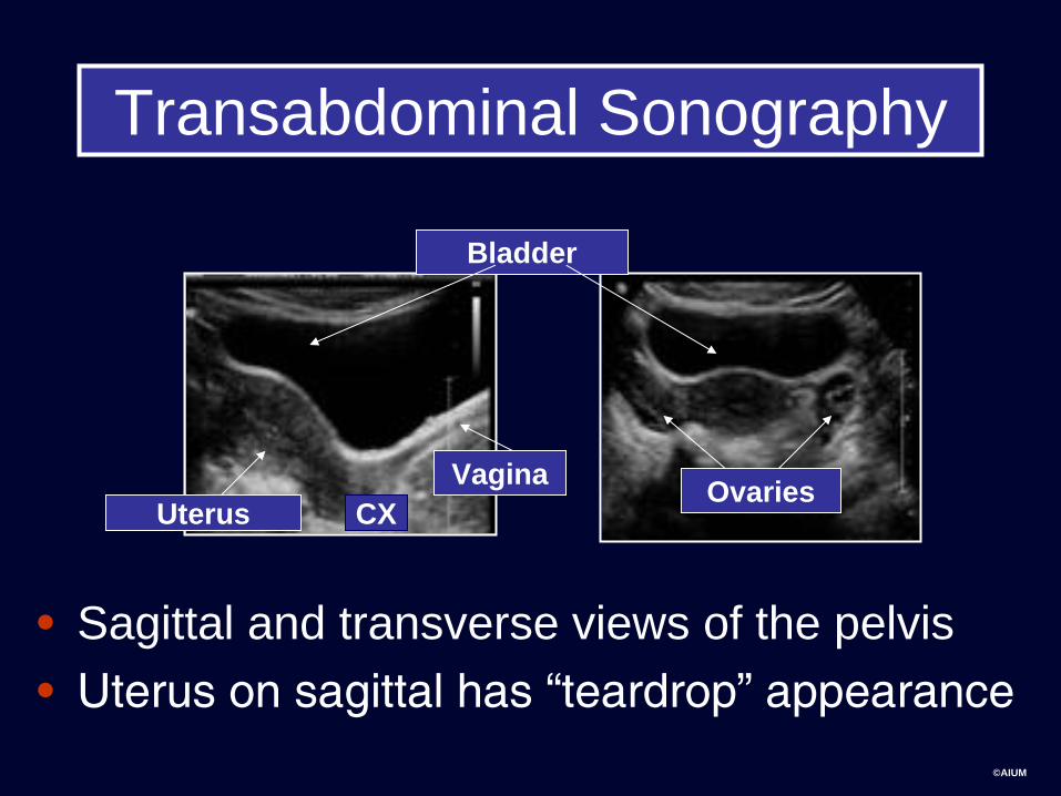
Transabdominal Sonography
• Sagittal and transverse views of the pelvis
• Uterus on sagittal has “teardrop” appearance
Bladder
Ovaries Uterus
Vagina
CX
Page 10
©AIUM
Technique
• Transvaginal sonography gives a
more detailed evaluation of pelvic
architecture using higher-frequency
transducers at closer proximity to
pelvic structures.
Page 11
©AIUM
Transvaginal Sonography
right left
anterior
posterior
anterior
posterior
cephalad
Page 12
©AIUM
Transvaginal US Transabdominal US
Page 13
©AIUM
Use all the information from the labeling that you are given to
orient yourself to anatomy
Long= longitudinal, usually sagittal relative to body.
Convention: patient’s head to left of screen.
Trans=transverse, usually axial relative to body. Convention:
patient’s right side to left of screen.
Page 14
©AIUM
Use all the information from the labeling that you
are given to orient yourself to anatomy and history
Page 15
©AIUM
The Normal Sonographic
Appearance of the
Nongravid Genital Tract
Page 17
©AIUM
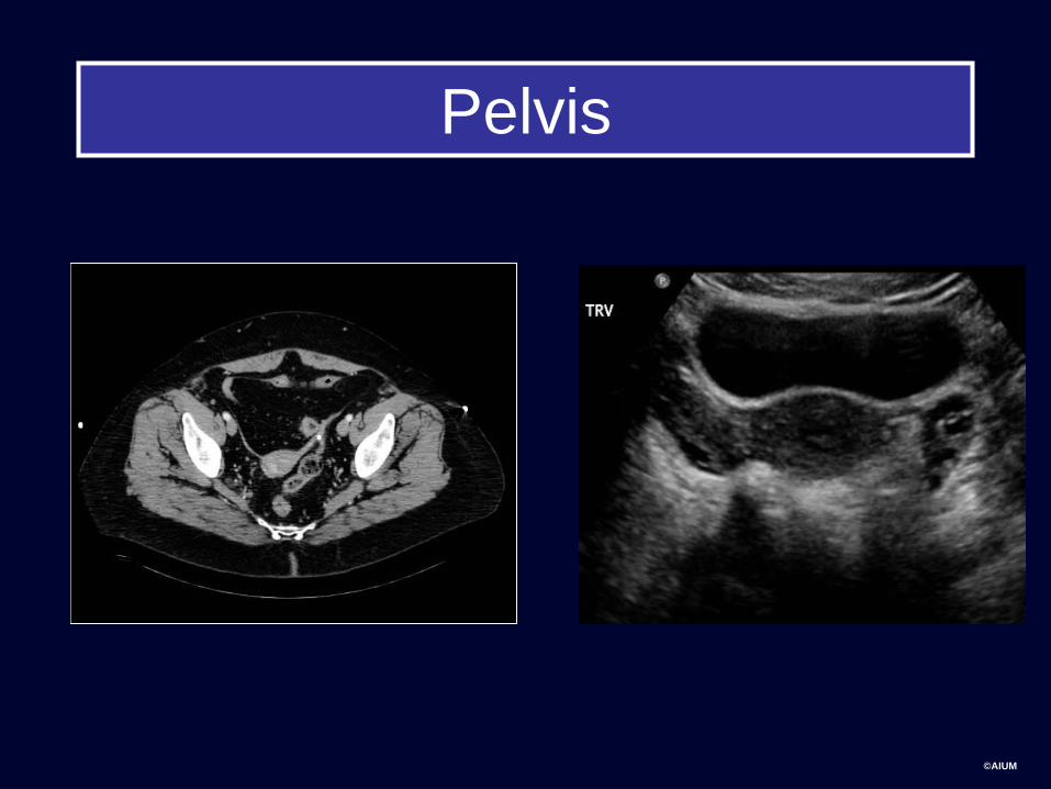
Anatomy Pelvis
Bladder
Vagina
R ovary L ovary
Uterus: cervix, body, fundus
Page 18
©AIUM
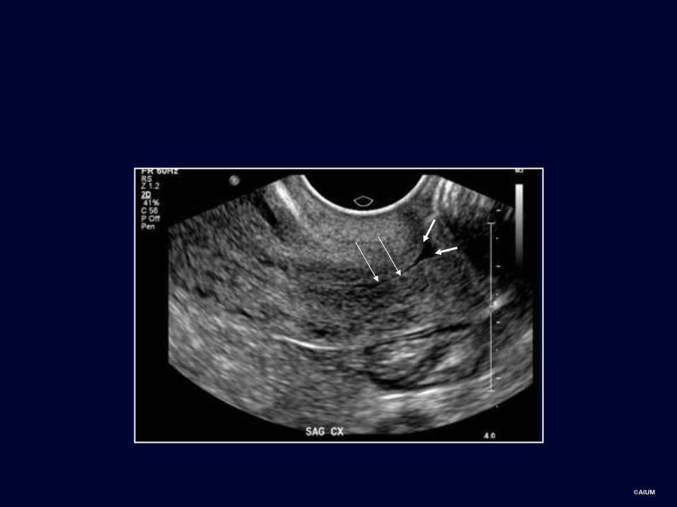
Premenopausal Endometrium
Proliferative Secretory
Uterine anatomy: myometrium vs. endometrium
Page 19
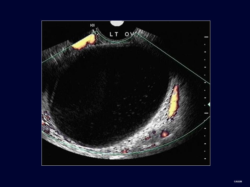
©AIUM
Ovary
• The ovaries are ellipsoid and can be
identified in menstruating females by
the presence of follicles.
Page 20
©AIUM
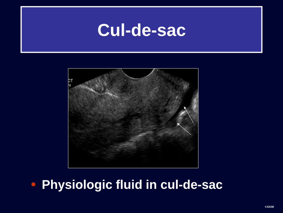
Cul-de-sac
• Physiologic fluid in cul-de-sac
Page 21
©AIUM
Basic obstetrical ultrasound
Page 22
©AIUM
LMP? Pregnant?
• In the female in the reproductive years, the physiologic as well as the pathologic processes are driven by the menstrual cycle and hormonal stimulation.
• Therefore, know the day of your patients’ day of the cycle, therefore…
• Know if your patient has a positive pregnancy test, and if so, what the quantitative serum beta hCG is.
Page 23
©AIUM
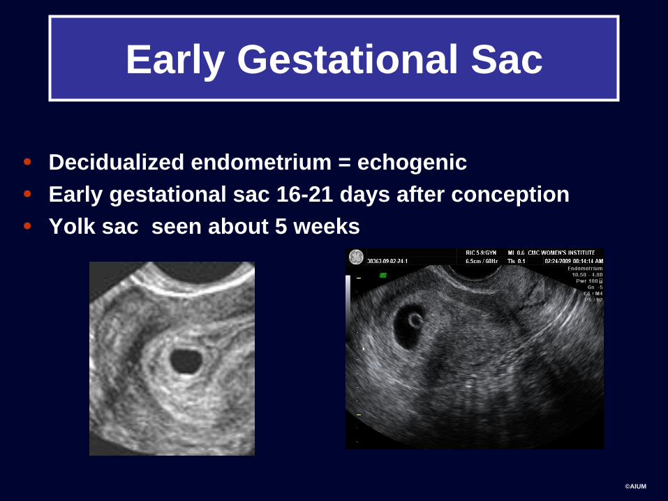
Early Gestational Sac
• Decidualized endometrium = echogenic
• Early gestational sac 16-21 days after conception
• Yolk sac seen about 5 weeks
Page 24
©AIUM
First Trimester
• By the 6th menstrual week, the early
embryo can be identified.
– Usually with cardiac activity
– The crown-rump length (CRL) is the best
estimation of GA once appears.
Page 25
©AIUM
Second Trimester
• After 13-14 weeks, measurements used
for dating are:
biparietal diameter (BPD), head
circumference (HC), abdominal
circumference (AC), and femur length
(FL)
Page 26
©AIUM
Fetal dating: BPD biparietal
diameter
BPD measured from outer to inner
Page 27
©AIUM
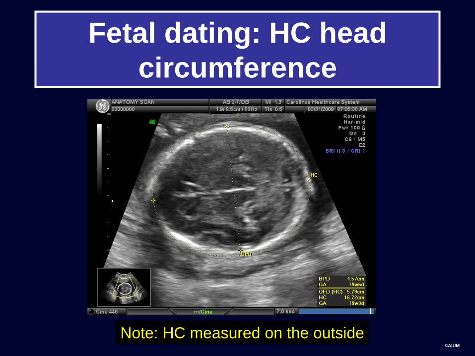
Fetal dating: HC head
circumference
Note: HC measured on the outside
Page 28
©AIUM
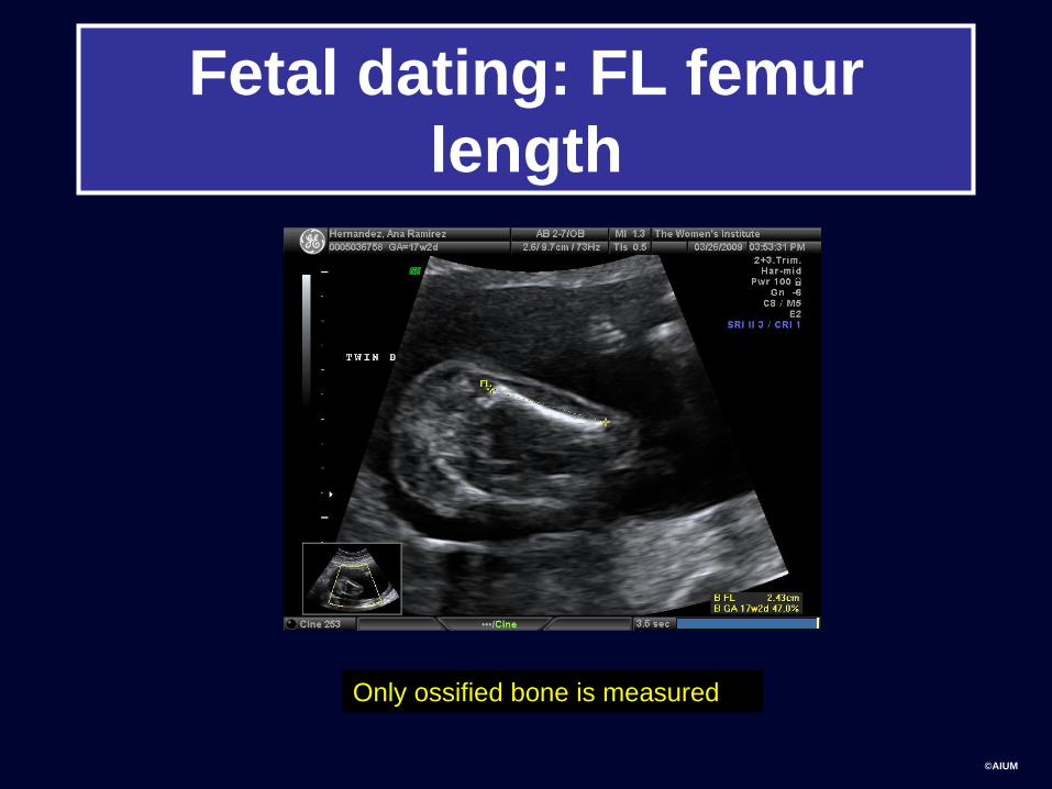
Fetal dating: FL femur
length
Only ossified bone is measured
Page 29
©AIUM
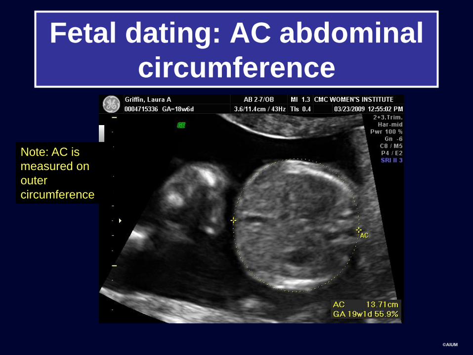
Fetal dating: AC abdominal
circumference
Note: AC is
measured on
outer
circumference
Page 30
©AIUM
Second Trimester
• Placenta
Placenta and cervix:
placenta previa
Page 31
©AIUM
Flow to the transducer is shown
in red and away in blue.
The Doppler sample volume
(oblique arrow) shows the
sampling site for pulsed Doppler
interrogation.
The right panel shows spectral
Doppler of umbilical artery flow.
As the flow is toward the
transducer, it is depicted as
positive or upward deflections. Umbilical artery Doppler waves
Cursor
Line
Page 32
©AIUM
Take Home Points
• US is first line modality to examine
female pelvis and gravid female pelvis
• US uses no ionizing radiation
• US uses sound waves, which travel best
through soft tissue or fluid
• US can be performed transabdominally
or transvaginally
• Conventional orientation for US images
is used
Page 33
©AIUM
Take Home Points
• Use terminology “hyperechoic” and
“hypoechoic”
• Fluid is black or anechoic on US
• We reviewed appearance of normal
uterine and ovarian anatomy
• We reviewed first and second trimester
pregnancy normal appearance
• Measurements used for fetal dating:
BPD, HC, AC, FL
Page 34
©AIUM
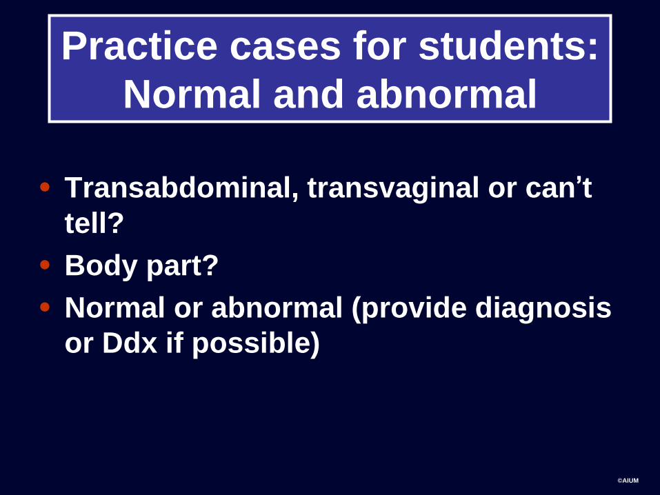
Practice cases for students:
Normal and abnormal
• Transabdominal, transvaginal or can’t
tell?
• Body part?
• Normal or abnormal (provide diagnosis
or Ddx if possible)