Normative values of clinical measurements around the scapula: assessment of the length of the pectoralis minor, scapular inclination and glenohumeral rotational range of motion Thomas Duyts Daan De Langhe Simon Dedecker Promotor: PT, PhD, Birgit Castelein PT, PhD, Ann Cools Master thesis submitted to achieve masters degree in rehabilitation sciences and physiotherapy Academic year: 2018-2019
Transcript
Normative values of clinical
measurements around the scapula:
assessment of the length of the
pectoralis minor, scapular inclination
and glenohumeral rotational range of
motion
Thomas Duyts Daan De Langhe Simon Dedecker
Promotor: PT, PhD, Birgit Castelein PT, PhD, Ann Cools
Master thesis submitted to achieve masters degree in rehabilitation sciences and physiotherapy
Academic year: 2018-2019
Acknowledgements The following words are an appreciation for the support we have received over the past two years in
accomplishing this study.
First, we would like to thank the University of Ghent for giving us the opportunity to do this research
and for providing all the necessary equipment. Secondly, our promotors PhD. Castelein Birgit and
PhD. Cools Ann should be acknowledged for the excellent guidance during this thesis. Their
knowledge and management of the whole process was an enormous contribution to the research
and its quality.
One last thing which cannot be forgotten in this acknowledgment, are the participants of the study.
They were a crucial factor in the research, without them doing what we did today would not have
been possible. Therefore, we would like to thank every single person who agreed to take part in the
testing procedure.
To end, thank you to everyone who made any contribution to this thesis, including ourselves.
Without the daily teamwork, patience and commitment finishing this work would not have been
Table 3.1 Descriptive statistics for men: PMI, ROM IR, ROM ER, Total ROM,
Inclination
P 20
Table 3.2 Descriptive statistics for women: PMI, ROM IR, ROM ER, Total ROM,
Inclination
P 21
Table 4.1 Descriptive statistics for men: scapular dyskinesis P 22
Table 4.2 Scapular dyskinesis for women: scapular dyskinesis P 22
Table 5 Statistical analysis P 23
Table 6 Significant results post hoc tests P 24
FIGURES
Figure 1 Scapular testing protocol P 13
Figure 2 Measurement of ER with digital inclinometer P 15
Figure 3 Measurement of IR with digital inclinometer P 15
Figure 4 Measurement of length of the pectoralis minor muscle with Digital Caliper P 15
Figure 5 Measurement of inclination of the scapula with digital inclinometer P 16
LIST OF ABBREVIATIONS
ROM Range Of Motion ER External Rotation IR Internal Rotation MT Middle trapezius LT Lower trapezius UT Upper trapezius SS Supraspinatus Kg Kilograms BMI Body Mass Index cm centimeter m2 square meter VAS Visual Analogue Scale HHD Handheld Dynamometer Dom Dominant NDom Non-dominant M Men or male F Female or women MD Mean Difference ICC Intraclass correlation coefficient SEM Standard error of the measurement SD Standard deviation MDC Minimal detectable change CI Confidence interval SAT Scapular Assistance Test SRT Scapular Retraction Test
8
ABSTRACT (English) Background: Shoulder pain is a prevalent symptom in the population. As the scapula is the central link
between the shoulder and the spine it forms the base of this functional unit. Within this unit the
balance between mobility and stability is easily disturbed. Optimal functioning of the scapula is
necessary to control this delicate balance. Normative values based on a good measurement protocol
are very useful for a critical evaluation of this function. In literature no normative values for clinical
evaluation of the shoulder-scapula unit are present neither is their consistency in the testing methods
to obtain these values.
Objectives: This study wants to offer a benchmark and easy to perform testing procedures for
clinicians. Four outcome parameters were measured, shoulder range of motion, length of the
pectoralis minor, scapular inclination and the presence of scapular dyskinesis.
Study design: Cross-sectional study.
Methods: 400 healthy (201 men, 199 women), non-overhead athletes, between 18 and 60 years of age
were recruited. All participants underwent measurements, for the four parameters, on both shoulders.
Scapular dyskinesis was assessed with the yes/no method. The length of the pectoralis minor was
measured with a caliper. A digital inclinometer was used for external/internal ROM and scapular
inclination. The data were then analyzed with linear mixed models, in order to find significant (p < 0.05)
interactions or significant main effects. Significant differences were further analyzed using post hoc
pairwise comparisons (Bonferroni). Normative values for age, side dominance, gender and presence
of dyskinesis were obtained this way.
Results: This study shows that the factors: age, gender, side dominance and presence of dyskinesis
have significant influence on the parameters. For the PMI, it was shown that the dominant side was
statistically shorter than the non-dominant side. Female have consequently greater ROM than male.
The same thing is noticed for the youngest age categories compared to the older. For IR the dominant
side has less ROM than the non-dominant, the opposite applies for ER. For Inclination, women without
scapular dyskinesis showed more upward rotation of the scapula compared to the same age categories
with dyskinesis. Scapular dyskinesis is present in almost half of the population.
Conclusion: This study created representative normative data, that can be used in a clinical setting to
evaluate the condition of the scapula in various populations. For further research in this topic, the
researchers advocate for consistency in the use of measurement protocols and the recruitment of a
Pm = Pectoralis minor; PMI = Pectoralis minor index; F = Force; Rom=Range of Motion; mm = millimeter; ° = Degrees; N = Newton; ICC = Intraclass correlation coefficient; SD = standard deviation; SEM = standard error of the measurement;
MDC = Minimal detectable change; SEM = SD √1 − 𝐼𝐶𝐶, MDC = 1.96 * SEM * √2
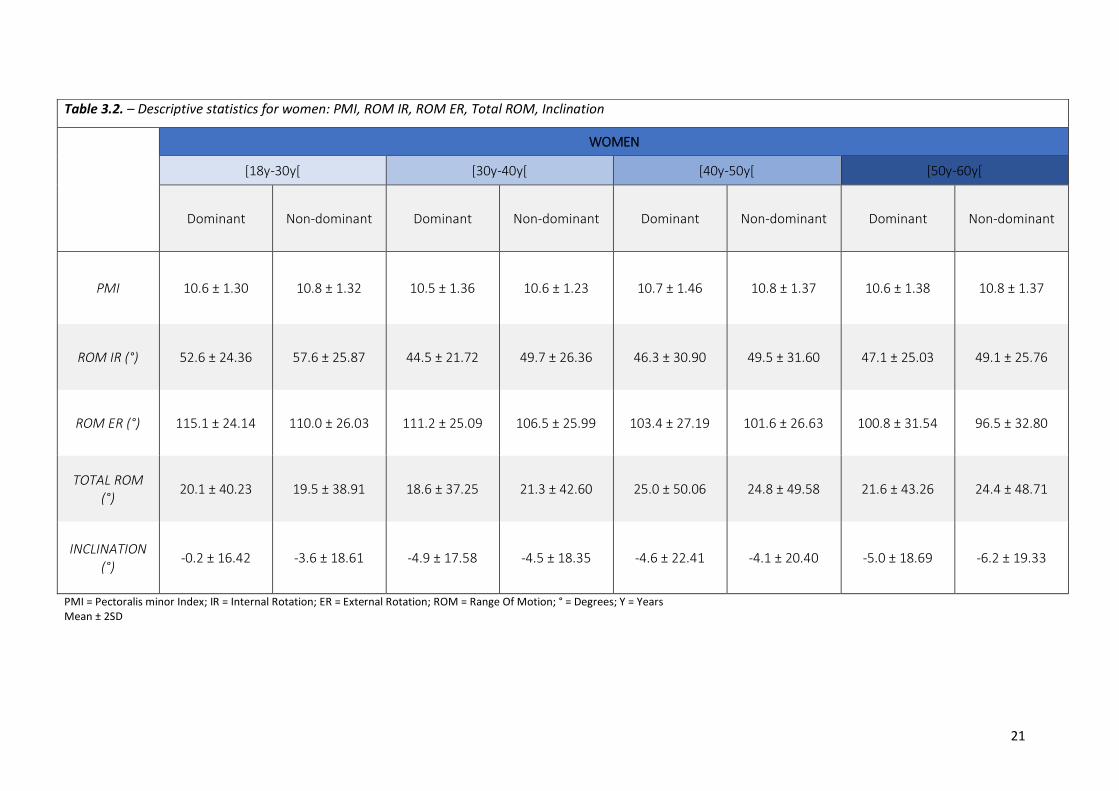
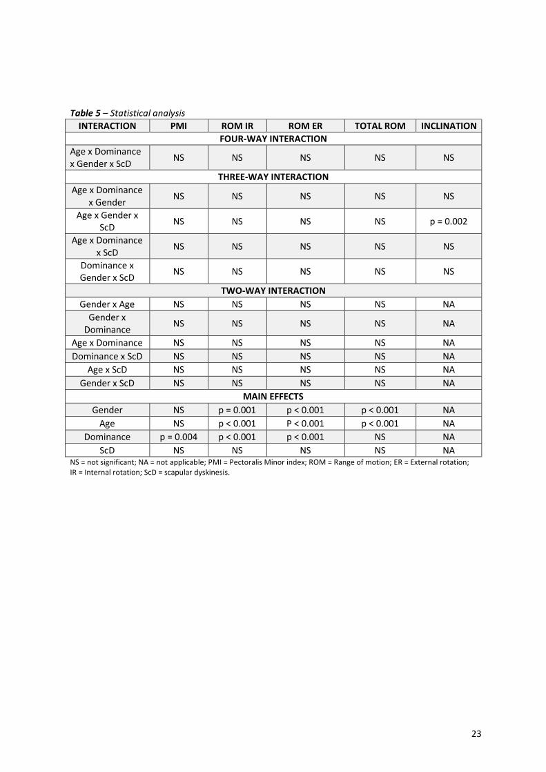
3.2. Synthesis of results Tables 3.1. and 3.2. show descriptive data (mean ± SD) for all measurements divided by sex, dominance
and age category. Results of statistical analysis of variance and post hoc Bonferroni analysis are
respectively represented in table 5. and 6.
3.2.1. PMI Statistical analysis showed no significant interactions but showed that the main effect “dominance”
had a significant (p = 0.004) influence on the PMI. Post-hoc tests showed that the PMI of the dominant
side is shorter than the non-dominant side. (p = 0.004; Mean difference (MD) Dom-NDom = -0.102)
3.2.2. ROM
• Internal rotation:
For internal rotation, it was shown that gender (p = 0.001), age (p < 0.001) and dominance (p < 0.001)
had significant main effects. Post-hoc tests showed that males have less IR than females (p = 0.001,
MD men-women = -4.015°) and age category 1 has the greatest mobility towards IR compared to the
19
other 3 age groups (1-2: p < 0.001, MD = 8.217°; 1-3: p > 0.001, MD = 7.898°; 1-4: p = 0.007, MD =
5.651°). ROM at the dominant side is less than on the non-dominant side (p < 0.001, MD Dom-NDom
= -3.990°).
• External rotation:
For external rotation, gender (p < 0.001), age (p < 0.001) and dominance (p < 0.001) are significant
main effects. Male have less ER than women (p < 0.001, MD men-women = -8.598°) and age category
1 has the greatest mobility towards ER compared to the other 3 age groups (1-2: p < 0.519, MD =
3.017°; 1-3: p < 0.001, MD = 8.259°; 1-4: p < 0.001, MD = 12.863°). ROM at the dominant side is greater
than on the non-dominant side (p < 0.001, MD = 4.055°).
• Total range of motion:
Gender (p < 0.001) and age (p < 0.001) are the significant main effects for total range of motion. Males
have less ROM than females (p < 0.001, MD men-women = -12.687°) and age category 1 has the
greatest mobility compared to the other 3 age groups (1-2: p = 0.001, MD = 11.254°; 1-3: p < 0.001,
MD = 16.164°; 1-4: p < 0.001, MD = 18.372°).
3.2.3. Scapular dyskinesis The descriptive results for scapular dyskinesis were separated for men and women into two tables,
represented by “Table 4.1 – Descriptive statistics for men: scapular dyskinesis” and “Table 4.2. –
Scapular dyskinesis for women: scapular dyskinesis”. In these two tables the population’s ratio for the
three different types of scapular dyskinesis and non-scapular dyskinesis were represented according
to the four different age categories. A differentiation between the dominant and non-dominant side
was created.
3.2.4. Inclination The results represent a three-way interaction between the factors age, gender and scapular dyskinesis
(P = 0.002) for the parameter inclination (Table 5.). Women in age-category one and three, without
scapular dyskinesis had a significantly more upward rotated scapula, compared to women in the same
age-categories with scapular dyskinesis (C1: p < 0.001, MD = 6.12; C3: p = 0.022, MD = 4.74).
20
Table 3.1. – Descriptive statistics for men: PMI, ROM IR, ROM ER, Total ROM, Inclination
TOTAL 37/199 33/199 7/199 122/199 30/199 55/199 6/199 108/199
18.6 % 16.6 % 3.5 % 61.3 % 15.1 % 27.6 % 3 % 54.3 % No ScD = Absence of scapular dyskinesis; Type 1 = Inferior dysfunction; Type 2 = Medial dysfunction; Type 3 = Superior dysfunction (Kibler et al.)
23
Table 5 – Statistical analysis
INTERACTION PMI ROM IR ROM ER TOTAL ROM INCLINATION
FOUR-WAY INTERACTION Age x Dominance x Gender x ScD
NS NS NS NS NS
THREE-WAY INTERACTION
Age x Dominance x Gender
NS NS NS NS NS
Age x Gender x ScD
NS NS NS NS p = 0.002
Age x Dominance x ScD
NS NS NS NS NS
Dominance x Gender x ScD
NS NS NS NS NS
TWO-WAY INTERACTION
Gender x Age NS NS NS NS NA
Gender x Dominance
NS NS NS NS NA
Age x Dominance NS NS NS NS NA
Dominance x ScD NS NS NS NS NA
Age x ScD NS NS NS NS NA
Gender x ScD NS NS NS NS NA
MAIN EFFECTS
Gender NS p = 0.001 p < 0.001 p < 0.001 NA
Age NS p < 0.001 P < 0.001 p < 0.001 NA
Dominance p = 0.004 p < 0.001 p < 0.001 NS NA
ScD NS NS NS NS NA NS = not significant; NA = not applicable; PMI = Pectoralis Minor index; ROM = Range of motion; ER = External rotation; IR = Internal rotation; ScD = scapular dyskinesis.
/ / / / F & C1 or C3: No ScD > ScD (C1: p < 0.001; C3: p = 0.022)
C1, C2, C3, C4 = Age-Category 1-4; M = Men; F = Women; PMI = Pectoralis minor Index; Rom = Range Of Motion; ER = External Rotation; IR = Internal Rotation; ScD = Scapular Dyskinesis; No ScD = Absence Of Scapular Dyskinesis; Dom = Dominant Side; NDom = Non-Dominant Side.
25
4. DISCUSSION The provided normative reference values for scapular evaluation are attained using the previous
described measurement protocols. The four evaluated parameters were scapular dyskinesis, muscle
length, ROM and inclination. The collected data is retrieved from 400 subjects, who were all tested as
reliable and homogeneous as possible, with cost-effective and practical devices. According to the work
from Cools et al. (2014), a constant subject position was kept for practical utility and to reveal
reproducible results (5). The reference values were benchmarked for the following population
factors: age, gender and side dominance (Table 3.1. & 3.2.). In the following part every parameter was
discussed based on the population factors and findings of previous research.
4.1. Summary of results
4.1.1. Range of motion (IR ROM, ER ROM, TOT ROM) According to the statistical analysis for the parameters IR ROM and ER ROM (table 5), statistically
significant differences within each of the three population factors were found. After comparison with
the MDC of 4.89° for IR and 4.17° for ER (Table 2), it turns out that for internal rotation the factor age
and for external rotation the factors gender and age (except for C1 - C2 comparison) were clinically
significant main effects. Based on the results from the post hoc tests, there could be assumed that
people younger than 30 years have a significant greater internal and external rotation mobility than
the older subjects. External rotation has an inversely proportional pattern, in which an increase in age
is accompanied by a decrease in ER ROM. For IR ROM the pattern was not fully clear. IR ROM showed,
apart from the first age category, a proportional pattern. Despite the opposite pattern in IR and ER
ROM, the distinctive decrease of ER ROM defined the pattern of TOT ROM.
IR ROM
The study by Cools et al. (2014) showed ROM differences based on the used equipment and position,
particularly for the measurement of IR in 90° abduction (5). Therefore, we should be careful in
comparing the results with other studies.
Dominance: The results from the statistical analyses, for IR ROM, showed a difference of
approximately 4° between the dominant and non-dominant side (3.99°, Dom < NDom). This side
difference is also reported in previous studies (35-38, 44). Garcia et al. (2013) reported a mean
difference of 4.7° (Dom < NDom), the subjects were measured in a side lying position (35). The testing
protocols by Myers et al. (2009) and Conte et al. (2009) were similar to the one used in this study which
made these protocols more relevant for comparison. They reported respectively a mean difference of
4.7° and 3.5° (Dom < NDom) (36, 38). These three studies (Garcia et al. (2013), Myers et al. (2009) and
26
Conte et al. (2009)) tested a young population, that varied between 20 and 29 years. The results were
pretty similar to those of the first age-category described in this study (35, 36, 38). Dover et al. (2003)
reported conflicting results in which the dominant side had slightly greater IR ROM compared to the
non-dominant side. Despite a similar testing procedure, the study reported divergently greater results
(Dom = 92.1°, NDom = 91.5°). An explanation could be that the measurement was actively performed,
and no external fixation/palpation was used. This means that movement performed was not an
isolated glenohumeral IR (37). Based on the findings of this study and of previous research, an
assumption could be made that the younger population ([18-30[) has 4° less mobility at dominant side
compared to the non-dominant side. For the other age categories more research is necessary.
Gender: Conflicting evidence is present for the gender based, 4° ROM difference this study found
(4.02°, M < F). Cools et al. (2014) and Garcia et al. (2013) reported no gender-based main effect (5, 35).
and used college aged participants. The studies by McKay et al. (2017) and Barnes et al. (2001) used a
broader age range. (39, 41). McKay et al. reported a similar difference of approximately 5°.
Unfortunately, McKay et al. did not describe the used measurement method in detail, but it was
mentioned that the measurement was performed actively (39). Barnes et al. used a similar testing
protocol, as this study, but a different device (goniometer). Barnes et al. reported that IR and ER ROM
showed a large difference in ROM, based on gender (41).
Age: The tendency that younger subjects have less internal rotation than older subjects was already
reported in 1985 by Murray et al. (42). In this study the same tendency was present. There was an
increase of 2.6° based on the mean values (C2 → C4). This was also shown by Roy et al. (2009) and
Barnes et al. (2001) who, despite the use of a goniometer, used a very similar study design compared
to this study (40,41).
ER ROM
Dominance: ER ROM presented an opposite pattern, with a similar side difference of approximately 4°
in favor of the dominant side, compared to IR ROM (4.06°, Dom > NDom). This finding was seen in
previous researches which compared the dominant side with the non-dominant (36-38, 40, 41, 44).
Myers et al. (2009), Conte et al. (2009) and Dover et al. (2003) also described a similar dominance-
based difference of respectively: 5°, 5.1° (women) and 3.7° (women), based on the mean values (36-
38). This significant difference was also reported by Barnes et al. (2001), Boon et al. (2000) and Roy et
al (2009).
27
Gender: For ER ROM women have a greater ROM, compared to men (8.6°, M < F). Note that the
significant difference for ER ROM is approximately two times higher, than IR ROM. This result is
conflicted by Cools et al. (2014) who described that there was no significant gender-based main effect
(5). Roy et al. (2009) reported that women had significantly higher ER ROM than men, especially in the
40-59 age category (40). Boon et al. (2000) and Barnes et al. also observed that women had greater ER
ROM than men (44,41).
Age: The inversely proportional tendency for ER ROM was noticed by several studies which used a
study design focusing on age (39-41, 44). A decrease of 12.9° between the youngest age-category and
the oldest age-category is shown in the results of this study (C1 → C4).
TOT ROM
Internal rotation is greater at the non-dominant side and increases until the age of 60. External rotation
is greater at the dominant side and decreases with an increasing age (until 60y). Because of the
opposite dominance-based differences for IR and ER ROM, dominance is not a significant main effect
for the TOT ROM. Based on previous literature and the results in this study, it appears that a greater
ER ROM and a lower IR ROM at the dominant side is common in the general population. This states
that the commonly used method of using the contralateral side as a baseline for comparison is not
always relevant and should therefore be performed with care. This statement emphasizes the need
and importance for gender-, age- and dominance-based normative values (41, 44, 46).
It is not new that an increase in age is attended with a decrease in ROM of the shoulder. The significant
decrease in TOT ROM is mostly affected by the decrease in ER ROM (12.9°) and slightly limited by the
increase in IR ROM (2.6°). Macedo et al. (2009) emphasized the importance of age-related decrease in
ER ROM. They concluded that among 11 movements (F, [18-59]), passive shoulder ER was the only
movement wherefore a distribution of reference values, based on age, were absolutely necessary (41,
45).
Previous research also showed that in a student population woman were more flexible than men (52).
Bassey et al. (1989) discussed that women had poorer abduction flexibility compared to men in the
older population (+65) (53). Although there is a great gender-based TOT ROM difference of 12.69° (F >
M), this does not suggest that for every shoulder movement and for every age women are the most
flexible. Further research is necessary to describe the outcomes of different shoulder movements
based on gender and age.
28
Differences in rotational ROM is mostly attributed to a variation in stiffness of the muscles or joint
capsule (43, 51). Hung et al. (2010) showed that stiffness of the Posterior Deltoid muscle had the
highest correlation with reduced IR ROM. Two other muscles who correlated significantly with reduced
IR ROM were the infraspinatus and teres minor (43). Myers et al. (2009) described that the difference
in glenohumeral rotation ROM is highly influenced by the amount of humeral torsion. They claimed
that a lower IR ROM and a higher ER ROM on the dominant side could be explained by more humeral
torsion (13°) compared to the non-dominant side (36). At this point there is no clear explanation for
the differences in glenohumeral ROM, further research is needed.
4.1.2. Scapular dyskinesis A lot of studies about scapular dyskinesis have been focusing on populations with a shoulder
impairment or overhead athletes (54, 55). This study shows presence of scapular dyskinesis in a healthy
population with a broad age range (Table 4.1. & 4.2.). Results show that within the male population
almost half of the subjects have dyskinesis. Dyskinesis itself is more present at the dominant side (Dom:
51%, NDom: 43%). The female population shows a lower presence of scapular dyskinesis and it occurs
more at the non-dominant side (Dom: 38.7%, NDom: 45.7%). These results were confirmed by other
studies, although they had smaller populations (47 - 49). The control group in Castelein et al. (2016)
showed that from the 19 tested women, 8 showed scapular dyskinesis (42%) (47). A study from Hannah
et al. (2017) found that even the majority of their population, 27 out of 40 people, had dyskinesis (48).
Uga et al. (2016) used a male population where 21 out of 40 shoulders showed dyskinesis. These results
probably suggest that scapular dyskinesis should not always be seen as divergent (49) and may be
considered as a common phenomenon in the population. Because of the remarkable presence of
scapular dyskinesis in the healthy population, there was opted to use scapular dyskinesis in the
statistical analyses as a factor and no longer as a parameter. Adding dyskinesis as a factor did not
change anything in the outcome of the statistical results except for inclination. This is not surprising as
scapular dyskinesis has an influence on the positioning of the scapula.
Causes for scapular dyskinesis have been comprehensively described in literature. When shoulder
pathology is present and scapular dyskinesis is detected, the link with scapular muscle imbalances or
weaknesses is often made (56). Though today, evidence to possibly refute this statement is present in
literature (48-50). One of the investigations undermining this theory is the one by Hibberd et al. (2012)
(16). The researchers showed that a program to strengthen the shoulder complex does not resolve
shoulder dyskinesis (16). Other factors such as neuromuscular control may be contributing to this
problem (62,63). Because scapular dyskinesis is so common in the healthy population, another way of
thinking is that every individual positions its scapula in an optimal way to generate maximal power
outputs. Therefore, dyskinesis is just a manner of scapular functioning. But this does not immediately
29
rule out the role of scapular dyskinesis in the rehabilitation of shoulder dysfunctions. The SRT (Scapular
Retraction Test) and SAT (Scapular Assistance Test) are excellent tools to detect if scapular dyskinesis
is involved in pathology (57, 58).
4.1.3. Pectoralis minor muscle length
The pectoralis minor length itself is clinically not so relevant therefore the PMI was calculated. Initially,
there was opted to divide the PMI into three categories based on the study from Borstad et al. (2005)
(22). Using the cut offs mentioned in the article none of the present PMI were divided into the ‘short’
category (PMI 7.5). A reason here fore might be that these cut off values were based on a pilot study
consisting of 6 people. This small and non-representative population may show irrelevant results.
Another study including 51 participants experienced the same problem, where no individual matched
the ‘short PMI’ criteria (61). For this reason, there was decided to calculate the cut off values with data
presented in this study. Following the method by Borstad et al. (2005) the group inclusion cut point
values for the present analysis were then set at 1SD from the mean PMI found in this study (short
9.86, middle 9,86-11.54, long 11.54).
Dominance: As shown in the results, dominance was the only significant main effect. The dominant
side had a lower PMI compared to the non-dominant side, but this was not clinically significant (Dom
< NDom: 0.102, MDC = 0.159). This side difference was also described by Struyf et al. (2014) who found
a lower PMI on the dominant side (9). An explanation for this observation could be that the dominant
side is more stiffened due to increased use. No evidence for this statement could be found in literature.
Relation with dyskinesis: A hypothesis was premised which said that people with lower PMI were more
likely to have scapular dyskinesis, especially Type 1. This assumption could be endorsed by the findings
of Borstad et al. (2005) which said that shorter PM length could cause a dysfunction of scapular
kinematics (22). Also, Yesilyaprak et al. (2016) found that a decline in PMI was related to a higher
possibility of scapular dyskinesis (33). In this study, results showed some similarities with the two
studies mentioned above. The group with the lowest PMI contained the highest percentage of people
with scapular dyskinesis (51.1%). Although the group of patients with high PMI values had a higher
percentage of scapular dyskinesis compared to the ones with ‘middle’ PMI (High: 49.5%, Middle:
42.4%). Following Yesilyaprak et al. PMI plays a determinative role in the presence of scapular
dyskinesis. These findings seem reasonably as the pectoralis minor muscle attaches directly to the
scapula and accordingly influence it.
30
4.1.4. Inclination The present three-way interaction showed a clinically significant difference (MDC = 4.35) between
women with and without scapular dyskinesis, in the first ([18-30y[) and third ([40y-50y[) age-category.
There is no previous research that mentioned an interaction between these factors. For both genders
the mean value is negative, which insinuates that the average population has a downward rotated
scapula. The results in this study do not match with these of Struyf et al. (2011) (26).
4.2. Limitations Although this study was conducted under supervision of professionals by the university of Ghent and
was performed with reliable instruments and reliable measurements, it still had some limitations.
The raters were rather inexperienced, and they got more familiar with the measurements during the
testing period. This could cause the latest measurements to be more accurate than the ones in the
beginning. On the other hand, they had a two-day training session and the measurements used were
shown reliable or were based on protocols used by other investigators.
A second limitation is that the measurements were performed by twelve raters in total. This could
cause different outcomes for different raters. To limit this margin of error every measurement was
clearly described in a video and every rater tried to reproduce the standardized measurement method
as accurate as possible. The measurements themselves showed good interrater reliability so this
should mitigate this remark.
As a third limitation, the exclusion criteria based on the hours of overhead sports performed is rather
lucratively chosen. The boundary was set with the intention to exclude competitive athletes, who could
have sport specific adaptations of the shoulder complex. In literature no consensus was found about
the hours of training necessary for those adaptations.
In the in- and exclusion criteria the professional activities of the subject were not kept in account. What
if they had very demanding professions for the shoulder complex (e.g. construction workers,
electricians, plasterers)? This was not seen as serious flaw as otherwise a great part of the general
healthy population would be excluded.
31
4.3. Conclusion This study emphasizes the importance of normative values as a base for clinical investigation of the
scapula and shoulder. For further research in this topic, the researchers advocate for consistency in
the use of measurement protocols and the recruitment of a representative population. Nevertheless,
this study created representative normative data, that can be used in a clinical setting to evaluate the
condition of the scapula in various populations. Abnormality’s compared to the reference values,
should be noticed and used as a guide for further investigation or evaluation. Further research is
necessary to link possible causes of pathology with marked deviations.
32
5. REFERENCES
1. Badcock, L. J., Lewis, M., Hay, E. M., McCarney, R., & Croft, P. R. (2002). Chronic shoulder pain in the community: a syndrome of disability or distress? Ann Rheum Dis, 61(2):128–31.
2. Burkhart, S.S., Morgan, C. D., & Kibler, W. B. (2003). The disabled throwing shoulder: spectrum of pathology Part III: The SICK scapula, scapular dyskinesis, the kinetic chain, and rehabilitation. Arthroscopy, 19(6):641-61.
3. Kibler, W. B., Uhl, T. L., Maddux, J. W., Brooks, P.V., Zeller, B., & McMullen, J. (2002). Qualitative clinical evaluation of scapular dysfunction: a reliability study. J Shoulder Elbow Surg, 11(6):550-6.
4. Dougherty, J., Walmsley, S., & Osmotherly, P. G. (2015). Passive range of movement of the shoulder: a standardized method for measurement and assessment of intrarater reliability. Journal of Manipulative and Physiological Therapeutics, 38(3):218-24.
5. Cools, A. M., De Wilde, L., Van Tongel, A., Ceyssens, C., Ryckewaert, R., & Cambier, D. C. (2014 Oct). Measuring shoulder external and internal rotation strength and range of motion: comprehensive intra-rater and inter-rater reliability study of several testing protocols. J Shoulder Elbow Surg, 23(10):1454-61.
6. McClure, P., Tate, A.R., Kareha, S., Irwin, D., & Zlupko, E. (2009). A Clinical Method for Identifying Scapular Dyskinesis, Part 1: Reliability. Journal of Athletic Training, 44(2):160-4.
7. Celik, D., Dirican, A., & Baltaci, G. (2012). Intrarater reliability of assessing strength of the shoulder and scapular muscles. J Sport Rehabil, 3:1-5.
8. Finley, M., Goodstadt, N., Soler, D., Somerville, K., Friedman, Z., & Ebaugh, D. (2017) Reliability and validity of active and passive pectoralis minor muscle length measures. Braz J Phys Ther, 21(3):212-8.
9. Struyf, F., Meeus, M., Fransen, E., Roussel, N., Jansen, N., Truijen, S., & Nijs, J. (2014 Aug) Interrater and intrarater reliability of the pectoralis minor muscle length measurement in subjects with and without shoulder impingement symptoms. Man Ther, 19(4):294-8
10. Cools, A. M., Witvrouw, E. E., Declercq, G. A., Danneels, L. A. & Cambier D. C. (2003 Jul-Aug) Scapular muscle recruitment patterns: trapezius muscle latency with and without impingement symptoms. Am J Sports Med, 31(4):542-9.
11. Huang, T. S., Huang, C. Y., Ou, H. L. & Lin, J. J. (2016 Dec) Scapular dyskinesis: Patterns, functional disability and associated factors in people with shoulder disorders. Man Ther, 26:165-171.
12. Ludewig, P. M. & Cook, T. M. (2000) Alterations in shoulder kinematics and associated muscle activity in people with symptoms of shoulder impingement. Phys Ther, 80(3):276-91.
13. Dickerson, C. R., Alenabi, T., Martin B. J. & Chaffin, D. B. (2018 Aug). Shoulder muscular activity in individuals with low back pain and spinal cord injury during seated manual load transfer tasks. Ergonomics, 61(8):1094-1101.
14. Michener, L. A., Sharma, S., Cools, A. M. & Timmons, M. K. (2016 Nov) Relative scapular muscle activity ratios are altered in subacromial pain syndrome. J Shoulder Elbow Surg, 25(11):1861-1867.
15. Hill, C. L., Gill, T. K., Shanahan, E. M. & Taylor, A. W. (2010 Aug) Prevalence and correlates of shoulder pain and stiffness in a population-based study: the North West Adelaide Health Study. Int J Rheum Dis, 13(3):215-22.
16. Luime, J. J., Koes, B. W., Hendriksen, I. J., Burdorf, A., Verhagen, A. P., Miedema, H. S. & Verhaar, J. A. (2004) Prevalence and incidence of shoulder pain in the general population; a systematic review. Scand J Rheumatol, 33(2):73-81.
17. Illyés, A. & Kiss, R. M. (2006) Kinematic and muscle activity characteristics of multidirectional shoulder joint instability during elevation. Knee Surg Sports Traumatol Arthrosc, 14:673–85.
33
18. Helgadottir, H., Kristjansson, E., Mottram, S., Karduna, A. R. & Jonsson, H. Jr. (2010) Altered scapular orientation during arm elevation in patients with insidious onset neck pain and whiplash associated disorder. J Orthop Sports Phys Ther, 40:784–91.
19. Ludewig, P. M. & Reynolds, J. F. (2009) The association of scapular kinematics and glenohumeral joint pathologies. J Orthop Sports Phys, 39:90–104.
20. Lewis, J., Green, A., Reichard, Z. & Wright, C. (2002 Feb) Scapular position: the validity of skin surface palpation. Man Ther, 7(1):26-30.
21. Struyf, F., Nijs, J., Mottram, S., Roussel, N. A., Cools, A. M. & Meeusen R. (2014 Jun) Clinical assessment of the scapula: a review of the literature. Br J Sports Med, 48(11):883-90.
22. Borstad, J. D. & Ludewig, P. M. (2005 Apr) The effect of long versus short pectoralis minor resting length on scapular kinematics in healthy individuals. J Orthop Sports Phys Ther, 35(4):227-38.
23. Borstad, J. D. (2006 Apr) Resting position variables at the shoulder: evidence to support a posture-impairment association. Phys Ther, 86(4):549-57.
24. Camargo, P. R., Phadke, V., Zanca, G. G. & Ludewig, P. M. (2018 Feb) Concurrent validity of inclinometer measures of scapular and clavicular positions in arm elevation. Physiother Theory Pract, 34(2):121-130.
25. Johnson, M. P., McClure, P. W. & Karduna, A. R. (2001 Feb) New method to assess scapular upward rotation in subjects with shoulder pathology. J Orthop Sports Phys Ther, 31(2):81-9.
26. Struyf, F., Nijs, J., Baeyens, J. P., Mottram, S. & Meeusen, R. (2011 Jun) Scapular positioning and movement in unimpaired shoulders, shoulder impingement syndrome, and glenohumeral instability. Scand J Med Sci Sports, 21(3):352-8.
27. Awan, R., Smith, J. & Boon, A. J. (2002 Sep) Measuring shoulder internal rotation range of motion: a comparison of 3 techniques. Arch Phys Med Rehabil, 83(9):1229-34.
28. Castelein, B., Dedecker, S., DeLanghe, D. & Duyts, T. (2018 May) Normative values of clinical measurements around the scapula: a systematic review. Ghent, Belgium: University of Ghent.
29. Borstad, J. D. (2008 Apr) Measurement of pectoralis minor muscle length: validation and clinical application. J Orthop Sports Phys Ther, 38(4):169-74.
30. Katoh M. (2015 Jun) Test-retest reliability of isometric shoulder muscle strength measurement with a handheld dynamometer and belt. J Phys Ther Sci, 27(6):1719-22.
31. Shahidi, B., Johnson, C. L., Curran-Everett, D. & Maluf, K.S. (2012) Reliability and group differences in quantitative cervicothoracic measures among individuals with and without chronic neck pain. BMC Musculoskelet Disord, 13:215.
32. Watson, L., Balster, S. M., Finch, C. & Dalziel, R. (2005) Measurement of scapula upward rotation: a reliable clinical procedure. Br J Sports Med, 39(9):599-603.
33. Yeşilyaprak S.S., Yüksel E. & Kalkan S. (2016) Influence of pectoralis minor and upper trapezius lengths on observable scapular dyskinesis. Phys Ther Sport., 19:7-13.
34. Koo, T. K. & Li, M. Y. (2016 Jun). A Guideline of Selecting and Reporting Intraclass Correlation Coefficients for Reliability Research. Journal of Chiropractic Medicine, 15 (2): 155–63.
35. Carcia, C. R., Cacolice, P. A. & Scibek, J. S. (2013) Sidelying glenohumeral passive internal rotation range of motion values in a healthy collegiate population. Int J Sports Phys Ther, 8(6):793-9.
36. Myers, J. B, Oyama, S., Goerger, B. M., Rucinski, T. J., Blackburn, J. T. & Creighton, R. A. (2009) Influence of Humeral Torsion on Interpretation of Posterior Shoulder Tightness Measures in Overhead Athletes. Clinical Journal of Sport Medicine, 19(5):366-71.
37. Dover, G. C., Kaminski, T. W., Meister, K., Powers, M. E. & Horodyski, M. (2003) Assessment of shoulder proprioception in the female softball athlete. Am J Sports Med, 31(3):431-7.
38. Conte, A. L. F., Marques, A. P., Casarotto, R. A. & Amado-Joao, S. M. (2009) HANDEDNESS INFLUENCES PASSIVE SHOULDER RANGE OF MOTION IN NONATHLETE ADULT WOMEN. Journal of Manipulative and Physiological Therapeutics, 32(2):149-53.
39. McKay, M. J., Baldwin, J. N., Ferreira, P., Simic, M., Vanicek, N. & Burns, J. (2017) Normative reference values for strength and flexibility of 1,000 children and adults. Neurology, 88(1):36-43.
40. Roy J. S., MacDermid, J. C., Boyd, J. U., Faber K. J., Drosdowech, D. & Athwal G. S. (2009) Rotational strength, range of motion, and function in people with unaffected shoulders from various stages of life. Sports Med Arthrosc Rehabil Ther Technol, 1:4.
41. Barnes, C. J., Van Steyn, S. J. & Fischer, R.A. (2001) The effects of age, sex, and shoulder dominance on range of motion of the shoulder. J Shoulder Elbow Surg, 10(3):242-6.
42. Murray, M. P., Gore, D. R., Gardner, G. M. & Mollinger, L. A. (1985) Shoulder motion and muscle strength of normal men and women in two age groups. Clin Orthop Relat Res, 268–273.
43. Hung, C. J., Hsieh, C. L., Yang, P. L. & Lin, J. J. (2010 Mar) Relationships between posterior shoulder muscle stiffness and rotation in patients with stiff shoulder. J Rehabil Med, 42(3):216-20.
44. Boon, A. J. & Smith, J. (2000) Manual scapular stabilization: its effect on shoulder rotational range of motion. Arch Phys Med Rehabil, 81(7):978-83.
45. Macedo, L. G. & Magee, D. J. (2009) Effects of age on passive range of motion of selected peripheral joints in healthy adult females. Physiother Theory Pract, 25(2):145-64.
46. Miller P. (1985) Assessment of joint motion. In: Rothstein J, editor. Measurement in physical therapy. New York: Churchill Livingstone.
47. Castelein, B., Cools, A., Parlevliet, T. & Cagnie, B. (2016 Dec) Are chronic neck pain, scapular dyskinesis and altered scapulothoracic muscle activity interrelated?: A case-control study with surface and fine-wire EMG. J Electromyogr Kinesiol, 31:136-143.
48. Hannah, D. C., Scibek, J. C. & Carcia, C. R. (2017 Jun) STRENGTH PROFILES IN HEALTHY INDIVIDUALS WITH AND WITHOUT SCAPULAR DYSKINESIS. Int J Sports Phys Ther, 12(3): 305–313.
49. Uga, D., Nakazawa, R. & Sakamoto, M. (2016 Apr) Strength and muscle activity of shoulder external rotation of subjects with and without scapular dyskinesis. J Phys Ther Sci, 28(4):1100-5.
50. Hibberd, E. E., Oyama, S. & Spang, J. T. (2012) Effect of a 6-week strengthening program on shoulder and scapular-stabilizer strength and scapular kinematics in division I collegiate swimmers. J Sport Rehabil, 21(3):253-265.
51. Clarke, G. R., Willis, L. A., Fish, W. W. & Nichols, P. J. (1975 Feb) Preliminary studies in measuring range of motion in normal and painful stiff shoulders. Rheumatol Rehabil, 14(1):39-46.
52. Schwartz, C., Croisier, J. L., Rigaux, E., Bruls, O., Denoel, V. & Forthomme, B. (2016) Gender effect on the scapular 3D posture and kinematic in healthy subjects. Clin Physiol Funct Imaging,36(3):188-96.
53. Bassey, E. J., Morgan, K., Dallosso, H. M. & Ebrahim, S. B. (1989) Flexibility of the shoulder joint measured as range of abduction in a large representative sample of men and women over 65 years of age. Eur J Appl Physiol Occup Physiol, 58(4):353-60.
54. Burn, M. B., McCulloch, P. C., Lintner, D. M., Liberman, S. R. & Harris, J. D. (2016) Prevalence of Scapular Dyskinesis in Overhead and Nonoverhead Athletes: A Systematic Review. Orthop J Sports Med, 17,4(2):2325967115627608.
55. Hickey, D., Solvig, V., Cavalheri, V., Harrold, M. & Mckenna, L. (2018 Jan) Scapular dyskinesis increases the risk of future shoulder pain by 43% in asymptomatic athletes: a systematic review and meta-analysis. Br J Sports Med, 52(2):102-110.
56. Dexel, J., Kopkow, C. & Kasten, P. (2014 Mar) Scapulothoracic dysbalance in overhead athletes. Causes and therapy strategies. Orthopade, 43(3):215-22.
57. Kibler, W. B., Sciascia, A. & Wilkes, T. (2012 Jun) Scapular dyskinesis and its relation to shoulder injury. J Am Acad Orthop Surg, 20(6):364-72.
35
58. Rabin, A., Chechik, O., Dolkart, O., Goldstein, Y. & Maman, E. (2018 Sep) A positive scapular assistance test is equally present in various shoulder disorders but more commonly found among patients with scapular dyskinesis. Phys Ther Sport, 34:129-135.
59. Yeşilyaprak, S. S., Yüksel, E. & Kalkan, S. (2016 May) Influence of pectoralis minor and upper trapezius lengths on observable scapular dyskinesis. Phys Ther Sport, 19:7-13.
60. Tsun-Shun Huang, Jiu-Jenq Lin, Hsiang-Ling Ou & Yu-Ting Chen (2017 Jul) Movement Pattern of Scapular Dyskinesis in Symptomatic Overhead Athletes. DSci Rep, 7: 6621.
61. Huang, T. S., Huang, C. Y., Ou, H. L. & Lin, J. J. (2016 Dec) Scapular dyskinesis: Patterns, functional disability and associated factors in people with shoulder disorders. Man Ther, 26:165-171.
62. Kim, S. H., Kwon, O. Y., Kim, S. J., Park, K. N., Choung, S. D., & Weon, J. H., (2014). Serratus anterior muscle activation during knee push-up plus exercise performed on static stable, static unstable, and oscillating unstable surfaces in healthy subjects. Physical Therapy in Sport, 15(1).
63. Park, S. Y., & Yoo, W. G. (2015). Activation of the serratus anterior and upper trapezius in a population with winged and tipped scapulae during push-up-plus and diagonal shoulder-elevation. Journal of Back and Musculoskeletal Rehabilitation, 28(1), 7–12.
36
6. Leken abstract Achtergrond: Binnen het schouder complex is de balans tussen mobiliteit en stabiliteit eenvoudig
verstoord. Optimaal functioneren van het schouderblad is noodzakelijk om deze delicate balans te
bewaren. Normatieve waarden gebaseerd op duidelijk omschreven metingen zijn interessant voor een
kritische evaluatie van deze functie.
Doelstellingen: Deze studie wenst een benchmark en eenvoudige meetprocedures aan te bieden. Het
betreft metingen van schouderbewegelijkheid, positie van het schouderblad, de lengte van de kleine
borstspier en eventuele bewegingsafwijkingen van het schouderblad.
Methode: In deze studie werden 400 gezonde personen, tussen de 18 en 60 jaar, die niet bovenhands
sporten, getest. Achteraf is een statistische analyse uitgevoerd om de invloed van leeftijd, geslacht en
armvoorkeur op de resultaten te onderzoeken.
Resultaten: Deze studie toont aan dat leeftijd, geslacht en armvoorkeur weldegelijk een invloed
hebben op de metingen. Zo heeft de voorkeursarm de kortste kleine borstspier. Hebben vrouwen en
jongere personen meer beweeglijkheid dan mannen en oudere individuen. Tenslotte is
bewegingsafwijking van het schouderblad aanwezig in bijna de helft van de populatie.
Conclusie: De studieresultaten kunnen door therapeuten gebruikt worden als basis voor hun
schouderonderzoek. Indien anderen rond dit onderwerp onderzoek wensen te verrichten is het
aangeraden om dezelfde test methodes en populatie te gebruiken.
37
38
39
40
41
42
43
44
45
46
7. APPENDIX 7.1. Scapular normative values
Scapular normative values (men): PMI, ROM IR, ROM ER, ROM Total, Inclination
MEN
[18y-30y[ [30y-40y[ [40y-50y[ [50y-60y[
Scapular Dyskinesis
No Scapular Dyskinesis
Scapular Dyskinesis
No Scapular Dyskinesis
Scapular Dyskinesis
No Scapular Dyskinesis
Scapular Dyskinesis
No Scapular Dyskinesis
Dom NDom Dom NDom Dom NDom Dom NDom Dom NDom Dom NDom Dom NDom Dom NDom
PMI 10.4 ± 1.51
10.7 ± 1.61
10.4 ± 1.52
10.5 ± 1.51
10.4 ± 1.35
11.0 ± 4.63
11.1 ± 3.77
10.6 ± 1.69
10.9 ± 1.58
11.1 ± 1.70
10.9 ± 0.93
10.9 ± 1.29
10.8 ± 1.49
10.9 ± 1.26
10.7 ± 1.36
10.8 ± 1.34
ROM IR (°) 46.8 ± 19.57
52.1 ± 25.93
49.6 ± 26.29
55.1 ± 28.52
41.1 ± 23.19
45.7 ± 29.50
39.8 ± 21.91
43.2 ± 9,5513
38.5 ± 19.10
42.8 ± 24.85
41.6 ± 24.83
45.7 ± 28.52
44.3 ± 22.33
46.4 ± 22.07
45.8 ± 26.37
48.7 ± 25.61
ROM ER (°) 100.5
± 26.09
99.0 ± 27.69
107.5 ±
26.48
102.0 ±
25.31
103.2 ±
25.41
97.8 ± 23.81
101.2 ±
22.08
97.0 ± 24.75
96.4 ± 22.25
94.6 ± 19.09
98.6 ± 26.45
93.4 ± 24.16
92.6 ± 31.24
87.3 ± 23.06
92.5 ± 27.99
88.4 ± 29.59
TOTAL ROM (°)
147.3 ±
37.71
151.0 ±
43.10
157.1 ±
39.88
157.1 ±
40.60
144.2 ±
39.77
143.5 ±
42.67
141.1 ±
33.97
140.1 ±
31.38
134.9 ±
33.80
137.4 ±
33.65
140.2 ±
45.26
139.2 ±
44.76
136.9 ±
40.96
133.8 ±
35.27
138.2 ±
42.86
137.1 ±
43.68
INCLINATION (°)
-5.3 ± 17.71
-8.2 ± 18.68
-6.1 ± 18.04
-6.9 ± 22.24
-7.3 ± 22.45
-9.9 ± 19.37
-7.3 ± 22.69
-5.2 ± 21.89
-4.6 ± 18.71
-7.9 ± 21.22
-5.5 ± 17.07
-4.5 ± 15.24
-7.6 ± 19.94
-2.6 ± 18.71
-4.3 ± 24.24
-6.4 ± 23.23
PMI = Pectoralis minor Index; IR = Internal Rotation; ER = External rotation; ROM = Range Of Motion; ° = Degrees; Y = Years; Dom = Dominant; NDom = Non-Dominant Mean ± 2SD
47
Scapular normative values (women): PMI, ROM IR, ROM ER, Total ROM, Inclination
WOMEN
[18y-30y[ [30y-40y[ [40y-50y[ [50y-60y[
Scapular Dyskinesis
No Scapular Dyskinesis
Scapular Dyskinesis
No Scapular Dyskinesis
Scapular Dyskinesis
No Scapular Dyskinesis
Scapular Dyskinesis
No Scapular Dyskinesis
Dom NDom Dom NDom Dom NDom Dom NDom Dom NDom Dom NDom Dom NDom Dom NDom
PMI 10.5 ± 1.30
10.6 ± 1.45
10.7 ± 1.32
11.0 ± 0.93
10.5 ± 1.25
10.7 ± 1.07
10.4 ± 1.46
10.6 ± 1.35
10.7 ± 1.57
11.1 ± 1.57
10.7 ± 1.42
10.6 ± 1.37
10.5 ± 1.44
10.6 ± 1.33
10.7 ± 1.32
10.9 ± 1.40
ROM IR (°) 54.9 ± 30.21
54.8 ± 27.94
50.7 ± 18.00
62.8 ± 18.02
43.1 ± 22.13
51.3 ± 26.34
45.1 ± 21.74
48.8 ± 26.63
46.0 ± 26.24
53.3 ± 26.28
46.4 ± 33.43
47.3 ± 34.07
47.2 ± 24.63
47.9 ± 21.11
47.1 ± 25.81
50.0 ± 29.28
ROM ER (°) 114.0
± 30.66
109.5 ±
27.65
115.9 ±
17.52
110.9 ±
23.44
110.2 ±
30.80
108.3 ±
28.38
111.6 ±
22.60
105.5 ±
44.87
103.1 ±
27.63
105.0 ±
24.39
103.5 ±
27.39
99.4 ± 27.43
101.1 ±
34.89
98.9 ± 35.42
100.5 ±
29.11
94.5 ± 30.53
TOTAL ROM (°)
169.0 ±
53.03
164.3 ±
42.90
166.6 ±
26.37
173.6 ±
27.37
153.3 ±
46.44
159.6 ±
49.25
156.7 ±
32.99
154.3 ±
38.97
149.1 ±
49.19
158.2 ±
46.20
149.9 ±
51.25
146.7 ±
50.25
148.3 ±
44.66
146.8 ±
45.88
148.2 ±
42.93
145.1 ±
51.84
INCLINATION (°)
-2.2 ± 13.71
-5.7 ± 17.39
1.5 ± 17.98
0.2 ± 18.81
-3.8 ± 16.46
-2.6 ± 18.98
-5.4 ± 18.20
-5.6 ± 17.98
-8.3 ± 26.22
-7.8 ± 25.34
-2.8 ± 19.56
-1.8 ± 15.24
-5.2 ± 14.62
-5.3 ± 18.33
-4.9 ± 21.60
-6.9 ± 20.36
PMI = Pectoralis minor Index; IR = Internal Rotation; ER = External rotation; ROM = Range Of Motion; ° = Degrees; Y = Years; Dom = Dominant; NDom = Non-Dominant Mean ± 2SD
48
7.2. Questionnaire
49
50
51
7.3. Scapula measurement protocol
7.3.1. Strength protocol For objectivation of the isometric strength, an HHD (HHD: compuFET; Hoggan Health Industries Inc,
West Jordan, Utah, USA) was used. This measurement was performed for seven parameters: ER 0°, IR
0°, ER 90°, IR 90°, abduction, lower trapezius and middle trapezius. The instructions during the
measurement were standardized as follow “3, 2, 1... YES! 1, 2… Comon ay! 5, 4, 3, 2, 1.”. From the
indication “YES!” until the end, the subject performed a slowly progressed isometric contraction (for 2
seconds) to maximal force (held for 5 seconds) over a period of approximately seven seconds. Only the
peak force during these 5 seconds was registered. At the end of the measurement the subject slowly
released the maximal contraction. As mentioned before, this was repeated for each side two times,
which results in 28 single measurements for each patient. The results were expressed in Newton with
one decimal.
For positioning the HHD, two marks were placed on each arm. The first mark was drawn two
centimeters proximal from the styloid process. The second mark was drawn 5 cm proximal from the
lateral epicondyle of the humerus.
The initial posture, for the movements that include internal rotation (IR
0° & 90°) (Figure 6 and 7) and external rotation (ER 0° & 90°) (Figure 8
and 9), was a supine position with the elbow of the testing side in 90°
flexion and the wrist in a neutral position. The forearm of the non-
testing side was placed under the lower back of the subjects, so it could
not be used for assistance/compensation. The shoulder was placed in
a 90° or 0° abduction starting position for external and internal
Figure 8. Measurement of ER strength in 0° of abduction
with HHD.
Figure 9. Measurement of ER strength in 90° of abduction
with HHD.
Figure 6. Measurement of IR
strength in 0° of abduction with HHD.
Figure 7. Measurement of IR strength in 90° of abduction with HHD.
52
rotation. The HHD was placed on the level of the first mark so that
optimal resistance could be applied. This protocol was based on Cools
et al. (5).
For abduction the same initial posture as for IR and ER was used, with
the shoulder in 0° abduction. From this position the subject needed to
generate as much abduction force as possible, resisted by the examiner
with an HHD placed on the second mark (Figure 10). The protocol was
based on the method described by Katoh et al. (30).
Lower trapezius and middle trapezius were measured with an initial
posture in prone, extended elbow, wrist in pronation. The other arm of the subject was placed in a
relaxed position next to the body. It was forbidden to use this arm for assistance. The shoulder was
positioned in 130° abduction for measuring the LT (Figure 11.) and 90° abduction for measuring the
MT (Figure 12.). The subject needed to perform a retraction of the scapula within the direction of the
fibers. The HHD was placed at the same level as the second mark, in the opposite direction of the line
of movement. This protocol is based on Shahidi et al. (31).
The seven strength measurements were performed in a randomized order to avoid bias by systematic
fatigue during testing. Here for each subject picked a folded card with one of the seven tests.
Figure 10. Measurement of abduction strength with HHD.
Figure 12. Measurement of middle strength with HHD.
Figure 11. Measurement of lower trapezius strength with
HHD.
53
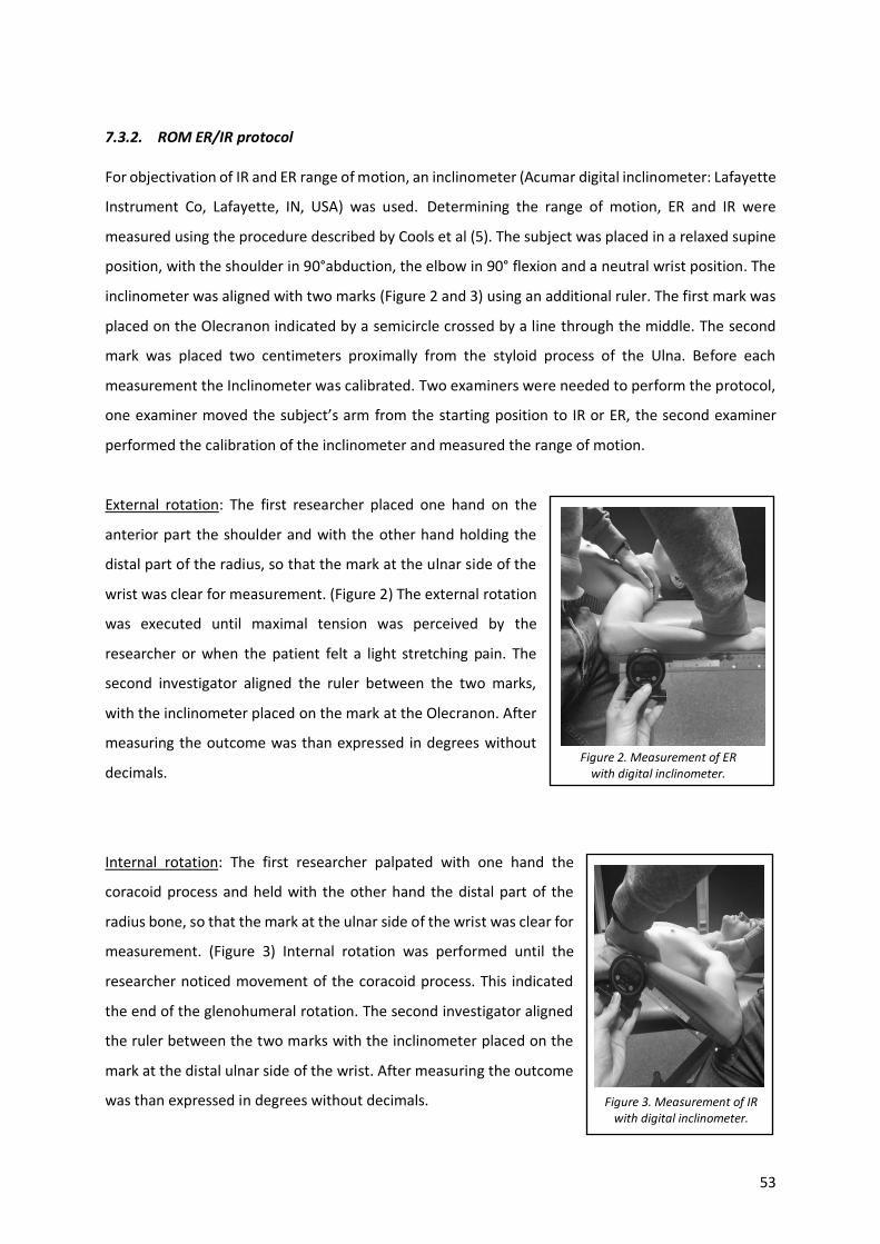
7.3.2. ROM ER/IR protocol For objectivation of IR and ER range of motion, an inclinometer (Acumar digital inclinometer: Lafayette
Instrument Co, Lafayette, IN, USA) was used. Determining the range of motion, ER and IR were
measured using the procedure described by Cools et al (5). The subject was placed in a relaxed supine
position, with the shoulder in 90°abduction, the elbow in 90° flexion and a neutral wrist position. The
inclinometer was aligned with two marks (Figure 2 and 3) using an additional ruler. The first mark was
placed on the Olecranon indicated by a semicircle crossed by a line through the middle. The second
mark was placed two centimeters proximally from the styloid process of the Ulna. Before each
measurement the Inclinometer was calibrated. Two examiners were needed to perform the protocol,
one examiner moved the subject’s arm from the starting position to IR or ER, the second examiner
performed the calibration of the inclinometer and measured the range of motion.
External rotation: The first researcher placed one hand on the
anterior part the shoulder and with the other hand holding the
distal part of the radius, so that the mark at the ulnar side of the
wrist was clear for measurement. (Figure 2) The external rotation
was executed until maximal tension was perceived by the
researcher or when the patient felt a light stretching pain. The
second investigator aligned the ruler between the two marks,
with the inclinometer placed on the mark at the Olecranon. After
measuring the outcome was than expressed in degrees without
decimals.
Internal rotation: The first researcher palpated with one hand the
coracoid process and held with the other hand the distal part of the
radius bone, so that the mark at the ulnar side of the wrist was clear for
measurement. (Figure 3) Internal rotation was performed until the
researcher noticed movement of the coracoid process. This indicated
the end of the glenohumeral rotation. The second investigator aligned
the ruler between the two marks with the inclinometer placed on the
mark at the distal ulnar side of the wrist. After measuring the outcome
was than expressed in degrees without decimals. Figure 3. Measurement of IR with digital inclinometer.
Figure 2. Measurement of ER with digital inclinometer.
54
7.3.3. Length of the pectoralis minor protocol For objectivation of the pectoralis minor muscle length, a caliper
(Digital Caliper, Mitutoyo BeNeLux,) was used. The assessment of
the length of the pectoralis minor muscle was based on the
protocol described by Borstad et al. (29), which showed to be
reliable. The subject was placed in a relaxed, neutral and supine
position with his upper body uncovered. Two marks were placed
on each side of the chest, directly distal of the coracoid process
and the distal part of the sternocostal articulation of the 4th rib. A
second investigator controlled the place of the marks, to make
sure it was linked with the right bony reference point.
Subsequently the distance between these two marking points was
measured, with a caliper (Figure 4). The results were expressed in millimeters and rounded to one
decimal. The whole protocol (placing the marks + measuring with the Caliper) was performed two
times on each side after which a mean value for each side was calculated.
7.3.4. Scapular dyskinesis protocol This parameter was examined during an arm elevation in the scapular plane, which was defined as 30°
in front of the coronal plane, while holding weights. Two poles were used to guide the participants
movement. The subject was standing straight in a neutral position with the palms of the hands facing
forward. The weight of the halters depended on the body mass of the person, people weighing under
68 kg had to lift 1.5 kg and people weighing over 68 kg, 2 kg. The subject performed 5 arm elevations
in a row, in order to have a clear interpretation of possible dyskinesis. Each time this parameter was
evaluated by the agreement of two examiners. Based on Kibler’s classification (3), a number from 1 to
4 was assigned, distinguishing 4 types of scapular dyskinesis: ‘1’ = Inferior prominence, ‘2’ = Medial
prominence, ‘3’ = Superior prominence ‘4’ = no scapular dyskinesis. McClure et al. showed that the
method used for assessing scapular dyskinesis proved satisfactory reliability for clinical use (6).
Figure 4. Measurement of length of the pectoralis minor muscle
with Digital Caliper.
55
7.3.5. Scapular inclination protocol For objectivation of scapular inclination, an inclinometer (Acumar digital
inclinometer: Lafayette Instrument Co, Lafayette, IN, USA) was used. The
Fourth parameter, scapular upward rotation, was measured following a
reliable method described by Watson et al. (32). The measurement was
performed in a neutral standing position with the arms relaxed. Two marks
were placed on the spine of the scapula, one near the posterior angle of the
acromion, the other directly lateral of the broad base of the scapular spine
(Figure 5.). After defining these marks, the inclinometer was placed on a
ruler connecting the two marks. A second investigator looked sideways at
the inclinometer to make sure it was positioned in the frontal plane. Data
was collected, in degrees without decimals. A ‘-’ (minus) was added if the scapula was rotated
downward and a ‘+’ (plus sign) for upward rotation.
Figure 5. Measurement of inclination of the scapula with