Norovirus Gastroenteritis in a Birth Cohort inSouthern IndiaVipin Kumar Menon1¤a, Santosh George1, Rajiv Sarkar1, Sidhartha Giri1,Prasanna Samuel2, Rosario Vivek1, Anuradha Saravanabavan1, Farzana Begum Liakath1,Sasirekha Ramani1¤b, Miren Iturriza-Gomara3¤c, James J. Gray3¤d, David W. Brown3, MaryK. Estes4, Gagandeep Kang1*
1 Department of Gastrointestinal Sciences, Christian Medical College, Vellore, India, 2 Department ofBiostatistics, Christian Medical College, Vellore, India, 3 Virus Reference Department, Centre for Infection,Health Protection Agency, London, United Kingdom, 4 Department of Molecular Virology and Microbiology,Baylor College of Medicine, Houston, Texas, United States of America
¤a Current address: MD Anderson Cancer Center, Houston, Texas, United States of America¤b Current address: Baylor College of Medicine, Houston, Texas, United States of America¤c Current address: Institute of Infection and Global Health, Liverpool, United Kingdom¤d Current address: Norwich Medical School, Norwich, United Kingdom* [email protected]
Abstract
Background
Noroviruses are an important cause of gastroenteritis but little is known about disease and
re-infection rates in community settings in Asia.
Methods
Disease, re-infection rates, strain prevalence and genetic susceptibility to noroviruses were
investigated in a birth cohort of 373 Indian children followed up for three years. Stool sam-
ples from 1856 diarrheal episodes and 147 vomiting only episodes were screened for noro-
virus by RT-PCR. Norovirus positivity was correlated with clinical data, secretor status and
ABO blood group.
Results
Of 1856 diarrheal episodes, 207 (11.2%) were associated with norovirus, of which 49(2.6%)
were norovirus GI, 150(8.1%) norovirus GII, and 8 (0.4%) were mixed infections with both
norovirus GI and GII. Of the 147 vomiting only episodes, 30 (20.4%) were positive for noro-
virus in stool, of which 7 (4.8%) were norovirus GI and 23 (15.6%) GII. At least a third of the
children developed norovirus associated diarrhea, with the first episode at a median age of
5 and 8 months for norovirus GI and GII, respectively. Norovirus GI.3 and GII.4 were the
predominant genotypes (40.3% and 53.0%) with strain diversity and change in the predomi-
nant sub-cluster over time observed among GII viruses. A second episode of norovirus gas-
troenteritis was documented in 44/174 (25.3%) ever-infected children. Children with the
G428A homozygous mutation for inactivation of the FUT2 enzyme (se428se428) were at a
significantly lower risk (48/190) of infection with norovirus (p = 0.01).
PLOS ONE | DOI:10.1371/journal.pone.0157007 June 10, 2016 1 / 18
a11111
OPEN ACCESS
Citation: Menon VK, George S, Sarkar R, Giri S,Samuel P, Vivek R, et al. (2016) NorovirusGastroenteritis in a Birth Cohort in Southern India.PLoS ONE 11(6): e0157007. doi:10.1371/journal.pone.0157007
Editor: Andres G. Lescano, Universidad PeruanaCayetano Heredia, PERU
Data Availability Statement: All relevant data arewithin the paper and its Supporting Information files.
Funding: This study was supported by the IndianCouncil of Medical Research (ICMR), award number:18/11/23/2006-ECD-I, grant recipient: GagandeepKang (http://icmr.nic.in/projects/projectsanc07-09.htm). The funders had no role in study design, datacollection and analysis, decision to publish, orpreparation of the manuscript.
Competing Interests: The authors have declaredthat no competing interests exist.
This is the first report of norovirus documenting disease, re-infection and genetic suscepti-
bility in an Asian birth cohort. The high incidence and apparent lack of genogroupII specific
immunity indicate the need for careful studies on further characterization of strains, asymp-
tomatic infection and shedding and immune response to further our understanding of noro-
virus infection and disease.
IntroductionNoroviruses are a leading cause of non-bacterial acute gastroenteritis (AGE) outbreaks in allage groups worldwide and are increasingly recognized as the second most common cause ofsporadic AGE in children after rotavirus [1–3]. Noroviruses are non-enveloped, single-stranded positive-sense RNA viruses belonging to the family Caliciviridae. The viral genomeconsists of three open reading frames (ORFs), with ORF 1 encoding six non-structural proteinsincluding the RNA dependent RNA polymerase (RdRp). ORF 2 encodes for the major capsidprotein (VP1) and ORF 3 encodes a minor capsid protein (VP2) [4].
Noroviruses are divided into 5 genogroups (GI-GV) of which GI, GII and GIV are knownto cause gastroenteritis in humans [5, 6]. Each genogroup is further divided into genetic clus-ters: 14 clusters in GI, 17 clusters in GII and 1 cluster in GIV have been described [7]. Amongnorovirus genogroups known to infect humans, GII noroviruses are the major genogroup caus-ing outbreaks and sporadic infections worldwide and also predominate in community studies[8, 9].
Recent reports on norovirus infections from India were hospital-based studies of acute gas-troenteritis [10–12], similar to the majority of reports in children from other parts of theworld. There are limited data on norovirus infections in the community mainly because of thedifficulty in conducting such studies, whether cross-sectional or longitudinal. Very recently, aPeruvian birth cohort study found that 71% of 189 children followed upto 2 years of age, expe-rienced norovirus associated diarrhea, that genogroup II infections were common and thatrepeat infections by the same genogroup were common, but repeat infections by the samegenotype were rare[13].We have previously reported a limited comparison of pediatric norovi-rus disease and strain distribution between children with diarrhea in the hospital and in thiscohort and asymptomatic children from this cohort that demonstrated the importance of noro-viruses as gastrointestinal pathogens in Indian children, the predominance of GII norovirusesand a low prevalence of GI [9].
Some studies have examined the course of norovirus infection, susceptibility and disease inhealthy adults through volunteer and outbreak studies, but few are population-based estimatesof norovirus disease burden in a community [8, 13]. The present study provides disease inci-dence and re-infection rates of norovirus genogroupsI and II, with evaluation of putative hostmarkers of susceptibility in an Asian population followed from birth to the age of 3 years.
Materials and Methods
Study DesignAll 1856 diarrheal stool samples and 147 available stool samples from the 155 vomiting onlyepisodes (collected within 0–15 days of a vomiting episode) from a birth cohort of 373 children,followed up to 3 years were screened for the presence of GI and GII norovirus. The enrollment
Norovirus Gastroenteritis in Indian Children
PLOS ONE | DOI:10.1371/journal.pone.0157007 June 10, 2016 2 / 18
criteria and methods for follow up of the cohort, which was established to study rotavirus infec-tions, included biweekly home visits, fortnightly surveillance stool sampling, monthly anthro-pometry (weight and length/height) measurements, in addition to collection of samples andclinical information for all diarrheal episodes [14, 15]. At least one fecal sample was collectedin 98% of diarrheal episodes within one week of onset. Venous blood or saliva was collectedfrom the children at the end of the study for genetic analysis. Household hygiene was assessedon a 24-point scale, using a structured questionnaire covering aspects of water, food and per-sonal hygiene; this questionnaire has previously been validated and used in the same commu-nity[16]. Written informed consent was obtained from parents of all children prior to theenrollment, and the study was approved by the Institutional Review Board of the ChristianMedical College, Vellore.
Definition, assessment of severity and etiology of diarrheaDiarrhea was defined as the passage of�3 loose watery stools during a 24‐hr period or inbreastfed children alone, a change in the number or consistency of stools as reported by themother. An episode was defined as at least one day of diarrhea, preceded and followed by�2days without diarrhea. The severity of diarrhea was assessed using the Vesikari scoring system[17] that was developed for rotavirus but has been used to score severity of norovirus diarrheain recent studies [8, 11, 18]. An episode was considered mild for a score�5, moderately severefor a score of 6–10 and severe for scores>10. Diarrheal stool samples were screened for para-site ova and cysts by microscopy, for bacterial pathogens by culture, and for rotavirus byenzyme-linked immunosorbent assay (Rota IDEIA; Dako, Ely, United Kingdom).
RNA extraction and cDNA synthesisViral RNA was extracted from 20% fecal suspension in Minimal Essential Medium using gua-nidium isothiocyanate and silica [19].The RNA was eluted into 50μl of diethylpyrocarbonatetreated water containing 40 units of RNase inhibitor (Invitrogen, Life Technologies, Paisley,UK). Complementary DNA (cDNA) was generated by reverse transcription in the presence ofrandom primers (hexamers) [Pharmacia Biotech, United Kingdom] using Moloney murineleukemia virus reverse transcriptase (Invitrogen, Life Technologies, Paisley, UK).
PCR for Norovirus GIScreening of all stool samples from diarrheal episodes and vomiting only episodes for NoV GIwas carried out using primers and conditions previously published [20]. The primers amplifyan 84 bp fragment of the ORF 1–2 regions. Samples that were positive were cloned into theTOPO TA cloning kit as per the manufacturer’s instructions (Invitrogen, Life Technologies,UK). Clones were purified using QIAprep Spin Miniprep Kit (Qiagen, Valencia, USA) and sub-jected to automated sequence analysis.
PCR for Norovirus GIITwo separate PCR reactions were carried out for all the samples using previously publishedprimers specific to the RNA dependent RNA polymerase (RdRp) and ORF 1–2 regions as perpreviously published cycling conditions, amplifying a 113bp and 468bp fragment, respectively[20, 21]. Samples with amplification using either primer set were considered positive for noro-virus and sequencing was attempted for all amplicons.
Norovirus Gastroenteritis in Indian Children
PLOS ONE | DOI:10.1371/journal.pone.0157007 June 10, 2016 3 / 18
Sequence AnalysisSequencing of the positive amplicons was carried out by using the ABI PRISM Big Dye Termi-nator cycle sequencing ready reaction kit (Applied Biosystems, CA, USA). The amplicons cor-responding to the ORF 1–2 junction region were sequenced when available; else the RdRpregion was sequenced. Sequences were resolved using an automated DNA sequencer ABIPRISM 310 Genetic Analyzer. The sequences were imported into BioEdit software (version 7).Phylogenetic analysis was carried out using reference sequences from the norovirus molecularepidemiology database (Noronet) at www.rivm.nl and sequences from GenBank. Variants ofNoV GII.4 were detected using the norovirus genotyping tool from http://www.rivm.nl/mpf/norovirus/typingtool. Neighbor joining method (10000 pseudo replicates) with MEGA soft-ware (version 4) was used to assign the samples to a genotype based on>90% homology at thenucleotide level with other strains within a given genotype [22].
Fucosyltransferase 2 (FUT2) genotyping for Secretor StatusGenomic DNA was isolated from buccal epithelial cells or blood using the QIAamp DNAMiniKit (Qiagen, Hilden, Germany) according to manufacturer’s instructions. The amplification ofa region of the FUT2 gene and genotyping for two single nucleotide polymorphisms (SNPs)was carried out using published methods [23, 24]. The TaqMan Genotyping Master Mix(Applied Biosystems) and Custom TaqMan SNP Genotyping Assay for the 428 and 385 SNPof the FUT2 gene designed by J. Le Pendu, France and M. Lay, USA (Applied Biosystems)respectively were used in a final volume of 20 μl.
ABO and secretor phenotyping from salivaSaliva samples that were boiled and cooled were used to coat 96-well high-binding plates(Costar, Corning, N.Y.) at room temperature for 4hrs and then blocked with 10% non-fat milk(blotto) overnight at 4°C. After washing, primary antibodies Anti-Lea Gamma-clone (1:100),Anti-A Gamma-clone (1:200) and Anti-B Gamma-clone (1:100) from ImmucorGamma, Geor-gia, USA, BG-6 (anti-Leb) antibody (1:100) from Covance, Texas, USA were added and platesincubated at 37°C for 1hr. This was followed by the addition of a Goat anti-Mouse IgG-HRP(Sigma, USA) and UEA-1 lectin-HRP (Sigma, USA) at 37°C for 1 hr. The detection was carriedout with TMB peroxidase substrate (KPL, Gaithersburg, MD) and the plates were read atOD450[25]. A pooled saliva sample and PBS were used as positive and negative controls,respectively.
Statistical AnalysisThe data were analyzed using STATA 10.0 for Windows (STATA Corp., TX, USA). Descriptiveanalysis was performed for all explanatory variables. Statistical significance of the observed dif-ferences in outcome between explanatory variables was assessed using Chi-square test or Fish-er’s exact test for categorical variables and two-tailed t-test or Mann-Whitney U test forcontinuous variables, depending on the distribution of the data. A p-value of<0.05 was consid-ered statistically significant. Seasonality of norovirus-associated diarrhea was assessed by fittinga sine curve to a time-series of the monthly stool positivity rates (proportion of diarrheal epi-sodes attributed to norovirus divided by the total number of diarrheal episodes). The intensityof the seasonal fluctuation was computed by calculating the peak-to-low ratio using a modifiedversion of the Edward’s technique [26].Association between distribution of Histo-Blood GroupAntigen and secretor status among the norovirus infected and non-infected individuals were
Norovirus Gastroenteritis in Indian Children
PLOS ONE | DOI:10.1371/journal.pone.0157007 June 10, 2016 4 / 18
ascertained through logistic regression analysis and odds ratios (ORs) with 95% confidenceintervals (CI) calculated.
Height-for age (HAZ), weight-for-height (WHZ) and weight-for-age (WAZ) z-scores werecalculated using the 2006 WHO child growth standards as the reference population [27], andchildren classified as stunted (HAZ<-2 SD), wasted (WHZ<-2 SD), underweight (WAZ<-2SD) or normal based on their respective z-scores.
ResultsA total of 1,856 episodes of diarrhea were experienced by the 373 children in the cohort, with apreviously published incidence of gastrointestinal illness of 3.6 per child year in infancy and 1.64and 1.16 per child year, respectively during the subsequent years [14]. The median (IQR) age ofthe first diarrheal episode was 3 (1–6) months. A total of 149 (149/373, 40%) children had 207episodes of diarrhea associated with norovirus detection (details available in S1 Dataset). Norovi-rus GI and GII were detected in 49episodes (49/1856, 2.6%) and 150 episodes (150/1856, 8.1%)of diarrhea respectively; eight (8/1856, 0.4%) episodes were mixed infections with both norovirusGI and GII. Of the available 147 stool samples from the 155 vomiting only episodes which werecollected within 0–15 days of vomiting, norovirus GI and GII were associated with 7 episodes (7/147, 4.8%) and 23 episodes (23/147, 15.6%) respectively, and there were no mixed infections.
Table 1 compares the clinical characteristics of diarrheal episodes associated with norovirusbetween GI and GII. The median (IQR) age at first symptomatic norovirus infection was 5 (3–7) months and 8 (5–15) months among norovirus GI and GII respectively. The median (IQR)duration of a diarrheal episode and proportion associated with vomiting and fever were similarfor the two viral genogroups (Table 1).
Re-infectionOne hundred and thirty of the 149 (87.2%) children who ever had a norovirus associated diar-rhea had only one episode during the 3-year follow-up period. Forty-four of 149 (29.5%) chil-dren had a re-infection with either genogroup. Children showed a higher rate of re-infectionwith norovirus GII (30/123, 24.3%) compared to norovirus GI (6/51, 11.8%). Eight of the 174children (4.6%) had both norovirus GI and GII infections. Among the 30 children with 2 ormore norovirus GII positive diarrheal episodes, three children experienced the second episode6, 8 and 9 days after the first episode, which may represent continued shedding.
Only one child (1.9%) with norovirus GI re-infection was re-infected with the same geno-type and the rest had a different genotype during re-infection (Fig 1). The duration betweenprimary and re-infection with the same genotype was 270 days. The shortest and longest dura-tion between re-infection with a different genotype was 26 and 811 days.
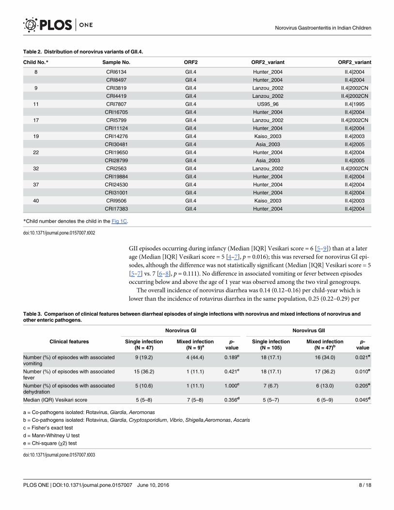
Of the 30 children with norovirus GII re-infection, genotype could not be identified for 7samples. Of the remaining, 12 children were re-infected with the same genotype, three ofwhom may have had continuous shedding as stated above, and 11 with a different genotype(Fig 1). Reinfections with GII.4 in six of nine re-infected children were with different variants(Table 2). The shortest and longest duration between re-infection with the same genotype,other than the three who were less than 10 days, were 74 and 534 days. Re-infection with differ-ent genotypes ranged from 16 days to 767 days.
Presence of co-pathogensMixed infections with other pathogens were seen in 56/207 (27%) norovirus-associated diar-rheal episodes, of which 52 episodes had a single co-pathogen and 4 had two co-pathogens.The commonest co-pathogen isolated was rotavirus, which was present in 36/56(64.2%)
Norovirus Gastroenteritis in Indian Children
PLOS ONE | DOI:10.1371/journal.pone.0157007 June 10, 2016 5 / 18
episodes of mixed infections, followed by Giardia in 9/56 (16.1%) episodes; other co-pathogenswere: Cryptosporidium (5/56, 8.9%), Aeromonas (4/56, 7.1%), Vibrio (3/56, 5.4%), Shigella (2/56, 3.6%) and Ascaris (1/56, 1.7%). The comparison of clinical features between diarrheal epi-sodes of single and mixed infections is presented in Table 3. Episodes of mixed infection hadhigher association with comorbidities such as vomiting and fever, and were more severe.
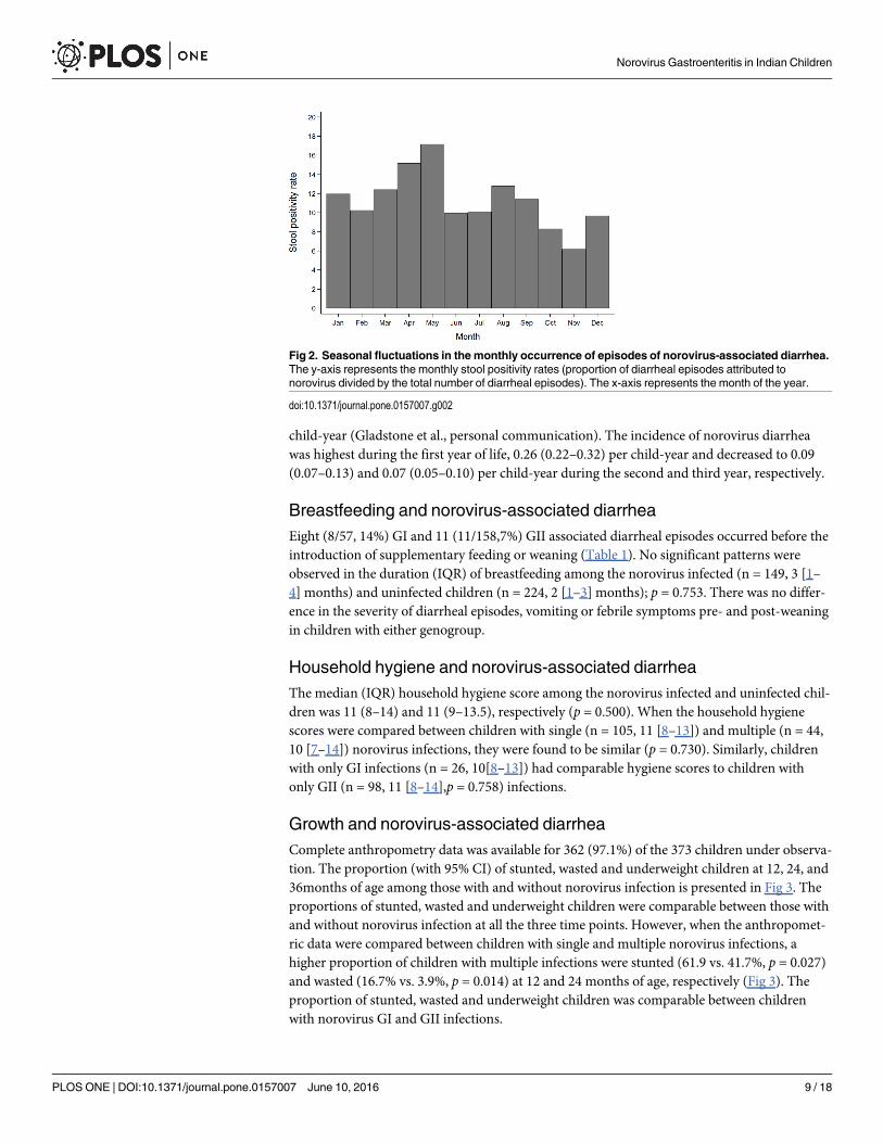
Seasonality of norovirus-associated diarrheaNorovirus-associated diarrhea was seen to occur during all months of the year, ranging from astool positivity rate of 6.2% in November to 17.2% inMay (Fig 2). The peak-to-low (95% CI) ratioof seasonal intensity of occurrence of norovirus diarrhea was 1.57 (1.05–2.20), suggestive of a mildincrease in the proportion of norovirus-associated diarrheal episodes during the summer season.
Age and norovirus-associated diarrheaA total of 50 (87.7%) and 98 (62%) episodes of diarrhea in children�1 year of age were associ-ated with norovirus GI and GII respectively. The median age of norovirus diarrhea was 7 (4.5–
Table 1. Comparison of clinical characteristics between norovirus GI and GII diarrheal episodes.
Variable NoV GI NoV GII p-value
Overall Number of episodesa 49 150 —
Median (IQR) duration (in days) 3 (2–4) 3 (2–5) 0.542b
Median (IQR) Vesikari score 5 (5–8) 5 (5–7) 0.749b
Number (%) of episodes with associated vomiting 12 (24.5) 33 (22.8) 0.804c
Number (%) of episodes with associated fever 14 (28.6) 33 (22.8) 0.443c
Before introduction of supplementary feeding Number of episodesa 8 11 —
Median (IQR) duration(in days) 4 (3–5.5) 3 (2–5) 0.399b
Median (IQR) Vesikari score 5 (4.5–6) 5 (5–7) 0.465b
Number (%) of episodes with associated vomiting 0 (0%) 3 (27.3) 0.228d
Number (%) of episodes with associated fever 1 (12.5) 2 (18.2) 1.000d
After introduction of supplementary feeding Number of episodesa 41 134 —
Median (IQR) duration(in days) 3 (2–4) 3 (2–5) 0.848b
Median (IQR) Vesikari score 6 (5–8) 5 (5–7) 0.475b
Number (%) of episodes with associated vomiting 12 (29.3) 30 (22.4) 0.367c
Number (%) of episodes with associated fever 13 (31.7) 31 (23.1) 0.268c
During infancy Number of episodesa 42 86 —
Median (IQR) duration(in days) 3 (2–4) 3 (2–5) 0.630b
Median (IQR) Vesikari score 5 (5–8) 6 (5–9) 0.291b
Number (%) of episodes with associated vomiting 9 (21.4) 23 (26.7) 0.514c
Number (%) of episodes with associated fever 11 (26.2) 22 (25.6) 0.941c
Post infancy (2nd and 3rd year of life) Number of episodes 7 59 —
Median (IQR) duration (in days) 3 (2–5) 2 (2–3) 0.229b
Median (IQR) Vesikari score 7 (6–8) 5 (4–7) 0.023b
Number (%) of episodes with associated vomiting 3 (42.9) 10 (16.9) 0.131d
Number (%) of episodes with associated fever 3 (42.9) 11 (18.6) 0.159d
a = Includes both primary and secondary infections
b = Mann-Whitney U test
c = Chi-square (χ2) test
d = Fisher’s exact test
doi:10.1371/journal.pone.0157007.t001
Norovirus Gastroenteritis in Indian Children
PLOS ONE | DOI:10.1371/journal.pone.0157007 June 10, 2016 6 / 18
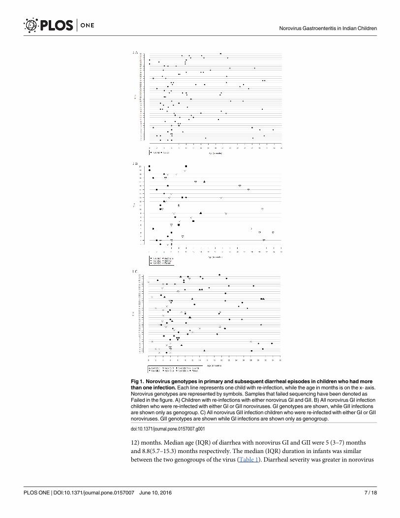
12) months. Median age (IQR) of diarrhea with norovirus GI and GII were 5 (3–7) monthsand 8.8(5.7–15.3) months respectively. The median (IQR) duration in infants was similarbetween the two genogroups of the virus (Table 1). Diarrheal severity was greater in norovirus
Fig 1. Norovirus genotypes in primary and subsequent diarrheal episodes in children who hadmorethan one infection. Each line represents one child with re-infection, while the age in months is on the x- axis.Norovirus genotypes are represented by symbols. Samples that failed sequencing have been denoted asFailed in the figure. A) Children with re-infections with either norovirus GI and GII. B) All norovirus GI infectionchildren who were re-infected with either GI or GII noroviruses. GI genotypes are shown, while GII infectionsare shown only as genogroup. C) All norovirus GII infection children who were re-infected with either GI or GIInoroviruses. GII genotypes are shown while GI infections are shown only as genogroup.
doi:10.1371/journal.pone.0157007.g001
Norovirus Gastroenteritis in Indian Children
PLOS ONE | DOI:10.1371/journal.pone.0157007 June 10, 2016 7 / 18
GII episodes occurring during infancy (Median [IQR] Vesikari score = 6 [5–9]) than at a laterage (Median [IQR] Vesikari score = 5 [4–7], p = 0.016); this was reversed for norovirus GI epi-sodes, although the difference was not statistically significant (Median [IQR] Vesikari score = 5[5–7] vs. 7 [6–8], p = 0.111). No difference in associated vomiting or fever between episodesoccurring below and above the age of 1 year was observed among the two viral genogroups.
The overall incidence of norovirus diarrhea was 0.14 (0.12–0.16) per child-year which islower than the incidence of rotavirus diarrhea in the same population, 0.25 (0.22–0.29) per
Table 2. Distribution of norovirus variants of GII.4.
Table 3. Comparison of clinical features between diarrheal episodes of single infections with norovirus andmixed infections of norovirus andother enteric pathogens.
PLOS ONE | DOI:10.1371/journal.pone.0157007 June 10, 2016 8 / 18
child-year (Gladstone et al., personal communication). The incidence of norovirus diarrheawas highest during the first year of life, 0.26 (0.22–0.32) per child-year and decreased to 0.09(0.07–0.13) and 0.07 (0.05–0.10) per child-year during the second and third year, respectively.
Breastfeeding and norovirus-associated diarrheaEight (8/57, 14%) GI and 11 (11/158,7%) GII associated diarrheal episodes occurred before theintroduction of supplementary feeding or weaning (Table 1). No significant patterns wereobserved in the duration (IQR) of breastfeeding among the norovirus infected (n = 149, 3 [1–4] months) and uninfected children (n = 224, 2 [1–3] months); p = 0.753. There was no differ-ence in the severity of diarrheal episodes, vomiting or febrile symptoms pre- and post-weaningin children with either genogroup.
Household hygiene and norovirus-associated diarrheaThe median (IQR) household hygiene score among the norovirus infected and uninfected chil-dren was 11 (8–14) and 11 (9–13.5), respectively (p = 0.500). When the household hygienescores were compared between children with single (n = 105, 11 [8–13]) and multiple (n = 44,10 [7–14]) norovirus infections, they were found to be similar (p = 0.730). Similarly, childrenwith only GI infections (n = 26, 10[8–13]) had comparable hygiene scores to children withonly GII (n = 98, 11 [8–14],p = 0.758) infections.
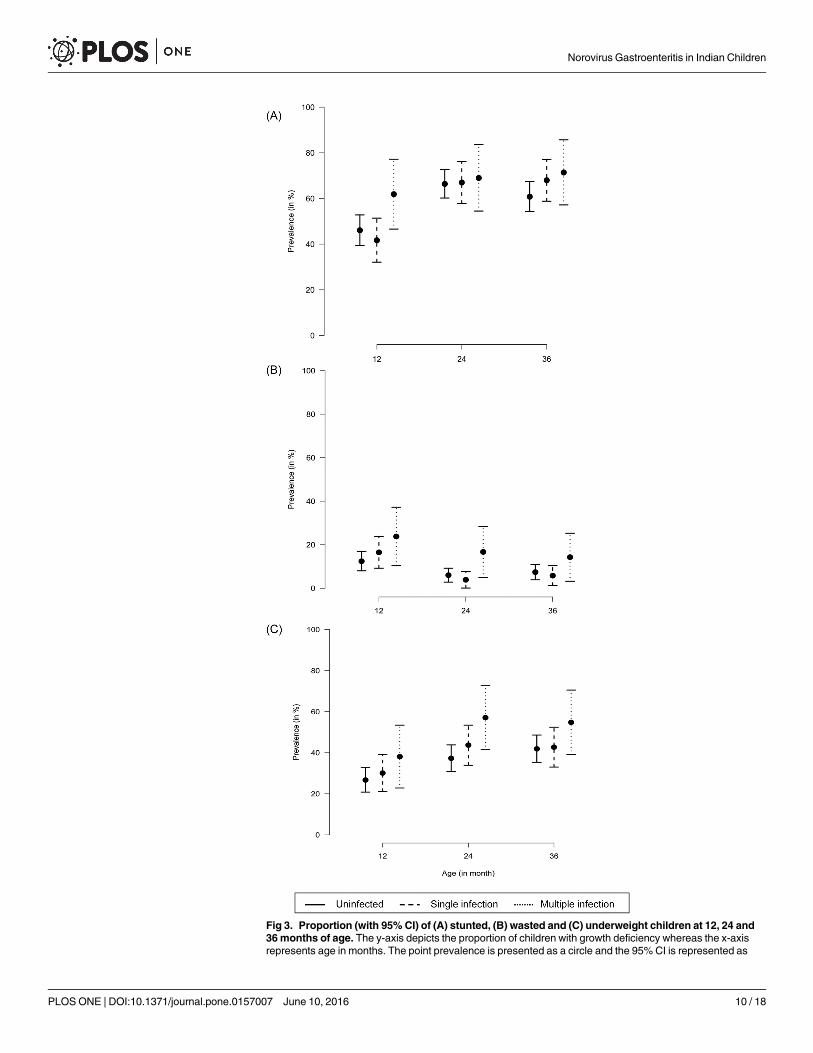
Growth and norovirus-associated diarrheaComplete anthropometry data was available for 362 (97.1%) of the 373 children under observa-tion. The proportion (with 95% CI) of stunted, wasted and underweight children at 12, 24, and36months of age among those with and without norovirus infection is presented in Fig 3. Theproportions of stunted, wasted and underweight children were comparable between those withand without norovirus infection at all the three time points. However, when the anthropomet-ric data were compared between children with single and multiple norovirus infections, ahigher proportion of children with multiple infections were stunted (61.9 vs. 41.7%, p = 0.027)and wasted (16.7% vs. 3.9%, p = 0.014) at 12 and 24 months of age, respectively (Fig 3). Theproportion of stunted, wasted and underweight children was comparable between childrenwith norovirus GI and GII infections.
Fig 2. Seasonal fluctuations in the monthly occurrence of episodes of norovirus-associated diarrhea.The y-axis represents the monthly stool positivity rates (proportion of diarrheal episodes attributed tonorovirus divided by the total number of diarrheal episodes). The x-axis represents the month of the year.
doi:10.1371/journal.pone.0157007.g002
Norovirus Gastroenteritis in Indian Children
PLOS ONE | DOI:10.1371/journal.pone.0157007 June 10, 2016 9 / 18
Fig 3. Proportion (with 95% CI) of (A) stunted, (B) wasted and (C) underweight children at 12, 24 and36months of age. The y-axis depicts the proportion of children with growth deficiency whereas the x-axisrepresents age in months. The point prevalence is presented as a circle and the 95% CI is represented as
Norovirus Gastroenteritis in Indian Children
PLOS ONE | DOI:10.1371/journal.pone.0157007 June 10, 2016 10 / 18
GenotypingA total of 215 samples were genotyped by sequencing of the amplicons. Three GI and 26 GIIsamples failed in sequencing reactions, but are included in the overall analysis because all PCRswere repeated at least twice, and band sizes re-confirmed with absence of non-specific bands.
Sequence analysis of the GI genogroups showed a predominance of GI.3 genotype in 23/57(40.3%) of the samples, followed by GI.1 (15/57, 26.3%), GI.2 (9/57, 15.7%) and GI.6 (6/57,10.5%). Phylogenetic analysis of norovirus GII genogroup of either the capsid or the RdRpregion (Fig 4) showed the predominance of GII.4 genotype in 70/132 (53.0%) of the samples,followed by GII.2 (28/132, 21.2%) and GII.3 (18/132,13.6%), respectively.
The GII.4 strains were placed in 3 sub-clusters when analyzed with reference sequences. Ofthe 35 samples identified as GII.4 at the ORF 1–2 capsid junctions, 17 strains with 96% nucleo-tide identity clustered in the Hunter sub-cluster, 6 with 93% nucleotide identity with the Far-mington sub-cluster and 12 with 98% nucleotide identity with the Sakai sub-cluster. Fifteenchildren between the ages of 2.5–8.3 months were infected with the Hunter sub-cluster, with 2children having two episodes of infection 17 and 159 days apart, respectively. Six childrenbetween the ages of 4.2–8.6 months were infected with the Farmington sub-cluster. The Hunterand Farmington sub-cluster infections were seen in children between the years 2002–2003, indi-cating the circulation of this sub-cluster in the community. However, there was a predominanceof the Sakai sub-cluster in 12 children who were infected between 2004–2005. Nine samples weregenotyped as GII.3 and clustered with 94% nucleotide identity to the Toronto strain. Ten sampleswere genotyped as GII.2 with 98% nucleotide identity to the SnowMountain strain.
Nucleotide sequences of positive samples from the study have been deposited in the Gen-Bank and the accession numbers are JN654720-JN654766.
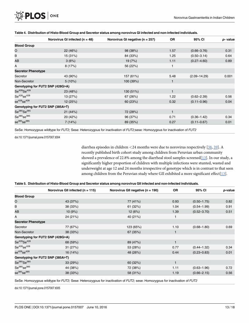
Association of blood group and secretor status with norovirus infectionABO blood group and secretor status were determined for 293 children from whom appropri-ate samples were available. The most common ABO blood group was O (39.3%), followed by B(32.5%), A (20.9%) and AB (7.2%). One hundred eighty-six children (186/305, 60.9%) weresecretor positive.
Secretor status was significantly associated with overall norovirus GI (Table 4), but not withnorovirus GII infection (Table 5). No significant association between the ABO blood group andnorovirus GI or GII infection was observed (Tables 4 and 5). Among the 48 children with GIinfection, those with GI.1 genotype (n = 15) were all secretor positive, whereas among the childrenwith non GI.1 infection (n = 33), 84.9% were secretor positives although the difference was notstatistically significant (p = 0.167). For the 115 children with GII infection, 75% of those withGII.4 (n = 56) were secretor positives, whereas only 57.8% of children with non GII.4 infection(n = 57) were secretor positives (p = 0.054). Moreover, genotyping for the FUT2 SNP showed thatchildren with the G428A homozygous mutation for inactivation of the FUT2 enzyme (se428se428)are significantly at a lower risk of infection with norovirus (Tables 4 and 5). There was no signifi-cant association between norovirus diarrhea with any genotype with any specific blood group.
DiscussionThis is the first longitudinal study of norovirus gastroenteritis in a birth cohort of children inIndia. Most estimates of disease burden are based on hospital studies, which sample the more
vertical lines. The solid vertical line represents uninfected children, the dashed and dotted lines representthose with single and multiple norovirus infections, respectively.
doi:10.1371/journal.pone.0157007.g003
Norovirus Gastroenteritis in Indian Children
PLOS ONE | DOI:10.1371/journal.pone.0157007 June 10, 2016 11 / 18
severe end of the spectrum of disease, and do not have community based denominator data. Inthis birth cohort from an urban slum in southern India, norovirus was a significant cause ofdiarrhea, associated with 11.2% of episodes, second only to rotavirus, and affecting almost halfthe cohort. In a recent study from India, 10.7% positivity was found in children<7 years ofage, with 40% positivity in the�1 year olds [10], but this was a cross-sectional study in chil-dren presenting to hospital with gastroenteritis. Community based studies from developedcountries like Europe have reported higher norovirus rates, 24.5% in children<5, and 28% inthose�1 year old in the UK [28], and in the Netherlands, 14% in children<4 years and 14.3%in children�1 year of age [29], but these findings are in settings where higher rates of viral gas-troenteritis are reported in children presenting to hospitals than in developing countries.
A majority of the norovirus reports from developing countries are hospital based and thedetection ranges from 5.5% in Vietnam, 15% in Nicaragua, 9.3% in Tunisia, 30% in Iraq, 3.7%in Brazil and 32.1% in urban Peru, 11.3% in Malawi and 14% in Guatemala [30–37]. Twocohort studies carried from Chile and Peruvian Amazon have shown 18% and 25% of acute
Fig 4. Dendrogram constructed using neighbor joining of nucleotide sequences corresponding tofragment of 468 bp of the ORF 1–2 region of the Norovirus capsid encoding gene. Bootstrap values for10000 pseudo replicates are shown. The reference strains included for comparison with accession numbersare: Hunter284/04/AU (DQ078794), Farmington Hills/02/US (AY502023), Sakai/04-179/05/JP (AB220922),Bristol/1993/UK (X76716), Maizuru/010426/2001/JP (EF547404), M7/1999/US (AY130761), Toronto/1991/CA (AAA18930), Seacroft/1990/UK (AJ277620), Hu/SaitamaU16/197/JP (AB039778), Erfurt546/2000/DE(AF427118), Mc37/2001/TH (AY237415), Wortley/1990/UK (AJ277618), Chitta/1996/JP (AB032758), SnowMountain/1976/US (AY134748), Melksham/95/UK (X81879), KY-89/1989/JP (L23828), Norwalk/1968/US(M87661).
doi:10.1371/journal.pone.0157007.g004
Norovirus Gastroenteritis in Indian Children
PLOS ONE | DOI:10.1371/journal.pone.0157007 June 10, 2016 12 / 18
diarrhea episodes in children<24 months were due to norovirus respectively [38, 39]. Arecently published birth cohort study among children from Peruvian urban communityshowed a prevalence of 22.8% among the diarrheal stool samples screened[13]. In our study, asignificantly higher proportion of children with multiple infections were stunted, wasted andunderweight at age 12 and 24 months irrespective of genotype which is in contrast to that seenamong children from the Peruvian study where GII exhibited a more significant effect[13].
Table 4. Distribution of Histo-Blood Group and Secretor status among norovirus GI infected and non-infected individuals.
Norovirus GI infected (n = 48) Norovirus GI negative (n = 257) OR 95% CI p- value
SeSe: Homozygous wildtype for FUT2; Sese: Heterozygous for inactivation of FUT2; sese: Homozygous for inactivation of FUT2
doi:10.1371/journal.pone.0157007.t005
Norovirus Gastroenteritis in Indian Children
PLOS ONE | DOI:10.1371/journal.pone.0157007 June 10, 2016 13 / 18
Children who had vomiting only episodes had high positivity for norovirus GI or GII (30/147, 20.4%) in stool compared to the proportion of diarrheal stools positive for norovirus (215/1856, 11.6%). Earlier studies on children have reported a higher percentage of vomiting epi-sodes than diarrheal episodes in norovirus associated outbreaks of gastroenteritis, described as'winter vomiting disease'[40].A study on norovirus gastroenteritis in military trainees from theUSA also reported a higher norovirus positivity in vomiting episodes (80%) compared to diar-rheal episodes (67%)[41].
The median Vesikari score for norovirus GI and GII diarrhea was 5, similar to studies fromother developing countries [8, 42], and was less severe than that associated with rotavirus dis-ease in this cohort, which had a median severity score of 7.6 [43]. The median age of the firstsymptomatic infection of norovirus GI and GII was 5 and 8 months respectively, slightly youn-ger than 9 months and 11 months as reported from studies in Chile and Brazil, respectively [8,44].In a study examining the global age distribution of pediatric norovirus cases, approximately70% of norovirus cases occurred in the age group between 6 months to 2 years [45]. The studyrecommended completion of norovirus vaccination in children by 6 months of age to poten-tially prevent a greater burden of disease compared to completion of vaccination at 12 months.The recommendation would hold true for our study population where the median age (IQR) ofnorovirus diarrhea was 7 (4.5–12) months. Early completion of immunization is likely to havea greater benefit in reducing disease burden, particularly for GII infections where the primaryinfection was more severe.
The severity and frequency of vomiting and fever associated with norovirus was higheramong the GII infected children (Table 1); this finding is similar to studies from Peru [13, 38].In human volunteer challenge studies and vaccine trials, the definition of norovirus illnessincludes individuals who experience only vomiting in the absence of diarrhea as a clinical end-point [46–48]. In this study, the examination of stool samples from children who presentedwith vomiting alone as a primary clinical symptom suggests that there may be an underestima-tion of norovirus disease burden by exclusion of cases presenting with vomiting only.
Symptomatic norovirus associated diarrhea was detected in children during the first 6months of age suggesting that breast feeding did not confer protection, unlike the absence ofsymptomatic infection among younger children in the Peruvian study [13]. Re-infection withnorovirus was associated with diarrhea in a quarter of ever-infected children, this is in contrastwith reports from the Chilean birth cohort where 40% (10/25) of re-infections with noroviruswere symptomatic, the lower number of re-infections seen could be due to the difference insize of the study population [8]. In the birth cohort from Peru, repeat infections by the samegenogroup of norovirus were seen, but the majority of the reinfections were by a different geno-type or variant of GII.4. The GII.4 variants described in Peru were 2006b, 2007, 2008 and 2010[13]. The data in this study showed higher rates of reinfections with the same genogroup andgenotype of norovirus than in Peru. Since these studies were carried out in distinct time periods(Vellore, 2002–2006 and Peru, 2007–2011), some of these differences may be due to the differ-ence in circulating GII.4 variants between the two studies (Table 2).Re-infection with the samegenogroup and genotype was observed in this study suggesting that lower genogroup or geno-type specific immunity may occur in India, in contrast to the Peruvian study where genotype-specific immunity was observed[13].
A high degree of genetic diversity was seen among circulating noroviruses, with four GIgenotypes and nine genotypes of norovirus GII detected. These data are similar to other studieswhere a higher diversity of norovirus has been documented in gastroenteritis in the communitywhen compared to outbreaks, generally in the adult and/or elderly population [4, 28, 49–53].GI.3 (40.3%) was the predominant strain detected in the cohort followed by GI.1 (26.3%), GI.2(15.7%) and GI.6 (10.5%). GII.4 was the predominant genotype while GII.2 (17.7%) and GII.3
Norovirus Gastroenteritis in Indian Children
PLOS ONE | DOI:10.1371/journal.pone.0157007 June 10, 2016 14 / 18
(11.4%) were also detected in significant proportions, similar to previous reports [28, 49].Other studies in India have reported a predominance of GII.4 strains; however, these were pre-dominantly outbreaks studies [9, 10]. The GII.4 strains detected in the current study clusteredinto3sub-clusters, with a temporal distribution of the Hunter sub-cluster being replaced by Far-mington and then by Sakai. Evolutionary analysis studies have shown that the newer strainsoriginate by amino acid changes due to positive selection and recombination which drives thepersistence of newer strains in the human population [54].
The link between the Histo-Blood Group Antigen (HBGA) binding and secretor status iswell documented for the prototype GI virus, Norwalk virus. In this study, there was no signifi-cant association between blood groups and norovirus GI infection, but higher infection rateswere seen among secretors compared to non-secretors, which is similar to previous studies ofvolunteer challenge and in vitro binding studies that have shown that carbohydrate binding isessential for GI infection [23, 55–57]. There was no significant association between bloodgroups or secretor status with norovirus GII infection which is consistent with other reportsthat have shown no association between host genetic factors and symptomatic infection of nor-ovirus GII[58–60]. Expression of the FUT2 gene and susceptibility to norovirus infection wasexamined by checking for two common mutations in Asian populations, which showed thatchildren with a nonsense mutation at G428A were significantly at a lower risk of infection withnorovirus, as has been previously shown by other studies [61, 62].
In summary, this report shows that norovirus infections are a common cause of pediatricgastroenteritis in southern India. A previous report from the same region found 15% of hospi-talized gastroenteritis cases in children<5 years of age to be positive for norovirus [9]. In thecommunity, norovirus infections were associated with mild disease, but symptomatic re-infec-tions were common. The continued replacement of strains in the population suggests the needfor strain specific vaccines, or identification of epitopes that are conserved across the norovirusvariants could be used to generate broadly protective vaccines. In order to assess the true rateof infection with noroviruses in this community and the role of prior infections in protectionfrom subsequent infection or disease, the investigation of asymptomatic shedding and immuneresponses would be of interest.
Supporting InformationS1 Dataset. Details of children with norovirus diarrhea.(XLSX)
Author ContributionsConceived and designed the experiments: GKMI-G MKE DWB JJG. Performed the experi-ments: VKM S. George S. Giri SR RV AS FBL. Analyzed the data: VKM RS PS SR MI-G MKEGK. Contributed reagents/materials/analysis tools: MI-G MKE GK. Wrote the paper: VKM S.George RS S. Giri PS SR MI-G DWB JJG MKE GK.
References1. ChengWX, Ye XH, Yang XM, Li YN, Jin M, Jin Y, et al. Epidemiological study of human calicivirus
infection in children with gastroenteritis in Lanzhou from 2001 to 2007. Arch Virol. 2010; 155(4):553–5.doi: 10.1007/s00705-010-0592-5 PMID: 20180141
2. Hall AJ, Lopman BA, Payne DC, Patel MM, Gastanaduy PA, Vinje J, et al. Norovirus disease in theUnited States. Emerg Infect Dis. 2013; 19(8):1198–205. doi: 10.3201/eid1908.130465 PMID:23876403
Norovirus Gastroenteritis in Indian Children
PLOS ONE | DOI:10.1371/journal.pone.0157007 June 10, 2016 15 / 18
3. Hall AJ, Wikswo ME, Manikonda K, Roberts VA, Yoder JS, Gould LH. Acute Gastroenteritis Surveil-lance through the National Outbreak Reporting System, United States. Emerg Infect Dis. 2013; 19(8):1305–9. doi: 10.3201/eid1908.130482 PMID: 23876187
4. Glass RI, Parashar UD, Estes MK. Norovirus gastroenteritis. N Engl J Med. 2009; 361(18):1776–85.doi: 10.1056/NEJMra0804575 PMID: 19864676
5. Ramani S, Kang G. Viruses causing childhood diarrhoea in the developing world. Curr Opin Infect Dis.2009; 22(5):477–82. doi: 10.1097/QCO.0b013e328330662f PMID: 19633550
6. Knipe DMH, Peter M., editor. Fields Virology. 5th ed. Philadelphia: Wolters Kluwer Health/LippincottWilliams &Wilkins; 2007.
7. Chung JY, Han TH, Park SH, Kim SW, Hwang ES. Detection of GII-4/2006b variant and recombinantnoroviruses in children with acute gastroenteritis, South Korea. J Med Virol. 2010; 82(1):146–52. doi:10.1002/jmv.21650 PMID: 19950237
8. O'Ryan ML, Lucero Y, Prado V, Santolaya ME, Rabello M, Solis Y, et al. Symptomatic and asymptom-atic rotavirus and norovirus infections during infancy in a Chilean birth cohort. Pediatr Infect Dis J.2009; 28(10):879–84. doi: 10.1097/INF.0b013e3181a4bb60 PMID: 19672213
9. Monica B, Ramani S, Banerjee I, Primrose B, Iturriza-Gomara M, Gallimore CI, et al. Human calici-viruses in symptomatic and asymptomatic infections in children in Vellore, South India. J Med Virol.2007; 79(5):544–51. PMID: 17385696
10. Chhabra P, Dhongade RK, Kalrao VR, Bavdekar AR, Chitambar SD. Epidemiological, clinical, andmolecular features of norovirus infections in western India. J Med Virol. 2009; 81(5):922–32. doi: 10.1002/jmv.21458 PMID: 19319938
12. Rachakonda G, Choudekar A, Parveen S, Bhatnagar S, Patwari A, Broor S. Genetic diversity of norovi-ruses and sapoviruses in children with acute sporadic gastroenteritis in New Delhi, India. J Clin Virol.2008; 43(1):42–8. doi: 10.1016/j.jcv.2008.05.006 PMID: 18602864
13. Saito M, Goel-Apaza S, Espetia S, Velasquez D, Cabrera L, Loli S, et al. Multiple norovirus infections ina birth cohort in a Peruvian peri-urban community. Clin Infect Dis. 2013.
14. Gladstone BP, Das AR, Rehman AM, Jaffar S, Estes MK, Muliyil J, et al. Burden of illness in the first 3years of life in an Indian slum. J Trop Pediatr. 2010; 56(4):221–6. doi: 10.1093/tropej/fmp116 PMID:20028725
15. Gladstone BP, Muliyil JP, Jaffar S, Wheeler JG, Le Fevre A, Iturriza-Gomara M, et al. Infant morbidity inan Indian slum birth cohort. Arch Dis Child. 2008; 93(6):479–84. PMID: 17916587
16. Brick T, Primrose B, Chandrasekhar R, Roy S, Muliyil J, Kang G. Water contamination in urban southIndia: household storage practices and their implications for water safety and enteric infections. Int JHyg Environ Health. 2004; 207(5):473–80. PMID: 15575563
17. Lukkarinen H, Eerola E, Ruohola A, Vainionpaa R, Jalava J, Kotila S, et al. Clostridium difficile ribotype027-associated disease in children with norovirus infection. Pediatr Infect Dis J. 2009; 28(9):847–8. doi:10.1097/INF.0b013e31819d1cd9 PMID: 19636284
18. Murata T, Katsushima N, Mizuta K, Muraki Y, Hongo S, Matsuzaki Y. Prolonged norovirus shedding ininfants <or = 6 months of age with gastroenteritis. Pediatr Infect Dis J. 2007; 26(1):46–9. PMID:17195705
19. Boom R, Sol CJ, Salimans MM, Jansen CL, Wertheim-van Dillen PM, van der Noordaa J. Rapid andsimple method for purification of nucleic acids. J Clin Microbiol. 1990; 28(3):495–503. PMID: 1691208
20. Kageyama T, Kojima S, Shinohara M, Uchida K, Fukushi S, Hoshino FB, et al. Broadly reactive andhighly sensitive assay for Norwalk-like viruses based on real-time quantitative reverse transcription-PCR. J Clin Microbiol. 2003; 41(4):1548–57. PMID: 12682144
21. Green J, Gallimore CI, Norcott JP, Lewis D, Brown DW. Broadly reactive reverse transcriptase polymer-ase chain reaction for the diagnosis of SRSV-associated gastroenteritis. J Med Virol. 1995; 47(4):392–8. PMID: 8636708
23. Hutson AM, Airaud F, LePendu J, Estes MK, Atmar RL. Norwalk virus infection associates with secretorstatus genotyped from sera. J Med Virol. 2005; 77(1):116–20. PMID: 16032732
24. Lay MK, Atmar RL, Guix S, Bharadwaj U, He H, Neill FH, et al. Norwalk virus does not replicate inhuman macrophages or dendritic cells derived from the peripheral blood of susceptible humans. Virol-ogy. 2010; 406(1):1–11. doi: 10.1016/j.virol.2010.07.001 PMID: 20667573
Norovirus Gastroenteritis in Indian Children
PLOS ONE | DOI:10.1371/journal.pone.0157007 June 10, 2016 16 / 18
25. Reeck A, Kavanagh O, Estes MK, Opekun AR, Gilger MA, GrahamDY, et al. Serological correlate ofprotection against norovirus-induced gastroenteritis. J Infect Dis. 2010; 202(8):1212–8. doi: 10.1086/656364 PMID: 20815703
26. Brookhart MA, Rothman KJ. Simple estimators of the intensity of seasonal occurrence. BMCMed ResMethodol. 2008; 8:67. doi: 10.1186/1471-2288-8-67 PMID: 18945366
27. GroupWHOMGRS.WHOChild Growth Standards based on length/height, weight and age. Acta pae-diatrica. 2006; 450:76–85. PMID: 16817681
28. Iturriza-Gomara M, Elliot AJ, Dockery C, Fleming DM, Gray JJ. Structured surveillance of infectiousintestinal disease in pre-school children in the community: 'The Nappy Study'. Epidemiol Infect. 2009;137(7):922–31. doi: 10.1017/S0950268808001556 PMID: 19017426
29. deWit MA, Koopmans MP, Kortbeek LM, Wannet WJ, Vinje J, van Leusden F, et al. Sensor, a popula-tion-based cohort study on gastroenteritis in the Netherlands: incidence and etiology. Am J Epidemiol.2001; 154(7):666–74. PMID: 11581101
30. Nguyen TA, Yagyu F, OkameM, Phan TG, Trinh QD, Yan H, et al. Diversity of viruses associated withacute gastroenteritis in children hospitalized with diarrhea in Ho Chi Minh City, Vietnam. J Med Virol.2007; 79(5):582–90. PMID: 17385670
31. Bucardo F, Nordgren J, Carlsson B, Paniagua M, Lindgren PE, Espinoza F, et al. Pediatric norovirusdiarrhea in Nicaragua. J Clin Microbiol. 2008; 46(8):2573–80. doi: 10.1128/JCM.00505-08 PMID:18562593
32. Hassine-Zaafrane M, Sdiri-Loulizi K, Kaplon J, Salem IB, Pothier P, Aouni M, et al. Prevalence andgenetic diversity of norovirus infection in Tunisian children (2007–2010). J Med Virol. 2013; 85(6):1100–10. doi: 10.1002/jmv.23552 PMID: 23532785
33. Al-Mashhadani MN, Nakagomi O, DoveW, Ahmed H, Nakagomi T, Hart CA, et al. Norovirus gastroen-teritis among children in Iraqi Kurdistan. J Med Virol. 2008; 80(3):506–9. doi: 10.1002/jmv.21099 PMID:18205231
34. Ribeiro LR, Giuberti RS, Barreira DM, Saick KW, Leite JP, Miagostovich MP, et al. Hospitalization dueto norovirus and genotypes of rotavirus in pediatric patients, state of Espirito Santo. Mem Inst OswaldoCruz. 2008; 103(2):201–6. PMID: 18425274
35. Parashar UD, Li JF, Cama R, DeZalia M, Monroe SS, Taylor DN, et al. Human caliciviruses as a causeof severe gastroenteritis in Peruvian children. J Infect Dis. 2004; 190(6):1088–92. PMID: 15319858
36. Trainor E, Lopman B, Iturriza-Gomara M, DoveW, Ngwira B, Nakagomi O, et al. Detection and molecu-lar characterisation of noroviruses in hospitalised children in Malawi, 1997–2007. J Med Virol. 2013; 85(7):1299–306. doi: 10.1002/jmv.23589 PMID: 23918547
37. Estevez A, Arvelo W, Hall AJ, Lopez MR, Lopez B, Reyes L, et al. Prevalence and genetic diversity ofnorovirus among patients with acute diarrhea in Guatemala. J Med Virol. 2013; 85(7):1293–8. doi: 10.1002/jmv.23578 PMID: 23595770
38. Yori PP, Schwab K, Gilman RH, Nappier S, Portocarrero DV, Black RE, et al. Norovirus highly prevalentcause of endemic acute diarrhea in children in the peruvian Amazon. Pediatr Infect Dis J. 2009; 28(9):844–7. doi: 10.1097/INF.0b013e3181a24730 PMID: 19636281
39. O'Ryan ML, Pena A, Vergara R, Diaz J, Mamani N, Cortes H, et al. Prospective characterization of nor-ovirus compared with rotavirus acute diarrhea episodes in chilean children. Pediatr Infect Dis J. 2010;29(9):855–9. doi: 10.1097/INF.0b013e3181e8b346 PMID: 20581736
41. Arness MK, Feighner BH, CanhamML, et al. Norwalk-like viral gastroenteritis outbreak in U.S. armytrainees. Emerging Infect Dis. 2000; 6:204–7. PMID: 10756159
42. Nakagomi T, Correia JB, Nakagomi O, Montenegro FM, Cuevas LE, Cunliffe NA, et al. Norovirus infec-tion among children with acute gastroenteritis in Recife, Brazil: disease severity is comparable to rotavi-rus gastroenteritis. Arch Virol. 2008; 153(5):957–60. doi: 10.1007/s00705-008-0060-7 PMID:18317870
43. Banerjee I, Iturriza-Gomara M, Rajendran P, Primrose B, Ramani S, Gray JJ, et al. Molecular character-ization of G11P[25] and G3P[3] human rotavirus strains associated with asymptomatic infection inSouth India. J Med Virol. 2007; 79(11):1768–74. PMID: 17854037
44. Barreira DM, Ferreira MS, Fumian TM, Checon R, de Sadovsky AD, Leite JP, et al. Viral load and geno-types of noroviruses in symptomatic and asymptomatic children in Southeastern Brazil. J Clin Virol.2010; 47(1):60–4. doi: 10.1016/j.jcv.2009.11.012 PMID: 20004146
45. Shioda K, Kambhampati A, Hall AJ, Lopman BA. Global age distribution of pediatric norovirus cases.Vaccine. 2015; 33(33):4065–8. doi: 10.1016/j.vaccine.2015.05.051 PMID: 26051514
Norovirus Gastroenteritis in Indian Children
PLOS ONE | DOI:10.1371/journal.pone.0157007 June 10, 2016 17 / 18
47. Atmar RL, Opekun AR, Gilger MA, Estes MK, Crawford SE, Neill FH, et al. Determination of the 50%human infectious dose for Norwalk virus. J Infect Dis. 2014; 209(7):1016–22. doi: 10.1093/infdis/jit620PMID: 24253285
48. Bernstein DI, Atmar RL, Lyon GM, Treanor JJ, ChenWH, Jiang X, et al. Norovirus vaccine againstexperimental human GII.4 virus illness: a challenge study in healthy adults. J Infect Dis. 2015; 211(6):870–8. doi: 10.1093/infdis/jiu497 PMID: 25210140
49. Nataraju SM, Pativada M, Chatterjee D, Nayak MK, Ganesh B, Bhattacharya MK, et al. Molecular epi-demiology of norovirus infections in children and adults: sequence analysis of region C indicatesgenetic diversity of NVGII strains in Kolkata, India. Epidemiol Infect. 2010:1–9.
50. Chhabra P, Walimbe AM, Chitambar SD. Molecular characterization of three novel intergenotype noro-virus GII recombinant strains from western India. Virus Res. 2010; 147(2):242–6. doi: 10.1016/j.virusres.2009.11.007 PMID: 19941918
51. Siebenga JJ, Vennema H, Zheng DP, Vinje J, Lee BE, Pang XL, et al. Norovirus illness is a global prob-lem: emergence and spread of norovirus GII.4 variants, 2001–2007. J Infect Dis. 2009; 200(5):802–12.doi: 10.1086/605127 PMID: 19627248
52. Bok K, Abente EJ, Realpe-Quintero M, Mitra T, Sosnovtsev SV, Kapikian AZ, et al. Evolutionarydynamics of GII.4 noroviruses over a 34-year period. J Virol. 2009; 83(22):11890–901. doi: 10.1128/JVI.00864-09 PMID: 19759138
53. Pang XL, Preiksaitis JK, Wong S, Li V, Lee BE. Influence of novel norovirus GII.4 variants on gastroen-teritis outbreak dynamics in Alberta and the Northern Territories, Canada between 2000 and 2008.PLoS One. 2010; 5(7):e11599. doi: 10.1371/journal.pone.0011599 PMID: 20661286
54. Lindesmith LC, Donaldson EF, Lobue AD, Cannon JL, Zheng DP, Vinje J, et al. Mechanisms of GII.4norovirus persistence in human populations. PLoS Med. 2008; 5(2):e31. doi: 10.1371/journal.pmed.0050031 PMID: 18271619
55. Lindesmith L, Moe C, Marionneau S, Ruvoen N, Jiang X, Lindblad L, et al. Human susceptibility andresistance to Norwalk virus infection. Nat Med. 2003; 9(5):548–53. PMID: 12692541
56. Huang P, Farkas T, ZhongW, Tan M, Thornton S, Morrow AL, et al. Norovirus and histo-blood groupantigens: demonstration of a wide spectrum of strain specificities and classification of two major bindinggroups among multiple binding patterns. J Virol. 2005; 79(11):6714–22. PMID: 15890909
57. Shirato H. Norovirus and histo-blood group antigens. Jpn J Infect Dis. 2011; 64(2):95–103. PMID:21519121
58. Nordgren J, Kindberg E, Lindgren PE, Matussek A, Svensson L. Norovirus gastroenteritis outbreakwith a secretor-independent susceptibility pattern, Sweden. Emerg Infect Dis. 2010; 16(1):81–7. doi:10.3201/eid1601.090633 PMID: 20031047
59. Bucardo F, Nordgren J, Carlsson B, Kindberg E, Paniagua M, Mollby R, et al. Asymptomatic norovirusinfections in Nicaraguan children and its association with viral properties and histo-blood group anti-gens. Pediatr Infect Dis J. 2010; 29(10):934–9. doi: 10.1097/INF.0b013e3181ed9f2f PMID: 20657344
60. Rockx BH, Vennema H, Hoebe CJ, Duizer E, Koopmans MP. Association of histo-blood group antigensand susceptibility to norovirus infections. J Infect Dis. 2005; 191(5):749–54. PMID: 15688291
61. Carlsson B, Kindberg E, Buesa J, Rydell GE, Lidon MF, Montava R, et al. The G428A nonsense muta-tion in FUT2 provides strong but not absolute protection against symptomatic GII.4 Norovirus infection.PLoS One. 2009; 4(5):e5593. doi: 10.1371/journal.pone.0005593 PMID: 19440360
62. Thorven M, Grahn A, Hedlund KO, Johansson H, Wahlfrid C, Larson G, et al. A homozygous nonsensemutation (428G—>A) in the human secretor (FUT2) gene provides resistance to symptomatic norovirus(GGII) infections. J Virol. 2005; 79(24):15351–5. PMID: 16306606
Norovirus Gastroenteritis in Indian Children
PLOS ONE | DOI:10.1371/journal.pone.0157007 June 10, 2016 18 / 18