Northwest Community Healthcare Paramedic Education Program AV Conduction Defects/AV Blocks Connie J. Mattera, M.S., R.N., EMT-P Reading assignments: Bledsoe Vol. 3: pp. 88-93; 120-121 (atropine, norepinephrine, dopamine) 125-128 (pacing) SOP: Bradycardia with a pulse National EMS Education Standard: AEMT Material PLUS: Anatomy, physiology, epidemiology, pathophysiology, psychosocial impact, presentations, prognosis, and management of complex depth, comprehensive breadth: Cardiac rhythm disturbances. KNOWLEDGE OBJECTIVES: Upon completion of the reading assignments, class, and homework questions, reviewing the SOPs, and working with their small group, each participant will independently do the following with at least an 80% degree of accuracy and no critical error: 1. Identify on a 6-second strip the following rhythms: a) First degree AV block b) Second degree AV block Mobitz I c) Second degree AV block Mobitz II d) Third degree AV block with a junctional and ventricular escape rhythm 2. Systematically evaluate each rhythm using the following discriminators: a) Rate: atria and ventricles, b) Rhythm: Regular/irregular, c) Presence/absence/morphology of P waves, d) R-R interval, P-P Interval, e) P-QRS relationship, and f) QRS duration. 3. Correlate the cardiac rhythm with patient assessment findings to determine the emergency treatment for each rhythm according to NWC EMSS SOPs. 4. Review the actions, prehospital indications, side effects, doses and contraindications of the following: a) Atropine b) Norepinephrine & Dopamine c) Transcutaneous pacing d) Glucagon CJM: F18
Transcript
Northwest Community Healthcare Paramedic Education Program
AV Conduction Defects/AV Blocks Connie J. Mattera, M.S., R.N., EMT-P
Reading assignments: Bledsoe Vol. 3: pp. 88-93; 120-121 (atropine, norepinephrine, dopamine) 125-128 (pacing) SOP: Bradycardia with a pulse National EMS Education Standard: AEMT Material PLUS: Anatomy, physiology, epidemiology, pathophysiology, psychosocial impact, presentations, prognosis, and management of complex depth, comprehensive breadth: Cardiac rhythm disturbances.
KNOWLEDGE OBJECTIVES: Upon completion of the reading assignments, class, and homework questions, reviewing the SOPs, and working with their small group, each participant will independently do the following with at least an 80% degree of accuracy and no critical error: 1. Identify on a 6-second strip the following rhythms:
a) First degree AV block b) Second degree AV block Mobitz I c) Second degree AV block Mobitz II d) Third degree AV block with a junctional and ventricular escape rhythm
2. Systematically evaluate each rhythm using the following discriminators:
a) Rate: atria and ventricles, b) Rhythm: Regular/irregular, c) Presence/absence/morphology of P waves, d) R-R interval, P-P Interval, e) P-QRS relationship, and f) QRS duration.
3. Correlate the cardiac rhythm with patient assessment findings to determine the emergency
treatment for each rhythm according to NWC EMSS SOPs. 4. Review the actions, prehospital indications, side effects, doses and contraindications of the
following:
a) Atropine b) Norepinephrine & Dopamine c) Transcutaneous pacing d) Glucagon
CJM: F18
NCH Paramedic Education Program Atrio-Ventricular Conduction defects/AV Blocks
Connie J. Mattera, M.S., R.N., EMT-P I. Conduction disturbances as sources of dysrhythmias
A. Etiology
1. Problems with electrical conduction through the heart.
2. With the AV blocks (AVBs), the conduction defect is usually in the AV node or Bundle of HIS which fail to act as a reliable bridge between the atria and the ventricles. Sometimes the impulses are just delayed, other times they are blocked entirely. This will cause a variation in the PR interval (PRI) and/or the P to QRS ratio.
3. With bundle branch blocks, the delay is in the ventricles causing a wide QRS following a P wave that usually has a normal PRI.
4. In all of the AV blocks, P waves are present and the P-P is usually regular as the SA node is still depolarizing normally and on time.
B. Classifications of atrioventricular blocks
1. Classified by the site of the block and the severity of the delay in descending order from mildest to the most severe form.
2. First Degree: All of the atrial impulses are conducted to the ventricles, they are just consistently delayed.
3. Second Degree types (or Mobitz) I and II: Some of the atrial impulses are conducted to the ventricles (P wave are tied to a QRS) and some are not.
4. Third Degree: None of the atrial impulses are conducted to the ventricles thus there is no correlation between P waves and QRS complexes.
C. Assessing for AV blocks
1. Assess rhythm regularity.
a. Determine if P waves are present and if P-P is regular. They may be a little difficult to see in some strips, but find two you can see and use that measure to look for subtle indications of P waves buried in other waves or segments. In all the AV blocks, the P-waves must be present and the P-P interval essentially regular.
b. R-R may be regular or irregular depending on degree of block
2. Assess the relationship of the P waves to the QRS complexes. More P waves than QRS complexes? It must be an AVB if the P-P is regular.
3. Measure all the PR intervals for the P waves immediately preceding each QRS. Are they fixed (same PRI for each complex) or variable?
4. Measure the width of the QRS. Is it normal or wide?
II. 1st Degree AV Block
A. Description
1. Impulse originates in the sinus node and is conducted normally through the atria. It is delayed longer than normal in the AV node (>0.20 sec to <0.40 sec), but all atrial impulses (P waves) are conducted through the AV node to the ventricles to create a QRS.
NCH Paramedic Education Program Page 2 AV Blocks
2. 1st degree AVB is a SUPRAHISIAN block. The AV node is the source of delay. 3. Not a rhythm in itself, but a condition superimposed on another rhythm. Interpret
the underlying rhythm first and then add...with first degree AV block.
B. Characteristics 1. Rate: May occur at any underlying heart rate 2. Rhythm: Generally regular if 1st degree block is the only abnormality 3. P waves
a. Present, upright in normal leads b. P-P regular c. P:QRS ratio is 1:1
4. P-R interval a. Consistently prolonged > 0.20 seconds but less than 0.40 seconds b. PRI is fixed
5. QRS duration: normal - 0.04-0.10 seconds
C. Etiology
1. Drugs: Quinidine, procainamide, digitalis, beta-blockers, calcium-channel blockers 2. Acute inferior wall MI causing ischemia of the AV junctional tissue 3. Increased vagal tone 4. ↑ K 5. Rheumatic fever; degenerative disease of conduction system 6. Congenital abnormality
D. Clinical significance
1. No danger in itself - all impulses are conducted to ventricles and cardiac output should be OK
2. Usually produces no symptoms unless very bradycardic
3. May be a forerunner of a more advanced block so continue ECG and SpO2 monitoring into the hospital.
E. Treatment:
1. IMC and treat for ischemia if chest pain present 2. If the heart rate is also slow and the patient is hypotensive and unstable, atropine
may be highly effective.
III. 2nd Degree Block – type 1 (Mobitz I or Wenckebach pattern)
A. Description
1. Well over 90-95% of 2nd degree AV blocks are Mobitz I which is a SUPRAHISIAN Intermittent block at the level of the AV node.
2. P waves come right on time, but the wave of depolarization has a progressive delay conducting through the AV node, until one atrial impulse does not get through to the ventricles at all and the pattern resets.
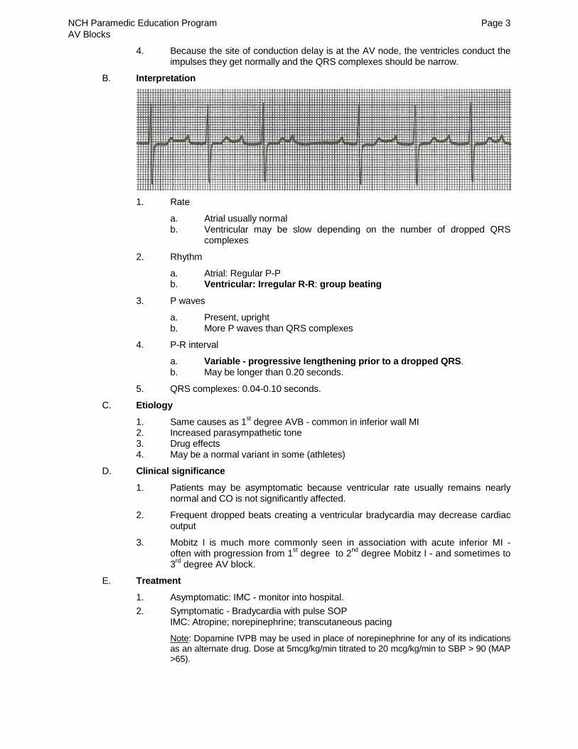
3. On the ECG, this is reflected as regular P-P intervals with P-R intervals that become progressively longer until one P wave is present without a QRS followed by a slight pause until the next P wave comes on time, and the pattern starts over again. Because conduction to the ventricles is progressively delayed until there is no QRS, the R-R intervals are irregular and the QRS complexes have a classic "group beating" appearance.
NCH Paramedic Education Program Page 3 AV Blocks
4. Because the site of conduction delay is at the AV node, the ventricles conduct the impulses they get normally and the QRS complexes should be narrow.
B. Interpretation
1. Rate
a. Atrial usually normal b. Ventricular may be slow depending on the number of dropped QRS
complexes
2. Rhythm
a. Atrial: Regular P-P b. Ventricular: Irregular R-R: group beating
3. P waves
a. Present, upright b. More P waves than QRS complexes
4. P-R interval
a. Variable - progressive lengthening prior to a dropped QRS. b. May be longer than 0.20 seconds.
5. QRS complexes: 0.04-0.10 seconds.
C. Etiology
1. Same causes as 1st degree AVB - common in inferior wall MI 2. Increased parasympathetic tone 3. Drug effects 4. May be a normal variant in some (athletes)
D. Clinical significance
1. Patients may be asymptomatic because ventricular rate usually remains nearly normal and CO is not significantly affected.
2. Frequent dropped beats creating a ventricular bradycardia may decrease cardiac output
3. Mobitz I is much more commonly seen in association with acute inferior MI - often with progression from 1st degree to 2nd degree Mobitz I - and sometimes to 3rd degree AV block.
E. Treatment
1. Asymptomatic: IMC - monitor into hospital. 2. Symptomatic - Bradycardia with pulse SOP
Note: Dopamine IVPB may be used in place of norepinephrine for any of its indications as an alternate drug. Dose at 5mcg/kg/min titrated to 20 mcg/kg/min to SBP > 90 (MAP >65).
NCH Paramedic Education Program Page 4 AV Blocks
IV. 2nd Degree AV Block type or Mobitz II
A. Etiology
1. INFRAHISIAN block: disease is below the AV node and may involve the bundle of HIS or both bundle branches
2. Much more serious than Mobitz I 3. Usually associated with acute anterior or anteroseptal MI and more likely in that
setting to be abrupt in onset 4. Degeneration of the electrical conduction system usually related to age 5. Same other causes as 1st degree AVB 6. It is very unlikely (though not impossible) to switch back-and-forth from Mobitz I
to Mobitz II.
B. Description
1. Intermittent block characterized by P waves that originate in the sinus node but are not all conducted to the ventricles.
a. The P-P is reg. as in all AV blocks, but conduction to the ventricles is intermittent resulting in a regular or irregular R-R depending on the number of blocked impulses.
b. Fixed PRI before each QRS that is present
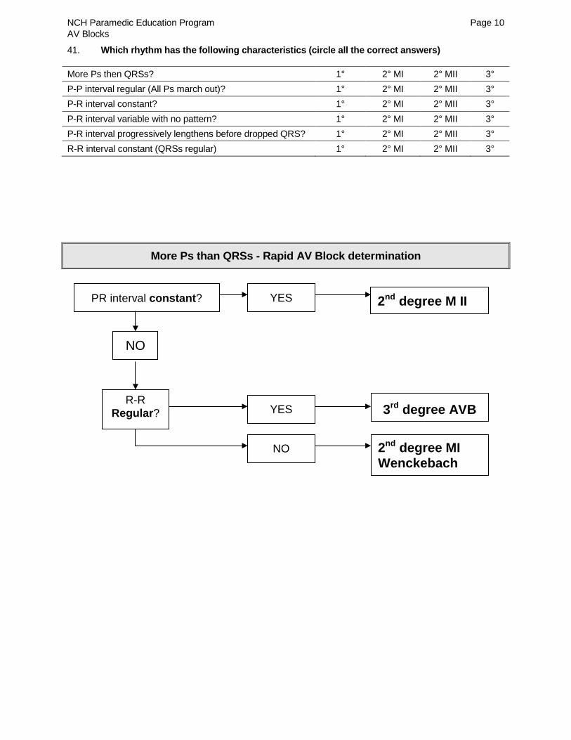
c. Seeing two or more P waves between QRS complexes should trigger an analysis of 2nd or 3rd degree ABV - look at PRI and regularity of the R-R. See graphic decision tree at end of handout:
(1) Variable PRI with Irregular R-R: 2nd degree Mobitz I (2) Fixed PRI and regular or irregular R-R::2nd degree Mobitz II (3) Variable PRI with regular R-R: 3rd degree AVB
2. QRS complexes may be narrow (if block is in the bundle of HIS) or wide (if block is in the bundle branches).
C. Interpretation
1. Rate
a. Atrial: Usually normal b. Ventricular: Depends on the number of impulses that conduct to the
ventricles - always less than the atrial rate.
2. Rhythm a. Atrial: P-P regular b. Ventricular: May be regular or irregular depending on conduction ratio.
3. P waves a. Present upright b. More P waves than QRS complexes
4. P-R interval a. Constant (fixed) for conducted beats b. May be normal or greater than 0.20 seconds.
5. QRS complex: May be normal or wide depending on site of block.
NCH Paramedic Education Program Page 5 AV Blocks
D. Clinical significance
1. Slow rate may compromise cardiac output 2. Frequently progresses to 3rd degree block or asystole with no warning.
E. Treatment
1. Continuous ECG, SpO2 and ETCO2 monitoring; apply pace-defib pads 2. IV/IO; IVF challenges if lungs clear; norepinephrine drip (atropine contraindicated) 3. Transcutaneous pacing if hypotensive and unresponsive to norepinephrine 4. Most patients with this AV block get implantable pacemakers at the hospital
V. 3rd Degree (Complete) AV Block
A. Description
1. INFRAHISIAN block: disease is in AV node and may involve bundle of HIS or both bundle branches
2. Most serious AVB - Can progress suddenly to asystole without warning
3. No atrial impulses are conducted to ventricles due to complete electrical block below the AV node.
4. Atria pace themselves (P-P regular)
5. Ventricles are paced by an escape pacemaker. This may be in the AV node (giving the appearance of a junctional rhythm) or the ventricles (giving the appearance of an Idioventricular rhythm) at a regular rhythm for that pacemaker. Thus the QRS may be narrow or wide and the ventricular rate will often depend on the escape pacemaker site.
6. No relationship between P waves and QRS complexes
7. PRI will be constantly changing (variable) as two different rhythms are superimposed on the same strip (atrial and ventricular)
B. Etiology
1. Inferior and anterior MI 2. Drug toxicity: Beta blocker, calcium blocker, digitalis 3. Degenerative conduction system disease 4. Following cardiac surgery 5. Elderly with degeneration of conduction system
C. Interpretation
1. Rate
a. Atrial: P wave rate usually WNL for SA node, may be slow if sinus bradycardia
b. Ventricular (R rate): (1) 40-60 if paced by AV node (2) 20-40 if paced by ventricles
2. Rhythm a. Atrial: P-P regular b. Ventricular: R-R regular
3. P waves a. Present; upright
NCH Paramedic Education Program Page 6 AV Blocks
b. More P waves than QRS complexes as atrial rate is usually faster c. P may be buried in or superimposed a QRS or T wave – measure P-P
regularity and look for evidence of a P wave where it should normally be present based on P-P rate.
4. PRI – totally variable; no correlation between Ps and QRSs
5. QRS complex a. Narrow if junctional escape pacemaker b. Wide if ventricular escape pacemaker
D. Clinical significance
1. Heart sounds variable in intensity
2. May have 3rd & 4th HS, murmurs or gallops (variable ventricular filling by atrial rhythm)
3. Cannon A waves: Jugular vein pulsations become momentarily massive – atria contract against closed AV valves. Blood cannot enter ventricles. Cause huge venous pulsations.
4. Relative or actual hypotension
5. Serious & potentially life threatening
6. Can progress to asystole without warning
7. May have severe hemodynamic compromise due to slow ventricular rate and ↓ CO
8. May experience dyspnea, HF, ↓ BP, chest pain or syncope
E. Treatment – same as 2nd AVB MII
1. Continuous ECG, SpO2 and ETCO2 monitoring; apply pace-defib pads 2. IV/IO; IVF challenges if lungs clear; norepinephrine drip (atropine contraindicated) 3. Transcutaneous pacing if hypotensive and unresponsive to norepinephrine 4. Most patients with this AV block get implantable pacemakers at the hospital 5. Transport ASAP
References Bledsoe, B., Cherry, R.A., Porter, R.S. (2017). Arrhythmias originating within the AV junction (AV blocks).
In, Bledsoe et. al. (Eds), Paramedic Care Principles and Practice (Fifth Edition) Volume 3; (pp. 88-93). NY: Pearson.
NCH Paramedic Education Program Page 7 AV Blocks
Homework questions 1. The term heart block describes disturbances in conduction through the 2. Which is the mildest form of AV block? 3. Which is the most extreme or severe AV block? 4. List four steps that are critical to accurately diagnosing AV blocks:
5. Which of these is characteristic of all first degree AV blocks?
A. All of the atrial impulses are conducted to the ventricles. B. Some of the atrial impulses are conducted to the ventricles and some are not. C. None of the atrial impulses are conducted to the ventricles.
6. The PR interval in first degree block is short / normal / prolonged. 7. The PR interval in first degree AV block is fixed / variable. 8. The QRS complex in first degree AV block should be normal / wide. 9. First degree block usually
A. causes no symptoms. B. results in syncope and hypotension.
10. What is the ratio of P waves to QRS complexes in first degree block?
A. 3:1 B. 2:1 C. 1:1
11. Which of these is characteristic of all second degree blocks?
A. All of the atrial impulses are conducted to the ventricles. B. Some of the atrial impulses are conducted to the ventricles and some are not. C. None of the atrial impulses are conducted to the ventricles.
12. Second degree block Mobitz I has a pattern of conduction known as 13. The P - P in 2° AVB MI is regular / irregular. 14. The PR interval in 2° AVB MI is fixed / variable. 15. The P-R interval in 2° AVB MI is progressively shortens / lengthens until a P wave occurs that is not
followed by a QRS. 16. This pattern causes the ventricular rhythm to be regular / irregular. 17. The repetitive ventricular pattern is called beating. 18. The QRS complex in 2° AVB MI should be normal / wide. 19. The P - P in a 2° AVB MII is regular / irregular.
NCH Paramedic Education Program Page 8 AV Blocks
20. The PR interval in 2° AVB Mobitz II is fixed / variable. 21. The ventricular pattern in a 2° AVB MII with a fixed conduction ratio is regular / irregular. 22. The location of the conduction disturbance in 2° AVB MII may be in the
or .
23. As a result, the QRS may be narrow if located in the
and wide if located in the . 24. 2° AVB MII is less / more serious than Mobitz I. 25. Mobitz II has a potential to progress suddenly to 26. The first treatment of choice for 2° AVB MII with a wide QRS is .
A. Atropine B. Norepinephrine
Why? 27. Which of these is characteristic of all 3° AV blocks?
A. All of the atrial impulses are conducted to the ventricles. B. Some of the atrial impulses are conducted to the ventricles and some are not. C. None of the atrial impulses are conducted to the ventricles.
28. In 3° AVB, the atria and ventricles beat synchronously with / independent of each other. 29. The P - P in a 3° AVB is regular / irregular. 30. The R - R in a 3° AVB is regular / irregular. 31. The P-R interval in a 3° AVB is fixed / variable. 32. In 3° AVB, P waves have direct / no relationship to the QRS complexes. 33. The QRS in 3° AVB may be narrow if the ventricles become paced by the
and wide if the escape pacemaker comes from the 34. Third degree block can progress suddenly to 35. List three physical symptoms commonly experienced by patients with 3° AVB
36. What is the first treatment of choice for 3° AVB?
A. Atropine B. Norepinephrine Why?
NCH Paramedic Education Program Page 9 AV Blocks
37.
Identify the rhythm:
38.
Identify the rhythm:
39.
Identify the rhythm:
40.
Identify the rhythm:
NCH Paramedic Education Program Page 10 AV Blocks
41. Which rhythm has the following characteristics (circle all the correct answers) More Ps then QRSs? 1° 2° MI 2° MII 3° P-P interval regular (All Ps march out)? 1° 2° MI 2° MII 3° P-R interval constant? 1° 2° MI 2° MII 3° P-R interval variable with no pattern? 1° 2° MI 2° MII 3° P-R interval progressively lengthens before dropped QRS? 1° 2° MI 2° MII 3° R-R interval constant (QRSs regular) 1° 2° MI 2° MII 3°