Page 1
Pharmacologyonline 3: 221-243 (2009) Newsletter Jagdish Kakadiya
221
NOVEL ANTI-ARRHYHTMIC AGENTS
Jagdish Kakadiya
Dharmaj Degree Pharmacy College, Sanskruti Sanraksha Charitable Trust, Dharmaj, Tal: Petlad, Dist: Anand.
Gujarat, INDIA.
ADDRESS FOR CORRESPONDENCE
Mr. Jagdish L. Kakadiya Dharmaj Degree Pharmacy College,
Sanskruti Sanraksha Charitable Trust, Petlad-Khambhat road,
Dharmaj, Tal: Petlad, Dist: Anand. Gujarat, INDIA.
Phone 09825882922
Page 2
Pharmacologyonline 3: 221-243 (2009) Newsletter Jagdish Kakadiya
222
CONTENTS
1 INTRODUCTION
2 PHYSIOLOGY OF CARDIAC MUSCLE
2.1 PROPERTIES OF CARDIAC
2.2 ELECTRICAL POTENTIAL IN CADIAC MUSCLE
3 DEFINITION CARDIAC ARRHTHMIA
4 CLASSIFICATION OF CARDIAC ARRHYTHMIA
5 MECHANISMS OF CARDIAC ARRHYTHMIA
5.1 ABNORMAL PACEMAKER ACTIVITY
5.2 AFTER DEPOLARIZATION
5.3 RE-ENTRY
5.4 FRACTIONATION OF IMPULSES
5.5 HEART BLOCK
6 GENERAL MECHANISMS OF ANTI ARRHYTHMIC
AGENTS
7 CLASSIFICATION
8 NOVEL ANTI ARRHYTHMIC AGENTS
8.1 CLASS- I AGENTS
8.2 CLASS- III AGENTS
8.3 OTHER AGENTS
8.4 NEWER TECHNIQUES
Page 3
Pharmacologyonline 3: 221-243 (2009) Newsletter Jagdish Kakadiya
223
1. INTRODUCTION
Approximately 50% of post MI fatalities are due to sudden cardiac death resulting from
ventricular tachycardia or ventricular fibrillation.
It is believed that these arrhythmias are arising from mechanical dysfunction and
ischemic events interacting within disordered electro physiologic milieu (1).
This has prompted active search for safe and effective treatment modalities and their
ultimate evaluation in clinical trials.
Currently ß-blockers are recommended for post MI patients with premature ventricular
contraction where as Ca2+channel blockers are not useful and sodium channel blockers
are actually harmful.
DRUGS DEVELOPMENT
In 1970 and early 1980s target or models used in drug development were based on
suppression of PVCs recorded in animal models or in patients with PVCs after MI.
Recently drug development has evolved in two areas
–Amiodarone analogues
–Delayed rectifier k+ channel blockers
Remarkable clinical efficacy of amiodarone in the treatment of wide varieties of
arrhythmia has lead to search for class-III drugs with better safety profile.
22.. PPHHYYSSIIOOLLOOGGYY OOFF CCAARRDDIIAACC MMUUSSCCLLEE ((22))
22..11 PPRROOPPEERRTTIIEESS OOFF CCAARRDDIIAACC MMUUSSCCLLEE
11.. EEXXCCIITTAABBIILLIITTYY::
TThhee aabbiilliittyy ooff ttiissssuuee ttoo ggiivvee rreessppoonnssee ttoo aa ssttiimmuulluuss iiss ccaalllleedd EExxcciittaabbiilliittyy..
IInniittiiaall sstteepp iiss tthhee ddeevveellooppmmeenntt ooff aaccttiioonn ppootteennttiiaall wwhhiicchh iiss ffoolllloowweedd bbyy ccoonnttrraaccttiioonn ooff
mmuussccllee ffiibbrreess..
22.. AAUUTTOOMMAATTIICCIITTYY ((RRHHYYTTHHMMIICCIITTYY))::
TThhee aabbiilliittyy ooff ttiissssuuee ttoo pprroodduuccee iittss oowwnn iimmppuullsseess rreegguullaarrllyy iiss ccaalllleedd RRhhyyhhmmiicciittyy
AAllll tthhee ttiissssuuee ooff tthhee hheeaarrtt ppoosssseesssseess tthhee pprrooppeerrttyy ooff rrhhyytthhmmiicciittyy
AAuuttoommaattiicciittyy ooff hheeaarrtt iiss mmaaiinnttaaiinn bbyy ppaacceemmaakkeerr
Page 4
Pharmacologyonline 3: 221-243 (2009) Newsletter Jagdish Kakadiya
224
PPAACCEEMMAAKKEERR::
IItt iiss aa ppaarrtt ooff hheeaarrtt ffrroomm wwhhiicchh tthhee iimmppuullsseess ffoorr hheeaarrttbbeeaatt aarree pprroodduucceedd nnoorrmmaallllyy..
33.. CCOONNDDUUCCTTIIVVIITTYY::
HHuummaann hheeaarrtt hhaass ssppeecciiaalliizzeedd ccoonndduuccttiivvee ssyysstteemm tthhrroouugghh wwhhiicchh tthhee iimmppuullsseess pprroodduucceedd
bbyy SSAA nnooddee aarree ttrraannssmmiitttteedd ttoo aallll ppaarrttss ooff hheeaarrtt..
The conductive system in human heart comprises:
1. AV node
2. Bundle of His
3. Right & left bundle branches
4. Purkinje fibers
4. CONTRACTILITY:
The ability of tissue to shorten in length (contraction) after receiving a stimulus is called
contractility
-The different contractile properties are as follows.
Page 5
Pharmacologyonline 3: 221-243 (2009) Newsletter Jagdish Kakadiya
225
1. All OR None law
When stimulus is applied to muscle it respond to its maximum OR does not give response at all
is called All OR None law
2. Staircase phenomenon
When stimuli are applied at base of the ventricles of quiescent frog heart at interval of two
second without changing the strength, for first few stimulus responses are increased & then
responses remain unchanged is called Staircase phenomenon
3. Summation of subliminal stimuli
When stimulus with a subliminal strength is applied to muscle it does not give any response but
few stimuli with a subliminal strength are applied to ventricles in succession it does response by
contraction. It is due to summation of subliminal stimuli.
4. Refractory period
Refractory period is the relative brief period of relaxation of a muscle during which its
excitability is depressed. The refractory period of the heart is long & is divided into 3 parts:
1. Absolute Refractory Period:
Coincides the period from the onset of depolarization phase to the repolarization up to threshold
potential. During this period even a strong stimulus fails to produce Response. Such a long
refractory period ensures enough time for recovery of cardiac muscle & so cardiac muscle can’t
be fatigued.
2. Relative refractory period:
Begins when transmembrane potential (Vm) has just reached about -60 mV & ends before
repolarization phase is ceased.
3. Supernormal Period:
It is time interval from the point of termination of the repolarization to the beginning of slow
diastolic depolarization phase.
2.2 ELECTRICAL POTENTIAL IN CADIAC MUSCLE
Resting membrane potential
In individual cardiac muscle fiber, the resting membrane potential is about -80 mV.
In SA node it is -55mV. In purkinje fibers, it is about -90 to-100 mV.
Page 6
Pharmacologyonline 3: 221-243 (2009) Newsletter Jagdish Kakadiya
226
Action potential
The electrical activity that takes place in the cardiac muscle is known as action potential.
Duration of action potential in cardiac muscle is 0.25 to 0.35sec.
1) PHASE O RAPID DEPOLARIZATION
Nearby -60 mV rapid depolarization occurs by the movement of Na+ ions through
selective channels that are activated in a voltage dependent manner when
The propagating cardiac impulse or
Spontaneous phase 4 depolarization causes the hypothetical m-gate in Na+ channel to
open.
2) PHASE 1 PARTIAL REPOLARIZATION
At the peak of upstroke there occurs a rapid repolarization in which membrane potential
returns towards 0 mV because of the rapid inactivation of Inward Sodium current
It occurs also due to activation of short-lived outward current carried by mainly
Potassium ions i.e. known as Transient outward current.
3) PHASE 2 ACTION POTENTIAL PLATEAU
• Conductance of all ion channels decreases rapidly giving net balance between inward
(depolarizing ) &outward (repolarizing ) ion currents.
• Increase ICa2+ is a major contributor to phase 2 & when membrane is depolarized to -40
mV, opening of voltage dependent Ca2+ channels causes increase ICa2+
• Cardiac muscle membrane assists plateau by its special property that is Inward going
Rectification – potassium conductance falls to a low level when membrane is
depolarized, so Outward K+ current,
• Can’t restore resting membrane potential during plateau.
4) PHASE 3 REPOLARIZATION:
It occurs as the calcium current inactivates &
Outward potassium current activates causing outward potassium current.
Such potassium current repolarizes the fiber to normal diastolic value of Vm
5) PHASE 4 PACEMAKERS POTENTIAL / DIASTOLIC DEPOLARIZATION
Caused by combination of increased inward & Decreased outward currents during diastole
Cells having property of Automaticity exhibit spontaneous phase 4 depolarization
Page 7
Pharmacologyonline 3: 221-243 (2009) Newsletter Jagdish Kakadiya
227
Cells of SA node have Greater background conductance to Na+ ions, so great inward
Na+ entry.
3. DEFINITION
Cardiac arrhythmia:
It is defined as disorder of disturbance in rate, rhythm, origin or conduction of cardiac
impulses within a heart.
Drug therapy of arrhythmias depends on
- Presence of arrhythmia and its type.
- Use of anti arrhythmic drugs on basis of mechanism of action (3).
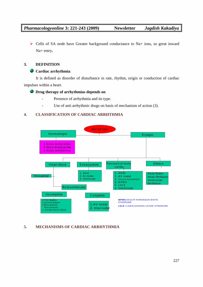
4. CLASSIFICATION OF CARDIAC ARRHTHMIA
Arrhythmia
EctopicNormotropic
1.Sinus tachycardia2.Sinus bradycardia3.Sinus arrhtythm ia
Heart block Extrasystole Paroxysm al tachy-cardia
Others
Sinoatrial
Atrioventricular
Incomplete Complete
Atrial flutter.Atrial fibrillationVentricularfibrillation
1.First degree.2.Second degree.3.W enckebach
Phenomenon.4..Bundle branch Block
1.AV nodal2. Internodal
1. Atrial2. Av nodal.3. Ventricular
1 .Atrial2 .AV nodal A. Circus movementB .W PW SC. LGLS3 .Ventricular
WPWS=W OLFF-PARKINSON W HITE SYNDROME
LGLS =LOW N-GANONG-LEVINE SYNDROME
5. MECHANISMS OF CARDIAC ARRHYTHMIA
Page 8
Pharmacologyonline 3: 221-243 (2009) Newsletter Jagdish Kakadiya
228
55..11 AABBNNOORRMMAALL PPAACCEEMMAAKKEERR AACCTTIIVVIITTYY ((44))
Under some pathological conditions pacemaker activity can arise in other parts of heart
than SA node and conducting tissue.
Predisposing factors are:
Catecholamines acting on ß1adrenorecepor increase the Rate of depolarization during
phase 4 &can cause normally quiescent part of heart to take on a spontaneous rhythm.
Pain (During myocardial infarction) increased sympathetic discharge releases adrenaline
from the adrenal gland.
Partial depolarization resulting from ischamic damage is caused by decreased activity of
electrogenic sodium pump and it will cause abnormal pacemaker activity.
This mechanism is mainly responsible for Sinus tachycardia, Atrial&Ventricular
extrasystoles, and Atrial flutter.
5.2 AFTER DEPOLARIZATION
Under some pathophysilogical conditions, normal cardiac action potential is interrupted or
followed by abnormal depolarization if this abnormal depolarization reaches threshold potential it may,
in turn give rise to secondary upstrokes which then can propagate and crate abnormal rhythms (5).
These abnormal secondary upstrokes occur can only after initial normal or triggering
upstroke & so are termed as triggered arrhythmias (5).
There are two types of After Depolarization.
1) Delayed After Depolarization (DAD)
2) Early After Depolarization (EAD)
1) Delayed After Depolarization (DAD)
A normal action potential may be followed by delayed after depolarization if this reaches
threshold a secondary triggered beat can occur.
It is due to increase in intracellular calcium load in condition like myocardial ischemia,
adrenergic stress, digitalis intoxication .
DAD amplitude is increase in vitro by rapid pacing and clinical arrhythmia thought to be
correspond to DAD mediated triggered beat are more frequent when underlying cardiac
rate is rapid (6).
It is responsible for train of Extrasystole, Tachycardia, Torsades de pointes (5).
Page 9
Pharmacologyonline 3: 221-243 (2009) Newsletter Jagdish Kakadiya
229
EAD mediated arrhythmias are common in condition like slow heart rate, low extra cellular
calcium and use of class III drugs.
When cardiac Repolarization markedly prolonged polymorphic ventricular tachycardia
with a long QT interval, known as Tosades de pointes syndrome may occur and is
thought to be caused by EAD and resultant triggering activity (7).
The congenital long QT syndrome, a disease in which Torsades de pointes is common is
now known to be caused by mutation in the genes encoding the sodium channels
underlying the repolazising current IKR and IKS.
2) Early After Depolarization (EAD)
It is due to marked prolongation of action potential when this occu
rs phase-3 Repolarization may be interrupted by an Early After Depolarization.
C) RE-ENTRY
Due to conduction abnormality, impulse may recirculate in the heart and cause repetitive
activation without the need for any new impulse to be generated.
Two types of RE-ENTRY
1) Circus movement type
2) Micro-Re entry Circuit.
1) CIRCUS MOVEMENT TYPE (5).
It occur in anatomically define circuit.
A premature impulse is temporary blocked in one direction by refractory tissue
and makes only one way transit around an obstacle i.e. natural orifice heart, Infracted
myocardium and that will find original spot in advance state of recovery and re excite it
Page 10
Pharmacologyonline 3: 221-243 (2009) Newsletter Jagdish Kakadiya
230
and cause recurrent activation of the adjacent myocardium. This mechanism may cause
Atrial flutter, PSVT, Atrioventricular reciprocal tachycardia in Wolff Parkinson White
syndrome.
2) MICRO-RE ENTRY CIRCUIT.
It is formed at a junction of purkinje fibers and ventricular fibers.
One of the branch of purkinje fibers sufficiently depolarized to cause unidirectional
block.
Extremely slow conduction due to slow channel depolarization and markedly
abbreviated, APD and ERP make re-entry possible ventricular Extra-systole, VT and VF.
For re-entry to occur the pathlength of circuit should be grater than the wavelength (ERP
x Conduction Velocity) of impulse.
Slow conduction in re-entrant circuit may be caused by:
Partial depolarization of membrane-decrease slope of phase 0.
Cells changing over from fast channel to slow channel depolarization which conducts
very slowly when fiber is depolarized to resting membrane potential of about –
50mV.Sodium channels are inactivated but calcium channels still able to produce
response
Page 11
Pharmacologyonline 3: 221-243 (2009) Newsletter Jagdish Kakadiya
231
3) FRACTIONATION OF IMPULSES
When atrial ERP is brief & inhomogeneous (under vagal overactivity), an impulse
generated early in diastole gets conducted irregularly over atrium i.e. it moves rapidly
through fibres with short ERP (which is completely recovered) slowly through fibres with
longer ERP (which is partially recovered) and not at all through those still
refractory.Thus, cause asynchronous activation of atrial fibres and cause Atrial
Fibrillation.
This arrhythmia must be initiated by premature depolarization but is self sustaining,
because passage of an irregular impulse leaves an irregular refractory trace and maintain
the inhomogenicity of ERPs.
Total number of impulses that can be sustained at any moment depends on -
Average of ERP
Mass of tissue
Thus atrial fibrillation is more common in dilated atria (Mitral stenosis).
4) HEART BLOCK
Even under physiological condition, conduction through SA and AV nodes are tardy.
It may further slow by ischemia causing partial to complete AV block or Sick sinus.
Two types of Heart block
1) Sinoatrial block.
When impulses from SA node are not transmitted to AV node due to defective internodal
fibers, is called sinoatrial block.
2) Atrioventricular block.
When impulses from atria are not transmitted to ventricle due to defective conductive
system is called atrioventricular block.
a) Incomplete H.block.
The transmissions of impulses from atria to ventricles are slow down but not blocked
completely.
i) First degree (Delayed conduction)
The conduction of impulses through AV node is very slow.
ii) Second degree (Partial H. block)
Page 12
Pharmacologyonline 3: 221-243 (2009) Newsletter Jagdish Kakadiya
232
In this some of impulses produced by SA node fail to reach to ventricles.
iii) Wenckebach phenomenon
It is a one type of heart bock characterized by progressive lengthening of conduction time
in AV node with ultimate missing of one beat. Afterwards, the conduction of impulses is
normal or slightly delayed.
iv) Bundle branch block
When Left or Right branch Bundle of His is affected then Left or Right Bundle branch
block occur respectively. During this, the impulse from atria reaches unaffected ventricle
first. Then from here impulse travel into affected side.
b) Complete H.block (Third degree H.block)
The impulses produced by SA node do not reach the ventricles. So, the ventricle beats in
their own rhythm independent of atrial beat. This is called Idioventricular rhythm.
6. GENERAL MECHANISMS OF ANTI-ARRHYTHMIC AGENTS (8)
A single arrhythmia may results from multiple mechanisms e.g. an automatic or
triggered beat may results in sustained re-entrant arrhythmia in patient with a potential re-entrant
circuit.
Anti-arrhythmic agents act by
1) Suppressing initiating mechanism
2) Altering the re-entrant circuit.
Drugs slow automatic rhythm by altering one of the four determinants.
1) Maximum diastolic potential, which is
-Increase by Ach and Adenosine.
2) Slope of phase 4, which is decreased by ß- blockers
3) Threshold potential, which is altered by class-I and class-IV drugs.
4) Action potential duration, which is prolonged by class-III drugs.
Arrhythmia due to after depolarization can be blocked by
i) Inhibition of development of after depolarization
ii) Interference with inward current, which is responsible for upstroke
Page 13
Pharmacologyonline 3: 221-243 (2009) Newsletter Jagdish Kakadiya
233
I.e. Arrhythmia due to Digitalis induced DADs can be treated by Verapamil (inhibit the
development of DADs) or by Quinidine (blocking sodium channel).
EAD induced triggered beat can be blocked by
i) Shortening of APD by class-1b drug.
ii) Use of magnesium.
In patients with congenitally prolonged QT interval, Torsades de pointes often
occur with adrenergic stress can be prevented by B blockers as well as rapid pacing.
Drugs that prolonged APD can terminate circus movement type re-entry
Drugs that prolonged refractoriness can terminate micro re-entry circuit
-- E.g: Class-I&Class-III drug in fast responsive tissue.
Class-IV drugs in slow responsive tissue.
Conduction usually fails in weak leak in circuit.
In condition like WPW-related arrhythmia, the weak leak is AV node and drug that
prolongs AV nodal refractoriness and slow AV nodal conduction such as class-I, Class-II
or digitalis glycosides are likely to be effective.
Drugs that interfere with cell-cell coupling can also prolong refractoriness in
multicellular preparation.
E.g. Amiodarone exerts this effecting diseased tissue.
Acceleration of conduction in area of slow conduction could also anti arrhythmic in re-
entry.
E.g. Lidocaine exerts such effect in some experimental condition.
7. CLASSIFICATION OF ANTI ARRHTHMIC AGENTS
CLASS I - Na+ channel blockers.
class Ia class 1b class Ic
-Quinidine -Phenytoin - Flecainide
-Disopyramide -Lignocaine - Encainide,
-Procainamide -Mexiletine - Propafenone
NOVEL CLASS-I AGENTS:
• SD-3212
•AZD 7009
Page 14
Pharmacologyonline 3: 221-243 (2009) Newsletter Jagdish Kakadiya
234
•MORICIZINE.
CLASS II- Anti adrenergic agents.
Propranolol
Bretylium
Esmolol
Sotalol
CLASS III- Potassium channel blockers
Amiodarone
Bretylium
Sotalol
NOVEL CLASS-III AGENTS:
Ibutilide Nibentan CX-1 Tedisamil
Dofetilide Azimilide AL-275
Trecetilide Dronedarone AZD-7009
CLASS IV- Calcium channel blockers:
Verapamil
Diltiazem
Other agents
Adenosine
Digitalis
Other novel agent:
RSD-1235
Newer techniques
Implantable Cardioverter Defibrillator
Pacemaker
Radiofrequency ablation
Surgery
1.Cryoablation
2.Maze Surgery
Page 15
Pharmacologyonline 3: 221-243 (2009) Newsletter Jagdish Kakadiya
235
Drugs used in Brady arrhythmias
1) Sympathetic agonist: Isoprenaline
2) Parasympathetic antagonist: Atropine
8. NOVEL ANTIARRHYTHMIC AGENTS
A) CLASS- I AGENTS:
1) SD-3212 (levo-semotiadil fumarate):
It acts by blocking sodium & calcium channels.
In animal models this drug suppressed Atrial tachyarrhythmias but not ventricular
tachyarrhythmias.
A scientific study indicates that SD-3212 is effective in interrupting canine atrial flutter
possibly by suppressing atrial conduction.
Use: It can be used for treatment of atrial tachyarrhythmias.
2) AZD-7009:
It is novel agent in early clinical development for treatment of atrial fibrillation.
It acts by blocking sodium & potassium channels.
Pre-clinical studies have shown that it is promising drug for converting AF to Sinus
rhythm.
An early clinical study shows that I.V AZD 7009 is well tolerated.
No cases of ‘Torsades de-pointes’ are reported.
3) MORICIZINE
It is a phenothiazine derivative with Class Ic antiarrhythmic properties
It under goes first pass metabolism.
PPB is 95%, B.A is 34-38%.
Recent clinical studies have shown that moricizine is slightly less effective than
encainide, flecainide but more effective than quinidine & disopyramide in suppressing
ventricular premature depolarization.
It can be used in treatment of VT &VF.
It has lower incidence of serious adverse effects than other Class- I drugs (Carnes CA
and Coyle JD, 2003).
Page 16
Pharmacologyonline 3: 221-243 (2009) Newsletter Jagdish Kakadiya
236
B) CLASS- III AGENTS
1) IBUTILIDE:
• It is a novel antiarrhythmic drug that was recently marketed for the rapid conversion of
atrial fibrillation and atrial flutter into Normal sinus rhythm.
•M/A:
•Prolongs action potential duration by activating a slow inward current, largely carried by
sodium ions.
•Blocks the Rapidly activating component of the delayed rectifier potassium current.
• It increases atrial and ventricular refractoriness by action potential prolongation in vivo.
Phamacokinetics: Not given oral route because of extensive first pass metabolism.
Route: Intravenous
t1/2-: 2-12 hrs
Linear kinetics: No
•Dose: I.V. 1mg over 10 minute periods in patients weighing 60kg.
Initial dose is 0.01mg/kg with second dose of same strength 10 minutes later in patients
weighing less than 60kg.
•Indication: Rapid conversion of atrial fibrillation and atrial flutter into sinus rhythm.
•Contraindication:
QTc Interval exceeding 440Ms
Bradycardia
Electrolyte disturbances
Other QT-prolonging drugs
• Adverse effects:
Torsades de pointes (5.1%)
Premature ventricular complexes (5.1%)
Monomorphic ventricular tachycardia (2.7%)
Hypertension (2%)
Bundle branch block (1.9%)
Nausea (1.9%)
Headache (3.6%)
Page 17
Pharmacologyonline 3: 221-243 (2009) Newsletter Jagdish Kakadiya
237
Bradycardia (1.2%)
2) DOFETILIDE:
•Class III anti-arrhythmic drug.
•M/A: Blocks particular potassium current in heart.
•It is recently approved by U.S. and its effectiveness was shown in large number of clinical trials
such as EMERALD and SAFIRE trials.
•These trials showed that patient given dofetilide were more likely to convert AFor AFL to
Normal sinus rhythm.
•Dofetilide was also evaluated in two large mortality trials known as DIAMOND trial in patients
were in hospital with CHF or Heart attack.
•These trial showed that this drug did not increase mortality in either of two patient groups.
•Dose: 125,250 or 500mcg twice a day
•A/E: Torsades de pointes
•Contraindication: Certain drug increases blood level of dofetilide and increase risk of Torsades
de pointes.
•E.g. Verapamil, Cimetidine, ketoconazole, trimethoprim, prochlorperazine etc.
•Uses: Atrial Fibrillation and Atrial flutter.
3) AZIMILIDE (NE10064):
•Azimilide is an investigational class-III antiarrhythmic that has been developed for treating both
supraventricular and Ventricular tachyarrhythmias.
•M/A: It mainly blocks the slowly activating (IKS) and rapidly activating (IKR)
Components which distinguishes it from most of the other potassium channel blockers such as
Sotalol and dofetilide, which block only IKR.
• Similar to other class-III drugs, azimilide prolongs myocardial depolarization in a dose
dependent manner by increasing the action potential during QT interval and effective refractory
period.
•In animal models, azimilide has shown to be effective in suppressing both atrial and ventricular
tachyarrhythmias, decreasing defibrillation energy requirement and preventing post myocardial
infarction, ventricular tachycardia and fibrillation.
Page 18
Pharmacologyonline 3: 221-243 (2009) Newsletter Jagdish Kakadiya
238
It has very predictable pharmacokinetic and has not significant drug interaction with Digoxin or
Wafarine.
•Side effects
Headache (most frequent)
Neutropenia
Torsades de pointes
4) DRONEDARONE:
•Dronedarone is new class-III antiarrhythmic drug under phase-III development by Sanofi-
Aventis for prevention and treatment of atrial fibrillation.
•Evidence of efficacy was first demonstrated in DAFNE trial, a phase-II b study that compared
dronedarone with placebo for maintenance of sinus rhythm after cardio version for AF.
•Dronrdarone proved effective in preventing recurrences of AF.
•No cases of Torsades de pointes & Pro-arrhythmias were reported.
5) TRECETILIDE:
•Trecetilide, a congener of ibutilide, is being evaluated in both IV and Oral preparations for the
termination and prevention of AF and AFL.
•In addition to blocking IKR, It seems to prolong repolarization through other mechanisms that
are still being delineated.
•It also significantly prolongs the action potential in animals and repolarization in humans
without exerting other electrophysiological effects.
6) NIBENTAN:
-M/A: It is a selective potassium channel blocker with class-III antiarrhythmic properties.
•At 0.1-0.25 mg/kg I/V. Nibentan produced dose dependent increases in atrial and ventricular
effective refractory periods and the QT interval without significant change in systolic or diastolic
atrial or left ventricular pressure, heart rate and left ventricular contractility.
•Nibentan was about 100 times more potent than quinidine and its effect was sustained as long.
CLINICAL STUDIES:
•Phase-I clinical trial was performed to evaluate safety of Nibentan in 67 patients with various
supraventricular & ventricular tachycardias.
Page 19
Pharmacologyonline 3: 221-243 (2009) Newsletter Jagdish Kakadiya
239
Nibentan produced effects characteristics of class-III drug appearance of additional “U” wave
with lowering of T wave amplitude, prolongation of QT interval by 34%.
•It significantly slows down sinus rhythm rate.
•Phase-II clinical trial, efficacy of nibentan was studied on 43 patients with cardiac diseases.
•Nibentan produce pronounced antiarrythmic effect in all patient with AFL or SVT & 73%
patients with AF
•Side effect
•. Torsades de pointes (5.5%)
7) TEDISAMIL:
•Tedisamil (pulzium) differs from other class-III anti arrhythmic agents in that it blocks ITo in
addition to IKR.
• Pulzium is an innovative drug for now in later stage of clinical development for treatment of
AF.
•Tedisamil has been shown to reduce the incidence of VF and AF in experimental studies.
• Tedisamil is a drug used by cardiologist to treat of rhythm disturbances of heart.
•S/E: It produces bradycardia, presumably by direct action on SA node, APD and prolongs the
QT intervals without affecting QRS complexes.
8) CX-1:
•ChanXpress is potent novel antiarrhythinic agent.
•Preclinical invitro test showed that CX-1 has the requisite properties of a satisfactory
antiarrhythmic drug.
•Animal studies have fully established that CX-1 reverses AF and also prevents recurrence of
AF.
•The safety of drug in human has fully established in phase-I clinical trials.
•Bioavailability of CX-1 is excellent and It can be given orally and intravenously.
•Drugs similar to CX-1 for the treatment of AF have proven to generate undesirable side effects
that limit their efficacy.
•This drug can be used for treatment of VF & VT
9) AL-275:
Page 20
Pharmacologyonline 3: 221-243 (2009) Newsletter Jagdish Kakadiya
240
•AL-275 is novel class-III anti arrhythmic drug, which is derivative of dicyclohexylamides of
amino carbonic acid.
•It mainly acts by blocking rapid (IKR) and slow (IKS) activating components of delayed
rectifier potassium current.
•It prolongs ventricular repolarization, lengthens atrial and ventricular ERPs, suppresses sinus
node automaticity, does not affect atrio-ventricular and intra-ventricular conduction.
C) OTHER AGENTS
RSD-1235:
•M/A: It is a novel, frequency dependent sodium and early-activating potassium channel
blacker under joint development by Cardiome and its partner.
•The agent is intended as an acute use via intravenous administration to terminate AF and restore
SR in AF patients.
•An oral formulation of RSD-1235 is also under development for the long-term maintenance of
normal SR following termination of AF.
Safety and side effects:
•During 30 days follow-up period after drug administration, 18.3% patient in placebo group
and 13.1% in the RSD-1235 group experienced a Serious Adverse Event (SAE).
•Most of these SAE’s were recurrence of AF requiring hospitalization
•There were no cases of drug related Torsades de pointes.
•During first 24 hrs after study drug infusion, the most common non-cardiac side effects
associated with RSD-1235 were,
Disgeusia (30%)
Paresthesia (11%).
Sneezing (16%).
Nausea (9%)
Cough (5%).
D) Newer techniques to treat arrhythmias
1.Implantable Cardioverter Defibrillator
This is a device that applies electric impulses or, if needed, a shock to restore a normal
heartbeat.
Page 21
Pharmacologyonline 3: 221-243 (2009) Newsletter Jagdish Kakadiya
241
The device's power source is implanted in a pouch beneath the skin of our chest or the area above
your stomach and connected to patches placed on our heart.
Use: Ventricular tachycardia and Ventricular fibrillation
Newer implantable devices are inserted through a blood vessel, which means that you do not
need open-chest surgery.
2.Pacemaker
A pacemaker is a matchbox size device that is run by a battery.
It is made up of two parts:
1. A pulse generator, which includes the battery and several electronic circuits.
2. Wires, called leads, which are attached to the heart wall.
Depending on the type of pacemaker you need, there may be one or two leads.
If only one lead is needed, it is placed inside the lower-right chamber (the right ventricle).
If two leads are needed, the other is placed in the upper-right chamber (the right atrium).
The leads are then attached to the pacemaker.
The pacemaker is surgically implanted near the bone below your neck (the collarbone).
If only one lead is needed, it is placed inside the lower-right chamber (the right ventricle).
If two leads are needed, the other is placed in the upper-right chamber (the right atrium).
The leads are then attached to the pacemaker.
Most pacemaker surgery is done under local anesthesia.
Page 22
Pharmacologyonline 3: 221-243 (2009) Newsletter Jagdish Kakadiya
242
Once the pacemaker is implanted, the leads carry signals back from the heart. The pulse
generator "reads" these signals and the batteries send electrical impulses to the heart to
help pace it.
Most pacemakers can sense the heart's rhythm and turn themselves off when the heartbeat
is above a certain level.
They will turn on again when the heartbeat is too slow. These types of pacemakers are
called demand pacemakers.
The pacemaker's batteries supply the electrical energy that acts like your heart's natural
pacemaker.
Uses:
Pacemakers can help pace the heart in cases of slow heart rate, fast and slow heart rate, or
a blockage in the heart's electrical system.
A pacemaker can pace the heart's upper chambers (the atria), the lower chambers (the
ventricles), or both.
Pacemakers may also be used to stop the heart from triggering impulses or from sending
extra impulses.
Page 23
Pharmacologyonline 3: 221-243 (2009) Newsletter Jagdish Kakadiya
243
3. Radiofrequency ablation
It is a procedure that uses a catheter and a device for mapping the electrical pathways of
the heart.
After you are given medicine to relax you, a catheter is inserted into a vein and guided to
your heart, where doctors use high-frequency radio waves to destroy (ablate) the
pathways causing the arrhythmia.
4.Surgery
1. Cryoablation
Using computerized mapping techniques, surgeons can find out which cells are
"misfiring.“
Then cryoablation can be used to eliminate tissue with a cold probe and destroy the
"misfiring" cells.
2. Maze Surgery
Maze surgery may be recommended if you have atrial fibrillation that has not responded
to medicines or electrical shock (cardioversion therapy).
Surgeons create a "maze" of new electrical pathways to let electrical impulses travel through
your heart without being blocked.
REFERENCES
1. Lucchesi BR, Chi L, Friedrichs GS, Black SC, Uprichard ACG. Antiarrhythmic versus antifibrillatory actions: inference from experimental studies. American Journal of Cardiology 1993 72: 25F-44F.
2. Sambulignum K and Sambulignum P. Arrhythmia In: Human anatomy 2005, 427-434, 458-463. 3. Barar FSK. Anti arrhythmic agents In: Essentials of Pharmacotherapeutics 2004, 8th Edition,
S.Chand and Company Ltd; 258-259. 4. Rang HP, Dale MM, Ritter JM, Moor PK, eds.The Heart, In: Pharmacology 2003, 5th Edition,
Churchill Livingstone, 268. 5. Tripathi KD. Anti arrhythmic agents In: Essentials of Medical Pharmacology 2005, 5th Edition,
Jaypee Brothers medical Publishers (p) Ltd; 506-506. 6. Rosen, M.R., and Reder, R.F.Does triggered activity have a role in the genesis of cardiac
arrhythmias Ann. Intern. Med., 1981, 94:794-801 7. Roden, D.M., and Hoffman, B.F. Action potential prolongation and induction of abnormal
automaticity by low quinidine concentrations in canine purkinje firbers, Relationship to potassium and cycle length circ. Res., 1985,56:857-867.
8. Goodman and Gilman’s. Anti arrhythmic agents In: The Pharmacological Basis of Therapeutics 2001, 10th Edition, Mc Graw Hill Publication, 938-942.