Novel biochemical markers in the diagnosis and management of early pregnancy problems Submitted to the University of Newcastle upon Tyne, Faculty of Medical Sciences as a thesis for the Degree of Doctor of Medicine Maya Chetty MBBS MRCOG January 2012
Transcript
Novel biochemical markers in the diagnosis and management of early pregnancy problems
Submitted to the University of Newcastle upon Tyne, Faculty of Medical Sciences as a thesis for the Degree of Doctor of Medicine
Maya Chetty MBBS MRCOG
January 2012
ii
Declaration
I hereby certify that the contents of this thesis are my own work and the
contributions of others have been acknowledged. No part of this work has been
previously submitted for a degree or other qualification to this or any other
University.
Maya Chetty
January 2012
iii
ABSTRACT
Background and purpose
Early pregnancy problems, including miscarriage, ectopic pregnancy and
pregnancy of unknown location, occur commonly and have significant medical,
psychological and economic consequences. Biochemical markers are
increasingly being used as an adjunct to ultrasonography and this thesis
describes three studies exploring the use of novel biochemical markers in the
diagnosis and management of early pregnancy problems.
Materials and methods
These are observational studies of women in early pregnancy recruited at
Sunderland Royal Hospital and King’s College Hospital. Serum samples were
taken from women in early pregnancy and lectin affinity chromatography used
to characterise the glycosylation of hCG by gestational age and by pregnancy
outcome. Women with a diagnosis of a miscarriage, ectopic pregnancy or
pregnancy of unknown location had serum levels of hCG, progesterone, inhibin
A, IGFBP-1 and inhibin proαC quantified, and statistical analysis was used to
see if spontaneous resolution of the pregnancies could be predicted.
Results
Lectin-affinity chromatography reveals five major glyco-isoforms of hCG in early
pregnancy, the expression of which changes with gestational age and by
pregnancy outcome. The novel markers of the luteo-trophoblastic axis inhibin
A, IGFBP-1 and inhibin proαC are found not to be clinically useful in the
prediction of spontaneously resolving PULs although when used in the decision
trees developed by Elson in 2005, they are useful for predicting spontaneous
resolution of miscarriages and failed pregnancies.
Conclusions
Novel biochemical markers have the potential to be a useful addition in the
management of early pregnancy problems. Further studies are required to
explore the physiological basis of these findings and the clinical applicability of
these tools.
iv
Acknowledgements
I am very grateful to all of the women who took part in these studies. Without
them, this clinical research would not have been possible.
I would also like to thank all those who have helped and supported me
throughout my scientific endeavours. In particular I wish to thank:
My supervisors: Dr Janine Elson for laying the foundations for the study, for
sharing her expertise in statistics, and her endless guidance in the design,
running and presentation of this research; and Dr John Chapman for his
infectious enthusiasm for hCG, his laboratory know-how and his
encouragement and support; to them both for their advice, guidance and
friendship.
Professor Chris Gray, my academic supervisor for his guidance, support and
patience.
My London colleagues: Dr Emma Sawyer for the King’s College Hospital patient
recruitment and data collection, and Ms Tracy Dew for her assistance with the
biochemical analysis; and Dr Stephen Butler at Middlesex University, for his
assistance with the H-hCG assay.
My colleagues at Sunderland Royal Hospital, particularly Judith Edmondson,
Denise Milford and Dr Andie Johnson for their assistance in patient recruitment,
their support and friendship and Gail Leadbitter for sharing her expertise in
biochemistry.
My parents, Udesh and Jill Chetty for their ever present support and
encouragement.
My husband, John Parker for his unfailing support, patience and optimism.
v
Contribution of Others The original research questions were devised by Dr J Elson, Dr J Chapman and
myself. I designed the studies with significant assistance from Miss J Elson and
Dr J Chapman. Under supervision, I set-up the studies and applied for COREC
approval. I undertook patient identification and data collection, with assistance
from Dr E Sawyer at King’s College Hospital. Biochemical assays were
performed by Ms G Leadbitter (Sunderland Royal Hospital), Ms T Dew (King’s
College Hospital), and Dr S Butler (Middlesex University). Dr J Chapman and
myself performed the chromatography. I managed the study data and Dr J
Elson provided statistical advice and assisted me with the statistical analysis.
List of Abbreviations ALP Alkaline phosphatase
CEMACH Confidential enquiry into maternal and child health
Con-A Concanavalin A
ELISA Enzyme linked immunoassay
EPAU Early pregnancy assessment unit
FSH Follicle stimulating hormone
GlcNAc N-acetyl-glucosamine
hCG Human chorionic gonadotrophin
H-hCG Hyperglycosylated human chorionic gonadotrophin
2. Literature Review .........................................................................................3 Normal Early Pregnancy .....................................................................................3 Early Pregnancy Problems................................................................................17
4. Glycosylation of maternal serum hCG in early pregnancy ....................63 Introduction........................................................................................................63 Study Design .....................................................................................................65 Methods.............................................................................................................66 Results ..............................................................................................................68 Discussion .........................................................................................................73 Conclusion.........................................................................................................77 Summary ...........................................................................................................77
5. Prediction of spontaneous resolution of pregnancies of unknown location using novel biochemical markers...................................................78 Introduction........................................................................................................78 Subjects & Methods ..........................................................................................80 Results ..............................................................................................................82 Discussion .........................................................................................................91 Conclusion.........................................................................................................94 Summary ...........................................................................................................95
viii
6. Prediction of successful expectant management of miscarriage and ectopic pregnancy using novel biochemical markers.................................96 Introduction........................................................................................................96 Study Design .....................................................................................................99 Results ............................................................................................................104 Discussion .......................................................................................................112 Conclusion.......................................................................................................115 Summary .........................................................................................................115
7. Discussion ................................................................................................116 Methodological challenges ..............................................................................116 Models in the diagnosis and management of early pregnancy problems .......117 Glycosylation of hCG.......................................................................................119
8. Conclusions and future research goals.................................................120
Figure 1. Schematic representation of concentration of human chorionic gonadotrophin (hCG) throughout gestation.........................................................6
Figure 2. Schematic representations of concentrations of progesterone during the course of human pregnancy........................................................................12
Figure 3. Gestational sac size correlated with menstrual age during the first 12 weeks ................................................................................................................16
Figure 4. Basic diagnostic algorithm for early pregnancy loss .........................18
Figure 5. Receiver Operating Characteristics curves demonstrating the performance of Hahlin's and the logistic regression model, serum progesterone, serum β-hCG and endometrial thickness in their ability in predicting correctly which pregnancies will resolve without the need for any intervention ...............22
Figure 6. Natural course of miscarriage, with opportunities for intervention ....24
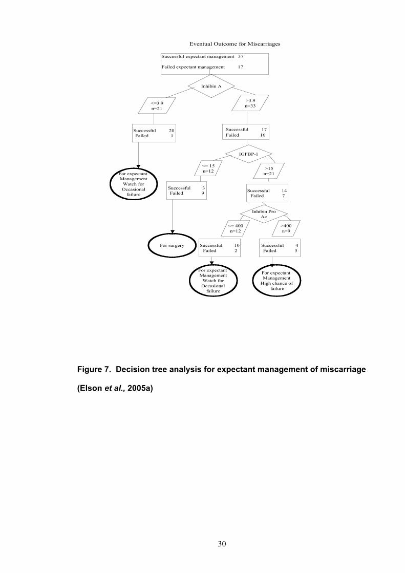
Figure 7. Decision tree analysis for expectant management of miscarriage....30
Figure 8. Schematic representation of the decision tree analysis employed in the study for the expectant management of tubal ectopic pregnancy ...............37
Figure 9. Longitudinal ultrasound image of the uterus .....................................42
Figure 10. Ultrasound measurement of retained products of conception.........43
Figure 11. Ultrasound measurements of an ectopic pregnancy ......................44
Figure 12. Schematic representation of measurement of ultrasound images of early pregnancy ................................................................................................45
Figure 13. Ultrasound picture of a missed miscarriage ....................................47
Figure 14. WGA lectin affinity chromatography by gestational age...................69
Figure 15. WGA lectin affinity chromatography by pregnancy outcome ..........72
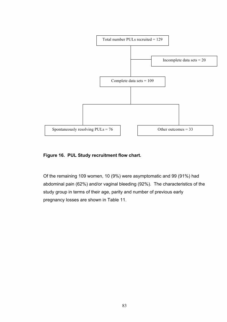
Figure 16. PUL study recruitment flow chart. ...................................................83
Figure 17. Receiver Operating Characteristics curves demonstrating the performance of the biochemical markers and the logistic regression model in their ability in predicting correctly which pregnancies will resolve without the need for intervention..........................................................................................89
Figure 18. Decision tree analysis for expectant management of PUL .............90
Figure 19. Flow-chart illustrating Trust Protocol “Management of Tubal mass”........................................................................................................................102
Figure 20. Miscarriage and ectopic pregnancy study recruitment flow chart. 105
x
Figure 21. Expectant management of miscarriage decision tree ....................110
Figure 22. Expectant management of failed pregnancies decision tree..........111
xi
INDEX OF TABLES
Table 1. Key chronological landmarks in the development of the embryo, as seen on transvaginal ultrasound examination ...................................................17
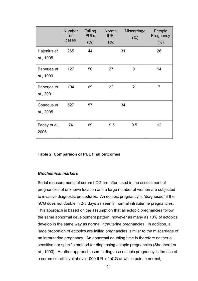
Table 2. Comparison of PUL final outcomes.....................................................20
Table 3. Guidelines for establishing the death of an embryo by ultrasound.....25
Table 4. Comparison of the cut-off levels of progesterone used in the diagnosis of early pregnancy failure ..................................................................................26
Table 5. Comparison of the cut-off levels of progesterone used in the diagnosis of viability in anembryonic pregnancy ...............................................................27
Table 6. Diagnostic criteria for diagnosis of miscarriage from CHS trust protocol ‘Management of Miscarriage’ ............................................................................46
Table 7. Comparison of median peak values between samples at <8 weeks (n=17) and >16-20 weeks gestation..................................................................70
Table 8. Comparison of median peak values between successive stages of early pregnancy.................................................................................................71
Table 9. Comparison of median peak values observed for samples at less than 8 weeks gestation between viable and failed pregnancies. ..............................73
Table 10. Protocol used for management of women with PUL ........................81
Table 12. Clinical diagnoses at completion of follow-up and time to diagnosis85
Table 13. Comparison of measured variables in spontaneous resolution and other outcomes..................................................................................................86
Table 14. Results of AUC analysis for the use of different hormonal variables to predict spontaneous resolution of PUL. ............................................................88
Table 15. Eligibility criteria for expectant management of miscarriage ..........101
Table 16. Comparison of measured variables in miscarriages with successful and failed expectant management ..................................................................107
Table 17. Comparison of measured variables in ectopic pregnancies with successful and failed expectant management ................................................108
1
CHAPTER 1. INTRODUCTION
Early pregnancy loss is perhaps the most common medical problem in women
of reproductive age. These conditions are a large burden to health services as
well as to the physical and psychological wellbeing of women and their partners.
In recent times there have been major changes in our approach to the
management of early pregnancy problems. With the introduction of early
pregnancy assessment units and transvaginal ultrasonography we have moved
away from an in-patient and surgical approach, to outpatient services and a
more conservative approach to management.
Transvaginal ultrasound is now used routinely in the assessment of early
pregnancy problems. This allows a more detailed assessment at earlier
gestations than was previous possible. These findings are often used in
combination with biochemical markers to give a more complete evaluation or
‘profile’ of the pregnancy. Serum levels of human chorionic gonadotrophin
(hCG) and progesterone are the most commonly used biochemical markers, the
optimum way to utilize them in the diagnosis and management of early
pregnancy problems remains contentious.
The move away from surgical treatment for early pregnancy problems has
economic and clinical advantages. Problems occur however when medical or
expectant management is unsuccessful and surgical intervention is required at
a later date, often in an ‘emergency’ situation. The lack of well-defined criteria
to differentiate between pregnancies that will spontaneously resolve and those
that will not is an ongoing problem for expectant management and is certainly
off-putting for patients and clinicians. Expectant management often takes
weeks to complete and success rates vary widely. Failure of expectant
management after prolonged follow-up is particularly disappointing for women
and reduces overall benefits of the management strategy.
A number of novel biochemical markers of the luteal-trophoblastic axis have
emerged in the last decade. The aim of this thesis was to investigate these
novel biochemical markers in the diagnosis and management of early
pregnancy complications. In particular I have tried to identify parameters, which
can reliably predict the success of expectant management in women with failing
pregnancies. The ability to do so would not only reduce the need for follow-up
2
but also decrease the need for surgery for both diagnostic and therapeutic
indications. The ability to estimate the likelihood of final outcome would allow
us to counsel patients appropriately and is likely to increase the uptake of
expectant management by both clinicians and patients. This would improve the
overall care of women with early pregnancy problems.
3
CHAPTER 2. LITERATURE REVIEW
2.1 NORMAL EARLY PREGNANCY
2.1.1 Fertilisation, implantation and early development
Following ovulation the ovum is taken up by the fimbrial end of the Fallopian
tube and is wafted medially by the rhythmical action of the cilia. Fertilisation of
the ovum by spermatozoa occurs either in the peritoneal cavity or within the
Fallopian tube. During the following 48 hours the conceptus travels along the
Fallopian tube and into the uterine cavity. Within 30 hours of fertilisation the
first cell division occurs, in which the fertilised ovum splits into two separate
cells. On the fifth day after conception, an additional round of division causes
the 32-celled morula to reach the blastocyst stage. This hollow ball is
composed of an inner cell mass, eventually giving rise to the fetal and
embryonic tissues, and an attached outer shell of cells known as the
trophoblast, which will ultimately give rise to the chorion. By the tenth day, the
invading trophoblast forms two distinct layers – the cytotrophoblast and the
syncytiotrophoblast. The cytotrophoblast (the inner layer) is composed of
individual, well defined and rapidly proliferating cells. The outer and thicker
layer, the syncytiotrophoblast, comprises of multinucleated cells with indistinct
cell borders (Anin et al., 2004).
The conceptus attaches to the secretory endometrium of the uterus by 5 days
post ovulation but it is not until day 12 that the blastocyst has burrowed into the
endometrium to such an extent that it is completely embedded. There is initial
decidualisation at the implantation site, which eventually extends to the whole
endometrium. The trophoblast cells produce a proteolytic enzyme which allows
invasion into the endometrium. The trophoblastic cells will form the extra-
embryonic tissue. The trophoblast differentiates in two ways – the villous and
the extravillous trophoblast. The villous trophoblast remains attached to the
villous membrane, this is responsible for maternal-fetal gas and nutrient
exchange, and hormone secretion. The cells of the extravillous trophoblast
proliferate from the tips of anchoring chorionic villi and migrate through the
maternal tissues towards decidual arterial walls (interstitial invasion) (Lyall,
2002), or infiltrate the lumens and walls of arteries to cause endovascular
4
invasion (Kaufmann et al., 2003). Endometrial arteries do not communicate
with the intervillous space before 12 weeks gestation because aggregates of
trophoblast cells derived from the cytotrophoblast shell plug their distal
segments. This protects the conceptus from high oxygen levels during this
critical stage of development (Burton et al., 1999). The definitive structure of
the placenta is apparent as early as day 21 post ovulation however the
uteroplacental circulation is not fully functional until the end of the first trimester
(Hustin & Jauniaux, 2000). Placentation, along with other early gestational
processes such as implantation, is one of the most important determinants of
pregnancy outcome.
2.1.2 Biochemistry in early pregnancy
The syncytiotrophoblast of the developing placenta plays a key part in hormone,
protein and growth factor production in early pregnancy. Human chorionic
gonadotropin (hCG) is secreted by the syncytiotrophoblast almost immediately
after implantation. This maintains the function of the corpus luteum which in
turn secretes hormones and growth factors which are essential for the
maintenance of early pregnancy. This activity of the corpus luteum decreases
after the seventh week of pregnancy at which time the trophoblast and decidua
take over as the main hormone-producing unit.
Human chorionic gonadotrophin
The glycoprotein hormone human chorionic gonadotrophin (hCG) has a
molecular weight of 36,700d (Midgley & Pierce, 1962) and was first identified in
1927 (Ascheim & Zondek, 1927). It consists of two dissimilar subunits, α and β,
which are glycosylated and non-covalently bound. The hCG-α subunit is
virtually identical to that of the human pituitary glycoprotein hormones,
luteinising hormone (LH), follicle stimulating hormone (FSH) and thyroid
stimulating hormone (TSH) (Bahl 1969a; Canfield et al., 1971). The
characteristic biological and immunological identities of these hormones are
conferred by their specific β-subunits (Pierce and Parsons, 1981). Synthesis of
hCG occurs predominantly within trophoblast cells of the blastocyst, the α and β
5
subunits being coded for separately on chromosomes 6 and 19 respectively
(Fiddes & Goodman, 1981; Boorstem et al, 1982). Post-translational
glycosylation comprises the addition of two N-linked oligosaccharides to each
subunit; on amino acids 52 and 78 (α subunit) and 13 and 30 (β subunit). In
addition to N-linked oligosaccharides, 4 O-linked oligosaccharides are located
within the hCG-β–COOH terminus (Bahl, 1969b). HCG is metabolised by the
kidney, it is desialated and then excreted into urine (Birken et al., 1996).
HCG secretion begins no later than day 7 in the blastocyst stage (Lopata &
Hay, 1989). It has been shown that its level in maternal serum doubles over
1.4-1.6 days from the time of first detection to the thirty-fifth day of pregnancy,
and then doubles over 2.0-2.7 days from the thirty-fifth to the forty-second day
(Pittaway et al., 1985). The pattern of hCG concentration throughout pregnancy
is shown in Figure 1. The half-life of hCG is 32 to 37 hours and the levels of
hCG are approximately 1000 IU/L at around 4 weeks of pregnancy, the time of
initial visualisation of a gestational sac on transvaginal ultrasound scan. HCG
secretion increases with advancing gestational age, reaching a maximal level of
50,000 to 100,000 IU/L at 10 weeks gestation. HCG levels decrease to around
10,000 to 20,000 IU/L by 20 weeks and this plateau is maintained for the rest of
pregnancy. Most commercially available monoclonal antibody-based urine
pregnancy tests can detect the presence of hCG at a level above 25 IU/L, which
corresponds to days 24 to 25 of a regular 28-day cycle. Many different
laboratory assay kits exist which are calibrated against different reference
preparations of hCG. βhCG may exist in the blood as part of the intact hCG
molecule (α and β subunits) or as free βhCG. Assay kits measure either intact
hCG, βhCG or total hCG (intact plus βhCG). These need to be taken into
account when comparing results from different investigators and appropriate
values need to be developed for individual medical centres.
In primate studies it is the exponential increase in hCG levels produced by the
implanting embryo and syncytiotrophoblast that appears to prolong the
functional lifespan of the corpus luteum (Zeleznik, 1998). In response to hCG,
the corpus luteum produces increasing concentrations of progesterone, 17 α-
hydroxyprogesterone (17-OHP) and oestradiol, and stimulates the secretion of
relaxin. hCG maintains the steroidogenesis of the corpus luteum until the ninth
to tenth week of pregnancy by which time placental steroidogenesis is
6
established and the role is entirely taken over by the placenta. It is also thought
that hCG produced by the placenta has a number of autocrine and paracrine
actions. These include involvement with the autoregulation of placental
steriodogenesis (Menon & Jaffe, 1973), and increasing syncytium formation
(Yang et al., 2003). There is also evidence that hCG modulates trophoblast
invasion by interfering with endometrial matrix metalloproteinases (MMPs) and
their tissue inhibitors (Licht et al., 2001) and influences endometrial
differentiation by modulating prolactin and insulin-like growth factor binding
protein-1 (IGFBP-1) at the implantation site (Fluhr et al., 2006).
Figure 1. Schematic representation of concentration of human chorionic
gonadotrophin (hCG) throughout gestation.
10 20 30 40
Weeks from LMP
Serum hCG (IU/L)
100,000
10,000
BIRTH
IMPLANTATION
7
Using antibodies to a linear epitope on the β-hCG-COOH terminus, a
hyperglycosylated (H-hCG) form of standard or regular hCG has been identified
as the predominant structure in early pregnancy (Sasaki et al., 2008). H-hCG
differs from hCG in both the branching and sialic acid content of N- and O-
linked oligosaccharides (Elliott et al., 1997; Cole et al., 2003). H-hCG is a
product of stem cytotrophoblast cells whereas hCG is produced by
syncytiotrophoblasts (Kovalevskaya et al., 2002a; Cole et al., 2006). The
expression of H-hCG as a proportion of total hCG declines rapidly in early
pregnancy from 92% of total hCG at 3 weeks gestation to <2% total hCG by the
second trimester (Cole et al., 2003). The relative proportion of H-hCG to total
hCG has been shown to be clinically significant. In particular implantation has
been shown to depend upon H-hCG. Low H-hCG levels are predictive of early
pregnancy loss (Sutton-Riley et al., 2006) and high concentrations are seen in
choriocarcinoma (Khanlian et al., 2003).
Inhibins
Inhibins are heterodimeric glycoprotein hormones consisting of disulfide-linked
alpha and beta subunits. Inhibin A has a molecular weight of 32kd and is
composed of inhibin α and βA subunits. It is produced by the corpus luteum
during the luteal phase of the ovarian cycle (Muttukrishna et al., 1994) and in
early pregnancy (Illingworth et al., 1996) and is also produced by the
syncytiotrophoblast in early pregnancy (Birdsall et al., 1997). There is
conflicting evidence about which is the major source of inhibin production in
early pregnancy. Santoro et al., (1992) looked at inhibin levels in women with
premature ovarian failure and donor in vitro fertilisation (IVF) pregnancies i.e.
aluteal women. They showed that there was no early rise in inhibin as seen in
normal pregnancies, although the levels did reach normal levels towards the
end of the first trimester. Lockwood et al., (1997) examined this further by
comparing blood samples of women who became pregnant following IVF with
fresh embryo transfer i.e. luteal women, with those who became pregnant
following IVF with frozen embryo transfer i.e. aluteal women. They concluded
that there was no difference in luteal and aluteal pregnancies and that therefore
the fetoplacental unit must be the major source of inhibin A in early pregnancy.
8
Treetampinich et al., (2000) however found that in IVF cycles inhibin A levels
were significantly lower in the absence of functioning ovaries and in natural
cycles compared with concentrations after ovarian stimulation. They also found
that inhibin A concentrations were not significantly different between singleton
and multiple pregnancies and therefore concluded that the corpus luteum is the
major source of circulating inhibin A in early pregnancy.
In 2002 Muttukrishna et al., found decreased maternal serum levels of inhibin A
in pregnancies that went on to miscarry and in 2006 Hwang et al., showed a
significant association between the number of fetuses and maternal inhibin A
levels in a study of singleton and multiple pregnancies following IVF and
embryo transfer. These also confirm that the trophoblast is the major source of
inhibin A after the luteo-placental shift in early pregnancy.
Circulating inhibin A levels are at detectable levels by 4 weeks gestation
(Lockwood et al., 1997) and climb to a peak at 8-10 weeks of gestation
(Tovanabutra et al., 1993; Illingworth et al., 1996, Phupong, Hanprasertpong &
Honsawek, 2008). Levels then fall slightly at 16 weeks and thereafter increase
progressively to maximal concentrations in week 36 (Fowler et al., 1998). The
clearance of inhibin A is fast with a short half-life of around 45 minutes
(Muttukrishna et al., 1997). Inhibin A is thought to be involved in regulating
placental hCG production by inducing changes in gonadotrophin-releasing
hormone (GnRH) secretion (Petraglia et al., 1987). It is also thought to play a
part in the cell signalling and therefore possibly trophoblast invasion (Debieve et
al., 2000). Animal studies have also suggested a role of inhibin A in maintaining
luteal progesterone output (Webley et al., 1994). Its function in humans
however, remains unclear. It has a shorter half-life than either hCG or
progesterone and therefore may be more sensitive at reflecting changes in the
trophoblast. Lower levels have already been demonstrated in women with
biochemical pregnancies and missed miscarriages (Glennon Phipps et al.,
2000; Muttukrishna et al., 2002). In women with induced pregnancy
termination, inhibin A levels have been shown to fall after the administration of
misoprostol, which interrupts trophoblastic blood flow and leads to expulsion of
the pregnancy (Lahiri et al., 2003). Illingworth et al., (1996) examined inhibin A
levels in ectopic pregnancies, complete and incomplete miscarriages, and
ongoing pregnancies, and found no significant differences between the groups.
9
Phipps et al., (2000) found that among dual biomarker combinations for
differentiating viable from nonviable pregnancies, the addition of inhibin A to
progesterone improved the specificity but not sensitivity of the test. A more
recent pilot study (Johns et al., 2007) however, using logistic regression
analysis, found that inhibin A alone is the best predictor of first trimester
miscarriage. Kirk et al. (2009) found that serum inhibin A levels may be of
some use in predicting failing PULs and IUPs in the PUL population.
Inhibin pro-αC, a pre-cursor protein of the inhibin α subunit, circulates as a
functionally inactive monomer and as part of high molecular weight functional
dimers. Inhibin pro-αC-related immunoreactivity (inhibin pro-αC-RI) is a
cumulative measurement of monomeric pro-αC subunit and pro-α containing
inhibins (Illingworth et al., 1996). Inhibin pro-αC-RI has been found to peak at
around day 16 after conception and then fall to a nadir at 16 weeks of gestation,
before increasing to a second peak at 36 weeks (Illingworth et al., 1996; Fowler
et al., 1998). Despite the secondary rise from the early second trimester,
absolute levels are at their highest in very early pregnancy (Fowler et al., 1998).
In their study of 334 women undergoing IVF, Tong et al. (2004) found that this
early peak in circulating inhibin pro-αC in very early pregnancy was a consistent
and specific feature of clinical pregnancy. Illingworth et al. (1996) also found
that inhibin pro-αC-RI concentration was an indicator of continuing pregnancy
viability, better than either hCG or inhibin A.
In their study of aluteal and luteal pregnancies Lockwood et al. (1997)
compared serial levels of pro-αC-RI in early pregnancy in these two groups.
They found that pro-αC-RI was significantly higher in those pregnancies with
multiple corpora lutea compared to those with single corpora lutea and
significantly lower in those women with conceptions from frozen embryos i.e.
aluteal than those with fresh embryos i.e. luteal. They therefore concluded that
the corpus luteum must be the major source in early pregnancy. Fowler et al.,
(1998) identified a small rise in inhibin A levels and a peak of hCG coinciding
with the fall in maternal venous pro-αC concentrations after week 9. These are
likely to reflect the luteal to placental shift in support for the pregnancy. Inhibin
pro-αC is secreted by the placenta into the fetal circulation at term, and is
thought to play a role as a paracrine and endocrine regulator of placental
10
function (Riley et al., 2000). IVF protocols involving complete ovarian
suppression (and therefore absence of luteal tissue) are compatible with
successful pregnancy (Lockwood et al., 1998), demonstrating that Inhibin pro-
αC is not, however, essential for successful pregnancy.
The early increase in inhibin pro-αC-RI in human pregnancy (Illingworth et al.,
1996, Lockwood et al., 1997) supports findings from animal models. Webley et
al., (1994) found that in the marmoset monkey circulating inhibin pro-αC-RI
were significantly elevated above ‘normal’ luteal phase concentrations as early
as 5 days after ovulation. Measurements of pro-αC-RI concentrations could
thus serve as a useful marker of luteal sufficiency during the establishment of a
pregnancy.
Steroids
Progesterone
Progesterone is a C-21 steroid hormone derived from cholesterol. It is one of
the primary products of the corpus luteum and plays a pivotal role in the
establishment and maintenance of pregnancy (Spencer & Bazer, 2004).
Progesterone acts on the uterus to stimulate and maintain uterine functions that
are permissive to early embryonic development, implantation, placentation and
successful fetal and placental development to term. These include endometrial
6 weeks Gestational sac (mean diameter 16 mm) and yolk sac with adjacent heart beat but small embryo (3 mm)
6 ½ weeks Embryo with crown-rump length of 6 mm with heart beat visible (rate 125 bpm)
7 weeks Embryo with crown-rump length of 10 mm with heart beat visible (rate 150 bpm)
8 weeks Embryo with crown-rump of 16 mm with separate amniotic sac and coelomic cavity with yolk sac. Fetal body movements visible, heart rate 175 bpm.
Table 1. Key chronological landmarks in the development of the embryo,
as seen on transvaginal ultrasound examination (Hately et al., 1995).
2.2 EARLY PREGNANCY PROBLEMS
2.2.1 Initial assessment
Early pregnancy failure is a large burden on health services because of its high
incidence and complex clinical management, which often requires the use of
multiple diagnostic tests and in-patient surgical treatment. Assessment of early
pregnancy is indicated in women with clinical symptoms suggestive of
miscarriage or ectopic pregnancy and in asymptomatic women who have
experienced miscarriage in the past and those at high risk of ectopic pregnancy
(Ankum et al., 1996). Assessment tools include ultrasonography and
measurement of biochemical markers (see Figure 4).
18
Figure 4. Basic diagnostic algorithm for early pregnancy loss (RCOG 2006)
pregnancy after sterilisation and with intrauterine contraceptive device (IUCD) in
situ, and smoking (Marchbanks et al., 1988; Ankum et al 1996; Bouyer et al.,
2003). In theory an abnormal conceptus could be predisposed to ectopic
implantation due to delayed migration but studies have not confirmed an
important role for chromosomal abnormalities in the aetiology of ectopic
pregnancies (Goddijn et al., 1996).
32
Biochemical markers
Biochemical markers have been used both to diagnose ectopic pregnancy and
in its management. Abnormal implantation leads to reduced levels of βhCG
being seen in ectopic pregnancies. Traditionally an ectopic pregnancy is
diagnosed if the hCG does not double in 2-3 days as seen in normal intrauterine
pregnancies. However, this may also be the case in a failing intrauterine
pregnancy. An abnormal doubling time is neither sensitive nor specific method
to diagnose ectopic pregnancy (Shepherd et al., 1990). Another approach used
to diagnose ectopic pregnancy is a serum cut-off level above 1000-1500 IU/L of
βhCG at which point an intrauterine pregnancy should be seen by transvaginal
sonography. However this fails to take into account the time to return to normal
of serum βhCG levels following miscarriage or the diagnostic accuracy of
ultrasound in the presence of uterine anomalies such as fibroids (Barnhart et
al., 1999). The serum level of hCG would appear to be higher in those women
with deeper trophoblastic invasion into the tubal wall than in those where the
ectopic trophoblast is limited to the lumen or tubal mucosa (Natale et al., 2003).
Several studies have demonstrated reduced progesterone and 17-OHP levels
in ectopic pregnancies (Hahlin et al., 1991; Choe et al., 1992; Stewart et al.,
1995). This is thought to be due to abnormal implantation thus affecting the
luteal-placental axis (Sauer et al., 1988). Progesterone and βhCG may
therefore be used together with ultrasound for the diagnosis and management
of pregnancies of unknown location as these pregnancies are likely to resolve
spontaneously regardless of location (Banerjee et al., 1999).
Only one small study has assessed the value of more novel biochemical
markers in the diagnosis of ectopic pregnancy. Illingworth et al., (1996)
examined eight women with ectopic pregnancies and compared their inhibin A
and pro-αC-RI levels with levels of eight women with ongoing intrauterine
pregnancies. There was no significant difference between the two groups.
Ultrasound
Traditionally the findings of a positive pregnancy test and an empty uterus seen
at the time of ultrasound scan have been synonymous with the presence of an
33
ectopic pregnancy. However with the use of transvaginal ultrasound,
approximately 85% of ectopic pregnancies can be visualised directly (Ofili-
Yebovi et al., 2003) and so transvaginal ultrasound has become the single
diagnostic tool of choice for ectopic pregnancy. Like laparoscopy, ultrasound
does not confer 100% sensitivity for the diagnosis of tubal ectopic pregnancy,
however it is safe, inexpensive and non-invasive, is acceptable by women and,
in trained hands, is highly reproducible (Condous, 2007).
The following transvaginal ultrasonagraphic criteria are used for the diagnosis of
ectopic pregnancy: (1) an inhomogenous adnexal mass (“blob sign”) (Condous
et al., 2005); (2) an empty extrauterine sac with a hyperechoic ring (“bagel-
sign”) (Goldstein & Timor-Tritsch, 1995); and (3) a yolk sac of fetal pole with or
without cardiac activity in an extrauterine sac. Recent studies using high-
resolution sonography have shown the most common morphology to be a solid
ectopic pregnancy (Elson et al., 2000) and in a meta-analysis of ten studies, a
non-cystic adnexal mass or an inhomogenous mass was diagnostic of an
ectopic pregnancy with a sensitivity and specificity of 84.4% and 98.9%
respectively (Brown & Doubilet, 1994).
Management
There are three main methods of currently managing an ectopic pregnancy:
surgical, medical and expectant. Surgical management is indicated in all
haemodynamically unstable patients and in selected other cases. Surgical
options include salpingectomy or salpingostomy, done either as an open
procedure or laparoscopically. Three randomised trials have shown that
laparoscopy is superior to laparotomy in haemodynamically stable patients
(Vermesh et al., 1989, Lundroff et al., 1991, Koninckx et al., 1991).
The folic acid antagonist methotrexate has been widely used for the medical
treatment of ectopic pregnancies since the late 1980s. Methotrexate inhibits
dihydrofolate reductase and so prevents the reduction of folic acid to
tetrahydrofolate, a key step in the synthesis of DNA and RNA precursors.
Methotrexate therefore leads to interference with DNA synthesis and cell
multiplication in the conceptus. It can be given either intramuscularly or by
34
direct injection into the ectopic pregnancy, either laparoscopically or under
ultrasound guidance. In the UK methotrexate is most commonly given as a
single intramuscular dose of 50 mg/m2. Single dose regimens have reported
success rates varying from 64-94% (Stovall & Ling, 1993, Stika et al., 1996).
Methotrexate treatment is more likely to be successful if the initial hCG level is
low. Ransom et al., (1994) also showed that ectopic pregnancies with serum
progesterone <10 nmol/L are more likely to be successfully treated by systemic
methotrexate injection.
It is now well recognised that not all ectopic pregnancies require treatment as
some will resolve spontaneously. Expectant management is becoming
increasingly important as the ability to detect small ectopic pregnancies and
tubal miscarriages increases. It is important that the ectopic pregnancy is
actually visualised to avoid mistakenly managing expectantly live or large
ectopic pregnancies where the risk of failure is high.
Large multicentre randomised controlled trials are currently underway in the UK
and Netherlands (The METEX study, van Mello et al., 2008) to compare
methotrexate and expectant management in haemodynamically stable patients
with an ectopic pregnancy and low serum hCG or PUL with low but plateauing
hCG concentrations. Results and guidance from these ongoing studies are
eagerly awaited.
Studies show that around a quarter of ectopic pregnancies will be suitable for
expectant management (Ylostalo et al., 1992; Elson et al., 2004). The selection
criteria for expectant management varies but those ectopic pregnancies with a
viable fetus or the presence of haemoperitoneum would be considered
unsuitable for all but surgical management. In expectant management once the
ectopic pregnancy is diagnosed management varies but consists of follow-up
with a combination of serial ultrasound scans, hCG and progesterone
measurements. The hCG levels are monitored until they drop below 20 IU/L
indicating spontaneous resorption of the pregnancy. An increase in the size of
the ectopic pregnancy or a rise in the serum hCG levels would be an indication
to consider surgery.
Success rates for expectant management vary between 50-100% (Lund, 1955;
Sauer et al., 1987). Several attempts have been made to examine the clinical,
35
ultrasound and biochemical parameters that can predict the success of
expectant management. Fernandez et al., (1988) looked at 14 patients with
ectopic pregnancies confirmed by laparoscopy. 64% of these resolved
spontaneously. This study found that a serum hCG below 1,000 IU/L appeared
to be the best marker for successful expectant management. Garcia et al.,
(1987) reported on 13 women with ectopic pregnancies of less than 4cm in size
diagnosed at laparoscopy. Only one case required surgical intervention. They
found that serum βhCG, progesterone and oestradiol levels were all below the
ranges expected for normal pregnancies but did not describe any threshold
levels. Shalev et al., (1995) examined 60 women with laparoscopically
diagnosed ectopic pregnancies. They found that the presenting level of hCG,
the rate of fall of hCG and the size of the ectopic pregnancy at laparoscopy
were significant factors in predicting successful expectant management. They
suggested that using a presenting level of hCG of <2,000 IU/L allowed a 60%
success rate.
Ylostalo et al., (1992) examined 83 patients, which represented 26% of all their
ectopic pregnancies over a 2-year period. They found that around 69% of the
83 cases, or 18% of all ectopic pregnancies, resolved spontaneously. They
also used 4 cm as a cut-off for the size of the ectopic pregnancy and included
those ectopic pregnancies with a fetal pole and no fetal heartbeat. Whilst they
also found that the hCG levels were significantly higher in the group with failed
expectant management than those who finally resolved spontaneously, there
were cases with high initial values with successful expectant management. No
attempt to define whether the morphology of the ectopic pregnancies
contributed to the final outcome was examined.
Sauer et al., (1987) compared the biochemical profiles of spontaneously
resolving ectopics, viable ectopics and normal intrauterine pregnancies. They
found that hCG, progesterone, 17-OHP, and oestradiol were all significantly
lower in ectopic pregnancies. By using a low threshold for progesterone of 4
nmol/L, they found that the ectopic pregnancies with progesterone below this
level had a shorter time to resolution. There was a high degree of correlation
between 17-OHP and progesterone. The fall in progesterone and 17-OHP
preceded the fall in hCG levels by 7-29 days.
36
Cacciatore et al., (1995) examined the sonographic findings and hCG levels in
expectantly managed ectopic pregnancies. They found that 69% of 71 patients
had spontaneously resolving ectopic pregnancies. They concluded that whilst
initial hCG and size of the ectopic pregnancy did not differ between the two
groups, a decrease in the size of the ectopic pregnancy by day 7 was a
significant predictor.
Elson et al., (2004) found significant differences in demographic, ultrasound and
biochemical findings between spontaneously resolving ectopics and those
requiring treatment. They devised a decision tree that may be used as a guide
to estimate the probability of successful expectant management in individual
cases (see Figure 8). This could predict outcome with a probability of 88% in
five out of the seven subgroups, which accounted for 69% of the study
population. As novel biochemical markers have been shown to be more
accurate indicators of the luteal-trophoblastic axis (Elson et al., 2005a) it is likely
that they may be useful for predicting the spontaneous resolution of ectopic
pregnancies more accurately.
Fertility rates after expectant management have been examined and patients
treated in this way have good long-term fertility outcomes with spontaneous
pregnancy rates of around 80% (Carp et al., 1986). The risk of repeat ectopic
pregnancies is low, around 4%.
37
Figure 8. Schematic representation of the decision tree analysis
employed in the study for the expectant management of tubal ectopic
pregnancy (Elson et al., 2004).
38
2.3 SUMMARY
It is clear from the literature review presented that the management of early
pregnancy problems has benefited from the introduction of transvaginal
ultrasound scanning and the adjunctive use of biochemical markers. We have
also identified a number of areas in need of further research to improve our
understanding of the pathophysiology of early pregnancy failure and to improve
the care received by patients with early pregnancy problems. Our research
questions include is the glycosylation of hCG involved in early pregnancy
failure? Can the successful expectant management of miscarriage and failed
pregnancies be predicted using novel biochemical markers as described by
Elson et al., (2005a, 2005b) in our own population? And will these biochemical
markers similarly predict the successful expectant management of pregnancies
of unknown location?
The original research presented endeavors to answer these questions. The
glycosylation of hCG and expectant management studies were conceived
separately and are not related apart from in their overarching theme ‘the use of
novel biochemical markers in the diagnosis and management of early
pregnancy problems’.
39
CHAPTER 3. MATERIALS AND METHODS
3.1 PATIENT RECRUITMENT
Patients for these prospective observational studies were recruited from the
Early Pregnancy Assessment Unit of Sunderland Royal Hospital during the
period August 2005 and June 2008. The Unit serves a local community of
330,000 residents including both inner city and rural areas with a high level of
socio-economic deprivation. This is a secondary referral unit seeing
approximately 1300 women per year. Women can be referred by their midwife,
general practitioner, accident and emergency department, family planning
department or hospital doctor if they have pain or bleeding in early pregnancy.
The unit also has an open access policy for women who have had a previous
ectopic pregnancy or two previous miscarriages.
Patients were also recruited from the Early Pregnancy Assessment Unit of
King’s College Hospital, London, for the pregnancy of unknown location and
ectopic pregnancy studies, during the period August 2006 and July 2007. The
Unit serves a racially mixed inner city population with a high level of socio-
economic deprivation. This is a tertiary referral unit seeing approximately 2,500
women per year. The unit has an open access policy and additionally sees
women referred by their general practitioner or hospital consultants.
In both units all women are triaged by a nurse and if appropriate undergo a
urine pregnancy test (Clearview HCG IITM, Unipath, Bedford, UK). This test is a
monoclonal antibody test which according to the manufacturers specifications
has a sensitivity of 99% at a urine β-hCG level greater than 25 IU/L. Those
women with a positive test then undergo ultrasound scanning and biochemical
testing as appropriate.
Clinically stable women with an ultrasound diagnosis of pregnancy of unknown
location, missed or incomplete miscarriage, or ectopic pregnancy, who were
suitable for and chose expectant management were eligible to take part in the
expectant management studies.
Informed written consent was taken from all women prior to inclusion in the
studies.
40
3.2 ETHICS COMMITTEE APPROVAL
Approval for recruitment of women into the glycosylation of hCG study was
granted by Sunderland Local Research Ethics Committee (SLREC 704) and for
the expectant management studies by Northumberland Regional Ethics
Committee (05/Q0902/63).
3.3 ULTRASOUND
3.3.1 Ultrasound Equipment
The equipment used at Sunderland Royal Hospital was a Toshiba Powervision
6000, with a Toshiba IPVM-651VT 6MHz transvaginal probe (Toshiba Medical
Systems Ltd, Tokyo, Japan) when a TVS was indicated. The equipment used at
King’s College Hospital was an Aloka ultrasound system with a 5MHz
transvaginal probe. (Aloka SSD-5000, Aloka Co. Ltd, Tokyo, Japan). The
mechanical index (MI) was continuously displayed during examination and it
was always kept <1.
3.3.2 Ultrasound Method
Ultrasounds were performed by sonographers and verbal consent for the
procedure was obtained in all cases. In accordance with departmental
guidelines initial scans were performed transabdominally when the estimated
gestational age was nine weeks or greater, and transvaginally when less than
nine weeks. Transvaginal scans were performed following transabdominal
scans when required. The bladder was emptied prior to transvaginal scanning.
The probe was introduced gently into the vagina and the cervix and uterus
demonstrated in the sagittal plane. The probe was then rotated through 90o and
the uterus examined in the coronal plane from fundus to the cervical region.
Whilst in the coronal plane the tip of the probe was tilted to the patients right
and the right ovary and adnexa examined in the coronal and sagittal planes.
The tip of the probe was then tilted to the other side and the left ovary and
adnexa then examined in the same way. Finally the pouch of Douglas was
inspected for the presence of free fluid.
41
3.3.3 Ultrasound measurements
All measurements were done on a frozen ultrasound image with callipers. The
endometrial thickness was measured from a longitudinal image through the
thickest area of the endometrium, from the outermost border of the
endometrium on one side to that on the other side.
42
Figure 9. Longitudinal ultrasound image of the uterus, with the measurement of endometrial thickness from outermost border of the
endometrium on one side to the other (image courtesy of J. Elson)
In the case of an incomplete miscarriage, the intrauterine diameter of the
retained products of conception was determined by taking two further
measurements in the coronal plane at the thickest area and calculating the
diameter as endometrial thickness x diameter 2 x diameter 3 divided by 3.
43
Figure 10. Ultrasound measurement of retained products of conception – longitudinal section (Endometrial thickness 22.5 mm)
Tubal ectopic pregnancies were diagnosed only when there was an adnexal
mass with morphological characteristics of an ectopic separate to the ovary and
corpus luteum. For ectopic pregnancies the average diameter of the ectopic
pregnancy was calculated by measuring the ectopic pregnancy in three
dimensions.
The morphology of the ectopic pregnancy was classified into four categories:
gestational sac with an embryo, gestational sac with a yolk sac, gestational sac
with no detectable embryonic structures and homogenous or solid tubal mass.
44
Figure 11. Ultrasound measurements of an ectopic pregnancy showing a solid left sided tubal mass
Measurements of intrauterine contents in missed miscarriages or normal
pregnancies are demonstrated in Figure 12.
45
Gestational (chorionic) sac - Measurements should be performed from the inner edges of trophoblast in three planes. The diameters measured correspond to those of the chorionic cavity. The maximum and mean diameters should be recorded. The volume is calculated using formula for ellipsoid V=AxBxCx 0.523.
Amniotic sac - The three perpendicular diameters should be measured and the mean diameter calculated. As the amnion is very thin the measurements should be taken from the centre of the membrane.
Yolk sac - Three diameters are measured from the outer wall of the yolk sac.
Crown- rump length - In early pregnancy this is the greatest length of the embryo as the crown and rump cannot be distinguished. From 7 weeks onwards the
measurement should be taken in the sagittal section, with care taken not to include the yolk sac.
Figure 12. Schematic representation of measurement of ultrasound images of early pregnancy showing gestational sac (GS), yolk sac (YS), amniotic sac (AS) and fetal pole (FP) (diagram courtesy of J. Elson)
YS
GS
AS
F P
46
Diagnosis of miscarriages were made in accordance with departmental
guidelines as outlined in Table 6.
The diagnosis of miscarriage can be made if ultrasound demonstrates one of
the following:
• Retained products of conception (>15 mm AP diameter)
• Gestation sac >20 mm (mean sac diameter) with no contents or fetal
pole
• Fetal pole >10 mm with no fetal heart pulsation
• No change in 2 scans with intrauterine pregnancy over 10 days
Table 6. Diagnostic criteria for diagnosis of miscarriage from CHS trust protocol ‘Management of Miscarriage’
47
Figure 13. Ultrasound picture of a missed miscarriage with no change in
two scans over 10 days
3.4 BIOCHEMISTRY
Blood samples were collected in plain tubes. All blood samples were
centrifuged for 10 minutes at 1,000 RPM at room temperature and the serum
extracted and frozen at -20oC. The hCG and progesterone assays for those
patients with either pregnancies of unknown location or ectopic pregnancies
were measured immediately. All others samples were frozen for later analysis
in batches.
The panel of novel biochemical markers was chosen to reflect all aspects of the
luteo-trophoblastic unit. Progesterone, inhibin pro-αC and 17-OHP are
products of and reflect the function of the corpus luteum. hCG is the major
product trophoblast and IGFBP-1 is secreted by the decidua. Inhibin A is
produced both by the trophoblast and the corpus luteum.
48
3.4.1 hCG assay
Serum hCG concentrations (intact hCG plus the hCG β-subunit) were quantified
using an automated immunoassay technique and expressed in IU/L using the
World Health Organisation Third International Reference 75/537. For the
glycosylation of hCG study the analysis was carried out using the Immulite 2000
(Diagnostic Products Corporation, Los Angeles, CA, USA). For the expectant
management studies the analysis was carried out using the Roche E170
(Roche Diagnostics, Mannheim, Germany) analyser at Sunderland Royal
Hospital and the Bayer Immuno1TM (Bayer Diagnostics, Basingstoke, UK) was
used at King’s College Hospital.
Immulite
Immulite hCG is a solid-phase, two site, chemiluminescent enzyme
immunometric assay designed for the quantitative measurement of hCG in
serum.
o 5 µL of sample, reagent (alkaline phosphatase conjugated to polyclonal
ovine anti-hCG in buffer) and beads are added together and incubated
for 30 minutes.
o Reagent is removed from beads by spinning the reaction tube at high
speed along its vertical axis.
o Beads are washed then chemiluminescent substrate is added.
o The light emitted is detected at 477 nm by a photomultiplier tube.
The inter and intra assay coefficients of variation are less than 10%.
Roche E170
The test principle is a competitive immunoassay using two incubations.
o 1st incubation: 30 µL of sample, biotinylated monoclonal hCG-specific
antibodies, and a monoclonal hCG specific antibody labelled with a
ruthenium complex react to form a sandwich complex.
o 2nd incubation: after addition of streptavadin-coated microparticles the
complex becomes bound to the solid phase via interaction of biotin and
streptavadin.
49
o The reaction mixture is aspirated into the measuring cell where the
microparticles are magnetically captured onto the surface of the
electrode. Unbound substances are then removed with ProCell.
Application of a voltage to the electrode then induces chemiluminescent
emission which is measured by a photomultiplier.
o Results are determined via a calibration curve which is instrument-
specifically generated by 2-point calibration and a master curve provided
via a reagent bar code.
Bayer Immuno1
The immunoassay technique used was a heterogenous sandwich magnetic
separation assay (MSA).
o The hCG antibody conjugate (R1) and the hCG antibody conjugate 2
(R2) are reacted with the patient sample and incubated at 37 0C.
o The monoclonal Immunomagnetic Particle (mIMP) reagent is added and
a second incubation period occurs during which the antibody complex is
bound. The mIMP/antibody complex is then washed and the para-
nitrophenyl phosphate (pNPP) substrate is added. The alkaline
phosphatase (ALP) in the antibody conjugate reacts with the pNPP to
form para-nitrophenoxide and phosphate. Increasing absorbance due to
formation of para-nitrophenoxide is monitored at 405 nm and 450 nm.
o The dose/response curve will be directly proportional to the hCG
concentration in the sample.
The inter and intra assay coefficients of variation are less than 10%.
A test series of 4 samples analysed at both sites (Sunderland Royal Hospital
and King’s College Hospital) shows an 11% between-method variation (range
0-17%).
3.4.2 Progesterone assay
Progesterone levels were quantified using an automated immunoassay and
expressed in nmol/L. At Sunderland Royal Hospital the analysis was carried
50
out using the Roche E170 (Roche Diagnostics, Mannheim, Germany) analyser
and at King’s College Hospital the Bayer Immuno1TM (Bayer Diagnostics,
Basingstoke, UK) was used.
Roche E170 The test principle is a competitive immunoassay using two incubations.
o 1st incubation: 30µL sample – in the presence of a biotinylated
monoclonal progesterone-specific antibody and a progesterone
derivative labelled with ruthenium complex – are incubated with Danazol
to release progesterone. Progesterone from the sample competes with
the labelled progesterone derivative for the antibody binding site.
o 2nd incubation: After addition of streptavidin-coated microparticles, the
complex becomes bound to the solid phase via interaction of biotin and
streptavidin. The amount of the labelled progesterone derivative bound
to the solid phase is inversely proportional to the progesterone content of
the sample.
o The reaction mixture is aspirated into the measuring cell where the
microparticles are magnetically captured onto the surface of the
electrode. Unbound substances are then removed with ProCell.
Application of a voltage to the electrode then induces chemiluminescent
emission which is measured by a photomultiplier.
o Results are determined via a calibration curve which is instrument-
specifically generated by 2-point calibration and a master curve provided
via the reagent barcode.
51
Bayer Immuno1
The immunoassay method used is a heterogenous competitive immunoassay.
o Anti-progesterone antibody (R1) is reacted with the patient sample and
incubated at 37oC.
o Progesterone enzyme conjugate (R2), which competes with the
progesterone in the sample for binding sites on the antibody is then
added followed by the mIMP. A second incubation occurs during which
the antibody/hapten complex is washed and the pNPP substrate is then
added. The alkaline phosphatase in the antibody conjugate reacts with
the pNPP to form para-nitrophenoxide and phosphate. Increasing
absorbance due to formation of para-nitrophenoxide is monitored at 405
nm and 450 nm.
o The colour production in the reaction is inversely proportional to the
progesterone concentration.
The inter and intra assay coefficients of variation are less than 10%.
Our test series of 4 samples comparing the two methods confirms a 2-3 nmol
between-method variation, as quoted by UK National External Quality
Assessment Service (NEQAS). This is taken into account during statistical
analysis.
3.4.3 17 - α-OH progesterone assay
This was quantified using an enzyme linked immunoassay DSL-10-6800
ACTIVE (Diagnostic Systems Laboratories, USA) and expressed as ng/mL.
This ELISA uses the competitive binding enzyme immunoassay format. In the
assay, standards, controls and unknowns containing 17α-OH progesterone (17-
OHP) are incubated with biotin-labeled 17-OHP and rabbit anti-17-OHP
antiserum in microtitration wells coated with goat anti-rabbit gamma globulin
where the unlabeled and biotin-labeled antigens compete for a limited number
of anti-17α-OH progesterone binding sites. After incubation and washing, the
wells are incubated with streptavidin-horseradish peroxidase (HRPO), which
binds to the biotinylated 17α-OH progesterone. The unbound streptavidin-
HRPO is washed away, followed by incubation with the substrate
52
tetramethylbenzidine (TMB). An acidic stopping solution is then added to stop
the competition reaction, and the degree of enzymatic turnover of the substrate
is determined by dual wavelength absorbance measurement at 450 and 620
nm. The intra-assay coefficients of variation were 5.1% at 0.49 ng/mL and
4.9% at 6.13 ng/mL, the inter-assay coefficients of variation were 6.76% at 0.49
ng/mL and 5.38% at 6.63 ng/mL.
Assay Procedure
1. Pipet 50 µL of the standards, controls and unknowns into the microtiter
wells.
2. Add 50 µL of the 17-OHP biotin conjugate solution to each well.
3. Add 100 µL 17-OHP antiserum to each well.
4. Incubate the wells shaking at a fast speed (500-700 rpm) at room
temperature (~25°C) for 1 hour.
5. Aspirate and wash each well 5 times with the wash solution and
blot dry.
6. Add 200 µL of streptavidin-enzyme conjugate solution to each well.
7. Incubate the wells shaking at a fast speed (500-700 rpm) on an orbital
microplate shaker, for 30 minutes at room temperature (~25°C).
8. Aspirate and wash each well 5 times with the wash solution and blot dry.
9. Add 100 µL of the TMB chromogen solution to each well.
10. Incubate the wells shaking at a fast speed (500-700 rpm) at room
temperature (~25°C) for 30 minutes. Avoid exposure to direct sunlight.
11. Add 100 µL of the stopping solution to each well.
12. Read the absorbance of the solution in the wells within 30 minutes, using a
microplate reader set to 450 nm.
53
3.4.4 Inhibin A assay
Inhibin A was quantified using an enzymatically amplified "two-step" sandwich-
type immunoassay DSL-10-28100 ACTIVE (Diagnostic Systems Laboratory,
USA) and expressed as pg/mL. In the assay, duplicates of standards, controls
and unknown serum samples are incubated in microtitration wells that have
been coated with anti-inhibin βA subunit antibody. After incubation and
washing, the wells are treated with another anti-inhibin alpha subunit detection
antibody labelled with the enzyme horseradish peroxidase (HRP). After a
second incubation and washing step, the wells are incubated with the substrate
tetramethylbenzidine (TMB). An acidic stopping solution is then added and the
degree of enzymatic turnover of the substrate is determined by dual wavelength
absorbance measurement at 450 and 620 nm. The absorbance measured is
directly proportional to the concentration of inhibin A present. A set of inhibin A
standards were used to plot a standard curve of absorbance versus inhibin A
concentration from which the inhibin A concentrations in the unknowns can be
calculated.
Assay Procedure
1. Pipet 50 µL of the standards, controls, and unknowns to the wells of the
microtitre plate.
2. Add 50 µL of Inhibin A sample buffer A to each well.
3. Add 50 µL of Inhibin A sample buffer B to each well.
4. Incubate the wells, shaking at 500-700 rpm on an orbital microplate shaker,
for 3 hours at room temperature.
5. Aspirate and wash each well 6 times with the Wash Solution and blot dry.
6. Add 100 µL of the Inhibin A antibody-enzyme conjugate solution to each
well.
7. Incubate the wells on an orbital microplate shaker set at 500-700 rpm for 1
hour at room temperature.
8. Aspirate and wash each well 6 times with the wash solution and blot dry
9. Add 100 µL of the TMB chromogen solution to each well
10. Incubate the wells on an orbital microplate shaker set at 500-700 rpm for 15
54
minutes at room temperature. Avoid exposure to direct sunlight.
11. Add 100 µL of the stopping solution to each well using a semi-automatic
dispenser.
12. Read the absorbance of the solution in the wells within 30 minutes, using a
microplate reader set to 450 nm.
3.4.5 Inhibin proαC-RI assay
Inhibin pro-αC was quantified using a solid phase sandwich ELISA (Oxford Bio-
Innovation MCA 1254KZZ), and expressed as pg/mL. The wells of a microtitre
plate come dry-coated with a monoclonal antibody specific for the pro region of
the alpha subunit of inhibin. Samples are incubated in the wells so that the
antigen binds to the ‘capture’ or immobilised antibody via its pro region of the
alpha subunit. Following washing of the plate a ‘second’ or detection antibody
is added. This is the Fab fragment of a monoclonal antibody specific to the
alpha subunit of inhibin coupled to alkaline phosphatase. Any unreacted
material is then removed by washing before the detection of alkaline
phosphatase using a sensitive amplified substrate reaction. This results in a
red reaction product with a colour intensity which is directly proportional to the
concentration of inhibin-pro-αC related materials present in the original sample.
The assay has less than 0.1% cross reactivity with inhibin A, inhibin B, activin A,
activin B and follistatin.
Assay Procedure
1. Add 50 µL of each sample and standard dilution in duplicate, to the wells of
the microtitre plate.
2. Add 50 µL of assay diluent in duplicate wells as a zero analyte sample.
Cover the plate with a plate sealer and incubate overnight at 4oC.
3. To 1 vial of MCA1254A alkaline phosphatase conjugated Fab mouse anti
human inhibin alpha subunit, add 1 mL of pro-αC assay diluent. Replace
the top and mix. Remove the contents and add to a further 5 mL of assay
diluent.
55
4. Wash the wells of the microtitre plate by filling each well to the top with Pro-
αC washing buffer allowing to stand for about 15 seconds and then
decanting or aspirating each well thoroughly. Repeat this step a further 3
times. Invert the plate to drain on absorbent paper.
5. Add 50 µL MCA1254A prepared in step 3 to each well of the microtitre plate.
6. Cover and seal the plate and incubate at room temperature for 1 hour.
7. Wash as in step 4 but with 8 cycles and ending with the wells filled with
buffer. Leave the plate to soak for 15 minutes at room temperature whilst
preparing the substrate.
8. Prepare the substrate by adding the substrate diluent to the lyophilised
substrate. Mix for 5 minutes.
9. Remove the buffer from the plate wells and further wash the plate for 2-3
cycles. Drain the plate dry by inversion on absorbent paper.
10. To each well of the plate add 50 µL of substrate solution.
11. Cover and seal the plate and incubate at room temperature for 2 hours.
12. Prepare the amplifier by adding the amplifier diluent to the lyophilised
amplifier. Mix for 5 minutes.
13. To each well of the plate add 50 µL of amplifier solution. Agitate gently to
mix.
14. Cover the plate and incubate at room temperature. Colour will appear quite
rapidly. Read the absorbance values, at 5 minute intervals, of each well at
490 nm. Preferably referencing at 620 nm.
15. Stop the reaction by the addition of 50 µL of STOP solution to each well
when the 200 pg/mL standard has reached an absorbance of 2.0 at 490 nm
(approximately 10-20 minutes depending on ambient temperature).
56
3.4.6 IGFBP-1 assay
IGFBP-1 levels were quantified using The ACTIVE Total IGFBP-1 ELISA
(Diagnostic Systems Laboratories, USA), an enzymatically amplified two-step
“sandwich” assay, and expressed as µg/L. In the assay, standards, controls
and samples are incubated in microtitration wells which have been coated with
anti-IGFBP-1 antibody. After incubation and washing, anti-IGFBP-1 detection
antibody labeled with enzyme- horseradish peroxidase (HRP) is added to each
well. After a second incubation and washing step, the substrate
tetramethylbenzidine (TMB) is added to the wells. The reaction is then
terminated by adding an acidic stopping solution. The degree of enzymatic
turnover of the substrate is determined by dual wavelength absorbance
measurement at 450 nm and between 600 and 630 nm. The absorbance
measured is directly proportional to the concentration of IGFBP-1 in the
samples. A set of IGFBP-1 standards were used to plot a standard curve of
absorbance versus IGFBP-1 concentration. The total IGFBP-1 concentrations
in the samples were then calculated from this standard curve. The inter- and
intra-assay coefficients of variation are less than 10%.
Assay Procedure
1. Pipette 25 µL of the standards, controls and samples assay into the
appropriate microtiter wells.
2. Add 50 µL of the assay buffer to each well.
3. Incubate the wells, shaking at 500-700 rpm on an orbital microplate shaker,
for one hour at room temperature.
4. Aspirate and wash the wells 5 times with the wash solution and blot dry.
5. Pipette 100 µL of the antibody-enzyme conjugate into the wells.
6. Incubate the wells, shaking at 500-700 rpm on an orbital microplate shaker
for 30 minutes at room temperature.
7. Wash the wells 5 times with the wash solution and blot dry.
8. Add 100µL of TMB chromogen solution into each well.
9. Incubate the wells, shaking at 500-700 rpm on an orbital microplate shaker
for 10 minutes at room temperature.
57
10. Stop the reaction by adding 100 µL of stopping solution into each well.
11. Measure the absorbance of the solution in the wells within 30 minutes,
using a microplate reader set to 450 nm.
3.4.7 H-hCG assay
A specific monoclonal antibody to hCG-H (antibody B152) was generated
against the hCG with 100% hexasaccharide O-linked structures (100% H-hCG)
produced by a single patient with choriocarcinoma by Cole et al., (1999). Using
this monoclonal antibody a microtiter plate two antibody (B152 plus anti-β
tracer) assay was established for detecting intact H-hCG. H-hCG was
quantified using this two-step sandwich-type ELISA, and expressed as µg/L.
The inter-assay variance is 8.9%. The microtiter plate assay recognizes H-hCG
Mann Whitney U test P=0.0003* P=0.0079* P=0.986 P=0.0259* P=0.0333*
*significant at P < 0. 05 Table 9. Comparison of median peak values observed for samples at less than 8 weeks gestation between viable (n=17) and failed (n=18)
pregnancies.
4.4.3 Hyperglycosylated hCG
60 samples from fractions at the peaks of hCG immunoactivity were assayed for
H-hCG, 30 from pregnancies which were successful and 30 from pregnancies
which failed. In all but 5 of these samples H-hCG was undetectable (<20
ng/mL).
4.5 DISCUSSION
In this study, five major glyco-isoforms of hCG have been identified in early
pregnancy on the basis of their differing affinities for WGA. These isoforms are
expressed throughout early gestations and display a well-ordered progression
through early pregnancy and a marked and rapid shift at around eight weeks of
gestation. The concentration of maternal serum hCG is known to vary with
gestation and pregnancy viability. We have taken this into account by
expressing results for each chromatography run as a percentage within each
74
fraction of the total hCG recovered from the column. Analysis of our data is not,
therefore, affected by variations in initial serum hCG concentration.
hCG is detectable during pregnancy in maternal urine and serum following
implantation (Skarulis et al., 1992). It consists of two dissimilar subunits, α and
β, which are glycosylated and non-covalently bound (Pierce & Parsons, 1981).
Glycosylation is considered to play an important role in the bio-availability and
bio-activity of glycoprotein hormones in general (Willey, 1999) and hCG in
particular (Channing et al., 1978; Moyle et al., 1975; Merz, 1988; Birken et al.,
1996). Post-translational glycosylation comprises the addition of two N-linked
oligosaccharides to each subunit; on amino acids 52 and 78 (α subunit) and 13
and 30 (β subunit). In addition to N-linked oligosaccharides, 4 O-linked
oligosaccharides are located within the hCG-β–COOH terminus (Bahl, 1969b).
As previously described H-hCG is the predominant glyco-isoform detected in
early pregnancy. This early H-hCG form declines rapidly to be replaced by
hCG, which then becomes the predominant form (Cole, 2007). Replacement of
H-hCG by hCG is a continuous process. Both hCG and H-hCG have potential
for multiple structural variations not only between but within their
oligosaccharide moieties (Kovalevskaya et al., 1999; Sasaki et al., 2008). By
the time of the luteal-placental shift the contribution of H-hCG to total hCG
immunoactivity is minimal, so the variable glycosylation of total hCG at this
gestation cannot be accounted for on the basis of H-hCG alone.
Glyco-isoforms of hCG have been demonstrated in normal pregnancy, failing
pregnancy and trisomy 21 pregnancy by a variety of methods (Skarulis et al.,
1992; Cole et al., 1997; Kovalevskaya et al., 2002b; Diaz-Cueto et al., 1996;
Nemansky et al., 1998). The structural basis for this heterogeneity has usually
been attributed to variation in glycosylation though the precise basis for these
differences has varied according to techniques employed and gestational age.
Analysis of serum hCG charge isoforms has indicated changes between 10-11
and 35-38 weeks gestation (Diaz-Cueto et al., 1996). Urine and serum hCG,
together with the free α-subunit, increase in core fucosylation and branching of
complex oligosaccharides as pregnancy progresses from 7-12 weeks through to
28-32 weeks gestation (Skarulis et al., 1992; Nemansky et al., 1998). These
studies report differences in hCG glycoslyation between temporarily distinct
75
stages of gestation. We have concentrated on short, consecutive stages of
early pregnancy in order to investigate how isoforms of hCG develop. Our data
show significant changes between early first and early second trimesters with a
marked shift at 8 - <12 weeks gestation.
The molecular basis for the resolution of these isoforms is a function of their
variable N-acetylneuraminic acid (NeuNAc) and/or GlcNAc content, as inferred
from the known oligosaccharide affinities of WGA. Saccharides which are
bound specifically by WGA are 2-acetamido-2-deoxy-D-glucose (GlcNAc) and
its β-1,4-linked oligomers, and N-acetylneuramic acid (NeuNAc) (Bhavanandan
& Katlic, 1979; Monsigny et al., 1980; Ming-Chuan, 1992). With reference to
hCG, GlcNAc residues are located within N- and O-linked oligosaccharides
(Bahl, 1969b) and there are two NeuNAC residues on the N-linked
oligosaccharides of the α- and β-subunits (Kessler et al., 1979).
We have shown an altered spectrum of hCG oligosaccharide structure at
around 8 weeks gestation in normal early pregnancy which appears to coincide
with the luteal-placental shift in progesterone production. This relatively sudden
change in five isoforms, identified by WGA affinity chromatography, contrasts
with the gradual change from H-hCG to hCG which is known to occur. This shift
to the left in isoform expression is not only observed with advancing gestation,
but is also seen when comparing pregnancies that fail with pregnancies that are
viable. Failed and failing pregnancies seem to make this shift prematurely. The
significance of these findings is yet to be established.
Other studies have demonstrated variable expression of a single glyco-isoform
in early successful and failed pregnancies. These studies have used specific
antibodies to C5 hCG (monoclonal antibody B152) which is a hyperglycosylated
structure related to choriocarcinoma hCG. Relatively high levels of this isoform
have been described in the first 5-6 weeks gestation, with a subsequent decline
in expression as pregnancy progresses but which persists in failing pregnancy
(Kovalevskaya et al., 1999; Kovalevskaya et al., 2002b; Yoshimoto et al., 1979;
Dean et al., 1980; Sutton-Riley et al., 2006). Hyperglycosylated hCG differs
from hCG in its cell of origin and putative functions (Cole, 2007). How the
expression of H-hCG relates, if at all, to changes in the physiological spectrum
of hCG isoforms in early, continuing pregnancy or contributes to the aberrant
76
pattern of hCG which we have described in failed pregnancies is not known.
We have shown that the progressive expression of hCG in the first 16 weeks of
continuing pregnancies does not represent any significant H-hCG. Changes in
H-hCG alone do not account for our findings; differences in relative expression
of all 5 isoforms are seen through normal and 3 of 5 failing pregnancies.
Whether over expression of H-hCG in failing pregnancy is an isolated
phenomenon or is associated with variable expression of other isoforms is not
known.
Variable binding of hCG to WGA has also been reported in quantitative rather
than qualitative work (Abushoufa et al., 2000). Isoelectric focusing studies have
also been reported, but this technique is not specific for glycosylation. Another
feature of previous studies is the use of urinary hCG, which is known to have
undergone desialation (Birken et al., 1996). Less data are available for serum
hCG glycoforms.
Our own data indicate a spectrum of WGA-bound glyco-isoforms each of which
undergoes changes between early first and second trimesters. The significance
of this altered spectrum of hCG oligosaccharide structure at around 8 weeks
gestation in normal early pregnancy is not clear. It appears to coincide with the
luteal to placental shift in progesterone production, which occurs at
approximately the 7th to the 9th week of gestation (Csapo et al., 1973).
Variation in hCG glycosylation has been shown to affect signal transduction but
not hormone-receptor binding (Puett et al., 1996) so, although heterogenous
hCG receptors have been described, it would seem that they are not a pre-
requisite for differences in hCG biological activity. The variations in hCG
glycosylation which we have described may result in different biological effect
whilst acting through the same hCG receptors. Changing hCG biological
activity in early pregnancy does not appear to correlate with the expression of
hyperglycosylated hCG (Cole, 2007). Our findings cannot be explained on the
basis of changes in the relative proportions of H-hCG to hCG alone nor on the
quantity of hCG applied to, or recovered from, WGA columns, and suggests that
the expression of appropriate hCG isoforms is a pre-requisite for a successful
outcome not only in very early pregnancy but also throughout the first trimester.
77
We have described a reproducible and clinically applicable method for
identifying hCG glyco-isoforms in maternal serum. The method that we have
developed is rapid and requires minimal sample volume. The concentration of
maternal serum hCG which we have shown can be resolved into isoforms (54-
109,274 U/L) encompasses the full range seen in clinical practice.
4.6 CONCLUSION
Using lectin-affinity chromatography we have demonstrated hCG glyco-isoforms
in maternal serum and have identified differences with gestational age and also
by pregnancy outcome. We have observed a shift to the left in isoform
expression with advancing gestation, which failed and failing pregnancies seem
to make prematurely. We have shown that changes in H-hCG alone do not
account for these findings.
This work provides us with further insight into the endocrinology of early
pregnancy and possible mechanisms of early pregnancy failure and the role of
hCG structure. It forms the basis of a means by which we can increase our
understanding of the role of hCG isoforms in the physiology of pregnancy.
4.7 SUMMARY OF GLYCOSYLATION OF MATERNAL SERUM HCG IN EARLY PREGNANCY
• Variable glycosylation of hCG can be identified by lectin-affinity
chromatography with WGA.
• hCG glyco-isoform expression changes with gestational age.
• hCG glyco-isoform expression differs in viable as compared to non-viable
pregnancies at similar gestations.
• The variations in glycosylation identified cannot be accounted for by
changes in H-hCG alone.
78
CHAPTER 5. PREDICTION OF SPONTANEOUS RESOLUTION
OF PREGNANCIES OF UNKNOWN LOCATION USING NOVEL BIOCHEMICAL MARKERS
5.1 INTRODUCTION
The introduction of transabdominal ultrasound to gynaecology significantly
improved the management of women with suspected ectopic pregnancy. The
combination of a positive pregnancy test and the absence of an intra-uterine
gestational sac on sonographic examination became generally accepted as an
indication for laparoscopy. These observations, however, are not specific to
ectopic pregnancies and can also be seen in intrauterine pregnancies (IUPs)
that are too early to visualise, and in complete miscarriages (Ankum et al.,
1993). With the introduction of high-resolution transvaginal probes the
diagnosis of ectopic pregnancy has become more accurate, with up to 90%
visualised ultrasonographically (Condous et al., 2005). However, in 8-31% of
patients presenting to an EPAU it is still not possible to confirm the pregnancy
site by TVS at the first visit (Hahlin et al., 1995; Banerjee et al., 1999; Ankum et
al., 1993). The situation, when the location of a pregnancy cannot be confirmed
as an IUP or an extrauterine pregnancy on the basis of a transvaginal scan, is
currently classified as a ‘pregnancy of unknown location’ (PUL). This is a
descriptive term rather than a pathological diagnosis.
In current practice PULs are managed conservatively (Sagili & Mohamed,
2008). Expectant management is advocated to avoid unnecessary surgical
intervention and reduce the number and frequency of follow-up visits to a
minimum. An expectant management approach based on serum hCG and
progesterone was initially evaluated by Hahlin and co-workers (Hahlin et al.,
1995). In their study of 80 women, 45 (56%) pregnancies resolved
spontaneously, 16 (20%) were ectopic, 7 (9%) miscarried and the remaining 12
(15%) developed into normal pregnancies. The surgical intervention rate was
29%, one third of these procedures were performed to treat miscarriage.
Banerjee et al., (1999), Facey et al., (2006) and Day et al., (2009) have
subsequently confirmed that an expectant approach based on hCG and
79
progesterone levels, is safe and has a high success rate, with intervention rates
of 4-9%.
The majority of PULs are failing pregnancies. These include both failing ectopic
pregnancies and failing intrauterine pregnancies, which are never visualised on
TVS. In a prospective observational study in 2001, Banerjee et al. found serum
progesterone alone to be as accurate as more complex diagnostic models for
the prediction of successful expectant management of PULs, with a positive
predictive value of 97% using a cut-off level of 20 nmol/L. For the group with
progesterone >20 nmol/L further investigations and follow-up visits are required.
Facey et al. (2006) in their retrospective evaluation of 103 PULs, identified 7
failed pregnancies in the 28 patients with an initial progesterone level >20
nmol/L. Day et al., (2009) have recently reported a 1.3% intervention rate in
women with PULs with progesterone </= 10 nmol/L, but less than 50% of their
PULs fell into this category.
The main difficulty therefore, with expectant management of pregnancies of
unknown location is the lack of robust selection criteria, which reliably predict
the likelihood of successful spontaneous resolution of the pregnancy,
particularly when the initial progesterone is >20 nmol/L.
hCG and progesterone are the most commonly used biochemical markers of
the luteal-trophoblastic axis, hCG is released by the trophoblast within 2 weeks
of fertilization and then maintains the progestagenic activity of the corpus lutem.
Over the last 10 years several new biochemical markers of the luteal-
trophoblastic axis have been described. IGFBP-1 is one of the most important
decidual secretory products, with important roles at the embryo-maternal
interface in the regulation of placental development (Matsumoto et al., 2008),
embryo implantation (Irwin & Giudice, 1998) and fetal growth (Ben Lagha et al.,
2006). Inhibin A is produced by the corpus luteum during the luteal phase of
the ovarian cycle (Muttukrishna et al., 1994) and is also produced by the
syncytiotrophoblast in early pregnancy. The clearance of inhibin A is fast with a
short half-life of around 45 minutes (Muttukrishna et al., 1997). Inhibin pro-αC
is produced by the corpus luteum (Lockwood et al., 1997) and is thought to play
a role as a paracrine and endocrine regulator of placental function. These
markers may be useful in monitoring the luteal-trophoblastic axis in pregnancy.
80
Elson et al. (2005a) found that the presence of a raised level of IGFBP-1 and
low levels of inhibin A were associated with an increased chance of successful
expectant management of miscarriage. Inhibin A has also been used to predict
pregnancy loss in women with recurrent miscarriage (Muttakrishna et al., 2002),
IVF pregnancies (Hauzman et al., 2004), and threatened miscarriage (Johns et
al., 2007). Kirk et al. (2009) found that serum inhibin A levels may be useful in
predicting failing PULs.
We have hypothesised that by combining clinical information, ultrasound
findings and these newer biochemical markers we could predict the
spontaneous resolution of pregnancies of unknown location. The aim of this
study was to test this hypothesis.
5.2 SUBJECTS & METHODS
This was a prospective observational study of 129 women from Sunderland
Royal Hospital, Sunderland, and King’s College Hospital, London. Clinically
stable women with a positive pregnancy test (Clearview HCG IITM) and no
evidence of an intrauterine or extrauterine pregnancy on transvaginal scan were
eligible to take part. Patients who chose to take part were provided with an
appropriate information leaflet, oral explanation and a consent form by one of
the investigative team. A sample of blood was taken, spun and frozen in the
laboratory and analysed for hCG, progesterone, inhibin A (Diagnostic Systems
Laboratories, USA), inhibin proαC (Oxford Bio-Innovation, Oxford, UK) and
IGFBP-1 (Diagnostic Systems Laboratories, USA).
Women were managed in accordance with the relevant unit policy. These are
based on the measurement of serum progesterone and β-hCG levels as
outlined in Table 10. Follow-up was continued until the pregnancy had
spontaneously resolved, viability was confirmed or other treatment was
required. Clinical data was collected from patient’s medical records and the
hospital patient database.
81
Progesterone (nmol/L)
hCG (IU/L) Likely diagnosis Follow-up
<20 >25 Resolving pregnancy
Urine pregnancy test in 7 days
20-60 >25(<1000) High risk of ectopic Serum hCG in 2 days
>60 <1000 Normal pregnancy Repeat scan when hCG > 1000
Table 10. Protocol used for management of women with PUL
5.2.1 Sample size
Eight variables were used in the regression analysis and therefore the sample
size calculated allowed for 10 events per variable i.e.: at least 80 pregnancies
(Altman, 1999).
5.2.2 Statistical analysis
A database was established to record the women’s age, parity, number of
previous early pregnancy losses, gestational age from menstrual dates, the
presence of pain and bleeding, endometrial thickness as measured on
ultrasound, serum biochemical measurements, pregnancy outcomes and time
to diagnosis and resolution of the pregnancy. The outcomes were dichotomised
into ‘spontaneous resolution of PUL’ and ‘other outcome’ categories and
statistical analyses were carried out using SPSS version 16.0 (SPSS, Chicago,
IL, USA). Normality was tested and comparison of means of continuous
variables was performed using Mann-Whitney or Student t-tests depending on
data distribution. Proportions were compared using Yates corrected χ2 test. A
value of P < 0.05 was considered statistically significant.
82
A decision tree was developed using the classification and regression trees
method. The stopping rules for the iterative process were that the tree should
have a maximum of five levels, a minimum of five cases were to be present for
a split to be calculated and any given split should not generate a group with less
than two cases. These allow sequential analysis of variables to predict whether
the PUL would spontaneously resolve or not.
The multivariate logistic regression analysis was performed with spontaneous
resolution as the dependent variable. The objective of the model building
process was to obtain a “good fit” for the data, with the least number of
independent variables.
The regression equation was derived by the forward stepwise selection of
variables using the likelihood ratio test for determining which variables to
include in the model. The goodness of fit for the model was tested using the
Hosmer and Lemeshow test. A non-significant P-value (0.978) suggested a
favourable goodness of fit.
Substitution into the regression model with actual values for each case allowed
the calculation of the probability of spontaneous resolution for each individual.
Receiver operating characteristic (ROC) curves were then constructed to
describe the relationship between the sensitivity and false-positive rate for
different values of these probabilities, and area under the ROC curves (AUC)
were computed.
5.3 RESULTS
129 women with a positive pregnancy test and no evidence of an intra-uterine
or extra-uterine pregnancy on transvaginal scan consented to take part in the
study. Data sets were incomplete in 20 cases and these were excluded from
further analysis. Of the 109 cases included in the data analysis, 77 (71%) were
recruited from Sunderland Royal Hospital, Sunderland, and 32 (29%) from
King’s College Hospital, London.
83
Figure 16. PUL Study recruitment flow chart.
Of the remaining 109 women, 10 (9%) were asymptomatic and 99 (91%) had
abdominal pain (62%) and/or vaginal bleeding (92%). The characteristics of the
study group in terms of their age, parity and number of previous early
pregnancy losses are shown in Table 11.
Total number PULs recruited = 129
Complete data sets = 109
Incomplete data sets = 20
Spontaneously resolving PULs = 76 Other outcomes = 33
84
Characteristic (n=109)
Maternal age (years)* 27.6 (7.2)
Parity# 1 (0-1)
Early pregnancy losses# 0 (0-1)
*Data distributed normally with values given as the mean and standard deviation; #data distributed non-parametrically with values given as the median (25th to the 75th interquartile range).
Table 11. Maternal characteristics
Of the 109 women 91% did not require any further treatment and 9% did require
medical (3%) or surgical (6%) intervention. The majority (70%) were
retrospectively labelled as spontaneously resolving PULs i.e. the pregnancy
was never visualised on TVS and resolved spontaneously. These took 7-18
days to spontaneously resolve (median 7 days) i.e. for the urinary pregnancy
test to become negative. The final clinical diagnoses made for all of the cases
and the time taken to make the diagnoses are shown in Table 12.
85
Clinical diagnosis Number (%) Time to diagnosis (days)
Resolving PUL 76 (70%) N/A
Persistent PUL# 2 (2%) 15. 5 (13-18)
Miscarriage* 7 (6%) 16. 3 (8. 3)
Ectopic pregnancy# 8 (7%) 6 (2. 3-9. 5)
Viable IUP* 16 (15%) 13. 1 (5. 6)
*Data distributed normally with values given as the mean and standard deviation; #data distributed non-parametrically with values given as the median (25th to the 75th interquartile range).
Table 12. Clinical diagnoses at completion of follow-up and time to
*Data distributed normally with values given as the mean and standard deviation; #data distributed non-parametrically with values given as the median (25th to the 75th interquartile range).
Table 13. Comparison of measured variables in spontaneous resolution and other outcomes
Table 13 shows the different variables in women when compared according to
outcome. There were significant differences in serum hCG, progesterone,
inhibin A and IGFBP-1 levels, and in gestational age and endometrial thickness
between women who required further treatment and those who did not. This
was not affected by adjusting the progesterone levels for the difference in
assays. There was no significant difference between the pre and post adjusted
progesterone data sets.
None of the women suffered from excessive vaginal bleeding or required blood
transfusion. A patient who presented with bleeding at 6 weeks gestation and
had an endometrial thickness of 13 mm required a laparotomy and admission to
87
ITU with a septic miscarriage 23 days later. There was one case of ruptured
ectopic pregnancy which was diagnosed 8 days after initial presentation with
serum progesterone level of 8 nmol/L and hCG 945 IU/L. This was managed
laparoscopically.
Serum progesterone was found to have a statistically significant coefficient and
was therefore included in a logistic regression model. Natural logarithm of
serum progesterone levels was used to achieve conformity to linear gradient.
The probability of successful expectant management was then calculated using
the formula:
Probability of spontaneously resolving PUL = 1/(1+e-z)
Where z = 1. 998 x lnprog – 6. 126
With this model, at a cut-off value of 10% probability of spontaneous resolution,
prediction of spontaneous resolution was made with sensitivity of 72% (95% CI
61-82) and specificity of 94% (95% CI 85-102). A comparison of ROC curves
(see Table 14 and Figure 17) showed that the serum progesterone alone
performs significantly better than the logistic regression model and all other
individual parameters.
The novel biochemical markers were not useful in the decision tree analysis for
predicting spontaneous resolution of PULs (see Figure 18). The decision tree,
which includes serum progesterone and hCG levels, predicts 94% of
17-OHP (ng/mL) # 0.43 (0.12-0.77) 0.41 (0.15-1.1) NS *Data distributed normally with values given as the mean and standard deviation; #data distributed non-parametrically with values given as the median (25th to the 75th interquartile range). **discrete data given as percentage of feature for each final outcome.
Table 16. Comparison of measured variables in miscarriages with successful and failed expectant management
IGFBP1 (µg/L)# 6.5 (2.8-24.4) 6.9 (5.5-44.4) NS *Data distributed normally with values given as the mean and standard deviation; #data distributed non-parametrically with values given as the median (25th to the 75th interquartile range). **discrete data given as percentage of feature for each final outcome.
Table 17. Comparison of measured variables in ectopic pregnancies with successful and failed expectant management
There was no significant difference in time to resolution by expectant
management outcome in either the miscarriage or ectopic pregnancy sets. In
expectant management of miscarriage the median time to spontaneous
resolution was 21 days (25th-75th interquartile range 20.5-21.5) and 31 days
(25th-75th interquartile range 12.5-43.5) in the failed expectant management
group. In the expectant management of ectopic pregnancies median time to
pregnancy resolution was 11 days (25th-75th interquartile range 7.25-14) in the
successful group and 16.5 days (25th-75th interquartile range 5.75-24.25) in the
group which required further treatment.
109
6.3.1 Miscarriages
When our data are put into Elson et al’s miscarriage decision tree (Elson,
2005a; 2005b) (see Figure 21), successful expectant management is predicted
with a sensitivity of 86% (95% CI 74-99) and a specificity of 29% (95% CI 5-62)
with a PPV of 83% (95% CI 3-64) and NPV 33% (95% CI 4-71). 91% of cases
with an unmeasurable inhibin A level were successfully managed expectantly,
compared with 95% in the original study. With higher levels of inhibin A 76%
had successful expectant management, compared with 51.5% in the original
study.
6.3.2 Failed pregnancies
When our miscarriage and ectopic pregnancy data are used together in Elson’s
failed pregnancy decision tree (Elson, 2005b) (see Figure 22), successful
expectant management is predicted with a sensitivity of 88% (95% CI 76-99)
and a specificity of 17% (95% CI 4-38) with a PPV of 74% (95% CI 3-64) and
NPV 33% (95% CI 4-71). If hCG < 1800 IU/L then the chance of successful
expectant management is 88%, compared with 89% in Elson’s original study.
When the hCG > 1800 IU/L the chance of successful expectant management is
69%, compared with 44% in the original study.
110
Successful expectant management 29 (37)
Failed expectant management 6 (17)
Inhibin A
<=3.9 <>3.9
Successful 8 (20)
Failed 0 (1)
Successful 21 (17)
Failed 6 (16)
For expectant management
Watch for occasional failure
<=15
Successful 4 (3)
Failed 2 (9)
Successful 17 (14)
Failed 4 (7)
For surgery Inhibin pro αC
<=400 >400
Successful 17 (10)
Failed 4 (2)
Successful 0 (4)
Failed 0 (5)
For expectant management
Watch for occasional failure
For expectant management
High chance of failure
IGFBP-1
>15
Successful 0 (4)
Failed 0 (5)
Decision tree developed by
Elson (2005b) with original
numbers shown in brackets
Figure 21. Expectant management of miscarriage decision tree decision treedecision tree
111
hCG <=1800
>1800
For expectant management
Watch for occasional failure
For surgery
For expectant management
Watch for occasional failure
For expectant management
Watch for occasional failure
For expectant management
High chance of failure
IGFBP-1
>15
Successful 20 (15)
Failed 8 (19)
Successful 13 (40)
Failed 3 (5)
Successful 13 (34)
Failed 3 (1)
Successful 0 (4)
Failed 0 (6)
Successful 5(3)
Failed 4 (14)
Successful 15 (12)
Failed 4 (5)
Successful 0 (2)
Failed 0 (1)
Successful 4 (1)
Failed 4 (13)
Successful 15 (9)
Failed 4 (1)
Successful 0(3)
Failed 0 (4)
Inhibin proαC
<=400 >400
<=42 >42
Average lesion
diameter
>25 <=25 <=15
Gestational age
For expectant management
High chance of failure
Successful expectant management 33 (55)
Failed expectant management 11 (24)
Decision tree
developed by Elson
(2005b) with
original numbers
shown in brackets
Figure 22. Expectant management of failed pregnancies decision tree
112
6.4 DISCUSSION
6.4.1 Miscarriages
In this study 83% of expectant management of miscarriage was successful.
Previous studies have found success rates ranging from 25% to 91% (Jurkovic
et al., 1998; Luise et al., 2002) although the type of miscarriage has been found
to be significant, with pregnancies with an intact gestation sac (i.e. missed
miscarriages) being less likely to resolve spontaneously. In Elson’s original
cohort of 54 cases of expectant management of miscarriage (2005a; 2005b),
the success rate was 69% with 41% having an initial diagnosis of a missed
miscarriage. Our success rate of 83%, with 77% having an initial diagnosis of a
missed miscarriage, is therefore better than expected. This may be a result of
increasing experience and confidence amongst clinicians with the expectant
management of miscarriage, or changing expectations amongst women with
miscarriages.
We did not find that type of miscarriage, or any other factor was significantly
different between the outcome groups. Inhibin A and IGFBP-1, used in Elson’s
original decision tree, were again useful for predicting successful expectant
management of miscarriages. This again suggests that low levels of inhibin A
indicate a small amount of functioning trophoblast (Birdsall et al., 1997;
Lockwood et al., 1997) and that inhibin A reflects trophoblastic activity more
accurately than hCG (Hauzman et al., 2004), presumably due to its shorter half-
life. Elson et al., (2005a) were the first to describe raised levels of IGFBP-1 in
association with miscarriage and suggested that that high levels of IGFBP-1
reflect a defect in attachment of the trophoblast to the decidua, thus the
products of conception are more easily expelled. This agrees with placental
work which has found higher levels of IGFBP-1 mRNA in placentas from
pregnancies complicated by pre-eclampsia, suggesting that IGFBP-1 might be
associated with impaired trophoblastic invasion (Shin et al., 2003). Our findings
do not confirm or disprove this theory.
Inhibin proαC was not a useful part of the decision tree in our cohort, as in the
cases with a high inhibin A and IGFBP-1, none had an inhibin proαC RI level of
more than 400 pmol/L. Previously Illingworth et al., (1996) found lower levels of
inhibin proαC-RI to reflect non-viability, and Elson et al., (2005a) found lower
113
levels of inhibin proαC-RI in successful expectant management of miscarriage.
We therefore expected to see lower levels in the successful expectant
management group, but this was not demonstrated in our cohort.
6.4.2 Ectopic pregnancies
Our findings confirm Elson’s original finding that in ectopic pregnancy, serum
hCG is the only significant variable between successful and failed expectant
management groups (Elson, 2005b). In our cohort of ectopic pregnancies, all
cases with hCG <400 IU/L resolved spontaneously. Lui et al., (1997), Atri et al.,
(2001), Elson et al., (2005a), Trio et al., (1995) and Korhonen et al., (1994) also
found lower initial levels of hCG to be associated with increased success of
expectant management. Trio et al., (1995) used receiver operating-
characteristics curve analysis to chose hCG < 1000 IU/L as the optimal cut-off,
and found it to detect 88% of ectopic pregnancies that resolve spontaneously.
In our cohort the success rate of expectant management when serum hCG
>1000 IU/L was 0% and when serum hCG < 1000 IU/L was 67%. Expectant
management failed however, in two cases with hCG < 1000 IU/L.
When looking at symptoms we noted that all of our ectopic pregnancies with
vaginal bleeding were successfully managed expectantly, although this
difference did not reach statistical significance. The presence of vaginal
bleeding has not previously been described as a marker of successful
expectant management and our findings differ greatly from Elson’s original
study (2005b) where 100% of the ectopic pregnancies requiring surgical
intervention had vaginal bleeding. Our observation may be spurious and
related to our small numbers, although the presence of bleeding may indicate a
tubal abortion, and therefore spontaneous resolution. We also noticed that a
higher proportion of our failed expectant management group complained of
pain, although again this was not statistically significant. It is likely that the
presence of pain contributed to the decision to proceed with active management
of the ectopic pregnancy. This therefore may lead to therapeutic bias in
interpretation of the results although is reflective of regular clinical practice.
114
Overall our rate of successful expectant management of ectopic pregnancy was
44%. Other studies have reported success rates of between 48% and 100%
(Elson et al., 2004; Ylostalo et al., 1992; Korhonen et al., 1994; Cacciatore et
al., 1995; Trio et al., 1995; Lui et al., 1997), however, considering our inclusion
criteria, our success rate was lower than expected. This again may be biased
by our small numbers, or may be the result of a lack of experience in expectant
management of ectopic pregnancy amongst clinicians.
6.4.3 Failed pregnancies
In Elson et al’s original studies hCG was the best discriminator for successful
expectant management of a failed pregnancy. The pregnancies with low hCG
levels and a small amount of pregnancy tissue on ultrasound scan were the
most likely to resolve spontaneously, both of these measures reflecting the
amount of functioning trophoblast. IGFBP-1 was the next best discriminator
with high levels reflecting defective attachment to the decidua and an increased
chance of successful expectant management. These findings are all confirmed
in our population. The rest of the failed pregnancy decision tree is less useful in
our cohort, however, as the cut-offs of lesions >25 mm, gestational age <42
days, and inhibin proαC >400 pmol/L were not discriminatory.
6.4.4 Models in the management of early pregnancy failure
Decision-making regarding the treatment of early pregnancy failure is complex.
Decision tree analysis produces an easy to follow-pathway which resembles the
clinical decision making process. The decision trees can be used for patient
selection and counselling, thus enabling women to make informed decisions.
Using the decision tree analysis for the management of miscarriages alone
successful expectant management could be predicted with sensitivity of 86%
and specificity of 29%. This means that it is excellent at predicting those
women who will have a successful management but not very accurate at
predicting those whose expectant management will fail. This decision-tree
performs nearly as well in our cohort as it did in the cohort from which it was
115
constructed. It also compares favourably to the logistic regression model
proposed by Nielsen et al., (1996) based on the traditional markers hCG and
progesterone, and to the use of colour Doppler as advocated by Schwarzler et
al., (1999).
The decision tree for failed pregnancies works less well with our data, with 88%
sensitivity and only 17% specificity. This again means that the decision tree is
good at predicting those women that will have a successful expectant
management but is poor at predicting those in which expectant management
will fail. In the group for which the decision-tree suggests surgery is indicated,
50% had successful expectant management.
6.5 CONCLUSION
We have shown that many of the discriminatory steps in Elson’s decision trees
to predict successful expectant management of failed pregnancies and
miscarriages are useful in our cohort, despite lack of significant differences
between the successful and failed expectant management groups. We have
seen that the novel biochemical markers inhibin A, inhibin proαC and IGFBP-1
may be useful in the decision trees, although these are not yet available in
routine clinical practice.
6.6 SUMMARY OF PREDICTION OF SPONTANEOUS RESOLUTION OF
MISCARRIAGES AND ECTOPIC PREGNANCIES USING NOVEL BIOCHEMICAL MARKERS
• The decision trees using novel biochemical markers inhibin A, IGFBP-1
and inhibin proαC are sensitive but not specific in their prediction of
spontaneous resolution of miscarriages and failed pregnancies.
• Serum hCG level remains the best single marker to predict successful
expectant management of ectopic pregnancy.
116
CHAPTER 7. DISCUSSION
The aim of this thesis was to investigate the use of novel biochemical markers
in the diagnosis and management of early pregnancy complications and models
for their use in clinical practice have been developed and tested. Although
there remains a lack of definitive evidence that novel biochemical markers are
clinically useful for the diagnosis and management of early pregnancy problems
in current practice, our data suggests that further research is necessary.
7.1 METHODOLOGICAL CHALLENGES
There are many methodological challenges when designing and conducting
research in patients with early pregnancy problems. The diagnosis of a
miscarriage, ectopic pregnancy or a PUL is a distressing and potentially
traumatic time for a woman. At the time of diagnosis there is a large amount of
clinical information to take in and decisions to make, many women feel unable
or unwilling to consider taking part in research for these reasons. The
symptoms of bleeding and pain in early pregnancy are very common and can
be very worrying for women, as a result many attend emergency services and
so not all are seen initially at Early Pregnancy Assessment Units, this can lead
to suitable cases being missed. The large number of patients assessed in Early
Pregnancy Assessment Units makes it challenging for a small research team to
identify all suitable patients, particularly during weekends and public holidays.
Excellent working relationships and communication with clinical colleagues are
essential.
Expectant management is a fairly novel concept in the management of
miscarriage and ectopic pregnancy and patients and clinicians have more
experience with medical and surgical management options. As a result, due to
counselling techniques and expectations, even when expectant management
inclusion criteria are fulfilled, patients often choose to pursue a more active
course of management. This has had implications on recruitment for these
studies. It already appears however, that this ‘culture’ is gradually changing, as
patients and clinicians build up more experience in expectant management
options.
117
In response to difficulties with recruitment for the expectant management of
PUL and ectopic pregnancy studies, we extended our recruitment to King’s
College Hospital, London. This improved our numbers but has introduced other
issues, which we have done our best to address. The study populations
covered by the two recruitment centres differ in a number of ways. Although
both are inner-city urban areas with high levels of socioeconomic deprivation,
the population in Sunderland is fairly static, with a small proportion of ethnic
minorities whereas King’s College Hospital covers an area with a highly racially
mixed and mobile population. Our findings are therefore based on a diverse
cohort but we cannot infer that they are applicable for general use.
When recruiting patients from multiple sites it is a concern that the clinical
management of the patients could differ between them and therefore that site of
recruitment affects the outcome of treatment. Although we cannot exclude this
as a possibility, we think this is unlikely to be a significant problem in our
studies. The Early Pregnancy Assessment Units at Sunderland Royal Hospital
and King’s College Hospital have similar approaches to the management of
early pregnancy problems and have similar guidelines. There will of course be
variation in the application of the guidelines by individual clinicians, but this is
reflective of clinical practice, and there was no difference in expectant
management outcome between the two recruitment centres.
In the PUL and ectopic pregnancy cohorts the hCG and progesterone analysis
was split between the two recruitment centres, and therefore analysed by
different methods. Comparisons of the methods by NEQAS and ourselves
confirmed that the hCG immunoassays were comparable, and there is a 2
nmol/L difference between the progesterone assays which did not confer any
significant difference.
7.2 MODELS IN THE DIAGNOSIS AND MANAGEMENT OF EARLY PREGNANCY PROBLEMS
In this thesis I have developed a decision tree for predicting spontaneously
resolving pregnancies of unknown location and have tested models for
predicting successful expectant management of miscarriage and failed
pregnancy. When formulating a diagnosis or management plan in clinical
118
practice, a range of factors are combined rather than considered in isolation.
This process is mirrored in decision-tree analysis, although the decision-making
steps are explicit and evidence-based.
For women found to have a ‘pregnancy of unknown location’ it is important to
balance the importance of reaching a diagnosis with the inconvenience, cost,
and potential risks associated with follow-up visits and surgical intervention.
Currently used models for the management of PUL use serum hCG ratios at 0
and 48 hours, or serum hCG and progesterone levels, to guide further care. The
use of the decision-tree, as described in Chapter 5, can aid with patient
selection and counselling. The flow-chart is easy to follow and guides the
clinician to the appropriate follow-up strategy based on initial hCG and
progesterone levels. Patients can be shown the decision-tree so that they can
understand the basis for the strategy of their care and make informed decisions.
Using this approach only 23% require ‘high-risk’ follow-up i.e. a repeat blood
sample in 48 hours, and the remaining 77% have an 89% probability of
spontaneous resolution. The need for repeat blood sampling at 48 hours is
therefore minimized without a reduction in sensitivity.
Expectant management is a useful alternative to medical or surgical intervention
in the treatment of miscarriages and ectopic pregnancies. It is an attractive
option for women who desire a natural solution and who prefer to avoid
hospitals, or fear operations. Many advantages of expectant management have
been demonstrated, including improved mental health scores in women
undergoing expectant management of miscarriage (Wieringa-de Waard et al.,
2002), and economic advantages of expectant management of miscarriage
over traditional surgical management. The main difficulty with expectant
management of both miscarriages and ectopic pregnancies, however, is the
lack of criteria which reliably predict the likelihood of spontaneous resolution of
the pregnancy, without requiring medical or surgical intervention. The decision-
tree models developed by Elson (2005b) for the expectant management of
miscarriage and failed pregnancies use serum levels of traditional and novel
biochemical markers. A woman can be given a probability of success based on
these levels and can therefore make a fully informed decision. We have tested
these models and have found them to be useful for predicting successful
119
expectant management in our cohort, despite differences in baseline factors
between this current study and the original findings.
The next step in the development of these models is to test them prospectively
in a range of centres, with implementation carefully audited to allow for the
differences between ultrasound operators and biochemistry laboratories. This
would enable necessary adjustments to be made in order to define the optimal
cut-offs for each individual unit and the most appropriate model for general use.
7.3 GLYCOSYLATION OF HCG
Measurements of serum hCG levels are the most commonly used biochemical
measurement in the management of early pregnancy problems. We have
identified variable glycosylation of hCG by lectin-affinity chromatography, with
changes in expression with gestational age and by pregnancy outcome. This
suggests that looking only at levels of hCG is too simplistic, and that by
analysing the structure of the hCG molecule, in particular the pattern of
glycosylation, much more useful information could be obtained. H-hCG, a
particular isoform of hCG predominant in very early pregnancy, has already
been found to be useful in the prediction of early pregnancy outcome (Sutton-
Riley et al., 2006). Using lectin-affinity chromatography to look at the overall
pattern of glyco-isoform expression has the potential to be a more reliable test,
and have wider applicability, it also provides a means by which we can increase
our understanding of the role of hCG isoforms in the physiology of pregnancy.
120
CHAPTER 8. CONCLUSIONS AND FUTURE RESEARCH
GOALS
This thesis describes three studies exploring the use of novel biochemical
markers in the diagnosis and management of early pregnancy problems:
Lectin-affinity chromatography reveals five major glyco-isoforms of hCG in early
pregnancy, the expression of which changes with gestational age and by
pregnancy outcome. Further work is required to explore the physiological basis
of these findings and the applicability of lectin-affinity chromatography as a
clinical test.
The novel markers of the luteo-trophoblastic axis inhibin A, IGFBP-1 and inhibin
proαC are found not to be clinically useful in the prediction of spontaneously
resolving PULs. A decision tree model using initial serum hCG and
progesterone levels is proposed to guide follow-up arrangements. A larger
prospective evaluation of this model is required before implementation into
clinical practice could be recommended. A large multicentre study could also
allow the use of inhibin A, IGFBP-1 and inhibin proαC in predicting specific
outcomes of PULs, to be investigated.
The decision trees developed by Elson (2005b), using inhibin A, IGFBP-1 and
inhibin proαC, are found to be useful for predicting spontaneous resolution of
miscarriages and failed pregnancies. A large prospective multi-centre study is
now required to validate these for general implementation. This could also
incorporate the multi-centre investigation, which would be needed to recruit
sufficient numbers, for an observational study into the use of these novel
biochemical markers in the prediction of spontaneous resolution of ectopic
pregnancies.
121
REFERENCES
ABRAHAM, G. E., ODELL, W. D., SWERDLOFF, R. S. & HOPPER, K. (1972)
Simultaneous radioimmunoassay of plasma FSH, LH, progesterone, 17-
hydroxyprogesterone, and estradiol-17 beta during the menstrual cycle. Journal
of Clinical Endocrinology & Metabolism, 34, 312-318.
ABUSHOUFA, R. A., TALBOT, J. A., BROWNBILL, K., RAFFERTY, B., KANE, J.
W. & ROBERTSON, W. R. (2000) The development of a sialic acid specific
lectin-immunoassay for the measurement of human chorionic gonadotrophin
glycoforms in serum and its application in normal and Down's syndrome
pregnancies. Clinical Endocrinology, 52, 499-508.
AKSOY, S., CELIKKANAT, H., SENOZ, S. & GOKMEN, O. (1996) The prognostic
value of serum estradiol, progesterone, testosterone and free testosterone levels
in detecting early abortions. European Journal of Obstetrics, Gynecology &
Reproductive Biology, 67, 5-8.
ALTMAN, D.C. (1999) Practical Statistics for medical research. Chapman & Hall;
London.
ANIN, S. A., VINCE, G. & QUENBY, S. (2004) Trophoblast invasion. Human
Fertility, 7, 169-74.
ANKUM, W. M., VAN DER VEEN, F., HAMERLYNCK, J. V. T. H. & LAMMES, F.
B. (1993) Transvaginal sonography and human chorionic gonadotrophin
measurements in suspected ectopic pregnancy: a detailed analysis of a
diagnostic approach. Human Reproduction, 8, 1307-1311.
ANKUM, W. M., MOL, B. W. J., VAN DER VEEN, F. & BOSSUYT, P. M. M.
(1996) Risk factors for ectopic pregnancy: a meta-analysis. Fertility & Sterility, 65,
1093-1099.
ANKUM, W. M., WIERINGA-DE WAARD, M. & BINDELS, P. J. E. (2001) Regular
review: Management of spontaneous miscarriage in the first trimester: an
example of putting informed shared decision making into practice. British Medical
Journal, 322, 1343-1346.
122
ASCHEIM, S. & ZONDEK, B. (1927) Hypophysenvorderlappenhormon und ovarial
hormone im harn von schwangeren. Klinische Wochenschrift, 6.
ASRM (2006) Medical treatment of ectopic pregnancy. Fertility & Sterility, 86,
S96-S100.
ATRI, M., CHOW, C. M., KINTZEN, G., GILLETT, P., A.A., A., THIBODEUA, M.,
REINHOLD, C. & BRET, P. M. (2001) Expectant treatment of ectopic
pregnancies: clinical and sonographic predictors. American Journal of
Roentgenology, 176, 123-127.
BAHL, O. P. (1969a) Human chorionic gonadotrophin i. Purification and
physiochemical properties. Journal of Biological Chemistry, 244, 567-574.
BAHL, O. P. (1969b) Human chorionic gonadotrophin ii: Nature of the
carbohydrate units. Journal of Biological Chemistry, 244, 575-583.
BAHL, O. P., CARLSEN, R. B., BELLISARIO, R. & SWAMINATHAN, N. (1972)
Human chorionic gonadotrophin: Amino acid sequence of the alpha and beta
subunits. Biochemical and Biophysical Research Communications, 48, 416-422.
BALIC, D., LATIFAGIC, A. & HUDIC, I. (2008) Insulin-like growth factor-binding
protein-1 in cervical secretions as a predictor of preterm delivery. Journal of
Maternal-Fetal & Neonatal Medicine, 21, 297-3000.
BALLAGH, S. A., HARRIS, H. A. & DEMASIO, K. (1998) Is curettage needed for
uncomplicated incomplete spontaneous abortion? American Journal of Obstetrics
& Gynecology, 179, 1279-1282.
BANERJEE, S., ASLAM, N., ZOSMER, N., WOELFER, B. & JURKOVIC, D.
(1999) The expectant management of women with pregnancies of unknown
location. Ultrasound in Obstetrics & Gynecology, 14, 231-236.
BANERJEE, S., ASLAM, N., WOELFER, B., LAWRENCE, A., ELSON, J. &
JURKOVIC, D. (2001) Expectant management of early pregnancies of unknown
location: a prospective evaluation of methods to predict spontaneous resolution
of pregnancy. British Journal of Obstetrics & Gynaecology, 108, 158-163.
123
BARNHART, K. T., SIMHAN, H., KAMELLE, S. A. (1999) Diagnostic Accuracy of
Ultrasound Above and Below the Beta-hCG Discriminatory Zone. Obstetrics &
Gynecology, 94, 583-587.
BARNHART, K., VAN MELLO, N. M., BOURNE, T., KIRK, E., VAN CALSTER, B.,
BOTTOMLEY, C., CHUNG, K., CONDOUS, G., GOLDSTEIN, S., HAJENIUS, P.
J., MOL, B. W., MOLINARO, T., O’FLYNN O’BRIEN, K. L., HUSICKA, R.,
SAMMEL, M., TIMMERMAN, D. (2011) Pregnancy of unknown location: a
consensus statement of nomenclature, definitions, and outcome. Fertility &
Sterility, 95, 857-866.
BAULIEU, E. E. (1989) Contragestion and other clinical applications of RU-486,
an antiprogesterone at the receptor. Science, 245.
BELL, S. C. (1988) Synthesis and secretion of protein by the endometrium and
decidua. IN CHARD, T. (Ed.) Implantation: Biology and Clinical Aspects. Berlin,
Springer-Verlag.
BEN LAGHA, N., SEURIN, D., LE BOUC, Y., BINOUX, M., BERDAL, A.,
MENUELLE, P. & BABAJKO, S. (2006) Insulin-Like Growth Factor Binding
Protein (IGFBP-1) Involvement in Intrauterine Growth Retardation: Study on
VERMESH, M., SILVA, P., ROSEN, G., STEIN, A. L., FOSSUM, G. T. & SAUER,
M. V. (1989) Management of unruptured ectopic gestation by linear
salpingostomy; a prospective randomised clinical trial of laparoscopy versus
laparotomy. Obstetrics & Gynecology, 73, 400-4.
WANG, X., CHEN, C., WANG, L., CHEN, D., GUANG, W. & FRENCH, J. (2003)
Conception, early pregnancy loss, and time to clinical pregnancy: a population
based prospective study. Fertility & Sterility, 79, 577-584.
WEBLEY, G. E., MARSDEN, P. L. & KNIGHT, P. G. (1994) Differential control of
immunoreactive alpha-inhibin and progesterone production by marmoset luteal
cells in vitro: evidence for a paracrine action of alpha-inhibin on basal and
gonadotrophin-stimulated progesterone production. Biology of Reproduction, 50,
1394-1402.
WIERINGA DE WAARD, M., VOS, J., BONSEL, G. J., BINDELS, P. J. E. &
ANKUM, W. M. (2002) Management of miscarriage: a randomised controlled trial
of expectant management versus surgical evacuation. Human Reproduction, 17,
2445-2450.
WILLEY, K. P. (1999) An elusive role for glycosyation in the structure and function
of reproductive hormones. Human Reproduction Update, 5, 330-355.
WONG, J. A. & CLARK, J. F. (1968) Correlation of symptoms with age and
location of gestation in tubal pregnancy. Journal of the National Medical
Association, 60, 221-3.
YANG, M., LEI, Z. M. & RAO, C. V. (2003) The central role of human chorionic
gonadotropin in the formation of human placental syncytium. Endocrinology, 144,
1108-20.
YEH, H. C., GOODMAN, J. D., CARR, L. & RABINOWITZ, J. G. (1986)
Intradecidual sign: a US criterion of early intrauterine pregnancy. Radiology, 161,
463-7.
146
YLOSTALO, P., CACCIATORE, B., SJORBERG, J., KAARIANEN, M.,
TENHUNEN, A. & STENMAN, U-H. (1992) Expectant Management of Ectopic
Pregnancy. Obstetrics & Gynecology, 80, 345-348.
YOSHIMOTO, Y., WOLFSEN, A. R. & ODELL, W. D. (1979) Glycosylation, a
variable in the production of hCG by cancers. American Journal of Medicine, 67,
414-420.
ZELEZNIK, A. J. (1998) In vivo responses of the primate corpus luteum to
luteinizing hormone and chorionic gonadotropin. Proceedings of the National
Academy of Sciences of the United States of America, 95, 11002-7.
147
APPENDICES
APPENDIX A. EXAMPLE PATIENT INFORMATION LEAFLET, CONSENT FORM, GP LETTER AND DATA COLLECTION SHEET
148
PATIENT INFORMATION LEAFLET
Study of the value of novel biochemical markers in the prediction of successful expectant management of pregnancy of unknown location
We are asking for your help with research that is being conducted in our hospital. Before you decide it is important that you understand why the research is being done and what it will involve. Please take time to read the following information carefully and discuss it with others if you wish. Please ask us if there is anything that is not clear or if you would like more information. Take time to decide whether or not you wish to take part. Thank you for reading this. What is the problem?
You have had an ultrasound scan to check that your pregnancy is developing normally today. Unfortunately we are unable to see your pregnancy on scan. This is a pregnancy of unknown location. There are three possible reasons for this:
1. A very small normal pregnancy. The pregnancy may be normal, but too small to be seen on the scan. This usually happens in women who are unsure of their dates or who have long menstrual cycles.
2. A miscarriage. The pregnancy may already have been lost with bleeding and
cannot be seen on scan. In these cases the pregnancy test may be positive for many weeks after the miscarriage.
3. An ectopic pregnancy. The pregnancy may be growing outside the womb
(uterus). These pregnancies are usually growing in the Fallopian tubes and may be difficult to see on scan. Sometimes an ectopic pregnancy can be dangerous and this is why we need to monitor you very carefully.
What is the purpose of the study?
In all women with a pregnancy of unknown location we take blood tests to measure pregnancy hormones (βhCG and progesterone) in the blood. This helps us to determine which of the above possibilities is most likely. We call this expectant management as it allows us to avoid unnecessary invasive tests and treatments in many cases. We think that this may be more accurate if we measure some new biochemical markers. These markers have been used in the diagnosis and treatment of other early pregnancy problems but not in pregnancy of unknown location before. If they are more accurate markers we would be able to counsel and treat women with pregnancy of unknown location more effectively.
149
Why have I been chosen?
Every woman who is found to have a pregnancy of unknown location will be approached to take part in this study. Do I have to take part? Participation is voluntary and you may decide not to take part or withdraw at any time without giving a reason. This will not affect your medical care in anyway. What will happen to me if I take part?
If you choose to take part in our study a blood sample will be taken for the new biochemical markers at the same time as the routine blood tests are taken. No ‘extra’ blood tests are required. We will then just observe your progress through until a diagnosis is made, and any treatment required is complete. Some of your details (including length of pregnancy, ultrasound scan findings, blood test results, diagnosis, and any treatments required) will be extracted from your medical records and recorded in a research database. Your care will not be any different from normal management, apart from the blood sample mentioned above. Will taking part in the study affect my care? As the value of these biochemical markers in investigating pregnancy of unknown location has not yet been investigated, these tests will not affect your care in any way. What are the possible benefits?
We hope that this study will prove that measuring these biochemical markers can effectively diagnose pregnancy of unknown location. The information we get from this study may help us to improve our care of future patients in your situation. This however cannot be guaranteed. Will my taking part in the study be kept confidential? All information the collected about you during the course of the research will be kept strictly confidential. Any information about you which leaves the hospital will have your name and address removed so that you cannot be recognised from it. Will I find out the results of the research project?
Once the study is complete we will contact all participants to let you know what the results were. What should I do if I get pain or bleeding? If you develop severe pain or bleeding you should contact us immediately. The ward number is 0191 569 9747. Until the final diagnosis is made we advise you not to travel outside of the local area and to avoid strenuous exercise.
150
What do I do next?
If you agree to take part in this research project we will ask you to sign a consent form. We will also give you a letter for your doctor. You will be given a copy of this information leaflet, with the contact details, and a signed consent form to keep. If you have any further questions before or during the study you may contact Dr Maya Chetty on 0191 565 6256, bleep 57519
Thank you for your help. Ethical approval no: 05/Q0902/63 Date approved: 03/02/06
151
Ethical approval number: 05/Q0902/63 Date approved: 03/02/06 Patient Identification Number for this study:
CONSENT FORM
Title of Project: Prediction of successful expectant management of pregnancy of unknown location
Name of Researcher: Dr Janine Elson, Dr Maya Chetty Please initial box 1. I confirm that I have read and understand the information sheet dated ............................ (version ............) for the above study and have had the opportunity to ask questions. 2. I understand that my participation is voluntary and that I am free to withdraw at any time, without giving any reason, without my medical care or legal rights being affected. 3. I understand that sections of any of my medical notes may be looked at by responsible
individuals from the research team and that certain details will be recorded in the research database. I give permission for these individuals to have access to my records.
4. I understand that the research notes and database may be looked at by regulatory authorities. I give
permission for these individuals to have access to these records. 5. I agree to take part in the above study.
________________________ ____________________ Name of Patient Signature Date _________________________ ____________________ Name of Person taking consent Signature Date (if different from researcher) _________________________ ____________________ Researcher Signature Date
1 for patient; 1 for researcher; 1 to be kept with hospital notes
152
Early Pregnancy Assessment Unit
Department of Gynaecology
Sunderland Royal Hospital
Tel: 0191 565 6256 ext 49779
Dear Doctor,
Your patient has consented to take part in our study “the value of novel biochemical markers in the prediction of successful expectant management of pregnancies of unknown location”. One blood sample has been taken after the initial ultrasound scan for the analysis of novel biochemical markers.
The study does not involve any additional procedures or hospital visits.
As the value of these markers has not yet been fully evaluated, the care of your patient will not be affected. However if they are found to be of value, they may benefit women in the future.
If you have any questions regarding the study, please contact Dr Maya Chetty on 0191 565 6256, bleep 57519.
Yours sincerely,
Dr Maya Chetty Miss Janine Elson
Research Fellow Consultant Obstetrician and Gynaecologist
Ethical approval no: 05/Q0902/63
Date approved: 03/02/06
153
Study number:
Hospital number:
Date recruited: Fertility pt: Y / N
Maternal age: Para +
LMP: Gestational age:
Bleeding 0 1
Pain 0 1
Endometrial thickness:
Progesterone
HCG
17-OHP
Inhibin A
Inhibin pro αC
IGFBP-1
Outcome: failed PUL miscarriage ectopic viable IUP
Time to diagnosis:
Time to pregnancy resolution:
Further treatment required 0 1
Medical /Surgical
Complications: 0 1
Details:
154
APPENDIX B. PUBLICATIONS AND PRESENTATIONS
The use of novel biochemical markers in predicting spontaneously resolving ‘pregnancies of unknown location’. Chetty M, Sawyer, E, Dew T, Chapman J, Elson J.
Human Reproduction, 2011; 26(6): 1318-1323.
Development of maternal serum hCG glycosylation in early pregnancy
Chetty M, Johnson AL, Nayar, R, Elson J, Butler SA, Chapman AJ
Manuscript in preparation
Novel biochemical markers to predict spontaneous resolution of pregnancies of unknown location.
Chetty M, Sawyer, E, Dew T, Chapman J, Elson J.
Ultrasound in Obstetrics and Gynaecology, 2009; 34 (suppl. 1): 16 (Abstract)
Platform presentation at 19th World Congress on Ultrasound in Obstetrics and
Gynaecology, Hamburg (September 2009).
Biochemistry in the diagnosis and management of abnormal early pregnancy
Chetty M, Elson J.
First trimester pregnancy complications symposium. Clinical Obstetrics and
Gynaecology, 2007; 50(1): 55-66 (Review).
Glycosylation of maternal serum hCG in early pregnancy. Chetty M, Johnson AL, Elson J, Chapman AJ
Human Fertility, 2006; 9(4): 263 (Abstract).
Platform presentation at British Fertility Society Summer College, Glasgow,