HAL Id: hal-02901906 https://hal-amu.archives-ouvertes.fr/hal-02901906 Submitted on 31 Aug 2020 HAL is a multi-disciplinary open access archive for the deposit and dissemination of sci- entific research documents, whether they are pub- lished or not. The documents may come from teaching and research institutions in France or abroad, or from public or private research centers. L’archive ouverte pluridisciplinaire HAL, est destinée au dépôt et à la diffusion de documents scientifiques de niveau recherche, publiés ou non, émanant des établissements d’enseignement et de recherche français ou étrangers, des laboratoires publics ou privés. Novel CAPN3 variant associated with an autosomal dominant calpainopathy Mathieu Cerino, Emmanuelle Salort-Campana, Alexandra Salvi, P Cintas, D. Renard, R Juntas Morales, C Tard, F. Leturcq, T Stojkovic, Nathalie Bonello-Palot, et al. To cite this version: Mathieu Cerino, Emmanuelle Salort-Campana, Alexandra Salvi, P Cintas, D. Renard, et al.. Novel CAPN3 variant associated with an autosomal dominant calpainopathy. Neuropathology and Applied Neurobiology, Wiley, 2020, 10.1111/nan.12624. hal-02901906
Transcript
HAL Id: hal-02901906https://hal-amu.archives-ouvertes.fr/hal-02901906
Submitted on 31 Aug 2020
HAL is a multi-disciplinary open accessarchive for the deposit and dissemination of sci-entific research documents, whether they are pub-lished or not. The documents may come fromteaching and research institutions in France orabroad, or from public or private research centers.
L’archive ouverte pluridisciplinaire HAL, estdestinée au dépôt et à la diffusion de documentsscientifiques de niveau recherche, publiés ou non,émanant des établissements d’enseignement et derecherche français ou étrangers, des laboratoirespublics ou privés.
Novel CAPN3 variant associated with an autosomaldominant calpainopathy
Mathieu Cerino, Emmanuelle Salort-Campana, Alexandra Salvi, P Cintas, D.Renard, R Juntas Morales, C Tard, F. Leturcq, T Stojkovic, Nathalie
Bonello-Palot, et al.
To cite this version:Mathieu Cerino, Emmanuelle Salort-Campana, Alexandra Salvi, P Cintas, D. Renard, et al.. NovelCAPN3 variant associated with an autosomal dominant calpainopathy. Neuropathology and AppliedNeurobiology, Wiley, 2020, �10.1111/nan.12624�. �hal-02901906�
Novel CAPN3 variant associated with an autosomal dominant calpainopathy
M. Cerino*†‡ , E. Campana-Salort*§, A. Salvi*, P. Cintas¶,1 , D. Renard**,1,R. Juntas Morales††‡‡, C. Tard§§¶¶ , F. Leturcq***, T. Stojkovic††† , N. Bonello-Palot*† ,S. Gorokhova*† , J. Mortreux*† , A. Maues De Paula*‡‡‡, N. L�evy*†, J. Pouget§,M. Coss�ee††§§§ , M. Bartoli* , M. Krahn*†,1 and S. Attarian*§,1
D�epartement de G�en�etique M�edicale, ‡APHM, Laboratoire de Biochimie, Hopital de la Conception, Marseille, §APHM,
centre de r�ef�erence des maladies neuromusculaires et de la SLA, CHU La Timone, Marseille, ¶Centre de r�ef�erence depathologie neuromusculaires, Hopital Purpan, CHU de Toulouse, Toulouse, **Service de Neurologie, CHU de Nımes,
Univ. Montpellier, Nımes, ††Laboratoire de G�en�etique de Maladies Rares, Universit�e de Montpellier, ‡‡Service deNeurologie, CHU de Montpellier, Montpellier, §§U1172, Service de Neurologie, CHU de Lille, Lille, ¶¶Centre der�ef�erence des maladies neuromusculaires Nord/Est/Ile de France, ***APHP, Laboratoire de g�en�etique et biologie
mol�eculaires, HUPC Cochin, †††APHP, Centre de r�ef�erence des maladies neuromusculaires Nord/Est/Ile de France,
Hopital Piti�e-Salpetri�ere, Paris, ‡‡‡APHM, Service d’anatomie pathologique et de neuropathologie, CHU La Timone,
Marseille and §§§Laboratoire de G�en�etique mol�eculaire, CHRU Montpellier, Montpellier, France
M. Cerino, E. Campana-Salort, A. Salvi, P. Cintas, D. Renard, R. Juntas Morales, C. Tard, F. Leturcq, T. Stojkovic, N. Bonello-Palot, S. Gorokhova, J. Mortreux, A. Maues De Paula, N. L�evy, J. Pouget, M. Coss�ee, M. Bartoli, M. Krahn and S. Attarian (2020) Neuropathology and Applied NeurobiologyNovel CAPN3 variant associated with an autosomal dominant calpainopathy
Aims: The most common autosomal recessive limb gir-dle muscular dystrophy is associated with the CAPN3 gene. The exclusively recessive inheritance of this disor-der has been recently challenged by the description of the recurrent variants, c.643_663del21 [p.(Ser215_-Gly221del)] and c.598_612del15 [p.(Phe200_Leu204-del)], associated with autosomal dominant inheritance. Our objective was to confirm the existence of autosomal dominant calpainopathies. Methods: Through our activ-ity as one of the reference centres for genetic diagnosis of calpainopathies in France and the resulting collabora-tions through the French National Network for Rare Neuromuscular Diseases (FILNEMUS), we identified four families harbouring the same CAPN3 heterozygous vari-ant with supposedly autosomal dominant inheritance. Results: We identified a novel dominantly inherited CAPN3 variant, c.1333G>A [p.(Gly445Arg)] in 14
affected patients from four unrelated families. The com-plementary phenotypic, functional and genetic findings correlate with an autosomal dominant inheritance in these families, emphasizing the existence of this novel transmission mode for calpainopathies. The mild pheno-type associated with these autosomal dominant cases widens the phenotypic spectrum of calpainopathies and should therefore be considered in clinical practice. Con-clusions: We confirm the existence of autosomal domi-nant calpainopathies as an entity beyond the cases related to the in-frame deletions c.643_663del21 and c.598_612del15, with the identification of a novel dom-inantly inherited and well-
documented CAPN3 missense variant, c.1333G>A [p.(Gly445Arg)]. In addition to the consequences for genetic counselling, the confirmation of an autosomal dominant transmission mode for cal-painopathies underlines the importance of re-assessing
Correspondence: Mathieu Cerino, Aix Marseille Univ, Inserm, MMG, Marseille Medical Genetics - Translational Neuromyology, U1251,Facult�e de M�edecine, 27 Bd Jean Moulin, 13385 Marseille, France. Tel: +04 91 32 49 06; Fax: +04 91 80 43 19; E-mail: [email protected]
c.1333G>A [p.(Gly445Arg)], CAPN3 heterozygous vari-ant associated with autosomal dominant inheritance as confirmed by the familial segregation analysis. There-fore, we describe here the first missense variant and the third well-documented CAPN3 variant associated with LGMDD4, underlining the importance of this clini-cal and genetic entity to be considered in future diag-nostic strategies for myopathies.
Materials and methods
Patients
Among our cohorts of patients investigated for a diag-nostic suspicion of calpainopathy, we identified four families harbouring the same CAPN3 heterozygous vari-ant with supposedly autosomal dominant inheritance. All families originated from France. Informed consent was obtained according to the Declaration of Helsinki. Phenotype and familial clinical history were systemati-cally reviewed for each proband. When possible, clinical examination was extended to family members. Other-wise, the review of the family medical records was undertaken. Serum creatine kinase (CK) levels were tested in probands and in available family members. Muscle MRI and/or CT-scan was also performed for the four index cases and four additional family members (Table 1 and Figure 2). Muscle biopsies were obtained for the four probands and two family members for histo-logical analyses and western blotting.
Molecular analyses
During the past decade, our molecular diagnosis strat-egy has been altered to include next-generation sequencing (NGS) technologies and follow the associ-ated recommendations [13]. The timeline of our study overlaps those changes in sequencing strategy. There-fore, mutational analysis was performed via different technologies ranging from Sanger sequencing [14] to whole-exome sequencing (WES).
Introduction
Calpainopathies, also known as Limb-Girdle Muscular Dystrophy Recessive type 1 (LGMDR1, formerly LGMD2A), are considered the most frequent recessive LGMD [1,2] and are typically associated with symmet-ric and progressive weakness of proximal limb-girdle muscles, but with significant clinical heterogeneity [3,4]. CAPN3 variants were initially linked to LGMDR1 in 1995 [5]. The CAPN3 gene encodes a calcium-de-pendent cysteine protease, calpain-3, that is expressed predominantly in skeletal muscle and could play an important role in processing various proteins involved in sarcomere maintenance [6-8].The majority of known CAPN3 pathogenic variants are loss-of-function resulting in a recessive disease. Recently, this
exclusive autosomal recessive transmis-sion mode has been reconsidered with the description of the recurrent variant, c.643_663del21, associated with an autosomal dominant inheritance in 10 families [9]. Shortly after, three additional families harbouring the same variant in an autosomal dominant transmission mode were described [10], followed by the reporting of 17 additional index cases carrying the c.643_663del21 variant [11]. Based on these observations, the LGMD classification was revised [12], introducing the CAPN3 associated Limb-Girdle Muscular Dystrophy Dominant type 4 (LGMDD4). Another CAPN3 variant with suppos-edly autosomal dominant inheritance, c.598_612del15, has since been reported in 15 index cases in a very large cohort of 4656 LGMD patients [11].However, no documented phenotype nor familial seg-regation analysis was available for these cases in order to confirm that
the c.598_612del15 variant caused the dominant form of the disease.Our activity as one of the reference centres for the genetic diagnosis of calpainopathies in France and the resulting
national collaborations through the French National Network for Rare Neuromuscular Diseases FILNEMUS (www.filnemus.fr) enabled us to identify four independent families harbouring the same,
disorders (=clinical exome) was performed for the index
case from family 1 (1/III.5). WES was performed for two
patients of family 1 (1/III.5 and 1/II.3).
The NGS approach used HaloPlex (Agilent, Santa
Clara, CA, USA), Nimblegen (Roche, Basel, Switzerland)
or ClearSeq Inherited Disease Panel (Agilent) target
enrichment systems, whereas WES was performed with
the SureSelect Human All Exon Kit version 5 (Agilent).
Sequencing was then done either with the MiSeq, the
Next Seq550 (Illumina, San Diego, CA, USA), the
HiSeq 2000 (Illumina) sequencers or on an Ion Proton
platform (Thermo Fisher Scientific).
Finally, complementary CAPN3 Multiplex Ligation-
dependent Probe Amplification (MLPA; MRC-Holland,
Amsterdam, the Netherlands) analysis was also done
for two patients (2/III.1 and 3/II.6).
All these results are detailed in Table 1.
Figure 1. Pedigrees and segregation analysis of the c.1333G>A CAPN3 variant for family 1 (A), family 2 (B), family 3 (C) and family 4
(D). (+) A nonmutated allele for the c.1333G>A CAPN3 variant; (�) a mutated allele for the c.1333G>A CAPN3 variant; (?) individuals
with unknown clinical status and/or ongoing clinical exploration; (*) clinically asymptomatic subject associated with muscular
impairment on MRI analysis; black colour indicates clinically symptomatic patients; grey colour indicates subjects with isolated
hyperCKaemia.
CAPN3 gene targeted Sanger sequencing was per-formed on a 3500XL Genetic Analyzer� (Thermo Fisher Scientific, Waltham, MA, USA) for 17 patients (Figure 1). Targeted NGS analysis used different genes panels,
depending on the genetic laboratory, but covered at least the initial ‘Limb Girdle Muscular Dystrophies’ clinical entry-diagnosis group defined by the FILNEMUS network guidelines, with the exception of a few newly involved genes added very recently to the corresponding genes list [13]. Therefore, targeted NGS sequencing was performed for 306 genes [15], 135 genes [16] or 38 genes associated to neuromuscular disorder [17], depending on the genetic laboratory, respectively for index cases from family 2 (2/III.1) and family 3 (3/II.6) as well as for a symptomatic patient from family 4 (4/II.1). See Supporting Information for detailed genes panels and genes lists (Appendix S1). Targeted NGS of 2742 genes known to cause inherited
1/5000 (ab5449, lot number GR178625-1; Abcam, Cambridge, UK) and anti-V5tag antibody 1/1000 (ab27671, lot number GR322548-6; Abcam).
Results
Diagnostic course and molecular testing
We undertook genetic analysis on DNA from the pro-band of family 1 (patient 1/III.5 in Table 1 and Fig-ure 1A). This patient had complained of diffuse myalgia since adolescence with high CK levels ranging from 1600 to 6000 IU/l. Clinical examination was nor-mal. The family history suggested autosomal dominant inheritance as the proband’s mother presented with a similar phenotype with an onset at 35 years of age. The proband’s daughter also had myalgia and hyperCKaemia.
For the proband, the clinical exome NGS analysis filtered initially on 44 genes (see Supporting Infor-mation, Appendix S1) associated with neuromuscular disorders [17] revealed only a heterozygous, c.1333G>A [p.(Gly445Arg)], CAPN3 variant. This variant was also identified in the affected mother and daughter of the pro-band, but not in the nonaffected son of the proband (Fig-ure 1A). The c.1333G>A CAPN3 variant was not initially considered as disease-causing due to the absence of a second CAPN3 variant. Complementary WES was thus performed for this index case and his mother (patient 1/II.3 in Figure 1A and Table 1) revealing no other alter-native molecular diagnosis. Therefore, the possibility of a CAPN3 autosomal dominant inheritance was considered, especially after the first description of a CAPN3 variant with that novel transmission mode [9] and strengthened by the consistent familial segregation analysis of the c.1333G>A variant (family 1 in Figure 1A).
Since the segregation data for this family was highly suggestive of an autosomal dominant role for the
c.1333G>A variant, we retrospectively reanalysed our suspected calpainopathy patient database and found two additional index cases harbouring this CAPN3 variant. Interestingly, these patients’ mutational status was also heterozygous for this variant, whereas familial clinical and molecular explorations for these two addi-tional families were consistent with autosomal domi-nant inheritance (Figure 1B,C and Table 1). A fourth family (Figure 1D) was subsequently identi-fied through the FILNEMUS network, resulting in a
Confirmation of variants identified by NGS as well as variant familial segregation analyses was undertaken by targeted Sanger sequencing.
Sequence variants were described using the Human Genome Variation Society recommendations [18] for the CAPN3 transcript reference NM_000070.2.
In vitro functional assay
Intramolecular and intermolecular autolytic activities of different calpain-3 forms were assessed by western blotting after transfection of different mutated calpain-3, including the c.1333G>A [p.(Gly445Arg)] CAPN3 variant, in human immortalized calpain-3 deficient myoblasts. These in vitro functional assays have been performed according to the experiments described by Milic et al. [19].
DNA constructs
The human calpain-3 (ENST00000397163.8) sequence was subcloned in pEGFPN1 (addgene #6085-1) to produce recombinant protein fused with green fluorescent protein (GFP). Site directed mutagenesis was performed using Quick Change II site-directed mutagenesis kit (Agilent).
Cell culture and transfection
Immortalized human skeletal muscle cells derived from a LGMDR1 patient harbouring a homozygous variant of exon 13, c.1699G>T [p.(Gly767Trp)], in the CAPN3 gene were generated in the Institute of Myology human cell immortalization platform, as previously described [20].
Cells were cultured in medium 199 and Dubelcco’s modified Eagle medium (Thermo Fisher Scientific) in a 1/ 4 ratio, supplemented with 20% (v/v) fetal bovine serum (Thermo Fisher Scientific), 25 µg/ml fetuin (Thermo Fisher Scientific), 5 ng/µl hEGF (Thermo Fisher Scientific), 0.5 ng/µl bFGF (Thermo Fisher Scientific), 5 µG/ml insu-lin (Sigma-Aldrich, Saint-Louis, MO, USA) and 0.2 µg/ml dexamethasone (Sigma-Aldrich). Two micrograms of each plasmids was transfected in cells with 6 µl Lipofectamine 2000 (Thermo Fisher Scientific). An equimolar concen-tration was used for co-transfection.
Western blot
Western blot analysis was performed on protein extracts from transfected cells with anti-GFP antibody
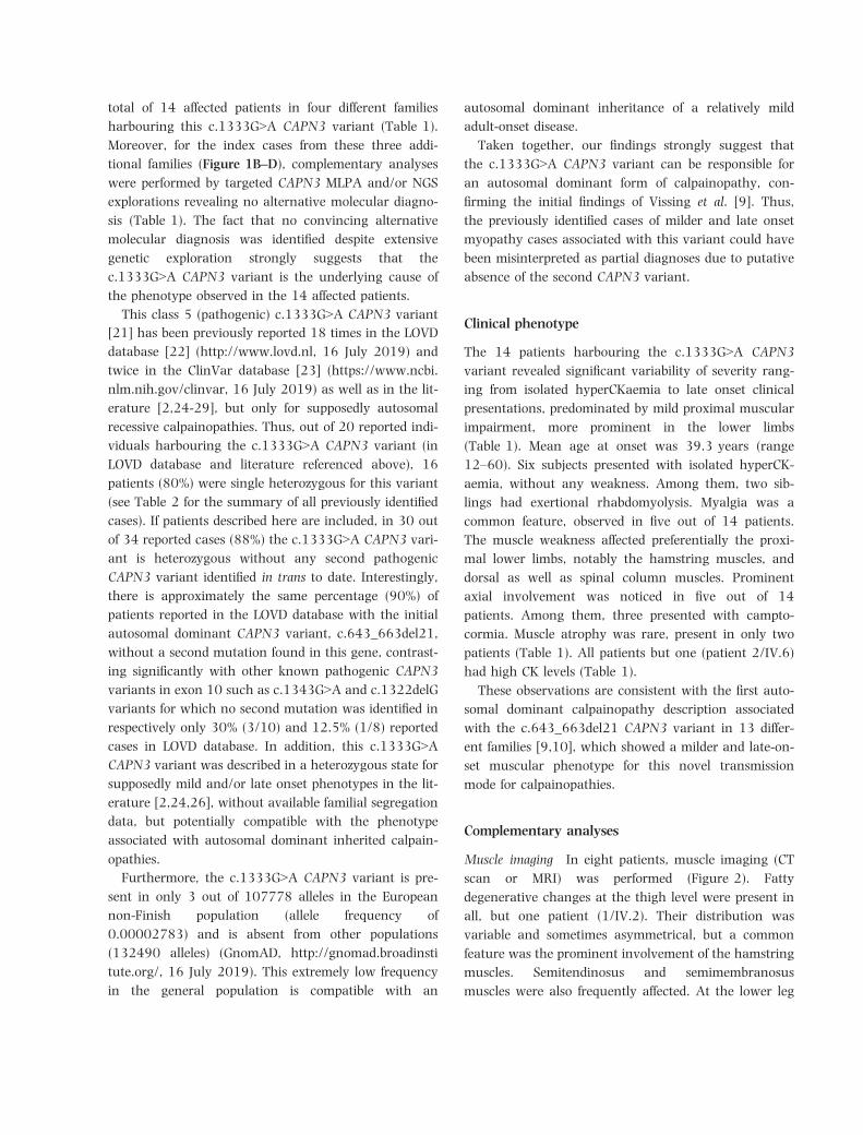
total of 14 affected patients in four different families
harbouring this c.1333G>A CAPN3 variant (Table 1).
Moreover, for the index cases from these three addi-
with the c.643_663del21 CAPN3 variant in 13 differ-
ent families [9,10], which showed a milder and late-on-
set muscular phenotype for this novel transmission
mode for calpainopathies.
Complementary analyses
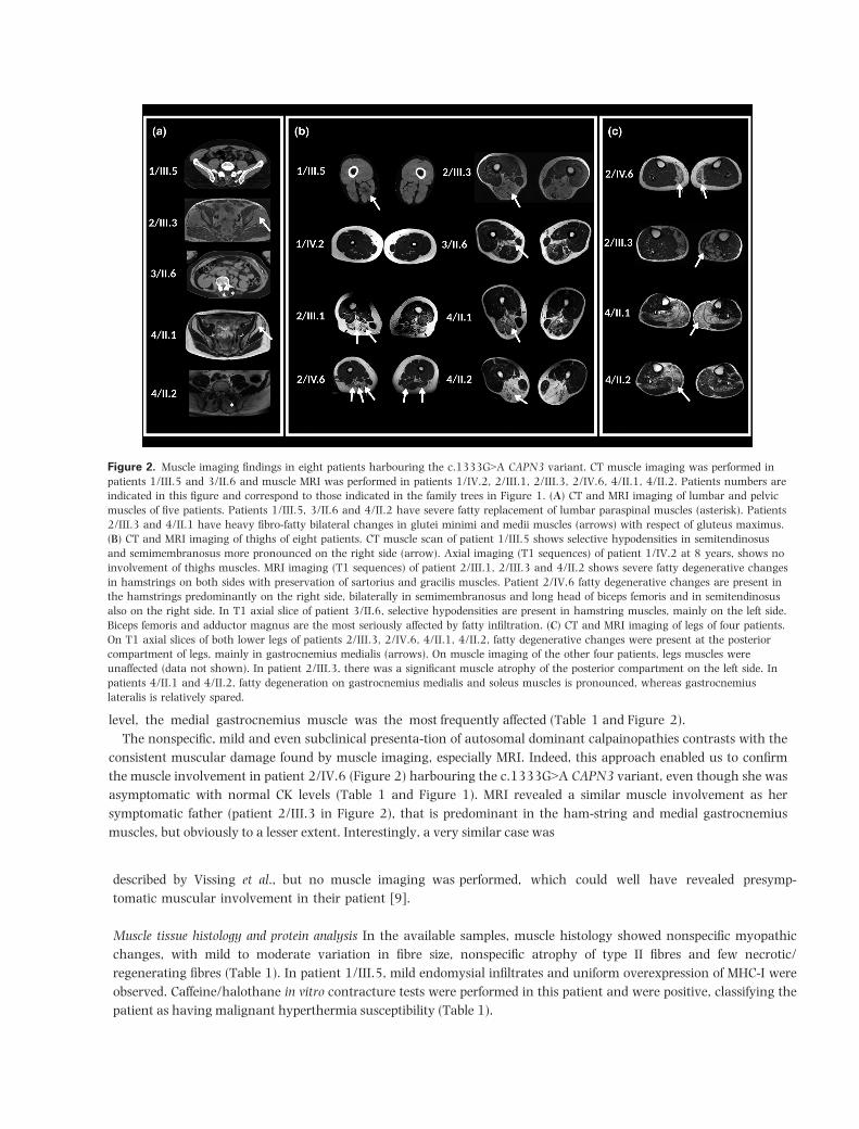
Muscle imaging In eight patients, muscle imaging (CT
scan or MRI) was performed (Figure 2). Fatty
degenerative changes at the thigh level were present in
all, but one patient (1/IV.2). Their distribution was
variable and sometimes asymmetrical, but a common
feature was the prominent involvement of the hamstring
muscles. Semitendinosus and semimembranosus
muscles were also frequently affected. At the lower leg
[21] has been previously reported 18 times in the LOVD database [22] (http://www.lovd.nl, 16 July 2019) and twice in the ClinVar database [23] (https://www.ncbi. nlm.nih.gov/clinvar, 16 July 2019) as well as in the lit-erature [2,24-29], but only for supposedly autosomal recessive calpainopathies. Thus, out of 20 reported indi-viduals harbouring the c.1333G>A CAPN3 variant (in LOVD database and literature referenced above), 16 patients (80%) were single heterozygous for this variant (see Table 2 for the summary of all previously identified cases). If patients described here are included, in 30 out of 34 reported cases (88%) the c.1333G>A CAPN3 vari-ant is heterozygous without any second pathogenic CAPN3 variant identified in trans to date. Interestingly, there is approximately the same percentage (90%) of patients reported in the LOVD database with the initial autosomal dominant CAPN3 variant, c.643_663del21, without a second mutation found in this gene, contrast-ing significantly with other known pathogenic CAPN3 variants in exon 10 such as c.1343G>A and c.1322delG variants for which no second mutation was identified in respectively only 30% (3/10) and 12.5% (1/8) reported cases in LOVD database. In addition, this c.1333G>A CAPN3 variant was described in a heterozygous state for supposedly mild and/or late onset phenotypes in the lit-erature [2,24,26], without available familial segregation data, but potentially compatible with the phenotype associated with autosomal dominant inherited calpain-opathies.
Furthermore, the c.1333G>A CAPN3 variant is pre-sent in only 3 out of 107778 alleles in the European non-Finish population (allele frequency of 0.00002783) and is absent from other populations (132490 alleles) (GnomAD, http://gnomad.broadinsti tute.org/, 16 July 2019). This extremely low frequency in the general population is compatible with an
Fanin et al., 2004 [25] +Nascimbeni et al., 2010 [27] (=IndividualLOVD ID: #00213950)*
Pathogenic c.1746-20C>G‡ LGMD2A
Saenz et al., 2005 [26] (=individualLOVD ID: #00214328)*
Pathogenic No second pathogenic variant
reported
Late-onset
LGMD2A
Schr€oder et al., 2013 [28] Pathogenic No second pathogenic variant
reported
LGMD2A
Nallamilli et al., 2018 [11] (=individualLOVD ID: #00221637)*
VUS No second pathogenic variant
reported
LGMD2A
LGMD2A, Limb-Girdle Muscular Dystrophy type 2A; VUS, variant of uncertain significance.
*Second description of a same individual in LOVD and/or literature.†Classification and phenotype suggested by source.‡Deleterious splicing effect later confirmed by Nascimbeni et al. [27].
described by Vissing et al., but no muscle imaging was performed, which could well have revealed presymp-
tomatic muscular involvement in their patient [9].
Muscle tissue histology and protein analysis In the available samples, muscle histology showed nonspecific myopathic changes, with mild to moderate variation in fibre size, nonspecific atrophy of type II fibres and few necrotic/regenerating fibres (Table 1). In patient 1/III.5, mild endomysial infiltrates and uniform overexpression of MHC-I were observed. Caffeine/halothane in vitro contracture tests were performed in this patient and were positive, classifying the patient as having malignant hyperthermia susceptibility (Table 1).
Figure 2. Muscle imaging findings in eight patients harbouring the c.1333G>A CAPN3 variant. CT muscle imaging was performed in
patients 1/III.5 and 3/II.6 and muscle MRI was performed in patients 1/IV.2, 2/III.1, 2/III.3, 2/IV.6, 4/II.1, 4/II.2. Patients numbers are
indicated in this figure and correspond to those indicated in the family trees in Figure 1. (A) CT and MRI imaging of lumbar and pelvic
muscles of five patients. Patients 1/III.5, 3/II.6 and 4/II.2 have severe fatty replacement of lumbar paraspinal muscles (asterisk). Patients
2/III.3 and 4/II.1 have heavy fibro-fatty bilateral changes in glutei minimi and medii muscles (arrows) with respect of gluteus maximus.
(B) CT and MRI imaging of thighs of eight patients. CT muscle scan of patient 1/III.5 shows selective hypodensities in semitendinosus
and semimembranosus more pronounced on the right side (arrow). Axial imaging (T1 sequences) of patient 1/IV.2 at 8 years, shows no
involvement of thighs muscles. MRI imaging (T1 sequences) of patient 2/III.1, 2/III.3 and 4/II.2 shows severe fatty degenerative changes
in hamstrings on both sides with preservation of sartorius and gracilis muscles. Patient 2/IV.6 fatty degenerative changes are present in
the hamstrings predominantly on the right side, bilaterally in semimembranosus and long head of biceps femoris and in semitendinosus
also on the right side. In T1 axial slice of patient 3/II.6, selective hypodensities are present in hamstring muscles, mainly on the left side.
Biceps femoris and adductor magnus are the most seriously affected by fatty infiltration. (C) CT and MRI imaging of legs of four patients.
On T1 axial slices of both lower legs of patients 2/III.3, 2/IV.6, 4/II.1, 4/II.2, fatty degenerative changes were present at the posterior
compartment of legs, mainly in gastrocnemius medialis (arrows). On muscle imaging of the other four patients, legs muscles were
unaffected (data not shown). In patient 2/III.3, there was a significant muscle atrophy of the posterior compartment on the left side. In
patients 4/II.1 and 4/II.2, fatty degeneration on gastrocnemius medialis and soleus muscles is pronounced, whereas gastrocnemius
lateralis is relatively spared.
level, the medial gastrocnemius muscle was the most frequently affected (Table 1 and Figure 2).The nonspecific, mild and even subclinical presenta-tion of autosomal dominant calpainopathies contrasts with the
consistent muscular damage found by muscle imaging, especially MRI. Indeed, this approach enabled us to confirm the muscle involvement in patient 2/IV.6 (Figure 2) harbouring the c.1333G>A CAPN3 variant, even though she was asymptomatic with normal CK levels (Table 1 and Figure 1). MRI revealed a similar muscle involvement as her symptomatic father (patient 2/III.3 in Figure 2), that is predominant in the ham-string and medial gastrocnemius muscles, but obviously to a lesser extent. Interestingly, a very similar case was
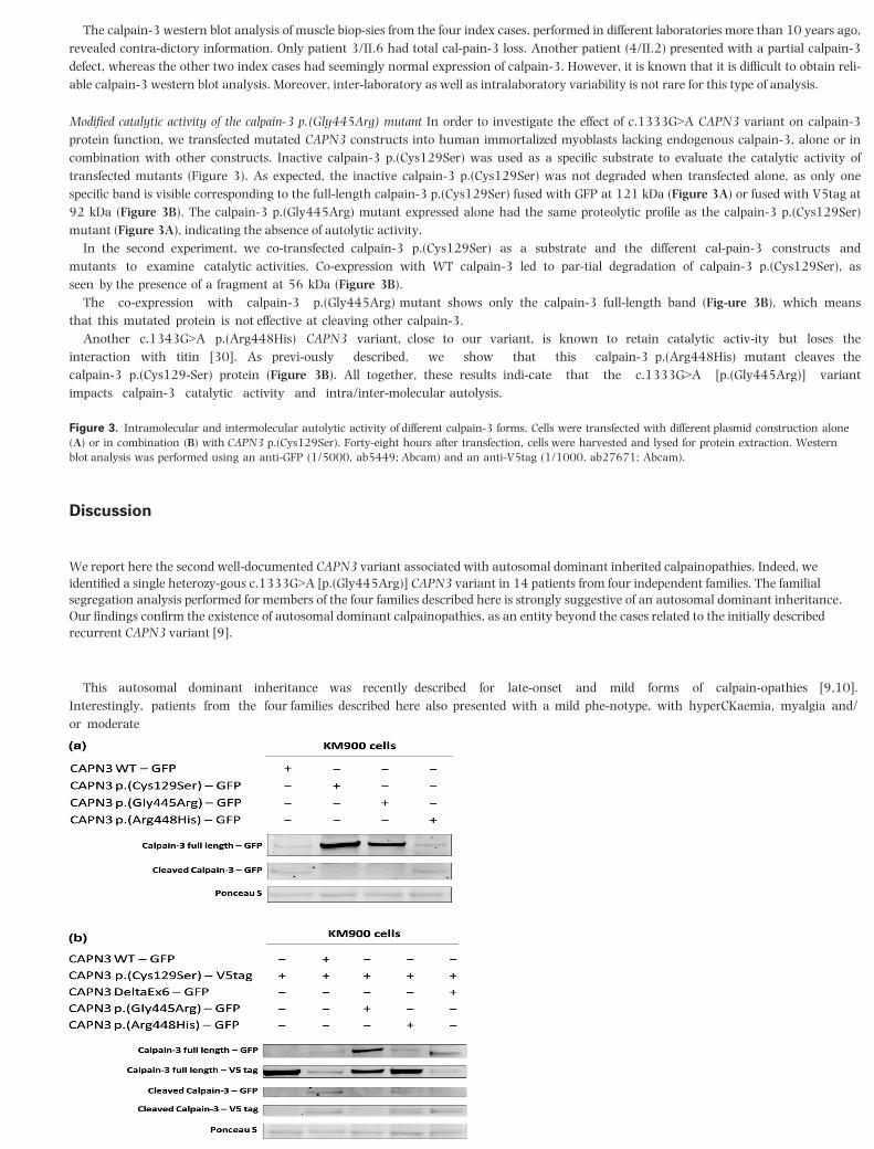
Figure 3. Intramolecular and intermolecular autolytic activity of different calpain-3 forms. Cells were transfected with different plasmid construction alone (A) or in combination (B) with CAPN3 p.(Cys129Ser). Forty-eight hours after transfection, cells were harvested and lysed for protein extraction. Western blot analysis was performed using an anti-GFP (1/5000, ab5449; Abcam) and an anti-V5tag (1/1000, ab27671; Abcam).
Discussion
We report here the second well-documented CAPN3 variant associated with autosomal dominant inherited calpainopathies. Indeed, we identified a single heterozy-gous c.1333G>A [p.(Gly445Arg)] CAPN3 variant in 14 patients from four independent families. The familial segregation analysis performed for members of the four families described here is strongly suggestive of an autosomal dominant inheritance. Our findings confirm the existence of autosomal dominant calpainopathies, as an entity beyond the cases related to the initially described recurrent CAPN3 variant [9].
This autosomal dominant inheritance was recently described for late-onset and mild forms of calpain-opathies [9,10]. Interestingly, patients from the four families described here also presented with a mild phe-notype, with hyperCKaemia, myalgia and/or moderate
The calpain-3 western blot analysis of muscle biop-sies from the four index cases, performed in different laboratories more than 10 years ago, revealed contra-dictory information. Only patient 3/II.6 had total cal-pain-3 loss. Another patient (4/II.2) presented with a partial calpain-3 defect, whereas the other two index cases had seemingly normal expression of calpain-3. However, it is known that it is difficult to obtain reli-able calpain-3 western blot analysis. Moreover, inter-laboratory as well as intralaboratory variability is not rare for this type of analysis.
Modified catalytic activity of the calpain-3 p.(Gly445Arg) mutant In order to investigate the effect of c.1333G>A CAPN3 variant on calpain-3 protein function, we transfected mutated CAPN3 constructs into human immortalized myoblasts lacking endogenous calpain-3, alone or in combination with other constructs. Inactive calpain-3 p.(Cys129Ser) was used as a specific substrate to evaluate the catalytic activity of transfected mutants (Figure 3). As expected, the inactive calpain-3 p.(Cys129Ser) was not degraded when transfected alone, as only one specific band is visible corresponding to the full-length calpain-3 p.(Cys129Ser) fused with GFP at 121 kDa (Figure 3A) or fused with V5tag at 92 kDa (Figure 3B). The calpain-3 p.(Gly445Arg) mutant expressed alone had the same proteolytic profile as the calpain-3 p.(Cys129Ser) mutant (Figure 3A), indicating the absence of autolytic activity.
In the second experiment, we co-transfected calpain-3 p.(Cys129Ser) as a substrate and the different cal-pain-3 constructs and mutants to examine catalytic activities. Co-expression with WT calpain-3 led to par-tial degradation of calpain-3 p.(Cys129Ser), as seen by the presence of a fragment at 56 kDa (Figure 3B).
The co-expression with calpain-3 p.(Gly445Arg) mutant shows only the calpain-3 full-length band (Fig-ure 3B), which means that this mutated protein is not effective at cleaving other calpain-3.
Another c.1343G>A p.(Arg448His) CAPN3 variant, close to our variant, is known to retain catalytic activ-ity but loses the interaction with titin [30]. As previ-ously described, we show that this calpain-3 p.(Arg448His) mutant cleaves the calpain-3 p.(Cys129-Ser) protein (Figure 3B). All together, these results indi-cate that the c.1333G>A [p.(Gly445Arg)] variant impacts calpain-3 catalytic activity and intra/inter-molecular autolysis.
as CAV3, GMPPB and more recently SGCA and TCAP [34-36]. Thus, autosomal dominant inherited calpain-opathy should be considered when faced by a wide range of initial phenotypic presentations from hyperCK-aemia (asymptomatic or not), myalgia with or without exertional rhabdomyolysis to mild LGMD.
This experience demonstrates that despite the advent of NGS, the simplest assumption remains the first one to consider; in the present case, the probable existence of an autosomal dominant form of calpainopathy asso-ciated with the c.1333G>A CAPN3 variant. Indeed, despite numerous molecular analyses, especially by NGS, no alternative molecular diagnosis could be estab-lished for these four different families, thus consolidat-ing the implication of this CAPN3 variant in an autosomal dominant inherited calpainopathy.
At the functional level, c.1333G>A [p.(Gly445Arg)] CAPN3 variant involves a modification of calpain-3 catalytic activity in cells, with a loss of intramolecular and intermolecular autolysis (Figure 3). The origin of this modification of activity could be an indirect loss of catalytic activity and possibly a modification of interac-tions with partners due to conformational changes in the protein structure. Indeed, using the available infor-mation regarding the calpain-3 structure, we locate thec.1333G>A [p.(Gly445Arg)] CAPN3 variant in the cal-pain b-sandwich domain (CBSW) interacting with the penta-EF-hand domain (PEF) supposedly involved in the dimerization of two calpain-3 units [37]. Interest-ingly, the c.1333G>A [p.
(Gly445Arg)] CAPN3 variant is located on a loop of the CBSW domain that has interactions with the PEF domain, and is expected to be involved in the communication of Ca2+-induced con-formational change throughout these domains. There-fore, the charge alteration associated with the
c.1333G>A [p.(Gly445Arg)] CAPN3 variant could dis-rupt communication between Ca2+-induced alteration in the PEF domain and the rest of the molecule [38]. Consequently, the c.1333G>A [p.(Gly445Arg)] CAPN3 variant could indirectly impair the catalytic activity of the mutated calpain-3 protein and secondarily, due to conformation changes, disrupt interactions with part-ners potentially resulting in the retention of calpain-3 in the sarcomere due to strong interaction with titin and/or to the other functional calpain-3 allele in case of homodimerization. This suggestion is reinforced by the fact that calpain-3 has already been shown to interact with titin in the region of the p.(Gly445Arg)
proximal muscle weakness, predominantly affecting the hamstrings as shown by muscle imaging (Figure 2), therefore expanding the phenotypic spectrum of cal-painopathies.Three subjects from our cohort had camptocormia. The same presentation of late-onset axial myopathy has been previously
described in a patient carrying a single heterozygous CAPN3 variant, c.759_761delGAA, suggesting a possible familial autosomal dominant cal-painopathy [31]. Moreover, among patients carrying the c.643_663del21 CAPN3 variant, paraspinal mus-cles were particularly affected on MRI studies [9].These observations suggest that an autosomal domi-nant calpainopathy should be considered when faced with a prominent
axial myopathy, widening the spec-trum of neuromuscular disorders for which MRI is use-ful in assisting genetic testing beyond the recent observations for distal myopathies [32].Thus, it is reasonable to conceive a broad continuum of clinical phenotypes for calpainopathies, from severe early childhood onset to
moderate adult-onset autoso-mal recessive forms [24,33], possibly depending on the CAPN3 mutational combination, overlapping with milder late-onset autosomal dominant forms that could extend to the extreme end of the spectrum to isolated hyperCKaemia. The evaluation of serum CK levels, despite its nonspecificity, seems to be of importance for autosomal dominant calpainopathies as it is a signifi-cant presenting finding. However, there is great vari-ability in terms of levels and positivity for this biomarker, as demonstrated by patient 2/IV.6 (Table 1 and Figure 1), similar to other cases reported in the lit-erature [9].It is likely that some of these late-onset mild clinical phenotypes could easily have been confused with sup-posedly natural
muscular senescence or even missed because of the patient’s premature death, only to be discovered recently as life expectancy gradually extended and novel biomarkers, notably CK measure-ment, as well as complementary diagnosis tool, such as muscle MRI, were available, enabling us to objectify and label these atypical clinical phenotypes.In addition, we describe exertional rhabdomyolysis in two siblings (3/III.1 and 3/III.2 in Figure 1). Although this clinical presentation
has to be confirmed for other reported cases before considering it a definite feature of calpainopathies, such a presentation has already been described for other genes associated with LGMD, such
Acknowledgements
We sincerely thank Nathalie Da Silva, Val�erie Delague,
Karine Bertaux, C�ecile Mouradian, Pierre Cacciagli,
JM, AMDP, NL, JP, MC, MB, MK and SA contributed to
drafting the manuscript and preparing tables and figures.
Funding information
FHU A*MIDEX project MARCHE n.ANR-11-IDEX-001-
02 funded by the “Investissement d’avenir” French
government program, managed by the French National
Research Agency (ANR).
Disclosure
The authors have no conflicts of interest to disclose.
Ethical approval
Informed consent was obtained for all patients, accord-
ing to the Declaration of Helsinki.
Data availability statement
The data that support the findings of this study are
available on request from the corresponding author.
The data are not publicly available due to privacy or
ethical restrictions.
References
1 Zatz M, Starling A. Calpains and disease. N Engl J Med
2005; 352: 2413–23
variant [39] and a modification of arginine in the nearby 448 amino acid position induces a loss of this interaction [30]. The c.1333G>A [p.(Gly445Arg)] vari-ant adds an arginine in proximity to this interaction site which could increase the interaction with titin, leading to retention of calpain-3 in the sarcomere. Therefore, our favoured hypothesis for the pathophysio-logical mechanisms associated with the c.1333G>A [p.(Gly445Arg)] CAPN3 variant is a dominant-negative effect, as also suggested for the c.643_663del21 CAPN3 variant [9].
Identification of the c.1333G>A [p.(Gly445Arg)] CAPN3 variant, initially considered pathogenic for autosomal recessive calpainopathies, also reveals an important issue for diagnostic genetics laboratories. Indeed, it can be very difficult to establish a causal link between a variant, described as pathogenic in the liter-ature and different databases, and an unexpected phe-notype and/or transmission mode for the considered gene. For instance this must be taken into account for mild and late-onset phenotypes or subclinical pheno-types such as isolated hyperCKaemia for which the genetic aetiology could legitimately be questioned. However, in the present case, isolated hyperCKaemia should not be neglected because it reveals a potential deleterious muscular process, that can stay clinically silent for the first fifth or sixth decades of life, only to be at a later stage associated to functional and clinical manifestations, thus revealing the variable limits of the muscular resilience for each individual, even within the same family. Furthermore, this autosomal dominant inheritance could be particularly prone to modifying genetic or environmental effects, therefore explaining the phenotypic heterogeneity and even the seemingly incomplete penetrance already observed for autosomal dominant inherited calpainopathies [9]. In fact, this point has already been raised at the time of the initial description by Vissing et al., regarding especially haplo-type combinations or cis-variants potentially affecting regulatory regions of the CAPN3 expression [40,41]. However, for the families described here, all available information clearly indicates an autosomal dominant inheritance associated with the c.1333G>A CAPN3 variant. Therefore, in addition to the clear impact for genetic counselling, these findings could also lead the way to the reassessment of other forms of myopathies where the genetic inheritance is, to date, considered to be strictly autosomal recessive.
2 Guglieri M, Magri F, D’Angelo MG, Prelle A, MorandiL, Rodolico C, et al. Clinical, molecular, and proteincorrelations in a large sample of genetically diagnosedItalian limb girdle muscular dystrophy patients. HumMutat 2008; 29: 258–66
3 Angelini C, Nardetto L, Borsato C, Padoan R, Fanin M,Nascimbeni AC, Tasca E. The clinical course of calpain-opathy (LGMD2A) and dysferlinopathy (LGMD2B).Neurol Res 2010; 32: 41–6
4 Luo S-S, Xi J-Y, Zhu W-H, Zhao C-B, Lu J-H, Lin J,et al. Genetic variability and clinical spectrum of Chi-nese patients with limb-girdle muscular dystrophy type2A. Muscle Nerve 2012; 46: 723–9
5 Richard I, Broux O, Allamand V, Fougerousse F, Chi-annilkulchai N, Bourg N, et al. Mutations in the prote-olytic enzyme calpain 3 cause limb-girdle musculardystrophy type 2A. Cell 1995; 81: 27–40
6 Kramerova I, Kudryashova E, Venkatraman G, SpencerMJ. Calpain 3 participates in sarcomere remodeling byacting upstream of the ubiquitin-proteasome pathway.Hum Mol Genet 2005; 14: 2125–34
7 Duguez S, Bartoli M, Richard I. Calpain 3: a key regu-lator of the sarcomere? FEBS J 2006; 273: 3427–36
8 Ono Y, Ojima K, Torii F, Takaya E, Doi N, NakagawaK, et al. Skeletal muscle-specific calpain is an intracel-lular Na+-dependent protease. J Biol Chem 2010; 285:22986–98
9 Vissing J, Barresi R, Witting N, Van Ghelue M, Gam-melgaard L, Bindoff LA, et al. A heterozygous 21-bpdeletion in CAPN3 causes dominantly inherited limbgirdle muscular dystrophy. Brain 2016; 139: 2154–63
10 Martinez-Thompson JM, Niu Z, Tracy JA, Moore SA,Swenson A, Wieben ED, et al. Autosomal dominantcalpainopathy due to heterozygous CAPN3C.643_663del21. Muscle Nerve 2018; 57: 679–83
11 Nallamilli BRR, Chakravorty S, Kesari A, Tanner A,Ankala A, Schneider T, et al. Genetic landscape andnovel disease mechanisms from a large LGMD cohortof 4656 patients. Ann Clin TranslNeurol 2018; 5:1574–87
12 Straub V, Murphy A, Udd B, Corrado A, Aym�e S,B€onneman C, et al. 229th ENMC international work-shop: limb girdle muscular dystrophies – nomenclatureand reformed classification Naarden, the Netherlands,17–19 March 2017. Neuromuscular Disord 2018; 28:702–10
13 Krahn M, Biancalana V, Cerino M, Perrin A, Michel-Calemard L, Nectoux J, et al. A National French con-sensus on gene lists for the diagnosis of myopathiesusing next-generation sequencing. Eur J Hum Genet2019; 27: 349–52
14 Krahn M, Lopez de Munain A, Streichenberger N, Ber-nard R, P�echeux C, Testard H, et al. CAPN3 mutationsin patients with idiopathic eosinophilic myositis. AnnNeurol 2006; 59: 905–11
15 Gorokhova S, Cerino M, Mathieu Y, Courrier S,Desvig-nes JP, Salgado D, et al. Comparing targeted exomeand whole exome approaches for genetic diagnosis ofneuromuscular disorders. Appl Transl Genom 2015; 7:26–31
16 Zenagui R, Lacourt D, Pegeot H, Yauy K, Juntas Mor-ales R, Theze C, et al. A reliable targeted next-genera-tion sequencing strategy for diagnosis of myopathiesand muscular dystrophies, especially for the giant titinand nebulin genes. J Mol Diagn 2018; 20: 533–49
17 Kaplan J-C, Hamroun D. The 2014 version of the genetable of monogenic neuromuscular disorders (nucleargenome). Neuromuscul Disord 2013; 23: 1081–111
18 den Dunnen JT, Dalgleish R, Maglott DR, Hart RK,Greenblatt MS, McGowan-Jordan J, et al. HGVS recom-mendations for the description of sequence variants:2016 update. Hum Mutat 2016; 37: 564–9
19 Milic A, Daniele N, Lochm€uller H, Mora M, Comi GP,Moggio M, et al. A third of LGMD2A biopsies havenormal calpain 3 proteolytic activity as determined byan in vitro assay. Neuromuscul Disord. 2007; 17: 148–56
20 Mamchaoui K, Trollet C, Bigot A, Negroni E, ChaouchS, Wolff A, et al. Immortalized pathological humanmyoblasts: towards a universal tool for the study ofneuromuscular disorders. Skelet Muscle 2011; 1: 34
21 Richards S, Aziz N, Bale S, Bick D, Das S, Gastier-Fos-ter J, et al. Standards and guidelines for the interpreta-tion of sequence variants: a joint consensusrecommendation of the American College of MedicalGenetics and Genomics and the Association for Molec-ular Pathology. Genet Med 2015; 17: 405–24
22 Fokkema IFAC, Taschner PEM, Schaafsma GCP, Celli J,Laros JF, den Dunnen JT LOVD vol 2.0: the next gen-eration in gene variant databases. Hum Mutat 2011;32: 557–63
23 Landrum MJ, Lee JM, Benson M, Brown GR, Chao C,Chitipiralla S, et al. ClinVar: improving access to vari-ant interpretations and supporting evidence. NucleicAcids Res 2018; 46: D1062–7
24 Richard I, Roudaut C, Saenz A, Pogue R, GrimbergenJ, Anderson L, et al. Calpainopathy-a survey of muta-tions and polymorphisms. Am J Hum Genet 1999; 64:1524–40
25 Fanin M, Fulizio L, Nascimbeni AC, Spinazzi M, PilusoG, Ventriglia V, et al. Molecular diagnosis in LGMD2A:mutation analysis or protein testing? Hum Mutat2004; 24: 52–62
26 S�aenz A, Leturcq F, Cobo AM, Poza JJ, Ferrer X, Otae-gui D, et al. LGMD2A: genotype-phenotype correla-tions based on a large mutational survey on thecalpain 3 gene. Brain 2005; 128: 732–42
27 Nascimbeni AC, Fanin M, Tasca E, Angelini C. Tran-scriptional and translational effects of intronic CAPN3gene mutations. Hum Mutat 2010; 31: E1658–69
28 Schr€oder T, Fuchss J, Schneider I, Stoltenburg-DidingerG, Hanisch F. Eosinophils in hereditary and inflamma-tory myopathies. Acta Myol 2013; 32: 148–53
29 Magri F, Colombo I, Del Bo R, Previtali S, Brusa R,Ciscato P, et al. ISPD mutations account for a smallproportion of Italian Limb Girdle Muscular Dystrophycases. BMC Neurol 2015; 15: 172
30 Ermolova N, Kudryashova E, DiFranco M, Vergara J,Kramerova I, Spencer MJ. Pathogenity of some limbgirdle muscular dystrophy mutations can result fromreduced anchorage to myofibrils and altered stabilityof calpain 3. Hum Mol Genet 2011; 20: 3331–45
31 Liewluck T, Goodman BP. Late-onset axial myopathyand camptocormia in a calpainopathy carrier. J ClinNeuromuscul Dis 2012; 13: 209–13
32 Bugiardini E, Morrow JM, Shah S, Wood CL, LynchDS, Pitmann AM, Lynch DS, Pitmann AM, et al. Thediagnostic value of MRI pattern recognition in distalmyopathies. Front Neurol 2018; 9: 456
33 Fanin M, Angelini C. Protein and genetic diagnosis oflimb girdle muscular dystrophy type 2A: the yield andthe pitfalls. Muscle Nerve 2015; 52: 163–73
35 Cabrera-Serrano M, Ghaoui R, Ravenscroft G, JohnsenRD, Davis MR, Corbett A, et al. Expanding the pheno-type of GMPPB mutations. Brain 2015; 138: 836–44
36 Rubegni A, Malandrini A, Dosi C, Astrea G, Baldacci J,Battisti C, et al. Next-generation sequencing approach
to hyperCKemia: a 2-year cohort study. Neurol Genet.2019; 5: e352
37 Ye Q, Campbell RL, Davies PL. Structures of humancalpain-3 protease core with and without bound inhi-bitor reveal mechanisms of calpain activation. J BiolChem 2018; 293: 4056–70
38 Jia Z, Petrounevitch V, Wong A, Moldoveanu T,Davies PL, Elce JS, Davies PL, Elce JS, et al. Mutationsin calpain 3 associated with limb girdle muscular dys-trophy: analysis by molecular modeling and by muta-tion in m-calpain. Biophys J 2001; 80: 2590–6
39 Ono Y, Torii F, Ojima K, Doi N, , Yoshioka K, Kawa-bata Y, et al. Suppressed disassembly of autolyzingp94/CAPN3 by N2A connectin/titin in a geneticreporter system. J Biol Chem 2006; 281: 18519–31
40 S�aenz A, L�opez de Munain A. Dominant LGMD2A:alternative diagnosis or hidden digenism? Brain 2017;140: e7
41 Vissing J, Duno M. Reply: dominant LGMD2A: alterna-tive diagnosis or hidden digenism? Brain 2017; 140: e8
Supporting information
Additional Supporting Information may be found in the
online version of this article at the publisher’s web-site:
Appendix S1. Detailed genes panels and genes lists.


![A novel homozygous ARL13B variant in patients with Joubert ... et al.pdf · (JS) [1, 3], a genetically heterogeneous autosomal recessive or X-linked disorder characterized by ataxia,](https://static.documents.pub/doc/80x56/5d01a78288c993a21e8cfaee/a-novel-homozygous-arl13b-variant-in-patients-with-joubert-et-alpdf-js.jpg)


