Novel Compound Heterozygous Nonsense PRX Mutations in a Korean Dejerine-Sottas Neuropathy Family
Ye Ji Choi,a Young Se Hyun,b Soo Hyun Nam,b Heasoo Koo,c Young Bin Hong,d Ki Wha Chung,b Byung-Ok ChoidaDepartments of Neurology and cPathology, Ewha Womans University School of Medicine, Seoul, Korea bDepartment of Biological Science, Kongju National University, Gongju, Korea dDepartment of Neurology, Samsung Medical Center, Sungkyunkwan University School of Medicine, Seoul, Korea
Received June 19, 2013Revised September 9, 2013Accepted September 13, 2013
CorrespondenceByung-Ok Choi, PhDDepartment of Neurology, Samsung Medical Center, Sungkyunkwan University School of Medicine, 81 Irwon-ro, Gangnam-gu, Seoul 135-710, KoreaTel +82-2-3410-1296Fax +82-2-3410-0052E-mail [email protected] Wha Chung, PhDDepartment of Biological Science, Kongju National University, 56 Gongjudaehak-ro, Gongju 314-701, KoreaTel +82-41-850-8506Fax +82-41-850-0957E-mail [email protected]
BackgroundzzMutations in the gene encoding periaxin (PRX) are known to cause autosomal recessive Dejerine-Sottas neuropathy (DSN) or Charcot-Marie-Tooth disease type 4F. Howev-er, there have been no reports describing Korean patients with these mutations.
Case ReportzzWe examined a Korean DSN patient with an early-onset, slowly progressive, demyelinating neuropathy with prominent sensory involvement. Whole-exome sequencing and subsequent capillary sequencing revealed novel compound heterozygous nonsense mutations (p.R392X and p.R679X) in PRX. One mutation was transmitted from each of the patient’s par-ents. No unaffected family member had both mutations, and the mutations were not found in healthy controls.
ConclusionszzWe believe that these novel compound heterozygous nonsense mutations are the underlying cause of DSN. The clinical, electrophysiologic, and pathologic phenotypes in this family were similar to those described previously for patients with PRX mutations. We have identified the first PRX mutation in a Korean patient with DSN. J Clin Neurol 2015;11(1):92-96
cc This is an Open Access article distributed under the terms of the Cre-ative Commons Attribution Non-Commercial License (http://creative-commons.org/licenses/by-nc/3.0) which permits unrestricted non-com-mercial use, distribution, and reproduction in any medium, provided the ori-ginal work is properly cited.
Introduction
Charcot-Marie-Tooth disease (CMT) is a clinically and geneti-cally heterogeneous disorder of the peripheral nervous sys-tem.1 Demyelinating CMT neuropathies are classified as either autosomal dominant CMT type 1 or autosomal recessive CMT type 4 (CMT4).2 CMT4 has been further divided into many subtypes from CMT4A to CMT4J according to clinical symp-toms and genetic causes.3 Dejerine-Sottas neuropathy (DSN), which is also known as CMT type 3, is characterized by early
age at onset, very slow motor nerve conduction velocities (MNCVs), and severe demyelinating neuropathy.4 Previous reports have described several genes that are relevant to DSN, including those encoding early growth response 2, myelin pro-tein zero, peripheral myelin protein 22, and periaxin (PRX).4-7
Mutations in PRX are known to cause DSN and CMT4F.6,7 Loss of periaxin function causes instability of Schwann cell myelination, which eventually leads to demyelination and pe-ripheral neuropathy. It has been shown that PRX-knockout mice develop a spectrum of morphologic changes to the my-elin sheath.8
Dozens of patients with PRX mutations have been de-scribed previously,9-14 but none of them were Korean. In this study we identified a Korean DSN patient with novel com-pound heterozygous nonsense mutations in PRX through
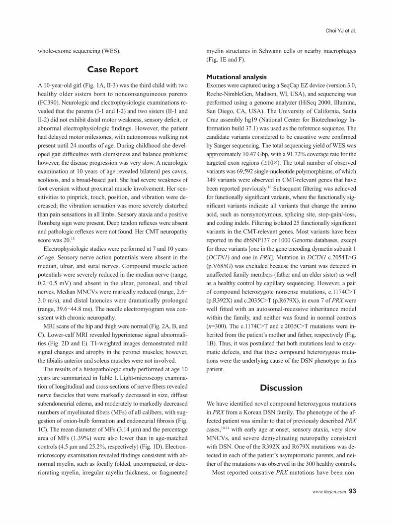
A 10-year-old girl (Fig. 1A, II-3) was the third child with two healthy older sisters born to nonconsanguineous parents (FC390). Neurologic and electrophysiologic examinations re-vealed that the parents (I-1 and I-2) and two sisters (II-1 and II-2) did not exhibit distal motor weakness, sensory deficit, or abnormal electrophysiologic findings. However, the patient had delayed motor milestones, with autonomous walking not present until 24 months of age. During childhood she devel-oped gait difficulties with clumsiness and balance problems; however, the disease progression was very slow. A neurologic examination at 10 years of age revealed bilateral pes cavus, scoliosis, and a broad-based gait. She had severe weakness of foot eversion without proximal muscle involvement. Her sen-sitivities to pinprick, touch, position, and vibration were de-creased; the vibration sensation was more severely disturbed than pain sensations in all limbs. Sensory ataxia and a positive Romberg sign were present. Deep tendon reflexes were absent and pathologic reflexes were not found. Her CMT neuropathy score was 20.15
Electrophysiologic studies were performed at 7 and 10 years of age. Sensory nerve action potentials were absent in the median, ulnar, and sural nerves. Compound muscle action potentials were severely reduced in the median nerve (range, 0.2–0.5 mV) and absent in the ulnar, peroneal, and tibial nerves. Median MNCVs were markedly reduced (range, 2.6–3.0 m/s), and distal latencies were dramatically prolonged (range, 39.6–44.8 ms). The needle electromyogram was con-sistent with chronic neuropathy.
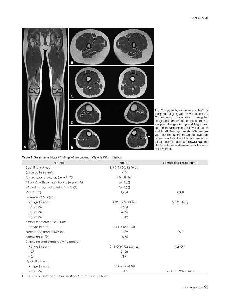
MRI scans of the hip and thigh were normal (Fig. 2A, B, and C). Lower-calf MRI revealed hyperintense signal abnormali-ties (Fig. 2D and E). T1-weighted images demonstrated mild signal changes and atrophy in the peronei muscles; however, the tibialis anterior and soleus muscles were not involved.
The results of a histopathologic study performed at age 10 years are summarized in Table 1. Light-microscopy examina-tion of longitudinal and cross-sections of nerve fibers revealed nerve fascicles that were markedly decreased in size, diffuse subendoneurial edema, and moderately to markedly decreased numbers of myelinated fibers (MFs) of all calibers, with sug-gestion of onion-bulb formation and endoneurial fibrosis (Fig. 1C). The mean diameter of MFs (3.14 μm) and the percentage area of MFs (1.39%) were also lower than in age-matched controls (4.5 μm and 25.2%, respectively) (Fig. 1D). Electron-microscopy examination revealed findings consistent with ab-normal myelin, such as focally folded, uncompacted, or dete-riorating myelin, irregular myelin thickness, or fragmented
myelin structures in Schwann cells or nearby macrophages (Fig. 1E and F).
Mutational analysisExomes were captured using a SeqCap EZ device (version 3.0, Roche-NimbleGen, Madison, WI, USA), and sequencing was performed using a genome analyzer (HiSeq 2000, Illumina, San Diego, CA, USA). The University of California, Santa Cruz assembly hg19 (National Center for Biotechnology In-formation build 37.1) was used as the reference sequence. The candidate variants considered to be causative were confirmed by Sanger sequencing. The total sequencing yield of WES was approximately 10.47 Gbp, with a 91.72% coverage rate for the targeted exon regions (≥10×). The total number of observed variants was 69,592 single-nucleotide polymorphisms, of which 349 variants were observed in CMT-relevant genes that have been reported previously.16 Subsequent filtering was achieved for functionally significant variants, where the functionally sig-nificant variants indicate all variants that change the amino acid, such as nonsynonymous, splicing site, stop-gain/-loss, and coding indels. Filtering isolated 25 functionally significant variants in the CMT-relevant genes. Most variants have been reported in the dbSNP137 or 1000 Genome databases, except for three variants [one in the gene encoding dynactin subunit 1 (DCTN1) and one in PRX]. Mutation in DCTN1 c.2054T>G (p.V685G) was excluded because the variant was detected in unaffected family members (father and an elder sister) as well as a healthy control by capillary sequencing. However, a pair of compound heterozygote nonsense mutations, c.1174C>T (p.R392X) and c.2035C>T (p.R679X), in exon 7 of PRX were well fitted with an autosomal-recessive inheritance model within the family, and neither was found in normal controls (n=300). The c.1174C>T and c.2035C>T mutations were in-herited from the patient’s mother and father, respectively (Fig. 1B). Thus, it was postulated that both mutations lead to enzy-matic defects, and that these compound heterozygous muta-tions were the underlying cause of the DSN phenotype in this patient.
Discussion
We have identified novel compound heterozygous mutations in PRX from a Korean DSN family. The phenotype of the af-fected patient was similar to that of previously described PRX cases,10-14 with early age at onset, sensory ataxia, very slow MNCVs, and severe demyelinating neuropathy consistent with DSN. One of the R392X and R679X mutations was de-tected in each of the patient’s asymptomatic parents, and nei-ther of the mutations was observed in the 300 healthy controls.
Most reported causative PRX mutations have been non-
DSN with PRX Mutation
94 J Clin Neurol 2015;11(1):92-96
E FFig. 1. Pedigree, sequencing analysis, and distal sural nerve biopsy of the proband with compound heterozygous mutations in PRX. A: Pedigree of patient’s family. Genotypes of both PRX mutations were indicated at the bottom of each examined individual. Open symbols, unaffected; filled symbol, affected; half-filled symbols, individuals possessing only a heterozygous mutation; asterisk, whole-exome se-quencing performed; and arrow, proband. B: Sequencing chromatograms from the proband and her parents. Vertical arrows indicate the mutation site. * (asterisk) and X indicate the mutation codon in I-2 and and I-1, respectively. C: Transverse semi-thin sections with toluidine blue stain (×400). Light microscopy images revealed nerve fascicles that were markedly decreased in size, diffuse subendoneurial edema, and moderately to markedly decreased numbers of myelinated fibers (MFs) of all calibers with suggestive onion bulb formation and endo-neurial fibrosis. D: Histogram of the sural nerve biopsy showing a unimodal distribution pattern. The mean diameter of MF (3.14 μm) and the percentage area of MFs (1.39%) were lower than in age-matched controls. E and F: Ultrastructural micrographs by electron microscopy (×1,000) revealed findings consistent with demyelinating neuropathy, such as focally folded, uncompacted, or deteriorating myelin, irregular myelin thickness, and fragmented myelin structures in Schwann cells or nearby macrophages.
C
700
600
500
400
300
200
100
0
Fiber
num
ber/
mm
2
0 1 2 3 4 5 6 7
Fiber diameter (µm)
8 9 10 11 12
D
I
1C/CC/T
1C/CC/C
2C/CC/C
3C/TC/T
*
c. 1174C>Tc.2035C>T
2C/TC/C
II
A
c.1174C>T (R392X)
I-1
I-2
II-3
c.2035C>T (R679X)
B
Choi YJ et al.
www.thejcn.com 95
Table 1. Sural nerve biopsy findings of the patient (II-3) with PRX mutation
Findings Patient Normal distal sural nerveCounting method EM (×1,000, 12 fields)
Thick MFs with axonal atrophy (/mm2) (%) 46 (3.65)
MFs with abnormal myelin (/mm2) (%) 76 (6.03)
MFs (/mm2) 1,484 9,800Diameter of MFs (μm)
Range (mean) 1.06–12.51 (3.14) 2–12.5 (4.5)
<3 μm (%) 57.54<6 μm (%) 96.65>8 μm (%) 1.12
Axonal diameter of MFs (μm)
Range (mean) 0.61–5.86 (1.94)
Percentage area of MFs (%) 1.39 25.2Axonal area (%) 0.45G-ratio (axonal diameter/MF diameter)
Range (mean) 0.18–0.84 (0.63±0.12) 0.6–0.7>0.7 31.28<0.4 3.91
Myelin thicknessRange (mean) 0.17–4.47 (0.60)
>2 μm (%) 1.12 At least 20% of MFsEM: electron microscopic examination, MFs: myelinated fibers.
Fig. 2. Hip, thigh, and lower calf MRIs of the proband (II-3) with PRX mutation. A: Coronal scan of lower limbs. T1-weighted images demonstrated no definite fatty or atrophic changes in hip and thigh mus-cles. B-E: Axial scans of lower limbs. B and C: At the thigh levels, MR images were normal. D and E: On the lower calf levels, we found mild fatty changes in distal peronei muscles (arrows), but, the tibialis anterior and soleus muscles were not involved.A
B
C
D
E
DSN with PRX Mutation
96 J Clin Neurol 2015;11(1):92-96
sense or frameshift mutations, and the novel mutations report-ed here in are also nonsense mutations.5-7,10-14 One exception was reported recently in a Japanese patient with a missense mutation in PRX;9 that patient exhibited an adult-onset pheno-type that was milder than in patients with nonsense mutations.
It is well known that PRX-associated neuropathies are char-acterized by slow clinical progression.7 In spite of markedly severe electrophysiologic defects, it is surprising that the pres-ent patient was able to walk without orthopedic foot devices at 10 years of age and had exhibited very slow disease progres-sion. Lower-limb MRIs revealed only mild fatty infiltrations of the bilateral peronei muscles, but the other muscles of the lower extremities were not involved. These MRI findings rep-resented evidence of a mild phenotype, which was consistent with the clinical manifestations.
To the best of our knowledge this is the first patient of Ko-rean origin with a PRX mutation. PRX mutation screening is recommended in Korean patients with early onset and slowly progressive demyelinating neuropathies.
Conflicts of InterestThe authors have no financial conflicts of interest.
AcknowledgementsThis study was supported by the grant of the Korean Health Technology R&D Project, Ministry of Health & Welfare, Republic of Korea (A120182).
REFERENCES1. Harding AE, Thomas PK. The clinical features of hereditary motor and
sensory neuropathy types I and II. Brain 1980;103:259-280.2. Hanemann CO. Hereditary demyelinating neuropathies: from gene to
disease. Neurogenetics 2001;3:53-57.3. Patzko A, Shy ME. Charcot-Marie-Tooth disease and related genetic
neuropathies. Continuum (Minneap Minn) 2012;18:39-59. 4. Roa BB, Dyck PJ, Marks HG, Chance PF, Lupski JR. Dejerine-Sottas
syndrome associated with point mutation in the peripheral myelin pro-tein 22 (PMP22) gene. Nat Genet 1993;5:269-273.
5. Boerkoel CF, Takashima H, Stankiewicz P, Garcia CA, Leber SM, Rhee-Morris L, et al. Periaxin mutations cause recessive Dejerine-Sot-tas neuropathy. Am J Hum Genet 2001;68:325-333.
6. Takashima H, Boerkoel CF, De Jonghe P, Ceuterick C, Martin JJ, Voit T, et al. Periaxin mutations cause a broad spectrum of demyelinating neuropathies. Ann Neurol 2002;51:709-715.
7. Marchesi C, Milani M, Morbin M, Cesani M, Lauria G, Scaioli V, et al. Four novel cases of periaxin-related neuropathy and review of the literature. Neurology 2010;75:1830-1838.
8. Gillespie CS, Sherman DL, Fleetwood-Walker SM, Cottrell DF, Tait S, Garry EM, et al. Peripheral demyelination and neuropathic pain behav-ior in periaxin-deficient mice. Neuron 2000;26:523-531.
9. Tokunaga S, Hashiguchi A, Yoshimura A, Maeda K, Suzuki T, Haruki H, et al. Late-onset Charcot-Marie-Tooth disease 4F caused by periax-in gene mutation. Neurogenetics 2012;13:359-365.
10. Kijima K, Numakura C, Shirahata E, Sawaishi Y, Shimohata M, Iga-rashi S, et al. Periaxin mutation causes early-onset but slow-progres-sive Charcot-Marie-Tooth disease. J Hum Genet 2004;49:376-379.
11. Otagiri T, Sugai K, Kijima K, Arai H, Sawaishi Y, Shimohata M, et al. Periaxin mutation in Japanese patients with Charcot-Marie-Tooth dis-ease. J Hum Genet 2006;51:625-628.
12. Kabzinska D, Drac H, Sherman DL, Kostera-Pruszczyk A, Brophy PJ, Kochanski A, et al. Charcot-Marie-Tooth type 4F disease caused by S399fsx410 mutation in the PRX gene. Neurology 2006;66:745-747.
13. Parman Y, Battaloglu E, Baris I, Bilir B, Poyraz M, Bissar-Tadmouri N, et al. Clinicopathological and genetic study of early-onset demyelinat-ing neuropathy. Brain 2004;127(Pt 11):2540-2550.
14. Guilbot A, Williams A, Ravisé N, Verny C, Brice A, Sherman DL, et al. A mutation in periaxin is responsible for CMT4F, an autosomal re-cessive form of Charcot-Marie-Tooth disease. Hum Mol Genet 2001; 10:415-421.
15. Shy ME, Blake J, Krajewski K, Fuerst DR, Laura M, Hahn AF, et al. Reliability and validity of the CMT neuropathy score as a measure of disability. Neurology 2005;64:1209-1214.
16. Choi BO, Koo SK, Park MH, Rhee H, Yang SJ, Choi KG, et al. Exome sequencing is an efficient tool for genetic screening of Charcot-Marie-Tooth disease. Hum Mutat 2012;33:1610-1615.