Novel CYP1B1 mutations in consanguineous Pakistani familieswith primary congenital glaucoma
Sabika Firasat, S. Amer Riazuddin, Shaheen N. Khan, Sheikh Riazuddin
(The first two authors contributed equally to this work.)
National Centre of Excellence in Molecular Biology, University of the Punjab, Lahore, Pakistan
Purpose: To identify the disease-causing mutations in three consanguineous Pakistani families with multiple membersaffected by primary congenital glaucoma.Methods: Blood samples were collected, and DNA was extracted. Linkage analysis for reported primary congenitalglaucoma loci was performed using closely spaced polymorphic microsatellite markers on genomic DNA from affectedand unaffected family members. All coding exons, the exon-intron boundaries, and the 5′ untranslated region ofCYP1B1 were sequenced.Results: The alleles of chromosome 2p markers segregate with the disease phenotype in all three families with positiveLOD scores. The sequencing results identified three novel mutations (L177R, L487P, and D374E) and one previouslyreported mutation (E229K) in CYP1B1 that segregate with the disease phenotype in their respective families. None ofthese sequence variations were present in 96 ethnically matched control samples.Conclusions: These results strongly suggest that missense mutations in CYP1B1 are most likely to be responsible forprimary congenital glaucoma in these families.
Glaucoma is the second leading cause of visual loss andaccounts for approximately 15% cases of blindnessworldwide [1]. It is estimated to affect 60 million people by2010 and predicted to rise to 80 million by 2020 [2]. Glaucomais a group of poorly understood neurodegenerative disordersthat are usually associated with elevated intraocular pressure[2]. It is clinically characterized by the degeneration of theoptic nerve, loss of retinal ganglion cells, and characteristicchanges in the visual field, which all lead to irreversible visionloss [3]. Although there has been much progress in findingnew genes and detecting disease-related mutations, little isknown about the function of the mutated gene products andthe underlying pathogenic mechanisms. Further, it isestimated that all the known loci/genes of glaucoma accountfor a minority of all cases of glaucoma, and hence, manyglaucoma genes remain to be identified.
Primary congenital glaucoma (PCG) is an inheritedocular congenital anomaly of the trabecular meshwork andanterior chamber angle [4-7]. This leads to the obstruction ofaqueous outflow and increased intraocular pressure (IOP),which results in optic nerve damage leading to blindness. Thedisease manifests in the neonatal or early infantile period withsymptoms of photophobia, epiphora, signs of enlargement ofthe globe, edema, opacification of the cornea, and breaks inthe Descemet's membrane. The mode of inheritance is largely
Correspondence to: Dr. Sheikh Riazuddin, PhD, National Centre ofExcellence in Molecular Biology, 87 West Canal Bank Road,Lahore, 53700, Pakistan; Phone: 92-42-542-1235; FAX:92-42-542-1316; email: [email protected]
autosomal recessive with variable penetrance, but rare casesof pseudo dominance are also seen in families with multipleconsanguinity [8-11]. Three chromosomal loci have beenlinked to PCG, GLC3A (2p21; OMIM 231300), GLC3B(1p36; OMIM 600975), and GLC3C (14q24.3) [12,13]. Todate, only mutations in the human cytochrome P450 gene,CYP1B1 (OMIM 601771), have been reported to cause PCG.
Here, we report three consanguineous Pakistani familiesdiagnosed with early onset primary congenital glaucoma.Linkage analysis with chromosome 2p21 markers that wereharboring CYP1B1 provided positive LOD scores.Sequencing of CYP1B1 identifies three novel mutations(L177R, L487P, and D374E) and one previously reportedmutation (E229K) that segregated with the disease phenotypein their respective families. None of these sequence variationswere present in 96 ethnically matched control samples.
METHODSThirteen consanguineous Pakistani families with PCG wererecruited to participate in a study to understand the geneticaspects of glaucoma at the National Centre of Excellence inMolecular Biology (Lahore, Pakistan). This study wasapproved by the internal review board (IRB) of the NationalCentre of Excellence in Molecular Biology. The participatingsubjects gave informed consent, consistent with the tenets ofthe Declaration of Helsinki. All three families described inthis study are from the Punjab province of Pakistan.
A detailed medical history was obtained by interviewingfamily members. All of the ophthalmological examinations
Molecular Vision 2008; 14:2002-2009 <http://www.molvis.org/molvis/v14/a236>Received 08 June 2008 | Accepted 27 October 2008 | Published 3 November 2008
including slit lamp biomicroscopy and applanation tonometrywere completed at Layton Rahmatullah Benevolent Trust(LRBT) hospital (Lahore, Pakistan). Diagnosis of PCG wasbased on established criteria that include measurement of IOP,measurement of corneal diameters, observation of optic nervehead where possible, and symptoms of corneal edemaincluding photophobia, buphthalmos, cloudy cornea, andexcessive tearing. Patients with elevated IOP associated withother systemic or ocular abnormalities were excluded. Bloodsamples were collected from affected and unaffected familymembers. DNA was extracted by a nonorganic methoddescribed by Grimberg et al. [14]
Genotype analysis: Genotype analysis was performedwith 12 highly polymorphic fluorescent markers for GLC3A,GLC3B, and GLC3C loci. Briefly, each reaction wasperformed in 5ml reaction mixture containing 40 ng genomicDNA, 1X PCR Buffer, 1mM dNTP mix, 2.5 mM MgCl2, and0.2 U Taq DNA polymerase (Ampli Taq Gold Enzyme;Applied Biosystems, Foster City, CA). Amplification wasperformed in a GeneAmp PCR System 9700 (AppliedBiosystems). Initial denaturation was performed for 5 min at95 °C followed by 10 cycles of 15 s at 94 °C, 15 s at 55 °C,and 30 s at 72 °C and then 20 cycles of 15 s at 89 °C, 15 s at55 °C, and 30 s at 72 °C. The final extension was performedfor 10 min at 72 °C followed by a final hold at 4 °C.Polymerase chain reaction (PCR) products from each DNAsample were pooled and mixed with loading cocktail HD-400containing size standards (Applied Biosystems). The resultingPCR products were separated in an ABI 3100 DNA Analyzerand analyzed by using GeneMapper software packages(Applied Biosystems).
Linkage analysis: Two-point linkage analysis wasperformed using the FASTLINK version of MLINK from theLINKAGE program package [15,16]. All linkage packagesare provided in the public domain by the Human GenomeMapping Project Resources Centre (Cambridge, UK).Maximum LOD scores were calculated using ILINK.Autosomal recessive PCG was analyzed as a fully penetranttrait with an affected allele frequency of 0.001. The markerorder and distances between the markers were obtained from
the Marshfield database and the National Center forBiotechnology Information (NCBI) chromosome 2 sequencemaps. Allele frequencies were estimated from 100 unrelatedand unaffected individuals from the Punjab province ofPakistan.
Mutation screening: Primer pairs for individual exons ofCYP1B1 were designed using the Primer3 program. Theprimers are listed in Table 1. Amplifications were performedin a 25 μl reaction containing 50 ng of genomic DNA, 2.5 μl1X PCR buffer, 8 pmoles of each primer, 2.5 mM dNTP, 2.5 mM MgCl2, and 0.2 U Taq DNA polymerase (AppliedBiosystems). Amplification was performed in a GeneAmpPCR System 9700 (Applied Biosystems). PCR amplificationconsisted of a denaturation step at 96 °C for 5 min, followedby 40 cycles, each at 96 °C for 45 s followed by 57 °C for 45s and 72 °C for 1 min. PCR products were analyzed on 2%agarose gel and purified by ethanol precipitation. The PCRprimers for each exon were used for bidirectional sequencingusing Big Dye Terminator Ready reaction mix (AppliedBiosystems) according to the manufacturer’s instructions.Sequencing products were precipitated, resuspended in 10 μlof formamide, and denatured at 95 °C for 5 min. Sequencingwas performed on an ABI 3100 Automated sequencer(Applied Biosystems). Sequencing results were assembledusing the ABI PRISM sequencing analysis software version3.7 and analyzed using Chromas software version 1.45.
RESULTSAll three families reported here, PKGL021, PKGL022, andPKGL026, are from the Punjab province of Pakistan. Adetailed medical history was obtained by interviewing familymembers. Ophthalmological examinations were completed atthe LRBT hospital in Lahore, Pakistan. The symptoms ofglaucoma in affected individuals in family PKGL021 werepresent at birth. Visual acuity is confined to hand motion orlight perception. The IOP is controlled with antiglaucomatreatments. Similarly, glaucoma in affected individuals offamily PKGL022 was diagnosed within the first month afterbirth. Clinical features include bilateral buphthalmos eyes,corneal opacity, and central corneal haze. Visual acuity was
TABLE 1. THE PRIMER SEQUENCES AND ANNEALING TEMPERATURES FOR CYP1B1.
Exon Forward Primer Reverse Primer Annealing Temp(°C)
reduced to counting fingers. The IOP of both affectedindividuals of PKGL022 was considerably higher than theaffected individuals of PKGL021 and PKGL026. The clinicalrecords indicate that individual 11 and 12 have 43/50 (OD/OS), and 23/37 (OD/OS) mm Hg of intraocular pressure,respectively. Lastly, ophthalmic examinations of affectedindividuals of PKGL026 show typical features of glaucomawith elevated IOP and visual acuity reduced to counting
fingers or light perception. The symptoms of glaucoma wereeither present at birth or appeared in the first six weeks afterbirth.
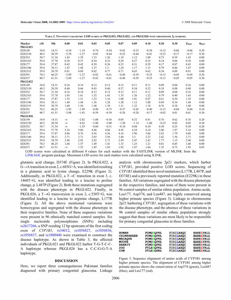
Two-point linkage analysis with chromosome 2p21markers provided positive LOD scores. For PKGL021, themaximum LOD scores of 1.43, 1.47, and 0.91 were obtainedwith markers D2S352, D2S1346, and D2S2331 at θ=0,respectively (Table 2). Similarly, for PKGL022, maximum
Figure 1. Family pedigrees. Pedigrees of A) PKGL021, B) PKGL022 and C) PKGL026. Squares denote males while circles denote females.Filled symbols indicate affected individuals. A double line between individuals signifies consanguinity, and a diagonal line through a symbolindicates a deceased family member. The haplotypes of six adjacent chromosome 2p21 microsatellite markers are shown with alleles formingthe risk haplotype shaded black, alleles cosegregating with PCG but not showing homozygosity shaded gray, and alleles not cosegregatingwith PCG shown in white.
LOD scores of 1.65, 1.35, 1.48, and 1.60 were obtained withmarkers D2S2163, D2S177, D2S1346, and D2S2331 at θ=0,respectively (Table 2). Lastly, for PKGL026, maximum LODscores of 5.16, 4.86, 3.71, and 2.96 were obtained withmarkers D2S2163, D2S177, D2S1346, and D2S2331 at θ=0,respectively (Table 2). Haplotype analysis show that all theaffected individuals of PKGL021, PKGL022, and PKGL026have homozygous alleles for D2S2163, D2S177, D2S1346,
and D2S2331 short tandem repeat (STR) markers whereas thenormal individuals are either heterozygous carriers of thedisease allele or are homozygous for the normal allele (Figure1).
All coding exons, exon-intron boundaries, and the 5′untranslated region of CYP1B1 were sequenced in all threefamilies. In PKGL021, a C→G transversion in exon 3, c.1122C>G, was identified resulting in an aspartic acid to
Figure 2. Sequence chromatograms.The forward and reverse sequencechromatograms of (A) unaffectedindividual 9 of PKGL021, (B)individual 5 of PKGL021, heterozygousand (C) individual 7 of PKGL021,homozygous for a C→G transversion inexon 3, c.1122C>G, resulting in a;p.D374E, (D) individual 8 of PKGL022,heterozygous and (E) individual 10 ofPKGL022, homozygous for G→Atransition in exon2, c.685G>A,resulting in mutation p.E229K, (F)individual 8 of PKGL022, heterozygousand (G) individual 10 of PKGL022,homozygous for a T→C transition inexon 3: c.1460T>C resulting in a;p.L487P (H) unaffected individual 21 ofPKGL026, (I) individual 7 ofPKGL026, heterozygous, and (J)individual 16 of PKGL026,homozygous for a T→G transversion inexon 2: c.530T>G: resulting in p.L177R.
glutamic acid change, D374E (Figure 2). In PKGL022, aG→A transition in exon 2, c.685G>A, was identified resultingin a glutamic acid to lysine change, E229K (Figure 2).Additionally, in PKGL022, a T→C transition in exon 3, c.1460T>G, was identified leading to a leucine to prolinechange, p. L487P (Figure 2). Both these mutations segregatedwith the disease phenotype in PKGL022. Finally, inPKGL026, a T→G transversion in exon 2, c.530T>G, wasidentified leading to a leucine to arginine change, L177R(Figure 2). All the above mentioned variations werehomozygous and segregated with the disease phenotype intheir respective families. None of these sequence variationswere present in 96 ethnically matched control samples. Sixsingle nucleotide polymorphisms (SNPs) includingrs2617266, a SNP residing 12 bp upstream of the first codingexon of CYP1B1, rs10012, rs1056827, rs1056836,rs1056837, and rs1800440 were examined to construct thedisease haplotype. As shown in Table 3, the affectedindividuals of PKGL021 and PKGL022 harbor T-G-T-C-C-A haplotype whereas PKGL026 has a C-C-G-G-T-Ahaplotype.
DISCUSSIONHere, we report three consanguineous Pakistani familiesdiagnosed with primary congenital glaucoma. Linkage
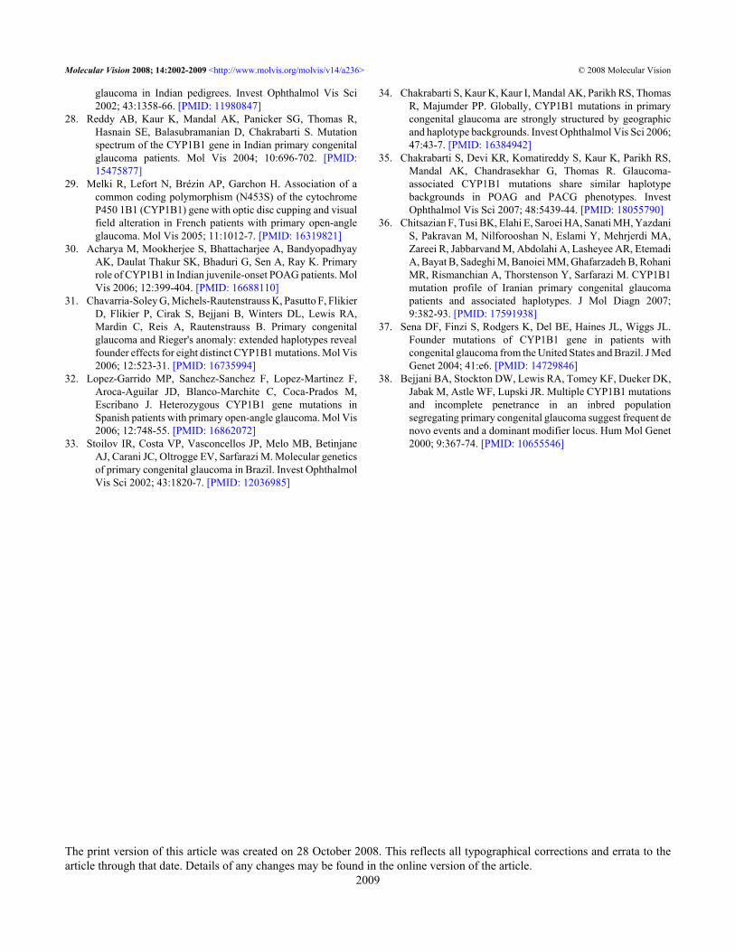
analysis with chromosome 2p21 markers, which harborCYP1B1, provided positive LOD scores. Sequencing ofCYP1B1 identified three novel mutations (L177R, L487P, andD374E) and a previously reported mutation (E229K) in thesefamilies. All variations segregated with the disease phenotypein the respective families, and none of them were present in96 control samples of similar ethnic population. Amino acids,Leu177, Asp374, and Leu487, are highly conserved amonghigher primate species (Figure 3). Linkage to chromosome2p21 harboring CYP1B1, segregation of these variations withthe disease phenotype, and the absence of these variations in96 control samples of similar ethnic population stronglysuggest that these variations are most likely to be responsiblefor primary congenital glaucoma in these families.
Figure 3. Sequence alignment of amino acids of CYP1B1 amonghigher primate species. The alignment of CYP1B1 among higherprimate species shows the conservation of Asp374 (green), Leu487(blue), and Leu177 (red).
TABLE 2. TWO-POINT PARAMETRIC LOD SCORES OF PKGL021, PKGL022, AND PKGL026 WITH CHROMOSOME 2p MARKERS.
LOD scores were calculated at different θ values for each marker with the FASTLINK version of MLINK from theLINKAGE program package. Maximum LOD scores for each marker were calculated using ILINK.
CYP1B1 is a member of the CYP450 super family, whichcontains 58 functional genes in the human genome [2]. Thegene product is a 543 amino acid protein that contains theNH2-terminal membranous region; a 10 residue long, proline-rich region; and a cytosolic globular domain [2]. Mutations inCYP1B1 are the predominant cause of PCG in patients fromvarious ethnic backgrounds. Previous studies have shown thatmissense mutations in CYP1B1 affect highly conserved andfunctionally important regions of CYP1B1, resulting insignificant structural changes and reduced CYP1B1 activity[11,17-21].
In PKGL021, we identified a homozygous missensechange that substitutes aspartic acid at position 374 withglutamic acid in affected individuals. Previously, aspartic acidto asparagine substitution at the same position has beenreported to lead to PCG phenotype in Saudi Arabian patients[22]. Amino acid 374 maps to helix K, one of the highlyconserved core structures thought to be involved in correctfolding and heme binding of the cytochrome P450 molecule[23]. Further mutations that map to helix K are reported to beresponsible for the severe PCG phenotype in Indian patients[21].
In PKGL022, a leucine to proline substitution at residue487 and a previously reported mutation, E229K, segregatedin an autosomal recessive pattern with the disease phenotype.L487P affects the highly conserved position marking the endof helix L, and the proline substitution at this position couldpotentially disrupt the helical structure and possibly affect thenative three-dimensional structures. Conversely, glutamicacid 229 resides in the vicinity of the substrate-binding region(SBR) and is reported to cause conformational changes inCYP1B1 [24]. Heterozygous carriers of this mutation developPCG and primary open-angle glaucoma phenotype [25-32].Interestingly, compound heterozygous carriers of E229K andL487P in PKGL022 show no symptoms of PCG. Noteworthy,the affected individuals of PKGL022 have higher IOPcompared with affected individuals examined during thisstudy. Mutation L177R was detected in PKGL026. Aminoacid 177 in CYP1B1 resides in a highly conserved NH2-capping region [21]. The non-conservative replacement of anon-polar amino acid, leucine, with positively chargedarginine is most likely to impair the native protein structureand consequently affect the protein function.
SNPs, especially rs2617266, rs10012, rs1056827,rs1056836, rs1056837, and rs1800440, have been used toconstruct haplotypes of CYP1B1 mutations [18,33,34].According to the proposed hypothesis of evolution, the T-G-T-C-C-A and C-C-G-G-T-A haplotypes are ancestral humanhaplotypes whereas the T-G-T-C-C-A haplotype iscomparatively recent [35,36]. To date, approximately 50% ofknown CYP1B1 mutations are associated with the C-C-G-G-T-A haplotype while the T-G-T-C-C-A and C-C-G-C-C-Ghaplotypes are less frequent and are associated with 9.7% and7% of CYP1B1 mutations, respectively [31,33,37]. Affectedindividuals of PKGL021 and PKGL022 harbor a T-G-T-C-C-A haplotype, which is indicative of their common ancestry.Noteworthy, the previously reported mutation, E229K, whichsegregates in PKGL022, has been associated with the T-G-T-C-C-A haplotype in French, German, Indian, and Iranianpatients [35,36]. The D374N mutation found in Saudi Arabianpatients is associated with the C-C-G-G-T-A haplotype,which is in contrast to the T-G-T-C-C-A haplotype coupledwith D374E mutation in PKGL021 [38]. Finally, the L177Rmutation detected in PKGL026 is present in the C-C-G-G-T-A haplotype, which is frequently associated with CYP1B1mutations.
Studies of pathogenic sequence variants in CYP1B1 willcontribute to better understanding of primary congenitalglaucoma. Identification of these mutations reaffirms thediverse allelic heterogeneity of CYP1B1 in the pathogenesisof PCG. This will further help in the elucidation of thestructure-function relationship of CYP1B1 and hence, lead tothe development of novel therapeutic approaches.Consequently, the association of specific haplotypes withpathogenic mutations will contribute to our knowledge ofhaplotype clustering of PCG associated with CYP1B1mutations. This will overall enhance our understanding ofprimary congenital glaucoma at the molecular level.
ACKNOWLEDGMENTSThe authors are grateful to all family members for theirparticipation in this study. We sincerely thank the staff ofLayton Rehmatullah Benevolent Trust (LRBT) hospital fortheir help in the clinical evaluation. This work was supportedin part by Higher Education Commission (Islamabad,Pakistan), Ministry of Science and Technology (Islamabad,
TABLE 3. SINGLE NUCLEOTIDE POLYMORPHISM (SNP) PROFILE OF PCG PATIENTS FROM ALL THREE FAMILIES LINKED TO CYP1B1.
Family rs2617266 rs10012 rs1056827 rs1056836 rs1056837 rs1800440PKGL021 T G T C C APKGL022 T G T C C APKGL026 C C G G T A
To trace CYP1B1 mutations identified in this study haplotypes for six intragenic SNPs including rs2617266 , which is 12 bpupstream of the first coding exon of CYP1B1, rs10012, rs1056827, rs1056836, rs1056837, and rs1800440 were constructed.
2. Vasiliou V, Gonzalez FJ. Role of CYP1B1 in glaucoma. AnnuRev Pharmacol Toxicol 2008; 48:333-58. [PMID: 17914928]
3. Wiggs JL, Lynch S, Ynagi G, Maselli M, Auguste J, Del BonoEA, Olson LM, Haines JL. A genomewide scan identifiesnovel early-onset primary open-angle glaucoma loci on 9q22and 20p12. Am J Hum Genet 2004; 74:1314-20. [PMID:15108121]
4. Allen L, Burian HM, Braley AE. A new concept of thedevelopment of the anterior chamber angle; its relationship todevelopmental glaucoma and other structural anomalies.AMA Arch Ophthalmol 1955; 53:783-98. [PMID: 14375435]
5. Maumenee AE. The pathogenesis of congenital glaucoma: anew theory. Trans Am Ophthalmol Soc 1958; 56:507-70.[PMID: 13647611]
6. Kupfer C, Kaiser-Kupfer MI. Observations on the developmentof the anterior chamber angle with reference to thepathogenesis of congenital glaucomas. Am J Ophthalmol1979; 88:424-6. [PMID: 484670]
7. Anderson DR. The development of the trabecular meshworkand its abnormality in primary infantile glaucoma. Trans AmOphthalmol Soc 1981; 79:458-85. [PMID: 7342408]
8. Francois J. Congenital glaucoma and its inheritance.Ophthalmologica 1980; 181:61-73. [PMID: 7219964]
9. Gencik A. Epidemiology and genetics of primary congenitalglaucoma in Slovakia. Description of a form of primarycongenital glaucoma in gypsies with autosomal-recessiveinheritance and complete penetrance. Dev Ophthalmol 1989;16:76-115. [PMID: 2676634]
10. Turacli ME, Aktan SG, Sayli BS, Akarsu N. Therapeutical andgenetical aspects of congenital glaucomas. Int Ophthalmol1992; 16:359-62. [PMID: 1428571]
11. Stoilov I, Akarsu AN, Alozie I, Child A, Barsoum-Homsy M,Turacli ME, Or M, Lewis RA, Ozdemir N, Brice G, AktanSG, Chevrette L, Coca-Prados M, Sarfarazi M. Sequenceanalysis and homology modeling suggest that primarycongenital glaucoma on 2p21 results from mutationsdisrupting either the hinge region or the conserved corestructures of cytochrome P4501B1. Am J Hum Genet 1998;62:573-84. [PMID: 9497261]
12. Stoilov I, Akarsu AN, Sarfarazi M. Identification of threedifferent truncating mutations in cytochrome P4501B1(CYP1B1) as the principal cause of primary congenitalglaucoma (Buphthalmos) in families linked to the GLC3Alocus on chromosome 2p21. Hum Mol Genet 1997;6:641-7. [PMID: 9097971]
13. Akarsu AN, Turacli ME, Aktan SG, Barsoum-Homsy M,Chevrette L, Sayli BS, Sarfarazi M. A second locus (GLC3B)for primary congenital glaucoma (Buphthalmos) maps to the1p36 region. Hum Mol Genet 1996; 5:1199-203. [PMID:8842741]
14. Grimberg J, Nawoschik S, Belluscio L, McKee R, Turck A,Eisenberg A. A simple and efficient non-organic procedure
for the isolation of genomic DNA from blood. Nucleic AcidsRes 1989; 17:8390. [PMID: 2813076]
16. Lathrop GM, Lalouel JM. Easy calculations of lod scores andgenetic risks on small computers 509. Am J Hum Genet 1984;36:460-5. [PMID: 6585139]
17. Achary MS, Reddy AB, Chakrabarti S, Panicker SG, MandalAK, Ahmed N, Balasubramanian D, Hasnain SE,Nagarajaram HA. Disease-causing mutations in proteins:structural analysis of the CYP1b1 mutations causing primarycongenital glaucoma in humans. Biophys J 2006;91:4329-39. [PMID: 16963504]
18. Bagiyeva S, Marfany G, Gonzalez-Angulo O, González-DuarteR. Mutational screening of CYP1B1 in Turkish PCG familiesand functional analyses of newly detected mutations. Mol Vis2007; 13:1458-68. [PMID: 17893647]
19. Mammen JS, Pittman GS, Li Y, Abou-Zahr F, Bejjani BA, BellDA, Strickland PT, Sutter TR. Single amino acid mutations,but not common polymorphisms, decrease the activity ofCYP1B1 against (-)benzo[a]pyrene-7R-trans-7,8-dihydrodiol. Carcinogenesis 2003; 24:1247-55. [PMID:12807732]
20. Mashima Y, Suzuki Y, Sergeev Y, Ohtake Y, Tanino T, KimuraI, Miyata H, Aihara M, Tanihara H, Inatani M, Azuma N,Iwata T, Araie M. Novel cytochrome P4501B1 (CYP1B1)gene mutations in Japanese patients with primary congenitalglaucoma. Invest Ophthalmol Vis Sci 2001; 42:2211-6.[PMID: 11527932]
21. Panicker SG, Mandal AK, Reddy ABM, Gothwal VK, HasnainSE. Correlations of Genotype with Phenotype in IndianPatients with Primary Congenital Glaucoma. InvestOphthalmol Vis Sci 2004; 45:1149-56. [PMID: 15037581]
22. Bejjani BA, Lewis RA, Tomey KF, Anderson KL, Dueker DK,Jabak M, Astle WF, Otterud B, Leppert M, Lupski JR.Mutations in CYP1B1, the gene for cytochrome P4501B1, arethe predominant cause of primary congenital glaucoma inSaudi Arabia. Am J Hum Genet 1998; 62:325-33. [PMID:9463332]
23. Graham-Lorence SE, Peterson JA. Structural alignments ofP450s and extrapolations to the unknown. Methods Enzymol1996; 272:315-26. [PMID: 8791791]
24. Panicker SG, Mandal AK, Reddy ABM, Gothwal VK, HasnainSE. Correlations of genotype with phenotype in Indianpatients with primary congenital glaucoma. InvestOphthalmol Vis Sci 2004; 45:1149-56. [PMID: 15037581]
25. Michels-Rautenstrauss KG, Mardin CY, Zenker M, Jordan N,Gusek-Schneider GC, Rautenstrauss BW. Primary congenitalglaucoma: three case reports on novel mutations andcombinations of mutations in the GLC3A (CYP1B1) gene. JGlaucoma 2001; 10:354-7. [PMID: 11558822]
26. Colomb E, Kaplan J, Garchon HJ. Novel cytochrome P450 1B1(CYP1B1) mutations in patients with primary congenitalglaucoma in France. Hum Mutat 2003; 22:496. [PMID:14635112]
27. Panicker SG, Reddy AB, Mandal AK, Ahmed N, NagarajaramHA, Hasanain SE, Balasubramanian D. Identification ofnovel mutations causing familial primary congenital
glaucoma in Indian pedigrees. Invest Ophthalmol Vis Sci2002; 43:1358-66. [PMID: 11980847]
28. Reddy AB, Kaur K, Mandal AK, Panicker SG, Thomas R,Hasnain SE, Balasubramanian D, Chakrabarti S. Mutationspectrum of the CYP1B1 gene in Indian primary congenitalglaucoma patients. Mol Vis 2004; 10:696-702. [PMID:15475877]
29. Melki R, Lefort N, Brézin AP, Garchon H. Association of acommon coding polymorphism (N453S) of the cytochromeP450 1B1 (CYP1B1) gene with optic disc cupping and visualfield alteration in French patients with primary open-angleglaucoma. Mol Vis 2005; 11:1012-7. [PMID: 16319821]
30. Acharya M, Mookherjee S, Bhattacharjee A, BandyopadhyayAK, Daulat Thakur SK, Bhaduri G, Sen A, Ray K. Primaryrole of CYP1B1 in Indian juvenile-onset POAG patients. MolVis 2006; 12:399-404. [PMID: 16688110]
31. Chavarria-Soley G, Michels-Rautenstrauss K, Pasutto F, FlikierD, Flikier P, Cirak S, Bejjani B, Winters DL, Lewis RA,Mardin C, Reis A, Rautenstrauss B. Primary congenitalglaucoma and Rieger's anomaly: extended haplotypes revealfounder effects for eight distinct CYP1B1 mutations. Mol Vis2006; 12:523-31. [PMID: 16735994]
33. Stoilov IR, Costa VP, Vasconcellos JP, Melo MB, BetinjaneAJ, Carani JC, Oltrogge EV, Sarfarazi M. Molecular geneticsof primary congenital glaucoma in Brazil. Invest OphthalmolVis Sci 2002; 43:1820-7. [PMID: 12036985]
34. Chakrabarti S, Kaur K, Kaur I, Mandal AK, Parikh RS, ThomasR, Majumder PP. Globally, CYP1B1 mutations in primarycongenital glaucoma are strongly structured by geographicand haplotype backgrounds. Invest Ophthalmol Vis Sci 2006;47:43-7. [PMID: 16384942]
35. Chakrabarti S, Devi KR, Komatireddy S, Kaur K, Parikh RS,Mandal AK, Chandrasekhar G, Thomas R. Glaucoma-associated CYP1B1 mutations share similar haplotypebackgrounds in POAG and PACG phenotypes. InvestOphthalmol Vis Sci 2007; 48:5439-44. [PMID: 18055790]
36. Chitsazian F, Tusi BK, Elahi E, Saroei HA, Sanati MH, YazdaniS, Pakravan M, Nilforooshan N, Eslami Y, Mehrjerdi MA,Zareei R, Jabbarvand M, Abdolahi A, Lasheyee AR, EtemadiA, Bayat B, Sadeghi M, Banoiei MM, Ghafarzadeh B, RohaniMR, Rismanchian A, Thorstenson Y, Sarfarazi M. CYP1B1mutation profile of Iranian primary congenital glaucomapatients and associated haplotypes. J Mol Diagn 2007;9:382-93. [PMID: 17591938]
37. Sena DF, Finzi S, Rodgers K, Del BE, Haines JL, Wiggs JL.Founder mutations of CYP1B1 gene in patients withcongenital glaucoma from the United States and Brazil. J MedGenet 2004; 41:e6. [PMID: 14729846]
38. Bejjani BA, Stockton DW, Lewis RA, Tomey KF, Dueker DK,Jabak M, Astle WF, Lupski JR. Multiple CYP1B1 mutationsand incomplete penetrance in an inbred populationsegregating primary congenital glaucoma suggest frequent denovo events and a dominant modifier locus. Hum Mol Genet2000; 9:367-74. [PMID: 10655546]
The print version of this article was created on 28 October 2008. This reflects all typographical corrections and errata to thearticle through that date. Details of any changes may be found in the online version of the article.