National Toxicology Program Toxicity Report Series Number 24 NTP Technical Report on Toxicity Studies of 1,6-Hexanediamine Dihydrochloride (CAS No. 6055-52-3) Administered by Drinking Water and Inhalation to F344/N Rats and B6C3F 1 Mice Charles D. Hébert, PhD, Study Scientist National Toxicology Program Post Office Box 12233 Research Triangle Park, NC 27709 NIH Publication 93-3347 March 1993 United States Department of Health and Human Services Public Health Service National Institutes of Health
Transcript
National Toxicology Program Toxicity Report Series
Number 24
NTP Technical Report on Toxicity Studies of
1,6-Hexanediamine Dihydrochloride
(CAS No. 6055-52-3)
Administered by Drinking Water and Inhalation
to F344/N Rats and B6C3F1 Mice
Charles D. Hébert, PhD, Study Scientist National Toxicology Program
Post Office Box 12233 Research Triangle Park, NC 27709
NIH Publication 93-3347 March 1993
United States Department of Health and Human ServicesPublic Health Service
National Institutes of Health
Foreword
The National Toxicology Program (NTP) is made up of four charter agencies of the United States Department of Health and Human Services (DHHS):
• the National Cancer Institute (NCI) of the National Institutes of Health; • the National Institute of Environmental Health Sciences (NIEHS) of the
National Institutes of Health; • the National Center for Toxicological Research (NCTR) of the Food and
Drug Administration; and • the National Institute for Occupational Safety and Health (NIOSH) of the
Centers for Disease Control. In July 1981, the Carcinogenesis Bioassay Testing Program was transferred from NCI to NIEHS. NTP coordinates the relevant Public Health Service programs, staff, and resources that are concerned with basic and applied research and with biological assay development and validation.
NTP develops, evaluates, and disseminates scientific information about potentially toxic and hazardous chemicals. This knowledge is used for protecting the health of the American people and for the primary prevention of disease.
To carry out its mission, NTP designs and conducts studies to characterize and evaluate the toxicologic potential of selected chemicals in laboratory animals (usually two species, rats and mice). Chemicals selected for NTP toxicology studies are chosen primarily on the bases of human exposure, level of production, and chemical structure. Selection per se is not an indicator of a chemical's toxic potential.
The studies described in this toxicity study report were performed under the direction of NIEHS and were conducted in compliance with NTP laboratory health and safety requirements. These studies met or exceeded all applicable federal, state, and local health and safety regulations. Animal care and use were in accord and compliance with the Public Health Service Policy on Humane Care and Use of Animals.
Single copies of this report are available without charge, while supplies last, from the NTP Public Information Office (telephone number 919/541-3991).
NTP Public Information Office NIEHS
Post Office Box 12233 Research Triangle Park, NC 27709
National Toxicology Program Toxicity Report Series
Number 24
NTP Technical Report on Toxicity Studies of
1,6-Hexanediamine Dihydrochloride
(CAS No. 6055-52-3)
Administered by Drinking Water and Inhalation
to F344/N Rats and B6C3F1 Mice
Charles D. Hébert, PhD, Study Scientist National Toxicology Program
Post Office Box 12233 Research Triangle Park, NC 27709
NIH Publication 93-3347 March 1993
United States Department of Health and Human ServicesPublic Health Service
National Institutes of Health
2 1,6-HEXANEDIAMINE DIHYDROCHLORIDE, NTP TOXICITY REPORT NUMBER 24
CONTRIBUTORS
This NTP Report on the toxicity studies of 1,6-hexanediamine dihydrochloride is based primarily on 2-week drinking water studies conducted in April, 1985, on 2-week inhalation studies conducted in January and February, 1986, and on 13-week inhalation studies that began in June 1987 and ended in September 1987 at Battelle Memorial Laboratories, Columbus, OH.
National Toxicology Program Evaluated experiment, interpreted results, and reported findings Charles D. Hébert, PhD, Study Scientist John R. Bucher, PhD Leo T. Burka, PhD Rajendra S. Chhabra, PhD Michael P. Dieter, PhD Michael Elwell, DVM, PhD John E. French, PhD Joel Mahler, DVM Robert R. Maronpot, DVM H. B. Matthews, PhD Morrow Thompson, DVM, PhD Gregory S. Travlos, DVM Errol Zeiger, PhD
Battelle Memorial Laboratories, Columbus Principal contributors Arthur Peters, DVM, Principal Investigator Milton Hejtmancik, PhD Lawrence E. Mezza, DVM Ronald Persing, DVM Betsy Carlton, PhD Ming J. W. Chang, PhD Peter L. Jepsen, DVM
Environmental Health Research and Testing, Inc Provided sperm morphology and vaginal cytology evaluation Dushant K. Gulati, PhD Susan Russell, BA
Experimental Pathology Laboratories, Inc Provided pathology quality assessment William MacKenzie, DVM, MS Jerry Hardisty, DVM
NTP Pathology Working Group Evaluated slides and prepared pathology report Michael Stedham, DVM, Chairperson
Pathology Associates, Inc. Micheal Jokinen, DVM
National Toxicology Program, Jeffery Everitt, DVM
Chemical Industry Institute of Toxicology Darlene Dixon, DVM, PhD
National Toxicology Program Michael Elwell, DVM, PhD
National Toxicology Program Roger Brown, DVM
Experimental Pathology Laboratories, Inc.
Analytical Sciences, Inc Provided statistical analyses Steven Seilkop, MS Janet Teague, MS Richard Morris, MS
National Institute of Environmental Health Sciences Provided toxicity report preparation Jane M. Lambert, BS Edison McIntyre, BA, BS Kristine Witt, MS
Oak Ridge Associated Universities
Biotechnical Services, Inc Provided supplemental toxicity report preparation Janet L. Elledge, BA, Principal Investigator Chad J. Fitz, MA Paula C. Higginson, BA Jennifer P. Rector, MAP
TABLE OF CONTENTS ABSTRACT ..................................................................................................................7
PEER REVIEW PANEL...................................................................................................10 SUMMARY OF PEER REVIEW COMMENTS .....................................................................11
INTRODUCTION............................................................................................................13 Uses, Production, Exposure, and Physical Properties.............................................13
Absorption, Metabolism, and Distribution .............................................................14 Toxicity..................................................................................................................15 Study Rationale and Design...................................................................................19
MATERIALS AND METHODS .........................................................................................21 Procurement and Characterization of 1,6-Hexanediamine .....................................21 Dose Formulations for Drinking Water Studies......................................................21 Aerosol Generation for Inhalation Studies..............................................................21
Concentration Monitoring......................................................................................22 Study Design.........................................................................................................23
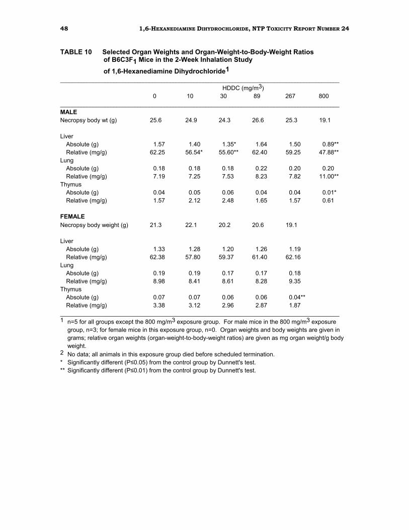
RESULTS ..................................................................................................................35 2-Week Drinking Water Study in F344/N Rats ......................................................35 2-Week Drinking Water Study in B6C3F1 Mice......................................................37 2-Week Inhalation Study in F344/N Rats ..............................................................39 13-Week Inhalation Study in F344/N Rats ............................................................41 2-Week Inhalation Study in B6C3F1 Mice .............................................................46 13-Week Inhalation Study in B6C3F1 Mice ...........................................................49 Mating Trials .........................................................................................................53 Genetic Toxicity Studies ........................................................................................54
3 1,6-HEXANEDIAMINE DIHYDROCHLORIDE, NTP TOXICITY REPORT NUMBER 24
TABLES Table 1 Experimental Design and Materials and Methods in the Drinking Water annd Inhalation Studies of 1,6-Hexanediamine Dihydrochloride ................................................ 29 Table 2 Survival, Weight Gain, Water Consumption, and Compound Consumption in F344/N Rats in the 2-Week Drinking Water Study of 1,6-Hexanediamine Dihydrochloride ................................................ 35 Table 3 Liver Weights and Liver-Weight-to-Body-Weight Ratios of F344/N Rats in the 2-Week Drinking Water Study of 1,6-Hexanediamine Dihydrochloride ................................................ 36 Table 4 Survival, Weight Gain, Water Consumption, and Compound Consumption in B6C3F1 Mice in the 2-Week Drinking Water Study of 1,6-Hexanediamine Dihydrochloride ................................................ 37 Table 5 Liver Weights and Liver-Weight-to-Body-Weight Ratios of B6C3F1 Mice in the 2-Week Drinking Water Study of 1,6-Hexanediamine Dihydrochloride ................................................ 38
Table 6 Survival and Weight Gain of F344/N Rats in the 2-Week Inhalation Study of 1,6-Hexanediamine Dihydrochloride ................................................ 39 Table 7 Survival and Weight Gain of F344/N Rats in the 13-Week Inhalation Study of 1,6-Hexanediamine Dihydrochloride ................................................ 41 Table 8 Incidence and Severity of Histopathologic Lesions in F344/N Rats in the 13-Week Inhalation Study of 1,6-Hexanediamine Dihydrochloride ................................................ 45 Table 9 Survival and Weight Gain of B6C3F1 Mice in the 2-Week Inhalation Study of 1,6-Hexanediamine Dihydrochloride ................................................ 46 Table 10 Selected Organ Weights and Organ-Weight-to-Body-Weight Ratios of B6C3F1 Mice in the 2-Week Inhalation Study of 1,6-Hexanediamine Dihydrochloride ................................................ 48 Table 11 Survival and Weight Gain of B6C3F1 Mice in the 13-Week Inhalation Study of 1,6-Hexanediamine Dihydrochloride ................................................ 49
4 1,6-HEXANEDIAMINE DIHYDROCHLORIDE, NTP TOXICITY REPORT NUMBER 24
TABLES (continued) Table 12 Selected Organ Weights and Organ-Weight-to-Body-Weight Ratios of B6C3F1 Mice in the 13-Week Inhalation Study of 1,6-Hexanediamine Dihydrochloride ................................................ 51 Table 13 Incidence and Severity of Histopathologic Lesions in B6C3F1 Mice in the 13-Week Inhalation Study of 1,6-Hexanediamine Dihydrochloride ................................................ 52
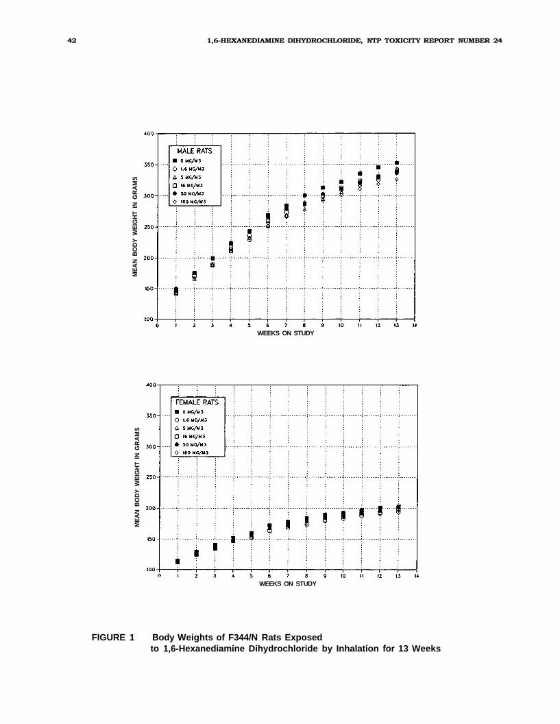
FIGURES Figure 1 Body Weights of F344/N Rats Exposed to 1,6-Hexanediamine Dihydrochloride by Inhalation for 13 Weeks .................................................................. 42 Figure 2 Body Weights of B6C3F1 Mice Exposed to 1,6-Hexanediamine Dihydrochloride by Inhalation for 13 Weeks .................................................................. 50
APPENDICES Appendix A Organ Weights and Organ-Weight-To-Body-Weight Ratios ..................A-1 Appendix B Hematology and Clinical Chemistry Results ........................................B-1 Appendix C Reproductive Tissue Evaluations and Results of Mating Trials ............C-1
Appendix D Genetic Toxicology ..............................................................................D-1
5 1,6-HEXANEDIAMINE DIHYDROCHLORIDE, NTP TOXICITY REPORT NUMBER 24
6 1,6-HEXANEDIAMINE DIHYDROCHLORIDE, NTP TOXICITY REPORT NUMBER 24
7 1,6-HEXANEDIAMINE DIHYDROCHLORIDE, NTP TOXICITY REPORT NUMBER 24
ABSTRACT
1,6-Hexanediamine Dihydrochloride
[H2N–CH2CH2 CH2CH2 CH2–NH2 ] 2 HCl
Molecular Formula C6H16N2•2HCl CAS Number 6055-52-3 Molecular Weight 185.2 Synonyms Hexamethylenediamine dihydrochloride;
1,6-Hexanediamine (HDA) is an aliphatic amine that is produced in large volumes in the
United States. HDA is widely used as a corrosion inhibitor in lubricants and as an
intermediate in the industrial synthesis of paints, resins, inks, and textiles. Toxicity
studies of the dihydrochloride salt of HDA (HDDC) were conducted in male and female Fischer 344/N rats and B6C3F1 mice by the drinking water (2-week studies only) and
whole-body inhalation routes (2-week and 13-week studies). Animals were evaluated for
histopathology, clinical chemistry, hematology, and reproductive toxicity. In addition,
the genetic toxicity of HDA was assessed in Salmonella typhimurium and in Chinese
hamster ovary cells in vitro; HDDC was evaluated in the mouse micronucleus assay in
vivo.
In the 2-week drinking water studies, groups of 5 rats of each sex received HDDC at doses of
0.75 to 6.7 mg/mL, and groups of 5 mice of each sex received doses of 0.2 to 3.0 mg/mL for
14 or 15 days. All animals survived to the end of the studies. No gross or microscopic
pathologic changes and no clinical abnormalities related to HDDC consumption were seen in
any dose group. The only statistically significant change was a slight decrease in absolute
and/or relative liver weights of female rats in the 1.7, 5.0, and 6.7mg/mL treatment groups,
in male rats in the 3.0 mg/mL treatment group, and in female mice in
8 1,6-HEXANEDIAMINE DIHYDROCHLORIDE, NTP TOXICITY REPORT NUMBER 24
the 0.8 mg/mL treatment group. Because there was no significant toxicity in these
studies, 13-week drinking water studies were not conducted.
In the 2-week inhalation studies, 5 rats and 5 mice of each sex were exposed to 0, 10,
30, 89, 267, or 800 mg HDDC/m3 for 6hours per day for 12 days. In the highest
exposure group (800 mg/m3), all male and female rats, all female mice, and 2 male
mice died before the end of the studies. In the remaining groups, there was a dose
dependent depression in body weight gain in male and female mice, but not in rats.
Clinical signs were primarily related to upper respiratory tract irritation and included
dyspnea and nasal discharge in rats and mice. Absolute and relative liver weights were
reduced in some male mice, but this did not occur in a dose-dependent manner. In
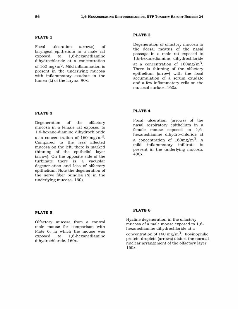
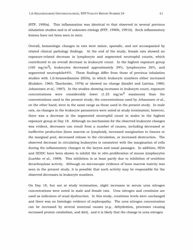
rats, histopathologic lesions that were considered related to chemical exposure
included inflammation and necrosis of laryngeal epithelium as well as focal
inflammation and ulceration of the respiratory and olfactory nasal mucosa. In mice,
focal areas of inflammation and necrosis were present in the respiratory mucosa of the
larynx and trachea in the 2 highest exposure groups. Nasal lesions, including focal
inflammation and ulceration, and degeneration and necrosis of the olfactory and
respiratory epithelium were also seen in mice. In addition, mild testicular degeneration
was present in 2 mice from the highest exposure group (800mg/m3).
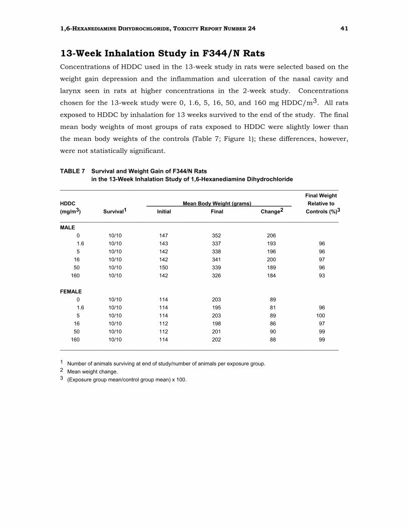
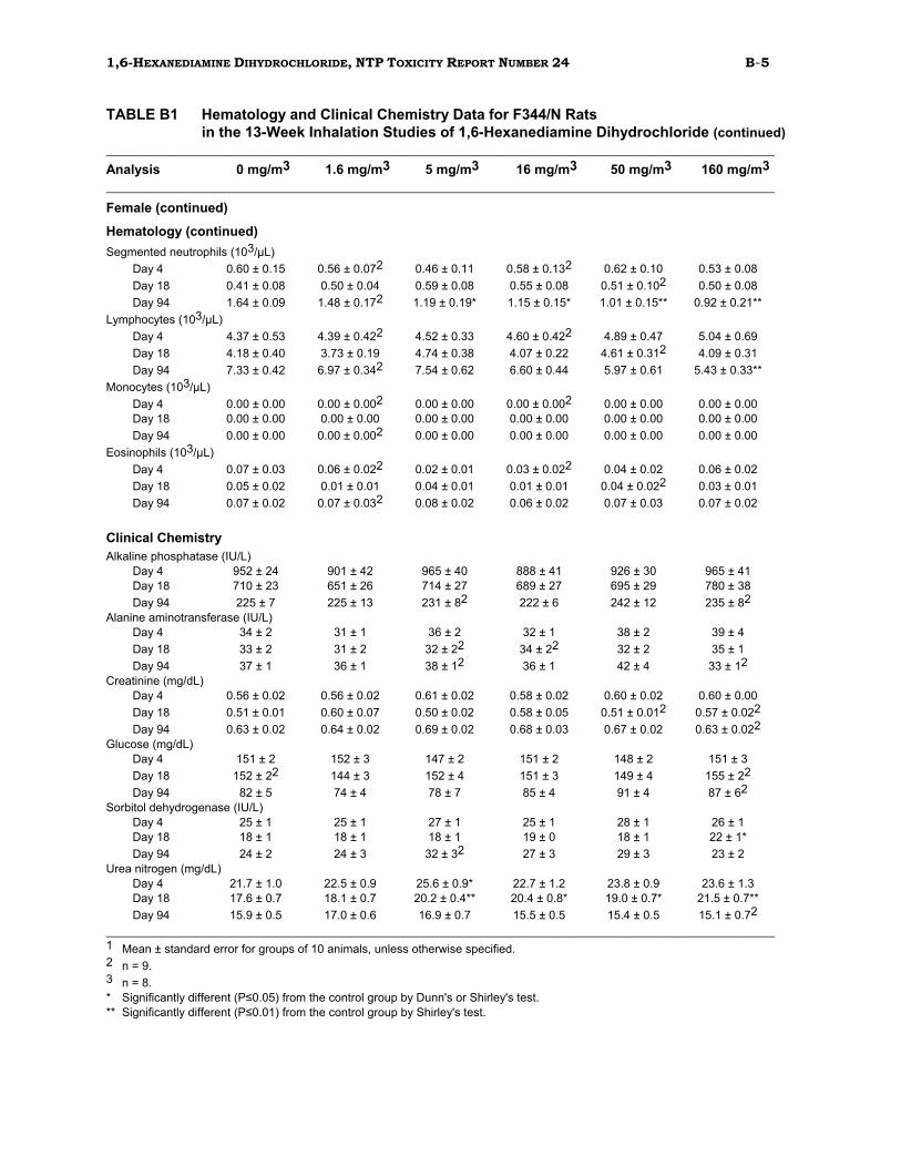
In the 13-week inhalation studies, 10 rats and 10 mice of each sex were exposed to 0,
1.6, 5, 16, 50, or 160 mg HDDC/m3 for 6 hours per day, 5 days per week for 13 weeks.
In addition special groups of 20 male and 40 female rats and mice (mating trial
animals) at each exposure level were included to assess the effect of HDDC on
reproduction. All rats and mice in the base-study groups survived to the end of the
studies, and there were no exposure-related changes in body weight. In the mating
trials, 3 female mice exposed to 16 mg/m3 and 1 female and 1 male mouse exposed to
50 mg/m3 died before scheduled termination. These deaths, however, were not
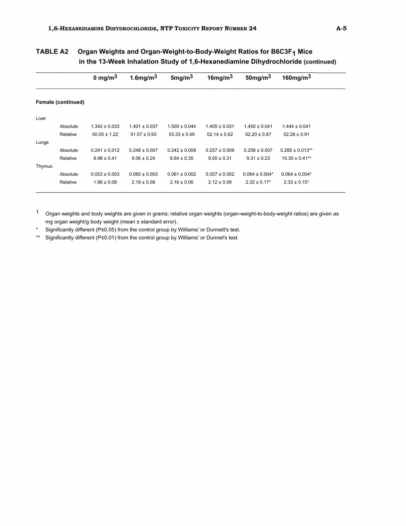
considered to be chemical related. In male mice in the base study, liver weights were
increased relative to controls in the 2highest exposure groups. No exposure-related
changes in absolute or relative organ weights and no exposure-related clinical signs or
gross lesions were seen in either species. In female rats, a dose-related decrease in
white blood cell count was observed. Chemical-related microscopic lesions in male and
female rats and mice were limited to the upper respiratory tract (larynx and nasal
passages) in the 2 highest exposure groups and were similar in both species. These
lesions included minimal to mild focal erosion/ulceration, inflammation, and
hyperplasia of the laryngeal epithelium as well as degeneration of the
9 1,6-HEXANEDIAMINE DIHYDROCHLORIDE, NTP TOXICITY REPORT NUMBER 24
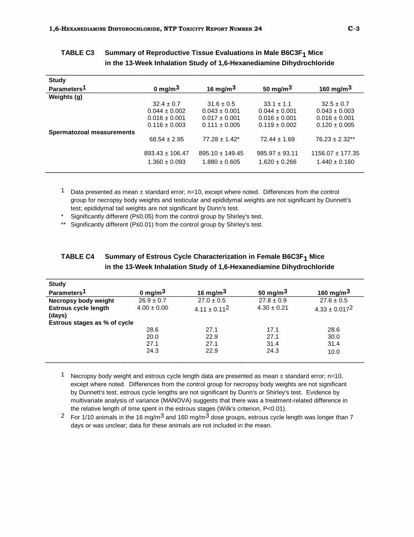
olfactory and respiratory nasal epithelium. HDDC caused no significant changes in
sperm morphology or in the length of the estrous cycle of rats or mice.
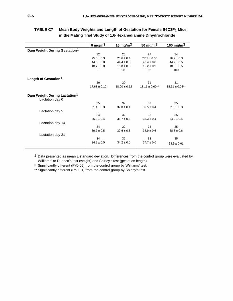
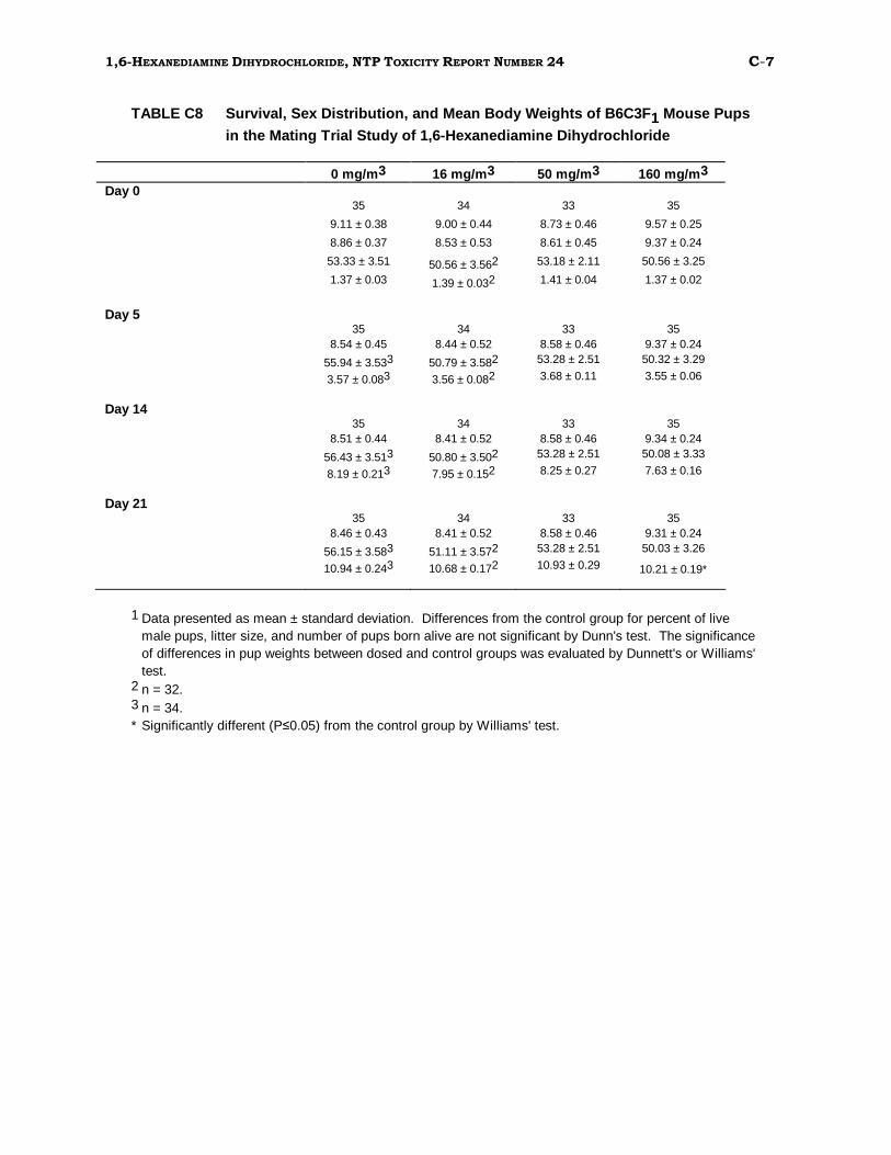
In mating trials, HDDC demonstrated no adverse effects on reproduction of rats. The
only statistically significant changes in reproductive parameters of mice were a slight
increase in gestation length in the 50 mg/m3 and 160 mg/m3 exposure groups and a
decrease in mean pup weight on Day 21 in the highest exposure group. These changes
were not considered to be biologically significant.
1,6-Hexanediamine was not mutagenic in 4 strains of Salmonella typhimurium, and it
did not induce sister chromatid exchanges or chromosomal aberrations in cultured
Chinese hamster ovary cells. These in vitro tests were conducted with and without
exogenous metabolic activation (S9). Negative results were also obtained in an in vivo
test that measured the frequency of micronucleated erythrocytes in peripheral blood of
male and female mice.
In summary, the toxicity of HDDC to rats and mice resulted from irritant properties of
the chemical and was consistent with the effects of other irritant chemicals
administered by inhalation. This toxicity was limited to the nose and airways. In the 2
week inhalation studies, deaths occurred in both rats and mice at the highest exposure
level (800 mg/m3). In the 13-week studies, the no-observed-adverse-effect-level
(NOAEL) for respiratory damage was 5 mg/m3 for rats and mice. HDDC had no
adverse effect on reproduction of either species and was not genotoxic.
________________
10 1,6-HEXANEDIAMINE DIHYDROCHLORIDE, NTP TOXICITY REPORT NUMBER 24
PEER REVIEW PANEL
The members of the Peer Review Panel who evaluated the draft report on the toxicity studies on 1,6-hexanediamine dihydrochloride on June 24, 1992 are listed below. Panel members serve as independent scientists, not as representatives of any institution, company, or governmental agency. In this capacity, panel members determine if the design and conditions of these NTP studies are appropriate and ensure that the toxicity study report presents the experimental results and conclusions fully and clearly.
Gary P. Carlson, PhD, Chair Department of Pharmacology and Toxicology
Purdue University West Lafayette, IN
Paul T. Bailey, PhD Environmental and Health Sciences Laboratory Mobil Oil Corporation
Princeton, NJ
Louis S. Beliczky*, MS, MPH, Principal Reviewer Department of Industrial Hygiene United Rubber Workers International Union
Akron, OH
Kowetha A. Davidson, PhD Health and Safety Research Division Oak Ridge National Laboratory Oak Ridge, TN
Harold Davis, DVM, PhD School of Aerospace Medicine Brooks Air Force Base, TX
Jay I. Goodman, PhD Department of Pharmacology and Toxicology Michigan State University East Lansing, MI
David W. Hayden, DVM, PhD Department of Veterinary Pathobiology College of Veterinary Medicine University of Minnesota St. Paul, MN
* Unable to attend
Curtis D. Klaassen*, PhD Department of Pharmacology and Toxicology University of Kansas Medical Center Kansas City, KS
Daniel S. Longnecker*, MD Department of Pathology Dartmouth Medical School
Barbara McKnight, PhD Department of Biostatistics University of Washington
Seattle, WA
Ellen K. Silbergeld, PhD, Principal Reviewer University of Maryland Medical School
Baltimore, MD
Matthew J. van Zwieten, DVM, PhD Department of Safety Assessment Merck, Sharpe & Dohme Research Laboratories West Point, PA
Lauren Zeise, PhD California Department of Health Services/RCHAS Berkeley, CA
11 1,6-HEXANEDIAMINE DIHYDROCHLORIDE, NTP TOXICITY REPORT NUMBER 24
Summary of Peer Review Comments
On June 24, 1992, the Technical Reports Review Subcommittee of the Board of
Scientific Counselors for the National Toxicology Program met in Research Triangle
Park, NC, to review the draft technical report on toxicity studies of 1,6-hexanediamine
dihydrochloride.
Dr. C.D. Hébert, NIEHS, introduced the short-term toxicity studies of 1,6
hexanediamine dihydrochloride by reviewing the uses and rationale for study of the
chemical, the experimental design, and results.
Dr. Silbergeld, a principal reviewer, said the test chemical is a derivative of the widely
used high-production volume chemical hexanediamine. She was concerned with the
decision to test hexanediamine dihydrochloride, rather than hexanediamine. She said
that the rationale appeared to be based on the ability to produce a rather stable
compound that could be handled under test conditions; however, public health
concerns relate to hexanediamine. Hexanediamine is reportedly toxic to humans, and
inhalation/ingestion studies have been conducted in rodents, although actual use of
the chloride in these studies could not be ruled out given the preparation method of the
chemical. Dr. Hébert responded that he did not have information concerning the actual
form of the compound as it occurs in the environment, or more specifically, in the
airway epithelium; however, based on its chemical properties, he would expect it to be
found as the mono- or dihydrochloride salt. In addition, because hexanediamine forms
a precipitate on the walls of the inhalation chambers, it was more practical to test the
dihydrochloride salt.
The comments of Mr. Beliczky, a second primary reviewer, who could not attend the
meeting, were read by Dr. L. G. Hart, NIEHS. Mr. Beliczky thought that considerable
toxicity data were available for hexanediamine, but that the current study still did not
provide adequate information on which to base a decision to conduct a 2-year study.
Dr. Hébert said that hexanediamine dihydrochloride had a low priority for further
studies.
Dr. J. Haartz, NIOSH, requested that the report include more information about the
generation and monitoring of inhalation aerosol. Dr. Carlson seconded this request
and stated on behalf of the committee that the report would be accepted with the
suggested changes.
12 1,6-HEXANEDIAMINE DIHYDROCHLORIDE, NTP TOXICITY REPORT NUMBER 24
______________
13 1,6-HEXANEDIAMINE DIHYDROCHLORIDE, NTP TOXICITY REPORT NUMBER 24
INTRODUCTION
1Uses, Production, Exposure, and Physical Properties1,6-Hexanediamine (HDA) is an aliphatic diamine that is widely used as an
intermediate in chemical processes, including the synthesis of nylon-type polyamide
resins (especially Nylon 66), the synthesis of oil-modified and moisture area types of
urethane coatings, and the manufacture of polyamides for printing inks, paints, dimer
acids, and textiles. HDA is also used as a corrosion inhibitor in oil and lubricants and
as a curing agent in epoxide resins. Commercially, HDA is prepared by the reduction of
adiponitrile with sodium and alcohol. According to the most recent available figures,
U.S. manufacturers of HDA produced 837 million pounds in 1972 and 749 million
pounds in 1975 (SRI, 1972, 1975); imports amounted to 370,000 pounds in 1972 and
16,000 pounds in 1975 (SRI, 1972, 1975). It has been estimated that as many as 12.8
million pounds of HDA were released into the environment each year in the mid-1970s.
According to NIOSH estimates, approximately 1100 workers per year were exposed
occupationally between 1972 and 1974 (NIOSH, 1972-1974). Occupational exposure
may occur by either dermal or inhalation routes. Estimates for nonoccupational
exposure to HDA are not available.
HDA is 1 of several odoriferous compounds produced by Arum lilies during flowering
(Smith and Meeuse, 1966). No other information on the natural occurrence of HDA was
found.
1 Numerous studies on the toxicity of 1,6-hexanediamine (HDA) have been reported in the literature. In many of these studies, the compound was studied in the form of aqueous solutions of HDA, which are highly basic. Some of these reports describe the HDA solutions as having been neutralized with hydrochloric acid (HCl) before use. Neutralization ofHDA with HCl leads to the formation of, first, the monohydrochloride salt and then, with further addition of HCl, the dihydrochloride salt. The majority of the literature reports do not give the pH of thefinal solutions. As a result, it is not possible to determine the exact form of the compound tested (i.e., free acid, monohydrochloride salt or dihydrochloride salt). Therefore, in the summary of literature, all studies are cited as having been conducted with HDA unless the papers specifically state that the dihydrochloride salt (HDDC) was used. In addition, many studies of HDA toxicity were published in Russian, and translations of the entire articles are unavailable. In those cases in which only the abstracts are available in English, it is difficult to assess the adequacy of study design, details of experimental method, interpretation of results, and accuracy of the abstracts. These citations are indicated as "Abstract" in the reference list.
14 1,6-HEXANEDIAMINE DIHYDROCHLORIDE, NTP TOXICITY REPORT NUMBER 24
HDA is a colorless compound with a molecular weight of 116.2, a melting point of 42°C,
and a boiling point of 205°C (Verschueren, 1979; NTP, 1991a). The compound in solid
form exists as colorless leaflets, which absorb water and carbon dioxide from the
atmosphere. HDA is soluble in water (≥ 100 mg/mL at 23°C), ethanol, dimethyl
sulfoxide, and benzene. Therefore, the technical product is supplied as a 70% aqueous solution. HDA is strongly alkaline, with a pKa of 10.7, and is irritating to skin and
mucous membranes. 1,6-Hexanediamine dihydrochloride (HDDC) is formed by the
neutralization of HDA with hydrochloric acid. HDDC has a molecular weight of 185.2, a
melting point of 248°C, and is freely soluble in water. In its solid form, HDDC
crystallizes as needles from water or ethanol. No other information on the physical
properties of HDDC was found in the literature.
HDA has not been found in U.S. or European drinking water supplies (Commission of
the European Communities, 1976; NAS, 1977) or in industrial effluent (US EPA, 1979).
No TLV (threshold limit value) for HDA has been established (ACGIH, 1990-91).
Absorption, Metabolism, and Distribution The pharmacokinetic profiles of HDA in humans and rodents have been investigated
experimentally. HDA (8.2 mg) was given orally to healthy human male volunteers on 2
occasions 3 months apart, and urinary excretion of the compound was monitored
(Brorson et al., 1990). Excretion of HDA was found to be virtually complete within 15
hours, with an estimated half-life of elimination of 1.5 hours. In addition to the parent
compound, the primary compounds found in the urine were the metabolites 6
aminohexanoic acid and N-acetyl-1,6-HDA.
Urine was found to be the principal route of excretion in adult male Fischer 344/N
rats dosed by gavage with 1,6-[14C]-hexanediamine (0.4 mg/kg body weight) (David
and Heck, 1983). Within 72 hours after dosing, urinary excretion accounted for
approximately 47% of the total dose; over a 72-hour period, 27% of the dose was excreted in feces; another 20% was recovered as exhaled 14CO2. Less than 1.5% of
the total dose was retained by the animals. Of the tissues examined, prostate gland
contained the highest specific activity of radiolabel, followed by kidney, liver,
intestine, and spleen. Similar results were obtained after intravenous
administration of [14C]-HDA to male and female rats. High levels of radioactivity
were found in the prostate gland, intestine, and liver of males 24 hours after dosing.
Uteri of female rats contained high concentrations of radiolabel 1 hour after
15 1,6-HEXANEDIAMINE DIHYDROCHLORIDE, NTP TOXICITY REPORT NUMBER 24
injection but not 24 hours after injection; ovaries contained little radioactivity at any
time. Gas and thin-layer chromatographic analyses of urine from orally dosed rats
indicated that 30% of the radioactivity was in the form of parent HDA. No further
analyses of the other metabolites were performed.
Decomposition of 1,6-hexanediamine is reported to occur in the presence of intact cells
or cell-free extracts of Bacillus subtilis (Roi, 1975; Garbara and Rotmistrov, 1982;
Gvozdyak et al., 1982). This is the principal method for removal of HDA found in waste
water effluents from polyamide fiber production plants in the former Soviet Union.
Bacterial diamine oxidases were shown to be incapable of metabolizing HDA (Tanzil and
Boenicke, 1969); however, porcine diamine oxidase was able to metabolize HDDC in
vitro (Bardsley et al., 1970). In addition, Subramanyam et al. (1989) found that the
metabolic fate of HDA is similar to that of putrescine and cadaverine in that HDA is
converted to 6-aminohexanoic acid and caprolactam by rat and rabbit liver aldehyde
oxidases via a cyclic intermediate.
Toxicity HUMAN EFFECTS
At least 2 incidents involving HDA poisoning in humans have been described in the
literature. Twenty workers at an Italian nylon manufacturing plant were exposed to
HDA and adiponitrile in air. HDA concentrations ranged from 2 to 5.5 mg/m3 during
normal plant operations and from 32.7 to 131.5 mg/m3 during autoclaving. Irritation
of the conjunctiva and respiratory tract was reported in 8 workers; 1 worker developed
contact dermatitis and acute hepatitis, which were believed to be due to HDA exposure.
No anemia was seen in any of the workers (Ceresa, 1948; Gallo and Ghiringhelli, 1958).
In the second incident, 488 workers in an epoxide resin plant were exposed to HDA. A
variety of symptoms, including itching, allergic rhinitis, bronchial asthma, impairment
of bronchial permeability, toxicoallergic hepatitis, gastritis, colitis,
hypergammaglobulinemia, increased serum transaminase activity, and eosinophilia of
peripheral blood, were reported after prolonged contact (Gul'ko, 1971).
ANIMAL TOXICITY
The toxicity of HDA in several mammalian and nonmammalian species has been examined
using various routes of administration. HDA is reported to be moderately toxic in most
16 1,6-HEXANEDIAMINE DIHYDROCHLORIDE, NTP TOXICITY REPORT NUMBER 24
rodent species tested. The single-dose oral LD50 for rats has variously been reported
as 750 mg/kg (unspecified strain and sex; Vernot et al., 1977), 750 and 800 mg/kg
(female and male Sprague-Dawley, respectively; Vernot et al., 1977), 792 and 1127
mg/kg (fasted and unfasted males, respectively; Dashiell and Kennedy, 1984) and 980
mg/kg (male and female Sprague-Dawley; Johannsen and Levinskas, 1987). Clinical
signs prior to death in these studies included weakness, malaise, salivation, diarrhea,
tremors, and weight loss. In addition, renal hyperemia and gastrointestinal
inflammation were reported.
No reports were found on the oral LD50 of HDA in mice. However, the LD50 in mice by
other routes is reported to be 180 mg/kg (intravenous; NDRC, 1942), 320 mg/kg
(intraperitoneal; Roi and Garbara, 1978) and 1300 mg/kg (subcutaneous; Izmerov et al., 1982). In addition, the inhalation LCLO is estimated at 750 mg/m3 for a 10 minute
exposure (Sax, 1984). Roi and Garbara (1978) also reported that the bacterial
degradation products of HDA were 1.7-fold less toxic in mice than the parent
compound, and that concentrations of HDA greater than 50 to 70 mg/L in water were
lethal to Daphnia and Cyclops within 5 to 7 minutes.
The single-dose oral LD50 for HDA in rabbits is 1110 mg/kg (Vernot et al., 1977).
Ceresa and DeBlasiis (1950) found HDA toxic when given orally (in pill form) and
subcutaneously (in solution) to guinea pigs. Five of 6 guinea pigs given 0.02 g
HDA/day orally died within 20 to 70 days, while the same dose given subcutaneously
killed 3 of 3 guinea pigs in 5 to 7 days. Clinical and pathologic findings included weight
loss, hemolytic anemia, and kidney and liver degeneration.
Subchronic and chronic studies of HDA toxicity have been conducted in rats and mice.
Male and female Sprague-Dawley rats given HDA in feed sufficient to provide daily
doses of up to 500 mg/kg body weight for 13 weeks experienced no toxic effects. There
were no changes in body weight or in several clinical chemistry parameters examined
(Johannsen and Levinskas, 1987). HDA given to rats and mice in drinking water for 1
to2years reportedly caused an increase in the mitotic index of lymphoid tissues at 0.1
and 1.0 mg/kg, but not at 10 mg/kg (Ponomareva and Merkushev, 1978). Marked
changes in hemodynamic and bioelectrical properties of the heart were seen in rats
exposed to 0.36mgHDA/m3 by inhalation for 3 months (Verich, 1979).
17 1,6-HEXANEDIAMINE DIHYDROCHLORIDE, NTP TOXICITY REPORT NUMBER 24
The toxic effects of HDA in rats after inhalation exposure have been characterized. Rats
exposed to 1.25 mg HDA/m3 for 4 hours each day for 8 days reportedly had reduced
nerve/muscle excitability, increased leukocyte counts, and impairment of hepatic
glycogen synthesis and renal excretory capability (Tkachenko, 1976). Izrailet and
Laivina (1980) saw no effects on hemoglobin or leukocyte parameters after chronic
exposure of rats to 1mgHDA/m3 for unspecified durations. The rat NOEL for
inhalation of HDA has been cited as 1 mg/L (equal to 1000 mg/m3) (15 days, 6 hours
per day) (Verschueren, 1979). However, albino rats exposed continuously to
concentrations of HDA up to 1 mg/m3 for 3 months were said to have experienced
growth retardation as well as a number of hematologic alterations, including increases
in reticulocytes and leukocytes and decreases in leukocyte phagocytic activity and
eosinophils (Kulakov, 1965). In a study published by Johannsen et al. (1987), male and
female Sprague-Dawley rats were exposed to HDA at concentrations of 0, 12.8, 51, or
215 mg/m3, 6 hours per day, 5 days per week for 13weeks. All rats in the 215 mg/m3
exposure group died or were killed moribund by Week 7 of the study. Inflammation of
the airways and lungs and conjunctival irritation were seen at exposure levels of 51
mg/m3 and greater. Body weights were significantly reduced only in the 215 mg/m3
group. After 5 weeks of exposure, erythrocyte counts and hemoglobin and hematocrit
values were elevated in the high-exposure group, suggesting possible hemopoietic
stimulation by HDA. However, there was no suggestion of hemoconcentration in this or
any other exposure group. Rats exposed to 12.8 or 51 mg/m3 showed no treatment
related hematologic changes. Microscopic lesions related to chemical exposure were
confined to the trachea, nasal passages, and lungs. The cause of death in rats that
died before the end of the study could not be determined.
HDA was shown to suppress immune function, both in vivo in rats and in vitro. Jobin
and Tremblay (1970) found that HDA was able to inhibit collagen- and latex-induced
aggregation of human platelets in vitro. Luebke et al. (1989) recently reported that
HDDC suppressed mitogen-stimulated proliferation of mouse lymphocytes in vitro; this
effect involved inhibition of ornithine decarboxylase (ODC) and polyamine activity, as
well as other unidentified processes. In a drinking water study in which HDA was
administered to rats for 12 months at concentrations of 0.1, 1.0, or 10.0 mg/L,
antibody production was inhibited and the volume of lymphoid tissue in the spleen was
reduced by approximately 40% (Shubik et al., 1978).
18 1,6-HEXANEDIAMINE DIHYDROCHLORIDE, NTP TOXICITY REPORT NUMBER 24
BIOCHEMICAL EFFECTS
HDA has been shown to inhibit the growth of human, monkey, and rodent cells in vitro
(Trakhtenberg et al., 1976; Chapman and Glant, 1980; Yano et al., 1981) and to induce
differentiation in sea urchin eggs (Lallier, 1966). Similarly, hexamethylene
bisacetamide, a derivative of HDA, has been shown to induce differentiation of murine
erythroid cells in culture (Reuben et al., 1976, 1978; Hozumi et al., 1979). Like other
diamines, HDA inhibits the activity of ornithine decarboxylase (ODC), an enzyme
necessary for synthesis of cellular polyamines. ODC inhibition has been demonstrated
in vivo (Guha and Janne, 1977; Pegg et al., 1978) and in vitro (Guha and Janne, 1977;
Kallio et al., 1977; Bethell and Pegg, 1979; Chapman and Glant, 1980), and is believed
to be at least partially responsible for HDA-induced inhibition of cell proliferation.
HDA and other polyamines stabilize the structure of polyribonucleotides and increase
their melting temperatures, presumably through association with double helical DNA
(Szer, 1966; Padmanabhan et al., 1991). This effect, too, may be related to the ability of
HDA to inhibit cellular proliferation.
REPRODUCTIVE AND DEVELOPMENTAL TOXICITY AND CARCINOGENICITY
A number of studies have been conducted to investigate the reproductive and
developmental toxicity of HDA in rats (David and Heck, 1983; Johannsen and
Levinskas, 1987; Short et al., 1991) and mice (Manen et al., 1983). These included a
double generation study of rats given HDA in the diet (Short et al., 1991). In rats, HDA
doses as high as 900 mg/kg/day (during gestation) or 150 mg/kg/day (over 2
generations) had no effect on copulatory behavior, gestation length, fertility, number of
corpora lutea, litter size, incidence of resorptions, pup survival, pup weight, sex ratios,
or nesting or nursing behavior of dams. Similarly, HDA did not affect testis weight or
copulatory behavior of male rats. HDA administered intraperitoneally to adult male
CD-1 mice caused no adverse reproductive effects. HDA given to pregnant female CD-1
mice at 0.89 mM/kg was not feticidal; however, it caused a decrease in fetal body
weight after administration on gestation Days 10, 11, or 12 (Manen et al., 1983) as well
as a retardation of supraoccipital bone development. No teratogenic effects of HDA
have been described, and no information was found in the literature on the
carcinogenic potential of HDA.
19 1,6-HEXANEDIAMINE DIHYDROCHLORIDE, NTP TOXICITY REPORT NUMBER 24
GENETIC TOXICITY
1,6-Hexanediamine was not mutagenic in any of several strains of Salmonella
typhimurium tested with a preincubation protocol in the presence or the absence of S9
activation (Mortelmans et al., 1986). 1,6-Hexanediamine was also tested (in the
dihydrochloride form, HDDC, pH 4.2) for direct mutagenic activity in 7 Salmonella
tester strains; HDDC was tested after reaction with sodium nitrite (Murphey-Corb et al.,
1983). In this study, HDDC failed to form a nitrosamine after incubation with nitrite,
and caused neither direct mutations in Salmonella nor frameshift activity in tester
strain TA1952.
Study Rationale and Design The U.S. Environmental Protection Agency nominated HDA for toxicity testing by the
National Toxicology Program because of the large production volume of the chemical,
the potential for occupational and nonoccupational human exposure, and the lack of
information on the toxicity, mutagenicity, teratogenicity, and carcinogenicity of HDA.
Inhalation and drinking water were chosen as administration routes because these are
the major routes of potential occupational and nonoccupational exposure, respectively,
in humans. Mating trials were included because inadequate data were available in the
literature to assess the potential reproductive effects of HDA.
In the NTP toxicity studies of HDA, all solutions were converted from the free diamine to
the dihydrochloride salt (HDDC) for the following reasons:
• HDA solutions are highly basic and, as such, are extremely caustic. HDA has a very high pKa (10.7) and would become protonated very rapidly upon contact with
tissues or fluids at physiologic pH, causing local necrosis.
• Nonneutralized HDA solutions have a pungent odor and would be unpalatable
in drinking water.
• HDA strongly absorbs carbon dioxide from the atmosphere. Stability studies
conducted by the NTP indicated that under inhalation exposure conditions, HDA
tended to deposit on the walls of the inhalation chambers and become converted to
the carbamate form. HDDC was found to be much more stable in the aerosol form
than HDA.
• HDDC has the same organic backbone as HDA and its use would allow
detection of any specific toxicity associated with that backbone while avoiding the
causticity, palatability, and stability problems that would be encountered with the
use of HDA.
20 1,6-HEXANEDIAMINE DIHYDROCHLORIDE, NTP TOXICITY REPORT NUMBER 24
Two-week drinking water studies were performed on male and female rats and mice
using HDDC. Because gross and histopathologic examinations of animals from the 2
week drinking water studies revealed no specific target tissues, 13-week drinking water
studies were not conducted. Two-week and 13-week whole body inhalation studies of
HDDC toxicity were conducted on male and female rats and mice. Gross and
histopathologic examinations and sperm morphology and vaginal cytology evaluations
were performed on rats and mice, and clinical pathology analyses were performed on
rats in the 13-week inhalation studies. In addition, supplemental groups of rats and
mice were exposed to HDDC by inhalation for 13 weeks and used in mating trials to
assess the reproductive toxicity of HDDC. The genetic toxicity of HDDC was evaluated
in the in vivo mouse micronucleus assay using mice in the 13-week inhalation study,
and the genetic toxicity of HDA was evaluated in in vitro assays in S. typhimurium and
in Chinese hamster ovary cells.
21 1,6-HEXANEDIAMINE DIHYDROCHLORIDE, NTP TOXICITY REPORT NUMBER 24
MATERIALS AND METHODS Procurement and Characterization of 1,6-Hexanediamine 1,6-Hexanediamine was purchased from E. I. DuPont de Nemours and Company, Inc.
(Wilmington, DE), in 2 lots (Lot No. PT-011882 and Lot No. PT-031985), and was
shipped to the study laboratory, Battelle Columbus (Columbus, OH), from Midwest
Research Institute (Kansas City, MO). Lot PT-011882 was used for the 2-week drinking
water and inhalation studies; Lot PT-031985 was used for the 13-week inhalation
studies. The chemical was identified as HDA by infrared spectroscopy. Purity analyses
performed by gas chromatography indicated a purity of 101% for Lot PT-011882
(purchased in solid form) and 70.9% for Lot PT-031985 (purchased as a 70% aqueous
solution). Bulk chemical was stored at room temperature in amber or foil-wrapped
bottles; periodic chemical reanalyses at 4-month intervals indicated no breakdown of
the chemical during storage.
Dose Formulations for Drinking Water Studies Drinking water solutions of hexanediamine were prepared in deionized water. All
solutions, including the dosed water for the control group, were adjusted to pH 4.5 to
5.5 with 5.0N hydrochloric acid. At this pH, virtually all the hexanediamine exists in
the form of the dihydrochloride salt (HDDC). Solutions were prepared weekly and
stored in Nalgene® containers in the dark at room temperature. Analyses of the dose
formulations were performed by gas chromatography; all dose formulations were found
to be within 10% of target concentrations.
Aerosol Generation for Inhalation Studies For the inhalation studies, 1,6-hexanediamine was converted to 1,6-hexanediamine
dihydrochloride (HDDC) by acidification with concentrated hydrochloric acid under a
stream of nitrogen. The final pH was adjusted within the range of 4.5 to 5.5 before
storage and again before use in the inhalation chambers.
The 70% aqueous HDDC solution was placed in a 9-liter glass reservoir and pressurized with N2 gas. HDDC was delivered to 5 Sonimist Ultrasonic Spray Nozzles (Model HS600
2, Heat Systems-Ultrasonics, Inc., Farmingdale, NY) by a positive displacement metering
22 1,6-HEXANEDIAMINE DIHYDROCHLORIDE, NTP TOXICITY REPORT NUMBER 24
pump. Up to this point, stainless steel lines carried the test substance. The nebulizer
reservoir was kept in a separate exposure chamber (H-1000, Hazelton Systems, Inc.,
Aberdeen, MD). This chamber served as a mixing plenum where large droplets and
nonnebulized liquid were impacted or sedimented out of the test atmosphere before the
aerosol was delivered to the inhalation chambers. The HDDC aerosol was mixed with
compressed breathing air that had been filtered through an ENMET (ENMET Air
Filtration Panel, Model AFP-82, Enmet Co., Ann Arbor, MI) and supplied at 50 psi to
generate an aerosol at a concentration equal to the highest exposure concentration.
The resulting aerosol was transported to the inhalation chambers through a manifold
constructed of 3-inch diameter PVC tubing. At each chamber, a metered amount of
aerosol was removed from the manifold and mixed with the appropriate amount of
HEPA/charcoal-filtered room air to obtain the desired test concentration, then delivered
to the inhalation chamber. After exiting the chambers, the test atmospheres were
delivered to a common duct and cleansed of the test substance by a Mystaire HS-7CM
scrubber (Heat Systems Ultrasonics).
Concentration Monitoring Concentrations of HDDC in the exposure chamber, exposure room, and exhaust were
monitored by measuring the forward light scatter with RAM-S real-time aerosol
monitors (GCA Corporation, Technology Division, Bedford, MA) and by gravimetric
analyses of filter samples collected from each exposure chamber. Six RAM-S readings
and 3 gravimetric samples were taken from each exposure chamber on each day of
exposure. Gravimetric sampling was conducted with 25 mm glass fiber filter paper
(Gelman Sciences, Inc., Ann Arbor, MI). Gravimetric analysis was performed on a
Perkin Elmer AS-2Zmicrobalance (Perkin Elmer, Norwalk, CT) by weighing filters to the
nearest 0.01 mg before and after sampling and again after storing the filters in a
desiccator overnight. Twice monthly during the 13-week studies, glass fiber filter
samples from each chamber were analyzed by gas chromatography with flame
ionization detection for total hexanediamine, using the technique supplied by Midwest
Research Institute. Measured concentrations of HDDC in the exposure chambers were
within 6% of the target concentrations in all samples.
Spatial homogeneity of the aerosol within the exposure chambers was determined using
the calibrated RAM-S monitors. Chamber concentrations were measured at 12 points
within each chamber and then were compared to a fixed reference point. Time spans
required to reach stable concentrations after start up and to reach background
concentrations at the end of exposure were determined by taking measurements of aerosol
23 1,6-HEXANEDIAMINE DIHYDROCHLORIDE, NTP TOXICITY REPORT NUMBER 24
concentrations every 60 seconds. The time span required after start up to reach 90% of
the target concentration was identified as the T90; the time span required after the end
of the exposure period to reach 10% of the target concentration was identified as the T10.
Triplicate particle size measurements were obtained for each exposure chamber once in
the first week and monthly thereafter, using an APS 3300 aerodynamic particle sizer
(TSI, Inc., Minneapolis, MN). In addition, a CFM Ambient Impactor (Flow Sensor,
McLean, VA) cascade impactor was used to determine the particle size distribution in
the highest exposure level chamber once during the 13-week studies. The mass
median aerodynamic diameter values for each chamber ranged from 1.62 to 1.72
microns, with a geometric standard deviation of 1.52 to 1.53. All control chamber
respirable mass concentration values were less than 0.005 mg/m3.
Study Design Fischer 344/N rats and B6C3F1 mice used for the 2-week drinking water studies were
obtained from Simonsen Laboratories, Inc. (Gilroy, CA). Animals used for the 2-week
inhalation studies were from Frederick Cancer Facility (Frederick, MD); those used in
the 13-week inhalation base studies and mating trials were from Taconic Laboratory
Animals and Services (Germantown, NY). Rats and mice were shipped to the study
laboratory at approximately 4 weeks of age, quarantined at the study laboratory for 11
to 14 days, and placed on study at 6 to 7 weeks of age. Blood samples were collected at
the beginning and end of the studies. Serum samples from 3 male and female rats and
mice in the 2-week inhalation studies were analyzed for viral titers, as were samples
from 5 male and female rats and mice in the 13-week studies. Data from 5 viral
screens performed in rats and 12 viral screens performed in mice (Boorman et al.,
1986; Rao et al., 1989a,b) showed no positive antibody titers. Additional details
concerning study design are provided in Table1.
After the quarantine period, rats and mice were weighed and randomly assigned to
exposure groups using a Xybion® computer program (Xybion Medical Systems Corp.,
Cedar Knolls, NJ).
In the 2-week drinking water studies, animals were housed individually in polycarbonate
cages suspended from stainless steel drawer-type racks; in the 2-week and 13-week
inhalation studies, animals were housed in individual compartments of multi-compartment
24 1,6-HEXANEDIAMINE DIHYDROCHLORIDE, NTP TOXICITY REPORT NUMBER 24
PA) and water were available ad libitum on a continuous basis in the drinking water
studies and during nonexposure periods in the inhalation studies. At all times (except
during exposure periods in the inhalation studies), animal rooms were maintained at
72° ± 3°F and 50% ± 15% relative humidity with 12 to 15 fresh air changes per hour
and 12 hours of subdued fluorescent light per day. In the inhalation studies, animals
were housed in Hazelton H-2000 stainless steel and glass exposure chambers (Hazelton
Systems, Inc., Aberdeen, MD) of 2 m3 volume, with 15 air changes per hour (500
L/min). During inhalation exposures, chambers were maintained at 72° to 78°F and
70% to 80% relative humidity.
In the drinking water studies, groups of 5 rats and 5 mice of each sex received drinking
water solutions containing 1,6-hexanediamine dihydrochloride ad libitum on a
continuous basis for 14 days (mice) or 15 days (rats). Doses were selected based on reported literature values for oral LD50 in rats (Vernot et al., 1977) and intraperitoneal
LD50 in mice (Roi and Garbara, 1978), and on estimated water consumption. The doses
for female rats were 11%higher than for males because female rats have been reported
to be slightly less sensitive to HDA toxicity than males (Vernot et al., 1977). The
concentrations used were: 0, 0.75, 1.5, 3.0, 4.5, and 6.0 mg/mL for male rats; 0, 0.83,
1.7, 3.3, 5.0, and 6.7 mg/mL for female rats; and 0, 0.2, 0.4, 0.8, 1.5, and 3.0 mg/mL
for male and female mice (Table1). Body weights were recorded on the day before
dosing began and on Days 8 and 15 of the study.
In the inhalation studies, animals were housed continuously in exposure chambers
with chamber doors closed except during animal husbandry procedures. In all
inhalation studies, rats and mice were treated in the same chambers and, therefore,
received the same exposure concentrations. For the 2-week inhalation studies, groups
of 5 rats and 5 mice of each sex were administered HDDC by whole-body inhalation
exposure for 12days, 6 hours plus T90 (30 minutes) per day, 5 days per week. The total
mass concentrations of aerosol for both rats and mice were 0, 31, 94, 282, 847, and
2540mg/m3 (equivalent to 0, 10, 30, 89, 267, and 800 mg HDDC/m3). These concentrations were chosen based on the reported inhalation LCLO of 750 mg/m3 in
mice and because of the lack of information on inhalation toxicity of HDDC in rats. For
the 13-week inhalation studies, HDDC was administered to 10
animals/sex/species/exposure group (Base Study Groups) and 20 male animals and
40 female animals/species/exposure group (Mating Trial Groups). Because of the
25 1,6-HEXANEDIAMINE DIHYDROCHLORIDE, NTP TOXICITY REPORT NUMBER 24
weight gain depression and the inflammation and ulceration of the nasal cavity and
larynx seen in both sexes of rats and mice at the higher concentrations in the 2-week
studies, the concentrations used in the 13-week studies were 0, 1.6, 5, 16, 50, and
160mg HDDC/m3. Exposures took place for 6 hours plus T90 (30minutes) per day, 5
days per week for 13 weeks. Body weights were recorded at study start, weekly, and at
the end of the studies. Clinical signs for animals in the base study groups were
recorded weekly.
At study termination, a complete necropsy was performed on all treated and control
animals in the 2-week drinking water studies and the 2-week and 13-week inhalation
base studies. The thymus, heart, right kidney, lungs, brain, liver, and right testis of
each animal were weighed. Organs and tissues were examined for gross lesions and
fixed in 10% neutral buffered formalin. Tissues to be examined microscopically were
trimmed, embedded in paraffin, sectioned, and stained with hematoxylin and eosin. No
chemical-related gross or microscopic lesions were identified in the 2-week drinking
water studies. For the inhalation studies, all tissues from control and high-exposure
groups were examined microscopically. On the basis of these examinations, the
mesenteric, mediastinal, mandibular, and peribronchiolar lymph nodes, spleen,
thymus, nose/nasal cavity, larynx, testis, ovary, pancreas, and trachea were examined
to a no-effect level in lower exposure groups in the 2-week inhalation studies. In the
13-week studies, nose/nasal cavity and larynx were examined in lower exposure groups
to a no-effect level. Nasal sections were taken at 3 standard sites (Levels I, II, and III)
for all animals (Boorman et al., 1990). Tissues and groups examined for rats and mice
are listed in Table 1.
Upon completion of the histologic evaluation by the laboratory pathologist, the slides,
paraffin blocks, and residual wet tissues were sent to the NTP Archives for inventory,
slide/block match, and wet tissue audit. The slides, individual animal data records,
and pathology tables were sent to an independent pathology laboratory for quality
assessment; the results were reviewed and evaluated by the NTP Pathology Working
Group (PWG). The final diagnoses represent a consensus of contractor pathologists and
the PWG. Details of these review procedures have been described by Maronpot and
Boorman (1982) and Boorman et al. (1985).
26 1,6-HEXANEDIAMINE DIHYDROCHLORIDE, NTP TOXICITY REPORT NUMBER 24
Supplemental Evaluations HEMATOLOGY AND CLINICAL CHEMISTRY
No clinical pathology studies were performed on rats or mice in the 2-week drinking
water studies or the 2-week inhalation studies, nor on mice in the 13-week inhalation
studies of HDDC. Clinical pathology analyses were performed on rats in the 13-week
inhalation studies, as described below.
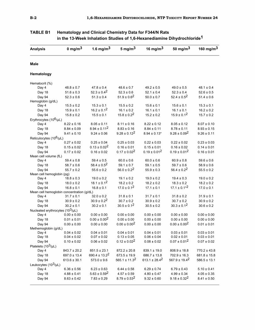
Blood samples were collected from all 13-week inhalation base-study rats at the end of
the study. In addition, blood samples were taken from 10mating-trial
rats/sex/exposure group after 3 and 13 exposures (Days 4 and 18). Animals were anesthetized with a CO2:O2 (70:30) gas mixture, and blood samples were drawn from
the retroorbital sinus. Blood for hematology was collected in Microtainers® (Becton-
Dickinson and Co., Rutherford, NJ) containing sodium EDTA as an anticoagulant.
Samples for clinical chemical chemistry evaluations were collected in serum separator
Microtainers® devoid of anticoagulant, allowed to clot at room temperature,
centrifuged, and the serum was removed. Blood for methemoglobin determination was
collected in Microtainers® containing heparin as anticoagulant.
Hematology determinations were performed with an Ortho ELT-8 Laser Hematology
Counter (Ortho Instruments, Westwood, MA). Smears of peripheral blood were stained
with Brechers stain and counterstained with a modified Romanowsky stain, then
examined microscopically for determination of differential leukocyte counts and
reticulocyte counts. Erythrocyte, leukocyte, and platelet morphologies were evaluated
during the leukocyte differential count. Methemoglobin concentrations were measured
with a Gilford spectrophotometer (Gilford Instrument Laboratories, Inc., Oberlin, OH).
Clinical chemistry variables were measured with an Hitachi Automatic Chemistry
Analyzer (Boehringer-Mannheim, Indianapolis, IN). Clinical pathology that were
evaluated are listed in Table 1.
REPRODUCTIVE SYSTEM EVALUATIONS
Sperm Morphology and Vaginal Cytology in Rats and Mice In the 13-week inhalation studies, sperm morphology and vaginal cytology evaluations
(SMVCE) were performed on base-study rats and mice from the control group and the
3highest exposure groups (0, 16, 50, and 160 mg/m3). To screen for potential
reproductive toxicity, epididymal sperm motility was evaluated at necropsy; vaginal
27 1,6-HEXANEDIAMINE DIHYDROCHLORIDE, NTP TOXICITY REPORT NUMBER 24
cytology was evaluated during the week preceding necropsy using procedures outlined
in the NTP's SMVCE protocol (modified October, 1984). Vaginal saline lavage was
performed on females for 7 consecutive days prior to scheduled termination. The
relative preponderance of leukocytes, nucleated epithelial cells, and large squamous
epithelial cells in the lavage fluid were used to identify the stages of the estrous cycle.
Sperm motility was evaluated at necropsy as follows: The right epididymal tail (cauda
epididymis) was removed at the junction of the vas deferens and the epididymal body
(corpus epididymis), and a small cut was made in the distal border of the epididymal
tail. A small amount of sperm was extruded, and the number of motile and nonmotile
sperm in 5 microscopic fields were counted. After sperm sampling for motility
evaluation, the epididymal tail was placed in sterile phosphate-buffered saline (PBS),
finely minced, and swirled to release the contents. The tissue was incubated in PBS
and then heat fixed at 65° C. Sperm density was determined using a hemacytometer.
Four sperm morphology slides were prepared for each animal evaluated. An aliquot of
killed sperm suspension was stained in a test tube, spread on a microscope slide,
coverslipped, and examined.
Mating Trials in Rats and Mice Mating trials were performed on rats and mice from the control group and from the 3
highest exposure groups (0, 16, 50, and 160 mg/m3) in the 13-week inhalation studies.
These exposure groups were selected based on the lack of significant clinical findings (body
weight changes or clinical signs of toxicity) in all exposure groups. Mating trial animals
were bred for 10 nights (approximately study days 68 to 80, weekdays only) prior to the
end of the 13-week exposure period. Females were removed from the inhalation chambers
and housed overnight in polycarbonate cages with males from the same treatment group (2
females per male). Trios selected for breeding were not altered during the mating trial.
These animals were returned to the inhalation chambers each day and exposed in the same
manner as the base-study animals. Each morning during the mating period, females were
examined for evidence of copulation by vaginal lavage. Females not showing evidence of
copulation were mated again each night until they were sperm positive or for a maximum of
10 nights. Day 0 of gestation was considered to be the day sperm were observed in the
lavage samples. Females not showing signs of copulation by the end of the breeding period
were monitored for signs of pregnancy for an additional 23 days. If no clinical signs of
pregnancy were seen, the animals were killed, and the uteri were examined for signs of
pregnancy. If implantation was not evident, the uterus was stained with
28 1,6-HEXANEDIAMINE DIHYDROCHLORIDE, NTP TOXICITY REPORT NUMBER 24
ammonium sulfide and was examined for signs of early implantation. Following the
last day of exposure, females were housed individually in polycarbonate cages until
parturition. Male rats were killed at the end of the breeding period and were discarded
without further examination. The day of parturition was considered to be lactation Day
0. Females and pups were killed on lactation Day 21.
Adult females were weighed on gestation Days 0 and 20. Adult males were weighed at
the end of the mating period. Dams and pups were individually weighed on lactation
Days 0, 5, 14, and 21. Pups were examined at birth for morphological abnormalities,
viability, and gender. The number of live/dead offspring, percent neonatal survival,
mean live pup weight, and sex ratio were recorded on lactation Days 0, 5, 14, and 21.
Necropsies were performed only on mating-trial females selected for breeding and
examined for pregnancy 23 days after the conclusion of breeding as described above.
Tissues from mating-trial animals were not fixed or retained.
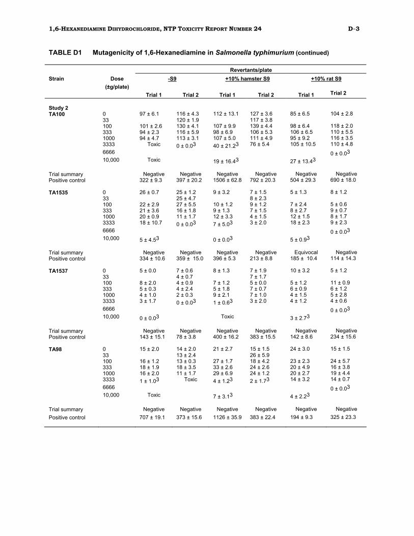
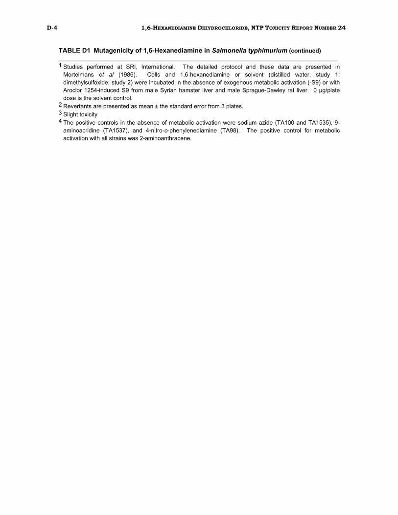
Genetic Toxicity Studies SALMONELLA TYPHIMURIUM MUTAGENICITY TEST PROTOCOL
Mutagenicity studies of 1,6-hexanediamine (HDA; CAS Number 124-09-4) in Salmonella
typhimurium were conducted as described in Mortelmans et al. (1986). Briefly, HDA
was supplied to the laboratory as a coded aliquot and was tested for mutagenicity in S.-
typhimurium strains TA98, TA100, TA1535, and TA1537, using a preincubation assay
in both the absence and presence of Aroclor 1254-induced S9 from male Syrian
hamster liver or male Sprague-Dawley rat liver. The compound was tested on 2
separate occasions in the same laboratory under different code numbers. HDA was
tested at doses up to 10,000 µg/plate in both studies.
CHINESE HAMSTER OVARY CELL CYTOGENETICS PROTOCOLS
Testing was performed as reported by Galloway et al. (1987). HDA was provided to the
testing laboratory as a coded aliquot. Chinese hamster ovary cells (CHO) were
incubated with HDA for induction of sister chromatid exchanges (SCEs) and
chromosomal aberrations (Abs), both in the presence and absence of Aroclor 1254
induced male Sprague-Dawley rat liver S9 and cofactor mix.
30 1,6-HEXANEDIAMINE DIHYDROCHLORIDE, NTP TOXICITY REPORT NUMBER 24
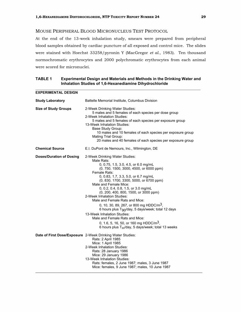
TABLE 1 Experimental Design and Materials and Methods in the Drinking Water and Inhalation Studies of 1,6-Hexanediamine Dihydrochloride (continued)
EXPERIMENTAL DESIGN (continued)
Date of Last Dose/Exposure 2-Week Drinking Water Studies: Rats: 16 April 1985 Mice: 15 April 1985
2-Week Inhalation Studies: Rats: 12 February 1986 Mice: 13 February 1986
13-Week Inhalation Studies: Rats: females, 2 September 1987; males, 3 September 1987 Mice: females, 9 September 1987; males, 10 September 1987
Necropsy Dates 2-Week Drinking Water Studies: Rats: 17 April 1985 Mice: 16 April 1985
2-Week Inhalation Studies: Rats: 13 February 1986 Mice: 14 February 1986
13-Week Inhalation Studies: Rats: females, 3 September 1987; males, 4 September 1987 Mice: females, 10 September 1987; males, 11 September 1987
Type and Frequency of Observation 2-Week Drinking Water Studies:
Observed 2 times per day. Animals were weighed just prior to being placed on study and on Days 8 and 15 of the study.
2-Week Inhalation Studies: Observed 2 times per day. Animals were weighed just prior to being placed on study and on Days 8 and 15 of the study.
13-Week Inhalation Studies: Body weights were recorded at study start, weekly, and at study termination. Clinical signs recorded weekly for animals in the basestudy groups.
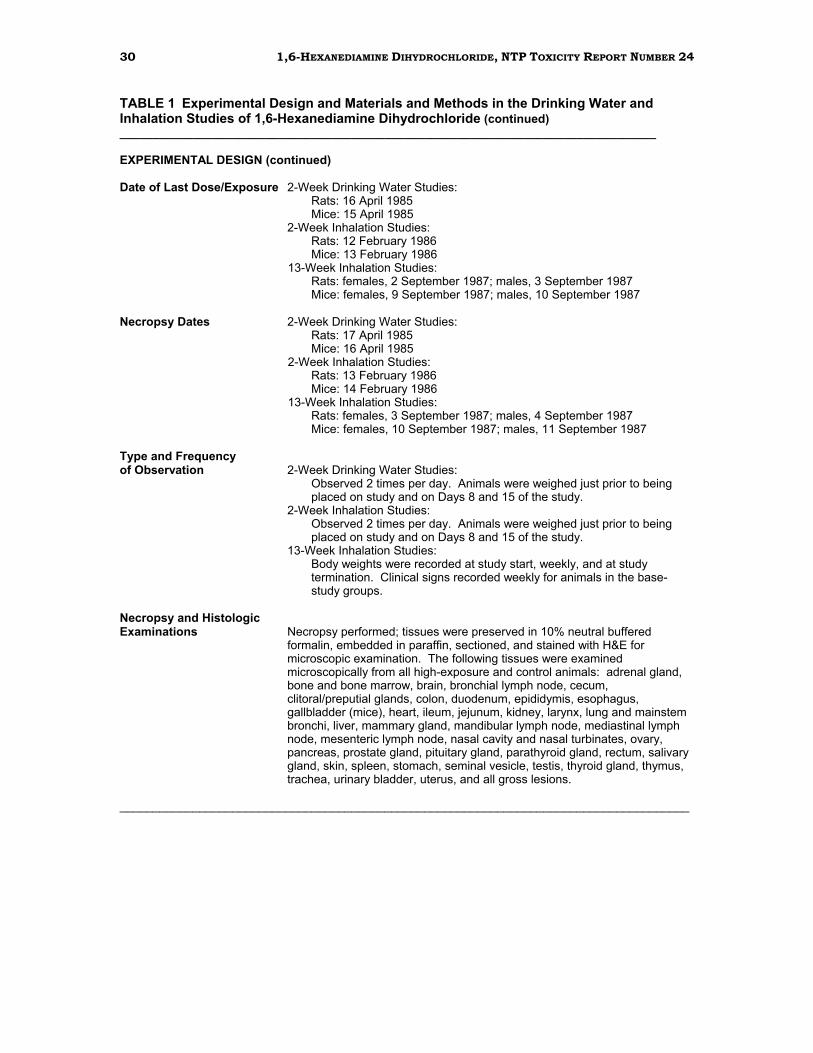
Necropsy and Histologic Examinations Necropsy performed; tissues were preserved in 10% neutral buffered
formalin, embedded in paraffin, sectioned, and stained with H&E for microscopic examination. The following tissues were examined microscopically from all high-exposure and control animals: adrenal gland, bone and bone marrow, brain, bronchial lymph node, cecum, clitoral/preputial glands, colon, duodenum, epididymis, esophagus, gallbladder (mice), heart, ileum, jejunum, kidney, larynx, lung and mainstem bronchi, liver, mammary gland, mandibular lymph node, mediastinal lymph node, mesenteric lymph node, nasal cavity and nasal turbinates, ovary, pancreas, prostate gland, pituitary gland, parathyroid gland, rectum, salivary gland, skin, spleen, stomach, seminal vesicle, testis, thyroid gland, thymus, trachea, urinary bladder, uterus, and all gross lesions.
31 1,6-HEXANEDIAMINE DIHYDROCHLORIDE, NTP TOXICITY REPORT NUMBER 24
TABLE 1 Experimental Design and Materials and Methods in the Drinking Water and Inhalation Studies of 1,6-Hexanediamine Dihydrochloride (continued)
EXPERIMENTAL DESIGN (continued)
Supplemental Evaluations Hematology and Clinical Chemistry: Blood samples were collected from all 13-week inhalation base-study rats at study termination. In addition, blood samples were taken from 10 mating-trial rats/sex/exposure group after 3 and 13 exposures. The following hematology parameters were evaluated: erythrocyte (RBC), leukocyte (WBC), and platelet (PLAT) counts, hemoglobin (HGB) concentration, hematocrit (HCT), mean corpuscular volume (MCV), mean corpuscular hemoglobin (MCH), mean corpuscular hemoglobin concentration (MCHC), and methemoglobin (METH). Smears of peripheral blood were stained with Brechers stain and counterstained with a modified Romanowsky stain, then examined microscopically. Leukocyte differentials were determined on 100 cells; the absolute counts for each leukocyte type were obtained as the product of the corresponding percentage and the total leukocyte count. Reticulocytes were counted from the slides prepared for the leukocyte counts. Relative numbers of reticulocytes, determined by microscopic examination of approximately 1000 erythrocytes, were converted to absolute counts based on the total erythrocyte count. Erythrocyte, leukocyte, and platelet morphologies were evaluated during the leukocyte differential count.
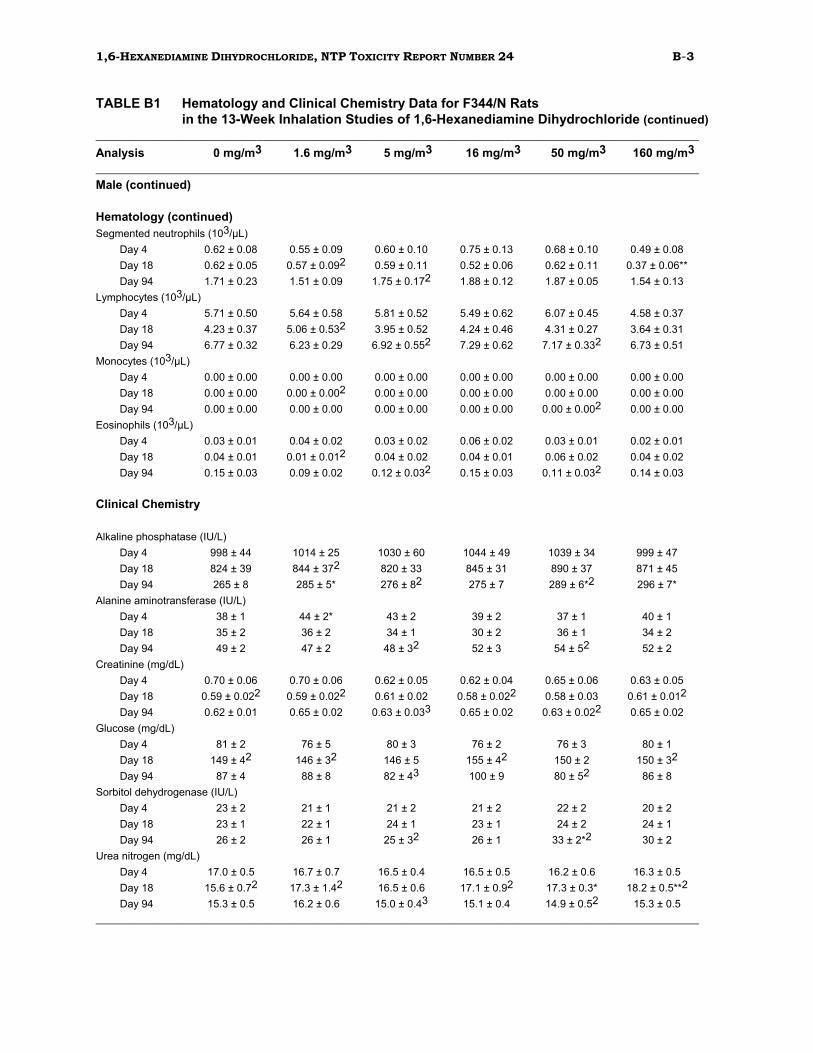
The following clinical chemistry assays were performed: urea nitrogen (UN), creatinine, alanine aminotransferase (ALT), alkaline phosphatase (AP), sorbitol dehydrogenase (SDH), and glucose.
Sperm Morphology/Vaginal Cytology and Mating Trials Sperm morphology and vaginal cytology evaluations (SMVCE) and mating trials were performed at the end of the 13-week studies. Sperm morphology and vaginal cytology were evaluated in base-study rats and mice from the control, 16, 50, and 160 mg HDDC/m3 exposure groups. Mating trials were performed on supplemental rats and mice exposed to 0, 16, 50, or 160 mg HDDC/m3.
ANIMALS AND ANIMAL MAINTENANCE
Strain and Species F344/N Rats B6C3F1 Mice
Animal Source 2-Week Drinking Water Studies: Simonsen Labs, Inc., Gilroy, CA
2-Week Inhalation Studies: Frederick Cancer Facility, Frederick, MD
13-Week Inhalation Studies: Taconic Farms, Inc., Germantown, NY
32 1,6-HEXANEDIAMINE DIHYDROCHLORIDE, NTP TOXICITY REPORT NUMBER 24
TABLE 1 Experimental Design and Materials and Methods in the Drinking Water and Inhalation Studies of 1,6-Hexanediamine Dihydrochloride (continued)
ANIMALS AND ANIMAL MAINTENANCE (continued)
Age When Killed 2-Week Drinking Water Studies: 8-9 weeks
2-Week Inhalation Studies: 8-9 weeks
13-Week Inhalation Studies: 19-20 weeks
Method of Animal Distribution Animals were weighed and randomized (by partitioning algorithm) into
groups by sex and assigned to cages; cages were assigned to dose groups.
Diet 2-Week Drinking Water Studies: NIH 07; available ad libitum
2-Week and 13-Week Inhalation Studies: NIH 07; available ad libitum except during exposure periods
Animal Room Environment 2-Week Drinking Water Studies: Temperature was maintained at 72° ± 3°F and relative humidity at 50% ± 15% with 12 - 15 room air changes per hour. Fluorescent light was provided for 12hours per day.
2-Week and 13-Week Inhalation Studies: Temperature was maintained at 72° ± 3°F and relative humidity at 50% ± 15% with 12 - 15 room air changes per hour. Fluorescent light was provided for 12 hours per day. During inhalation exposure, temperature was maintained at 72° - 78°F and relative humidity at 70% - 80%.
Statistical Methods ANALYSIS OF CONTINUOUS VARIABLES
In the 13-week studies, two approaches were employed to assess the significance of
pairwise comparisons between exposed and control groups in the analysis of
continuous variables. Organ and body weight data, which are approximately normally
distributed, were analyzed using the parametric multiple comparisons procedures of
Williams (1971, 1972) and Dunnett (1955). Clinical chemistry and hematology data,
which typically have skewed distributions, were analyzed using the nonparametric
multiple comparisons methods of Shirley (1977) and Dunn (1964). Jonckheere's test
(Jonckheere, 1954) was used to assess the significance of dose-response trends and to
determine whether a trend-sensitive test (Williams, Shirley) was more appropriate for
pairwise comparisons than a test capable of detecting departures from monotonic dose
response (Dunnett, Dunn). If the P-value from Jonckheere's test was greater than or
equal to 0.10, Dunn's or Dunnett's test was used rather than Shirley's or Williams' test.
33 1,6-HEXANEDIAMINE DIHYDROCHLORIDE, NTP TOXICITY REPORT NUMBER 24
The outlier test of Dixon and Massey (1951) was employed to detect extreme values. No
value selected by the outlier test was eliminated unless it was at least twice the next
largest value or, at most, half of the next smallest value.
ANALYSIS OF VAGINAL CYTOLOGY DATA
Because vaginal cytology data are proportions (the proportion of the observation period
that an animal was in a given estrous state), an arcsine transformation was used to
bring the data into closer conformance with normality assumptions. Treatment effects
were investigated by applying a multivariate analysis of variance (Morrison, 1976) to the
transformed data to test for the simultaneous equality of measurements across dose
levels.
ANALYSIS OF MATING TRIAL DATA
Data from the mating trials were grouped into 3 categories and analyzed statistically.
Continuous, quantitative data, such as body weights, were analyzed by Dunnett's t-test
for multiple comparisons to a single control group. Discrete, counting data, such as
litter counts, were analyzed by the Mann-Whitney U nonparametric test. Percentage
data, such as the fertility and survival indices, were analyzed by the Chi Square test.
ANALYSIS OF MUTAGENICITY IN SALMONELLA TYPHIMURIUM
A positive response in the Salmonella typhimurium assay was defined as a reproducible,
dose-related increase in histidine-independent (revertant) colonies in any 1
strain/activation combination. An equivocal response was defined as an increase in
revertants that was not dose related, not reproducible, or not of sufficient magnitude to
support a determination of mutagenicity. A negative response was obtained when no
increase in revertant colonies was observed following chemical treatment. There was no
minimum percentage or fold increase required for a chemical to be judged positive or
weakly positive.
ANALYSIS OF CHINESE HAMSTER OVARY CELL CYTOGENETICS DATA
For the SCE data, statistical analyses were conducted on the slopes of the dose-response
curves (Galloway et al., 1987). An SCE frequency 20% above the concurrent solvent
control value was chosen as a statistically conservative positive response. The probability
of this level of difference occurring by chance at 1 dose point is less than 0.01; the
34 1,6-HEXANEDIAMINE DIHYDROCHLORIDE, NTP TOXICITY REPORT NUMBER 24
probability for such a chance occurrence at 2 dose points is less than 0.001. An
increase of 20% or greater at any single dose was considered weak evidence of activity;
increases at 2 or more doses resulted in a determination that the trial was positive. A
statistically significant trend (P≤0.05) in the absence of any responses reaching 20%
above background led to a call of equivocal.
Chromosomal aberration data are presented as percentage of cells with aberrations.
Statistical analyses were conducted on both the dose-response curve and individual
dose points (Galloway et al., 1987). For a single trial, a statistically significant (P≤0.05)
difference for 1 dose point and a significant trend (P≤0.015) were considered weak
evidence for a positive response; significant differences for 2 or more doses indicated
the trial was positive. A positive trend test in the absence of a statistically significant
increase at any 1 dose resulted in an equivocal call (Galloway et al., 1987).
ANALYSIS OF MOUSE PERIPHERAL BLOOD MICRONUCLEUS DATA
Log transformation of the normochromatic erythrocyte (NCE) data, and testing for
normality by the Shapiro-Wilk test and for heterogeneity of variance by Cochran's test
were performed before statistical analyses. The frequency of micronucleated cells
among NCEs was analyzed by analysis of variance using the SAS GLM procedure. The
NCE data for each dose group were compared with the concurrent solvent control using
Student's t-test. The frequency of micronucleated cells among polychromatic
erythrocytes (PCEs) was analyzed by the Cochran-Armitage trend test, and individual
dose groups were compared to the concurrent solvent control by Kastenbaum
Bowman's (1970) binomial test. The percentage of PCEs among total erythrocytes was
analyzed by an analysis of variance on ranks (classed by sex) and individual dose
groups were compared with the concurrent solvent control using a t-test on ranks.
Quality Assurance The studies of 1,6-hexanediamine dihydrochloride were performed in compliance with
the United States FDA Good Laboratory Practices regulations (21 CFR 58). The Quality
Assurance Unit of Battelle Columbus Laboratories performed audits and inspections of
protocols, procedures, data, and reports throughout the course of the studies. The
operations of the Quality Assurance Unit were monitored by the NTP.
35 1,6-HEXANEDIAMINE DIHYDROCHLORIDE, TOXICITY REPORT NUMBER 24
RESULTS
2-Week Drinking Water Study in F344/N Rats All rats survived to the end of the study. No clinical abnormalities related to chemical
exposure occurred. Water consumption was reduced for male rats in the 2 highest
dose groups (4.5 and 6.0mg/mL) and for females in the 3highest dose groups (3.3, 5.0,
and 6.7 mg/mL). This decrease in water consumption was attributed to poor
palatability of the drinking water solutions. The total estimated dose of HDDC
consumed by each dose group, based on average water consumption and mean body
weights, is given in Table 2. Although the total amount of compound consumed did
increase with larger concentrations of HDDC in the water, this increase was not linear
because of the reduced water intake at the higher concentrations. Mean body weight
gains of treated rats were similar to those of controls (Table 2).
TABLE 2 Survival, Weight Gain, Water Consumption, and Compound Consumption in F344/N Rats in the 2-Week Drinking Water Study of 1,6-Hexanediamine Dihydrochloride
Final Weight Average Water Average Dose Mean Body Weight (grams) Relative to Consumption Dose of HDDC (mg/mL) Survival1 Initial Final Change2 Controls (%)3 (g/day) (mg/kg/day)
n=5. Liver weights and body weights are given in grams; relative liver weights (liver-weight-to-body-weight ratios) are given as mg liver weight/g body weight.
* Significantly different (P≤0.05) from the control group by Dunnett's test. ** Significantly different (P≤0.01) from the control group by Dunnett's test.
37 1,6-HEXANEDIAMINE DIHYDROCHLORIDE, TOXICITY REPORT NUMBER 24
2-Week Drinking Water Study in B6C3F1 Mice All mice survived to the end of the 2-week drinking water study. No clinical
abnormalities related to HDDC exposure were observed in any dose group. Water
consumption by treated mice was similar to that of controls. The estimated daily dose
of HDDC consumed by each treatment group is shown in Table 4. There was a linear
increase in total estimated chemical intake with increasing concentration of HDDC in
the drinking water. Mean body weights and body weight gains of treated mice were
similar to those of controls (Table 4). The only statistically significant difference in
absolute or relative organ weights was a decrease in the relative liver weight of females
in the 0.8 mg/mL dose group (Table5). No chemical-related gross or microscopic
lesions were present.
TABLE 4 Survival, Weight Gain, Water Consumption, and Compound Consumption in B6C3F1 Mice in the 2-Week Drinking Water Study of 1,6-Hexanediamine Dihydrochloride
Final Weight Average Water Average Dose Mean Body Weight (grams) Relative to Consumption Dose of HDDC (mg/mL) Survival1 Initial Final Change2 Controls (%)3 (g/day) (mg/kg/day)
1 n=5. Liver weights and body weights are given in grams; relative liver weights (liver-weight-to-bodyweight ratios) are given as mg liver weight/g body weight.
** Significantly different (P≤0.01) from the control group by Dunnett's test.
39 1,6-HEXANEDIAMINE DIHYDROCHLORIDE, TOXICITY REPORT NUMBER 24
2-Week Inhalation Study in F344/N Rats The exposure concentrations used in the 2-week inhalation study in rats (0, 10, 30, 89, 267, and 800 mg HDDC/m3) were chosen based on the reported inhalation LCLO of
750 mg/m3 in mice and because of the lack of information on inhalation toxicity of
HDDC in rats. Survival and mean body weight data for rats in the 2-week inhalation
study of HDDC are presented in Table 6. On Day 8 of the study (after 6 days of
exposure to HDDC), the body weights of female rats exposed to the highest
concentration of HDDC (800 mg/m3) were notably less than those of the controls. One
of the 5 female rats in the 800 mg/m3 exposure group died on Day 3; the remaining 4
females in this exposure group died on Day11. Although the body weights of male rats
in the highest exposure group were not reduced when the rats were weighed on Day 8,
4 of the 5 males in this group died on Day11, and the remaining male rat died on Day
15. The body weights of male and female rats exposed to 267 mg HDDC/m3 were also
less than those of controls, although this difference was not statistically significant. No
deaths occurred in any other exposure group.
TABLE 6 Survival and Weight Gain of F344/N Rats in the 2-Week Inhalation Study of 1,6-Hexanediamine Dihydrochloride
Final Weight HDDC Mean Body Weight (grams) Relative to (mg/m3) Survival1 Initial Day 8 Day 17 Change2 Controls (%)3
1 Number surviving at 2 weeks/number of animals per exposure group. For groups with no survivors, no Day 17 body 2 Mean weight change. 3 (Exposure group mean/control group mean) x 100. 4 Day of death: 11, 11, 11, 11, 15. 5 Day of death: 3, 11, 11, 11, 11. 6 n=4.
40 1,6-HEXANEDIAMINE DIHYDROCHLORIDE, NTP TOXICITY REPORT NUMBER 24
Clinical signs related to exposure included nasal discharge, rales and dyspnea,
diarrhea, ocular discharge, and hypoactivity. These signs were seen only in male and
female rats exposed to the highest concentration of HDDC. At necropsy, there were no
exposure-related gross lesions.
There were no statistically significant chemical-related changes in the liver, thymus,
heart, right testis, kidney, lung, or brain weights of male or female rats in any group.
Exposure-related histopathologic lesions were present in the nasal passages and
larynx. In the larynx, there were focal areas of inflammation and necrosis with
ulceration of laryngeal epithelium at exposure levels as low as 10 mg/m3 in males and
89 mg/m3 in females. Nasal lesions also were present in both males and females at
exposure levels of 89 mg/m3 and greater. Microscopic changes included focal areas of
inflammation and ulceration in both respiratory and olfactory portions of the nasal
mucosa. Degeneration characterized by thinning (atrophy) and necrosis of the olfactory
and respiratory epithelium was frequently associated with areas of inflammation and
ulceration.
A number of histopathologic lesions were present in rats that died or were killed
moribund. These included lymphoid depletion and necrosis in the thymus, lymph
nodes, and spleen as well as depletion of pancreatic zymogen granules and arrest of
ovarian follicular development. None of these lesions resulted from a direct toxic effect
41 1,6-HEXANEDIAMINE DIHYDROCHLORIDE, TOXICITY REPORT NUMBER 24
13-Week Inhalation Study in F344/N Rats Concentrations of HDDC used in the 13-week study in rats were selected based on the
weight gain depression and the inflammation and ulceration of the nasal cavity and
larynx seen in rats at higher concentrations in the 2-week study. Concentrations
chosen for the 13-week study were 0, 1.6, 5, 16, 50, and 160 mg HDDC/m3. All rats
exposed to HDDC by inhalation for 13 weeks survived to the end of the study. The final
mean body weights of most groups of rats exposed to HDDC were slightly lower than
the mean body weights of the controls (Table 7; Figure 1); these differences, however,
were not statistically significant.
TABLE 7 Survival and Weight Gain of F344/N Rats in the 13-Week Inhalation Study of 1,6-Hexanediamine Dihydrochloride
Final Weight HDDC Mean Body Weight (grams) Relative to (mg/m3) Survival1 Initial Final Change2 Controls (%)3
MALE 0 10/10 147 352 206
1.6 10/10 143 337 193 96 5 10/10 142 338 196 96
16 10/10 142 341 200 97 50 10/10 150 339 189 96
160 10/10 142 326 184 93
FEMALE 0 10/10 114 203 89
1.6 10/10 114 195 81 96 5 10/10 114 203 89 100
16 10/10 112 198 86 97 50 10/10 112 201 90 99
160 10/10 114 202 88 99
1 Number of animals surviving at end of study/number of animals per exposure group. 2 Mean weight change. 3 (Exposure group mean/control group mean) x 100.
42 1,6-HEXANEDIAMINE DIHYDROCHLORIDE, NTP TOXICITY REPORT NUMBER 24
ME
AN
BO
DY
WE
IGH
T IN
GR
AM
S
WEEKS ON STUDY
WEEKS ON STUDY
ME
AN
BO
DY
WE
IGH
T IN
GR
AM
S
FIGURE 1 Body Weights of F344/N Rats Exposed to 1,6-Hexanediamine Dihydrochloride by Inhalation for 13 Weeks
43 1,6-HEXANEDIAMINE DIHYDROCHLORIDE, TOXICITY REPORT NUMBER 24
No clinical signs of toxicity related to HDDC exposure were seen in the 13-week rat
study. Nasal discharge occurred in male rats in the 5 and 16 mg/m3 exposure groups
and in female rats in all exposure groups (including the control group) except those in
the 160mg/m3 group. Similarly, rales occurred in all female groups but not in exposed
males. However, because these signs appeared late in the study and because the
incidence was not dose related, the signs were not considered to be the result of specific
HDDC toxicity.
The only consistent changes in organ weights seen in rats were decreases in absolute
and relative lung weights compared to those of the controls. However, all control male
and female rats had inflammatory lesions in the lungs and had lung weights that were
greater than those of historical controls (NTP, 1990a). Similar inflammatory lesions
have been seen in other inhalation studies and are of unknown etiology (NTP, 1990b,
1991b). Sporadic changes in other absolute and relative organ weights were seen but
did not appear to be chemical related (Appendix A).
At Day 4, the only change noted in the hematology parameters of rats exposed by
inhalation to HDDC was a slight decrease in the mean platelet count in female rats in
the lowest exposure group (Table B1). At Day 18, hematocrit values were increased in
female rats in the 2 highest exposure groups and segmented neutrophil counts were
decreased minimally in male rats in the highest exposure group. By Day 94, there was
a significant decrease in leukocyte and lymphocyte counts in females in the highest
exposure groups, and in segmented neutrophil counts in females in the 3 highest
exposure groups (16, 50, and 160 mg/m3). Female rats in the 2 lowest exposure
groups had increased hematocrit values. A slight decrease in erythrocyte count was
noted in male rats in the 16mg/m3 exposure group, and a minor increase in mean cell
hemoglobin values occurred in female rats in the 160 mg/m3 exposure group and male
rats in the 50 mg/m3 exposure group.
Clinical chemistry changes on Day 4 included a small increase in alanine
aminotransferase activity in male rats in the lowest exposure group (1.6 mg/m3) and a
slight increase in the urea nitrogen level in female rats in the 5 mg/m3 exposure group
(Table B1). By Day18, concentrations of urea nitrogen increased in male rats in the 2
highest exposure groups (50 and 160 mg/m3) and female rats in the 4 highest exposure
groups (5, 16, 50, and 160 mg/m3). Sorbitol dehydrogenase (SDH) activity was slightly
elevated in female rats in the highest exposure group. At Day 94, alkaline phosphatase
activity was slightly increased in male rats in several exposure groups (1.6, 50, and
44 1,6-HEXANEDIAMINE DIHYDROCHLORIDE, NTP TOXICITY REPORT NUMBER 24
160mg/m3), and SDH activity was elevated in males in the 50 mg/m3 exposure group.
No other significant clinical chemistry changes occurred in male or female rats at Day
94.
There were no gross lesions attributed to HDDC exposure. Chemical-related
microscopic lesions were limited to the upper respiratory tract (larynx and nasal
passages) of male and female rats in the 2 highest exposure groups (Table 8). The
morphology, incidence, and severity of microscopic lesions were similar for males and
females, and there was a dose-related increase in the incidence and severity of these
lesions. In the larynx, minimal to mild focal erosion/ulceration was present in rats at
the 160 mg/m3 exposure level as a result of necrosis of the laryngeal epithelium (Plate
1). A minimal to mild inflammatory infiltrate in the mucosa was associated with these
erosions; sometimes the infiltrate extended into the lumen of the larynx. In 2 rats with
laryngeal erosion/ulceration and inflammation, there was also hyperplasia of the
laryngeal epithelium. In the nasal passages, chemical-related lesions were present in
the olfactory and respiratory regions, primarily in Levels I and II of the anterior and mid
portions of the nasal passages. Degeneration of the olfactory epithelium was generally
limited to the 160 mg/m3 exposure animals. The olfactory epithelium in the dorsal
meatus of Level II was more commonly affected, but in some rats lesions were also
present on the ethmoid turbinates of Level III. Degeneration was characterized by focal
areas with thinning of the olfactory epithelial layer (Plate 2). This normally
pseudostratified columnar epithelium was sometimes reduced to only 1 cell layer in
thickness; in some areas a respiratory or nonkeratinizing squamous epithelium
replaced the olfactory epithelium (metaplasia). Frequently a vacuolar change in the
olfactory epithelium was a part of the degenerative lesion, and in the more severely
affected areas there was degeneration of the underlying olfactory nerve bundles (Plate
3). Chemical-related lesions in the respiratory epithelium of the nasal passages
included degeneration and focal erosion/ulceration of the mucosa in Levels I and II.
Degeneration was characterized by loss of cilia and decreased height of the columnar
epithelium; squamous metaplasia (nonkeratinizing) was present in 160 mg/m3
exposure animals. In Levels I and II, the incidence of inflammation in the respiratory
mucosa was increased slightly in the higher exposure groups but the severity (minimal
to mild) was not dose related.
Administration of HDDC to rats by inhalation caused no changes in any of the sperm
morphology or vaginal cytology parameters evaluated (Appendix C).
1 n=10 for all groups. The average severity score ( ) was based on the number of animals with lesions from each group; 1 = minimal, 2 = mild, 3 = moderate, 4 = marked.
1 Number surviving at end of study/number of animals per group. For groups with no survivors, no final mean body weights or body weight changes are given. 2 Mean weight change. 3 (Exposure group mean/control group mean) x 100. 4 Day of death: 3,9. 5 n=4. 6 n=3. 7 Day of death: 9, 10, 10, 10, 13.
47 1,6-HEXANEDIAMINE DIHYDROCHLORIDE, TOXICITY REPORT NUMBER 24
Clinical signs of HDDC toxicity in male and female mice were related to irritation of the
upper respiratory tract and included dyspnea and nasal discharge. Other abnormal
signs noted were typical of those seen in moribund animals. These were seen primarily
in the highest exposure group (800 mg/m3) and included ruffled fur, abnormal posture,
hypoactivity, tremors, prostration, and decrease in body mass compared to controls. At
necropsy, the only chemical-related gross finding was a small spleen in male and
female mice exposed to the highest concentration of HDDC (800 mg/m3).
Exposure-related changes in absolute and relative organ weights of mice in the 2-week
inhalation study are shown in Table10. For surviving male mice in the highest
exposure group (800 mg/m3), absolute and relative liver weights were significantly
reduced. There were no surviving females in the 800 mg/m3 exposure group. Absolute
liver weight of male mice in the 30 mg/m3 exposure group and relative liver weights of
male mice in the 30 and 10 mg/m3 exposure groups also were reduced. Thymus
weights of male and female mice were significantly reduced in the 800 mg/m3 and 267
mg/m3 exposure groups respectively. Relative lung weights were increased in male
mice in the highest exposure group. No other statistically significant differences in
absolute or relative organ weights were seen.
Microscopic lesions attributed to HDDC exposure were present in the nasal passages,
larynx, testis, and trachea. In the larynx and trachea, focal areas of inflammation and
necrosis with ulceration of the respiratory mucosa were present at the 2 highest
exposure levels (267 and 800 mg/m3). The nasal lesions included degeneration
characterized by a thinning (atrophy) and necrosis of the olfactory and respiratory
epithelium. This was sometimes associated with focal inflammation and ulceration.
Mild testicular degeneration was present in 2 mice from the highest exposure group
(800 mg/m3); 1 of these mice died on Day 9 of the study. Other microscopic lesions
were considered secondary to stress or moribund condition.
1 n=5 for all groups except the 800 mg/m3 exposure group. For male mice in the 800 mg/m3 exposure group, n=3; for female mice in this exposure group, n=0. Organ weights and body weights are given in grams; relative organ weights (organ-weight-to-body-weight ratios) are given as mg organ weight/g body weight.
2 No data; all animals in this exposure group died before scheduled termination. * Significantly different (P≤0.05) from the control group by Dunnett's test. ** Significantly different (P≤0.01) from the control group by Dunnett's test.
1 Number of animals surviving at end of study/number of animals per exposure group. 2 Mean weight change. 3 (Exposure group mean/control group mean) x 100.
50
1.,6- HEXANEDIAMINE DIHYDROCHLORIDE, NTP TOXICITY REPORT NUMBER 24
ME
AN
BO
DY
WE
IGH
T IN
GR
AM
S
WEEKS ON STUDY
ME
AN
BO
DY
WE
IGH
T IN
GR
AM
S
WEEKS ON STUDY
FIGURE 2 Body Weights of B6C3F1 Mice Exposed to 1,6-Hexanediamine Dihydrochloride by Inhalation for 13 Weeks
1 n=10. Organ weights and body weights are given in grams; relative organ weights (organ-weight-to-bodyweight ratios) are given as mg organ weight/g body weight.
* Significantly different (P≤0.05) from the control group by Williams' or Dunnett's test. ** Significantly different (P≤0.01) from the control group by Williams' or Dunnett's test.
n=10 for all groups. The average severity score ( ) was based on the number of animals with lesions from each group; 1 = minimal, 2 = mild, 3 = moderate, 4 = marked.
1
53 1,6-HEXANEDIAMINE DIHYDROCHLORIDE, TOXICITY REPORT NUMBER 24
Necrosis of the laryngeal epithelium and minimal to mild focal erosion/ulceration were
present in the larynx of mice exposed to 160 mg/m3. Focal necrosis or hyperplasia of
the adjacent epithelium was associated with some erosions. In the nasal passages,
exposure-related lesions were present in the respiratory and olfactory regions, primarily
in Levels II and III of the mid-portion of the nasal passages. In the respiratory
epithelium, the primary change was hyaline degeneration characterized by the
accumulation of an eosinophilic proteinaceous material. Minimal to mild focal areas of
erosion/ulceration and inflammation were present in the 160mg/m3 exposure group
(Plate 4). Hyaline degeneration of the olfactory epithelium was generally limited to the
50 and 160 mg/m3 exposure levels. This was characterized by the accumulation of
eosinophilic proteinaceous material in the olfactory sustentacular cells and resulted in
a loss of olfactory sensory cells (Plates 5 and 6).
Administration of HDDC to mice by inhalation caused no changes in the sperm
morphology parameters evaluated (Table C3), with the exception of an increase in
sperm motility in the 16 and 160 mg/m3 exposure groups. However, this change was
not dose related, and the values for sperm motility were all well within the range for
historical controls for NTP studies. Consequently, the increase in sperm motility was
not interpreted as an adverse effect.
Mating Trials The results of mating trials conducted on rats and mice exposed to HDDC by inhalation for
13 weeks appear in Appendix C. In rats, HDDC demonstrated no reproductive toxicity.
There was no effect on male or female fertility, body weights or body weight gains, gestation
length, litter size, neonatal survival, pup weights, sex ratios of pups, or pup morphology in
rats exposed to HDDC. Similarly, reproductive effects of HDDC on mice were minimal.
There was no effect on male or female body weights or body weight gains, and no effect on
male or female fertility. Three female mice exposed to 16 mg/m3 and 1female and 1 male
mouse exposed to 50 mg/m3 died before scheduled termination; however, these deaths
were not considered compound related. A statistically significant increase in the mean
gestation length of mice in the 50 and 160 mg/m3 exposure groups was noted; however, in
the absence of other reproductive toxicity, this effect was not considered biologically
significant. HDDC had no effect on litter size, neonatal survival, sex ratio of pups, or pup
morphology in mice. Pups in the 160 mg/m3 exposure group had mean weights similar
to that of controls at birth and on lactation Day 5; however, mean
54 1,6-HEXANEDIAMINE DIHYDROCHLORIDE, NTP TOXICITY REPORT NUMBER 24
weights for pups in this exposure group were lower than that of controls on lactation
Days 14 and21.
Genetic Toxicity Studies 1,6-Hexanediamine (33 to 10,000 µg/plate) was tested in 2 independent studies for
induction of mutations in Salmonella typhimurium strains TA100, TA1535, TA1537,
and TA98, with and without Aroclor 1254-induced male Sprague-Dawley rat and Syrian
hamster liver S9 (Mortelmans et al., 1986; Table D1). In the first study, distilled water
was the solvent for 1,6-hexanediamine; in the second study, dimethylsulfoxide was
used as the solvent. The chemical was toxic at doses of 3333 µg/plate and greater in
both studies. No increases in mutations were observed in any of the 4 S. typhimurium
tester strains after incubation with 1,6-hexanediamine.
In cytogenetics tests with cultured Chinese hamster ovary (CHO) cells, no significant
increases in sister chromatid exchanges or chromosomal aberrations (Abs) were
observed after exposure to 1,6-hexanediamine, with or without S9 (Tables D2 and D3).
In the Abs test with S9, both trials showed an increase in total aberrations at the
highest concentration tested (500 µg/ml). In both of these trials, however, these
aberrations were concentrated in fewer than 5% of the total cells scored. Hence, the
percentage of cells with aberrations (the endpoint that is evaluated in this test) was not
sufficiently elevated for a positive response.
The frequency of micronucleated erythrocytes was measured in peripheral blood smears
of male and female mice exposed to 1,6-hexanediamine dihydrochloride in the 13-week
inhalation studies (Table D4). No significant increases were seen in the frequencies of
micronucleated normochromatic erythrocytes (NCEs) or polychromatic erythrocytes
(PCEs) in male or female mice. The percentage of PCEs among the total erythrocyte
population was increased at the highest exposure levels for male and female mice.
1,6-HEXANEDIAMINE DIHYDROCHLORIDE, TOXICITY REPORT NUMBER 24 55
56 1,6-HEXANEDIAMINE DIHYDROCHLORIDE, NTP TOXICITY REPORT NUMBER 24
PLATE 2PLATE 1
Focal ulceration (arrows) of laryngeal epithelium in a male rat exposed to 1,6-hexanediamine dihydrochloride at a concentration of 160 mg/m3. Mild inflammation is present in the underlying mucosa with inflammatory exudate in the lumen (L) of the larynx. 90x.
PLATE 3
Degeneration of the olfactory mucosa in a female rat exposed to 1,6-hexane-diamine dihydrochloride at a concen-tration of 160 mg/m3. Compared to the less affected mucosa on the left, there is marked thinning of the epithelial layer (arrow). On the opposite side of the turbinate there is a vacuolar degener-ation and loss of olfactory epithelium. Note the degeneration of the nerve fiber bundles (N) in the underlying mucosa. 160x.
PLATE 5
Olfactory mucosa from a control male mouse for comparison with Plate 6, in which the mouse was exposed to 1,6-hexanediamine dihydrochloride. 160x.
Degeneration of olfactory mucosa in the dorsal meatus of the nasal passage in a male rat exposed to 1,6-hexanediamine dihydrochloride at a concentration of 160mg/m3. There is thinning of the olfactory epithelium (arrow) with the focal accumulation of a serum exudate and a few inflammatory cells on the mucosal surface. 160x.
PLATE 4
Focal ulceration (arrows) of the nasal respiratory epithelium in a female mouse exposed to 1,6hexanediamine dihydro-chloride at a concentration of 160mg/m3. A mild inflammatory infiltrate is present in the underlying mucosa. 400x.
PLATE 6
Hyaline degeneration in the olfactory mucosa of a male mouse exposed to 1,6hexanediamine dihydrochloride at a concentration of 160 mg/m3. Eosinophilic protein droplets (arrows) distort the normal nuclear arrangement of the olfactory layer. 160x.
1,6-HBXANEDIAMINE DIHYDROCHLORIDB, TOXICITY REPORT NUMBER 24 57
PLATE 1 PLATE 2
PLATE 3 PLATE 4
PLATE 5 PLATE 6
58 1,6-HEXANEDIAMINE DIHYDROCHLORIDE, NTP TOXICITY REPORT NUMBER 24
1,6-HEXANEDIAMINE DIHYDROCHLORIDE, NTP TOXICITY REPORT NUMBER 24 59
DISCUSSION The toxic effects of 1,6-hexanediamine dihydrochloride (HDDC) in F344/N rats and B6C3F1 mice after inhalation exposure were restricted almost exclusively to the upper
airways and resulted from the strong irritant properties of the compound. At HDDC
concentrations of 16 mg/m3 and greater, inflammation and necrosis were present in
the nasal and laryngeal epithelium. These lesions were sometimes accompanied by
ulceration and compensatory hyperplasia or metaplasia of the epithelial lining. The
distribution of lesions was similar in rats and mice. Aside from the respiratory tract
lesions described above, no specific target organ for HDDC toxicity was identified in
these studies.
In the 2-week drinking water studies, no changes in survival or body weight, no gross
or microscopic pathologic changes, and no clinical abnormalities related to HDDC
consumption were seen. At the highest HDDC concentrations, water intake was
reduced due to the poor palatability of the dosed water. Because no significant toxic
effects were noted in these studies and because higher concentrations of HDDC would
probably have caused ill effects resulting from decreased water consumption, further
drinking water studies were not conducted.
In the 2-week inhalation studies, ulceration, necrosis, and inflammation were present
in the larynx and nasal passages of rats and mice; both respiratory and olfactory
epithelium in the nasal mucosa were affected. Because of mortality and clinical signs
of respiratory distress in the highest exposure group and severity of respiratory tract
lesions in animals administered HDDC at levels of 89 mg/m3 and greater, an upper
exposure concentration of 160 mg/m3 was selected for the 13-week studies in each
species. Following 13 weeks of exposure to HDDC, essentially the same lesions seen in
the 2-week studies were present in the larynx and nose/nasal passages of each species.
Almost all treatment-related lesions in male and female rats in the 13-week study were
limited to the 160 mg/m3 exposure group. No apparent sex-related differences in the
incidence, severity, or character of these lesions were observed. The site-specific and
morphological effects of HDDC on the upper respiratory tract (larynx and nasal cavity) were
consistent with those produced by other irritant chemicals administered by inhalation
(Giddens and Fairchild, 1972; Jiang et al., 1983; Buckley et al., 1984; Boorman et al.,
1987; Morgan and Monticello, 1990; NTP, 1991b). In a report on the toxicity of
60 1,6-HEXANEDIAMINE DIHYDROCHLORIDE, NTP TOXICITY REPORT NUMBER 24
HDDC in rats treated by inhalation for 13 weeks (Johannsen et al., 1987), the authors
described lesions similar to those seen in the present NTP study. Inflammation and
squamous metaplasia of the nasal respiratory epithelium, trachea, and lungs were
seen. These lesions were generally more severe than those seen in the present study
and occurred only at the highest HDDC concentration tested (215 mg/m3). No gross or
microscopic lesions were seen in rats administered 12.8 or 51 mg/m3 HDDC. High
mortality in the 215 mg/m3 exposure group of the Johannsen study resulted in
discontinuation of this group after 7weeks of exposure. As in the NTP study, the cause
of early mortality in the highest exposure group of the Johannsen study could not be
determined.
In mice, the character of the nasal lesions was slightly different than in rats. At the
highest exposure concentrations in the 2-week study, laryngeal lesions and
degeneration with necrosis and ulceration of the respiratory and olfactory epithelium
resembled those in rats. However, in the 13-week study, inflammation was less
prominent in mice and degenerative changes in the olfactory and respiratory epithelium
were characterized by the accumulation of an eosinophilic hyaline material in the
cytoplasm of respiratory and olfactory cells. These intracytoplasmic hyaline droplets,
believed to represent a proteinaceous secretory material, have been described in both
treated and control rats and mice (Monticello et al., 1990). The amount and
distribution of these droplets are often increased in chemical-exposed animals, and this
change has been described as an adaptive response to certain toxicants (Buckley et al.,
1985; Monticello et al., 1990). In mice, the olfactory mucosa in Level III was frequently
affected by this hyaline degeneration, while in rats the olfactory degeneration occurred
primarily in the dorsal meatus of LevelII. Mice appeared to be somewhat more sensitive
to the irritant properties of HDDC; degenerative lesions in mice generally occurred at
lower doses and with higher incidence than in rats. The severity of lesions, however,
was similar in both species. Similar results have been seen in previous NTP studies
(NTP, 1991b).
No chemical-related lesions were seen in the lungs of rats or mice exposed to HDDC. The
absolute and relative lung weights of HDDC-exposed rats in this study appeared to be
reduced. However, the lung weights of exposed animals were well within the normal range,
and the apparent reduction is considered to be an artifact resulting from the greater lung
weights of the controls. Inflammation was present in the lungs of control male and female
rats, resulting in increased lung weights in control rats compared to historical controls
1,6-HEXANEDIAMINE DIHYDROCHLORIDE, NTP TOXICITY REPORT NUMBER 24 61
(NTP, 1990a). This inflammation was identical to that observed in several previous
inhalation studies and is of unknown etiology (NTP, 1990b, 1991b). Such inflammatory
lesions have not been seen in mice.
Overall, hematologic changes in rats were minor, sporadic, and not accompanied by
related clinical pathology findings. At the end of the study, female rats showed an
exposure-related decrease in lymphocyte and segmented neutrophil counts, which
contributed to an overall decrease in leukocyte count. In the highest exposure group
(160 mg/m3), leukocytes decreased approximately 29%, lymphocytes 26%, and
segmented neutrophils44%. These findings differ from those of previous inhalation
studies with 1,6-hexanediamine (HDA), in which leukocyte numbers either increased
(Kulakov, 1965; Tkachenko, 1976) or showed no change (Izrailet and Laivina, 1980;
Johannsen et al., 1987). In the studies showing increases in leukocyte count, exposure
concentrations were considerably lower (1.25 mg/m3 maximum) than the
concentrations used in the present study; the concentrations used by Johannsen et al.,
on the other hand, were in the same range as those used in the present study. In male
rats, no changes in the leukocyte parameters were noted at study termination; however,
there was a decrease in the segmented neutrophil count in males in the highest
exposure group at Day 18. Although no mechanism for the observed leukocyte changes
was evident, decreases can result from a number of causes, including decreased or
ineffective production (bone marrow or lymphoid), increased margination to tissues or
the marginal pool, decreased release to the circulation, or increased destruction. The
observed decrease in circulating leukocytes is consistent with the margination of cells
during the inflammatory changes in the larynx and nasal passages. In addition, HDA
and HDDC have been shown to inhibit the in vitro proliferation of mouse lymphocytes
(Luebke et al., 1989). This inhibition is at least partly due to inhibition of ornithine
decarboxylase activity. Although no microscopic evidence of bone marrow toxicity was
seen in the present study, it is possible that such activity may be responsible for the
observed decreases in leukocyte numbers.
On Day 18, but not at study termination, slight increases in serum urea nitrogen
concentrations were noted in male and female rats. Urea nitrogen and creatinine are
used as indicators of renal dysfunction. In this study, creatinine levels were unchanged
and there was no histologic evidence of nephropathy. The urea nitrogen concentration
can be increased by several nonrenal causes (e.g., dehydration, processes causing
increased protein catabolism, and diet), and it is likely that the change in urea nitrogen
62 1,6-HEXANEDIAMINE DIHYDROCHLORIDE, NTP TOXICITY REPORT NUMBER 24
concentration was related to 1 or more of these causes. Slight increases in urea
nitrogen concentration, which were also noted in the highest exposure group of the
study by Johannsen et al. (1987), were attributed to a marked decrease in food intake
with a resulting increase in endogenous protein catabolism.
In summary, under the exposure conditions employed in these studies, inhalation of
HDDC by rats and mice produced lesions that could be attributed to the irritant effects
of the compound. The observed NOAEL for respiratory damage was 5 mg/m3 for rats
and mice. No significant systemic toxicity was observed, and no specific target organs
were identified. HDDC and HDA were not genotoxic, and inhalation of HDDC did not
cause any adverse reproductive effects.
1,6-HEXANEDIAMINE DIHYDROCHLORIDE, NTP TOXICITY REPORT NUMBER 24 63
REFERENCES AMERICAN CONFERENCE OF GOVERNMENTAL INDUSTRIAL HYGIENISTS (ACGIH) (1990
91). Threshold Limit Values for Chemical Substances and Physical Agents and
Biological Exposure Indices. Cincinnati, OH.
BARDSLEY, W. G., HILL, C. M., AND LOBLEY, R. W. (1970). Reinvestigation of the
substrate specificity of pig kidney diamine oxidase. Biochem. J. 117, 169-176.
BETHELL, D. R., AND PEGG, A. E. (1979). Effects of diamines on ornithine
decarboxylase activity in control and virally transformed mouse fibroblasts.
Biochem. J. 180, 87-94.
BOORMAN, G. A., MONTGOMERY, C. A., JR., EUSTIS, S. L., WOLFE, M. J., MCCONNELL, E.
E., AND HARDISTY, J. F. (1985). Quality assurance in pathology for rodent
carcinogenicity studies. In Handbook of Carcinogen Testing (H. A. Milman and E. K.
Weisburger, Eds.), pp. 345-357. Noyes Publications, Park Ridge, NJ.
BOORMAN, G. A., HICKMAN, R. L., DAVIS, G. W., RHODES, L. S., WHITE, N. W., GRIFFIN,
T. A., MAYO, J., AND HAMM, T. E., JR. (1986). Serological titers to murine viruses in
90-day and 2-year studies. In Complications of Viral and Mycoplasmal Infections in
Rodents to Toxicology Research and Testing (T. E. Hamm, Jr., Ed.), pp 11-23.
Hemisphere, New York.
BOORMAN, G. A., BROWN, R., GUPTA, B. N., URAIH, L. C., AND BUCHER, J. R. (1987).
Pathologic changes following acute methyl isocyanate inhalation and recovery in
BOORMAN, G. A., MORGAN, K. T., AND URIAH, L. C. (1990). Nose, larynx, and trachea.
In Pathology of the Fischer Rat. Reference and Atlas (G. A. Boorman, S. L. Eustis, M.
R. Elwell, C. A. Montgomery, Jr., and W. F. MacKenzie, Eds.), pp. 315-338.
Academic Press, Inc., San Diego, CA.
64 1,6-HEXANEDIAMINE DIHYDROCHLORIDE, NTP TOXICITY REPORT NUMBER 24
BRORSON, T., SKARPING, G., SANDSTRÖM, J. F., AND STENBERG, M. (1990). Biological
monitoring of isocyanates and related amines. I. Determination of 1,6
hexamethylene diamine (HDA) in hydrolysed human urine after oral administration
of HDA. Int. Arch. Occup. Environ. Health 62, 79-84.
BUCKLEY, L. A., JIANG, X. Z., JAMES, R. A., MORGAN, K. T., AND BARROW, C. S. (1984).
Respiratory tract lesions induced by sensory irritants at the RD50 concentration.
Toxicol. Appl. Pharmacol. 74, 417-429.
BUCKLEY, L. A., MORGAN, K. T., SWENBERG, J. A., JAMES, R. A., HAMM, T. E., JR., AND
BARROW, C. S. (1985). The toxicity of dimethylamine in F-344 rats and B6C3F1
mice following a 1-year inhalation exposure. Fundam. Appl. Toxicol. 5, 341-352.
CERESA, C. (1948). Observations on the picture of blood constitution in some workers
employed in preparation of nylon. Med. Lavoro. 39, 162-165. (In Italian, English
Abstr.)
CERESA, C., AND DEBLASIIS, M. (1950). Experimental research on intoxication with hexamethylenediamine (H2N-(CH2)6-NH2). Med. Lavoro. 4, 78-85. (In Italian,
English Abstr.)
CHAPMAN, S. K., AND GLANT, S. K. (1980). Antiproliferative effects of inhibitors of
polyamine synthesis in tumors of neural origin. J. Pharm. Sci. 69, 733-735.
CODE OF FEDERAL REGULATIONS (CFR) 21, Part 58. Good Laboratory Practice for
Nonclinical Laboratory Studies.
COMMISSION OF THE EUROPEAN COMMUNITIES (1976). European Cooperation and
Coordination in the Field of Scientific and Technical Research, COST-Project 64b, A
Comprehensive List of Polluting Substances which have been Identified in Various
Fresh Waters, Effluent Discharges, Aquatic Animals and Plants, and Bottom
Sediments, 2nd.ed.
DASHIELL, O. L., AND KENNEDY, G. L., JR. (1984). The effects of fasting on the acute
oral toxicity of nine chemicals in the rat. J. Appl. Toxicol. 4, 320-325.
65 1,6-HEXANEDIAMINE DIHYDROCHLORIDE, NTP TOXICITY REPORT NUMBER 24
DAVID, R. M., AND HECK, H. D'A. (1983). Localization of 1,6-[14C]diaminohexane
(HMDA) in the prostate and the effects of HMDA on early gestation in Fischer-344
rats. Toxicol. Lett. 17, 49-55.
DIXON, W. J., AND MASSEY, F. J., JR. (1951). Introduction to Statistical Analysis, 1st ed.,
pp. 145-147. McGraw-Hill Book Company, New York.
DUNN, O. J. (1964). Multiple comparisons using rank sums. Technometrics 6, 241
252.
DUNNETT, C. W. (1955). A multiple comparison procedure for comparing several
treatments with a control. J. Am. Stat. Assoc. 50, 1096-1121.
GALLO, G., AND GHIRINGHELLI, L. (1958). Occupational exposure to
hexamethylenediamine. Med. Lavoro. 49, 683-689. (In Italian, English Abstr.)
GALLOWAY, S. M., ARMSTRONG, M. J., REUBEN, C., COLMAN, S., BROWN, B., CANNON,
C., BLOOM, A. D., NAKAMURA, F., AHMED, M., DUK, S., RIMPO, J., MARGOLIN, B. H.,
RESNICK,M.A., ANDERSON, B., AND ZEIGER, E. (1987). Chromosome aberrations
and sister chromatid exchanges in Chinese hamster ovary cells: Evaluations of 108
GUL'KO, S. N. (1971). Damage to respiratory organs under the occupational effects of
epoxide resins. Klin. Med. 49, 107-109. (In Russian, English Abstr.)
66 1,6-HEXANEDIAMINE DIHYDROCHLORIDE, NTP TOXICITY REPORT NUMBER 24
GVOZDYAK, P. I., ROI, A. A., DATSENKO, I. N., DENIS, A. D., LYASKOVSKII, A. S.,
NIKONENKO, V.U., AND VERENYA, N. P. (1982). Pilot plant trials of a microbiological
method for removing hexamethylenediamine from wastewater. Khim. Tekhnol.
Vody. 4, 68-70. (In Russian, English Abstr.)
HOZUMI, T., NOMURA, J., AND ISHIZAWA, M. (1979). Induction of erythroid
differentiation in murine erythroleukemia cells by nitrogen substituted
polymethylene diamides. Int. J. Cancer 23, 119-122.
IZMEROV, N. F., SANOTSKY, I. V., AND SIDOROV, K. K. (1982). Toxicometric Parameters of
Industrial Toxic Chemicals Under Single Exposure, p.74. Centre of International
Projects, GKNT, Moscow.
IZRAILET, L. I., AND LAIVINA, E. (1980). Comparative evaluation of the effect of chemical
substances on animals having different immunological reactivities. Gig. Prof. Zabol.
123-126. (In Russian, English Abstr.)
JIANG, X. Z., BUCKLEY, L. A., AND MORGAN, K. T. (1983). Pathology of toxic responses
to the RD50 concentration of chlorine gas in the nasal passages of rats and mice.
Toxicol. Appl. Pharmacol. 71, 225-236.
JOBIN, F., AND TREMBLAY, F. (1970). Platelet reactions and immune processes. II.
Inhibition of platelet aggregation by complement inhibitors. Thromb. Diath.
Haemorrh. 22, 466-481.
JOHANNSEN, F. R., AND LEVINSKAS, G. J. (1987). Toxicological profile of orally
administered 1,6-hexane diamine in the rat. J. Appl. Toxicol. 7, 259-263.
JOHANNSEN, F. R., LEVINSKAS, G. J., BEN-DYKE, R., AND HOGAN, G. K. (1987).
Subchronic inhalation toxicity of hexamethylenediamine in rats. Fundam. Appl.
Toxicol. 9, 504-511.
JONCKHEERE, A. R. (1954). A distribution-free k-sample test against ordered
alternatives. Biometrika 41, 133-145.
67 1,6-HEXANEDIAMINE DIHYDROCHLORIDE, NTP TOXICITY REPORT NUMBER 24
KALLIO, A., POESOE, H., GUHA, S. K., AND JAENNE, J. (1977). Polyamines and their
biosynthetic enzymes in Ehrlich ascites-carcinoma cells. Modification of tumor
polyamine pattern by diamines. Biochem. J. 166, 89-94.
KASTENBAUM, M. A., AND BOWMAN, K. O. (1970). Tables for determining the statistical
significance of mutation frequencies. Mutat. Res. 9, 527-549.
KULAKOV, A. E. (1965). The effect of small concentrations of hexamethylenediamine on
experimental animals under conditions of chronic inhalation poisoning. Gig. Sanit.
30, 15-20. (In Russian, English Abstr.)
LALLIER, R. (1966). Relation between the structure of polyamines and their action on
the differentiation of the sea urchin Paracentrotus lividus egg. C. R. Hebd. Seances
Acad. Sci., Ser. D. 262, 1460-1463. (In French, English Abstr.)
LUEBKE, R. W., COPELAND, C. B., IRSULA, O., RIDDLE, M. M., ROGERS, R. R., LAU, C.,
AND SMIALOWICZ, R. J. (1989). Suppression of lymphocyte proliferation by
hexamethylene diamine. Toxicology 56, 301-313.
MACGREGOR J. T., WEHR, C. M., AND LANGLOIS, R. G. (1983). A simple fluorescent
staining procedure for micronuclei and RNA in erythrocytes using Hoechst 33258
and pyronin Y. Mutat. Res. 120, 269-275.
MANEN, C-A., HOOD, R. D., AND FARINA, J. (1983). Ornithine decarboxylase inhibitors
and fetal growth retardation in mice. Teratology 28, 237-242.
MARGOLIN, B. H., RESNICK, M. A., RIMPO, J. V., ARCHER, P., GALLOWAY, S. M., BLOOM,
A. D., AND ZEIGER, E. (1986). Statistical analyses for in vitro cytogenetic assays
using Chinese hamster ovary cells. Environ. Mutagen. 8, l83-204.
MARONPOT, R. R., AND BOORMAN, G. A. (1982). Interpretation of rodent hepatocellular
proliferative alterations and hepatocellular tumors in chemical safety assessment.
Toxicol. Pathol. 10, 71-80.
68 1,6-HEXANEDIAMINE DIHYDROCHLORIDE, NTP TOXICITY REPORT NUMBER 24
MONTICELLO, T. M., MORGAN, K. T., AND URAIH, L. C. (1990). Nonneoplastic nasal
lesions in rats and mice. Environ. Health Perspect. 85, 249-274.
MORGAN, K. T., AND MONTICELLO, T. M. (1990). Airflow, gas deposition, and lesion
distribution in the nasal passages. Environ. Health Perspect. 88, 209-218.
MORRISON, D. F. (1976). Multivariate Statistical Methods, pp. 170-179. McGraw-Hill
Book Company, New York.
MORTELMANS, K., HAWORTH, S., LAWLOR, T., SPECK, W., TAINER, B., AND ZEIGER, E.
(1986). Salmonella mutagenicity tests: II. Results from the testing of 270 chemicals.
Environ. Mutagen. 8 (Suppl 7), 1-119.
MURPHEY-CORB, M., KONG, H-L. AND MURRAY, M. L. (1983). Mutagenic activity from
nitrosation of oligoamines. Environ. Mutagen. 5, 101-109.
NATIONAL ACADEMY OF SCIENCES (NAS) (1977). Drinking Water and Health. National
Academy of Sciences, Washington.
NATIONAL DEFENSE RESEARCH COMMITTEE (NDRC) (1942). Progress Report - Toxicity
Laboratory - University of Chicago. Office of Scientific Research and Development.
Vol. NDCrc - 132, p. 8.
NATIONAL INSTITUTE FOR OCCUPATIONAL SAFETY AND HEALTH (NIOSH) (1972-1974).
National Occupational Health Surveys. Cincinnati, OH.
NATIONAL TOXICOLOGY PROGRAM (NTP) (1984). Technical Protocol for Sperm
Morphology and Vaginal Cytology Evaluations in Toxicity Testing for Rats and Mice,
10/31/82 version (updated, October 1984). Research Triangle Park, NC.
NATIONAL TOXICOLOGY PROGRAM (NTP) (1990a). Reference Values for 13-Week Studies of F344 Rats and B6C3F1 Mice, p. 14. Analytical Sciences, Inc., Durham, NC.
69 1,6-HEXANEDIAMINE DIHYDROCHLORIDE, NTP TOXICITY REPORT NUMBER 24
NATIONAL TOXICOLOGY PROGRAM (NTP) (1990b). Toxicity Studies of Ethylbenzene (CAS No. 100-41-4) In F344/N Rats and B6C3F1 Mice (Inhalation Studies). Toxicity
Report Series No. 10. NIH Publication No. 92-3129. U.S. Department of Health and
Human Services, Public Health Service, National Institutes of Health, Research
Triangle Park, NC.
NATIONAL TOXICOLOGY PROGRAM (NTP) (1991a). Chemical Repository, Radian
Chemical Corporation, Research Triangle Park, NC.
NATIONAL TOXICOLOGY PROGRAM (NTP) (1991b). NTP Technical Report on Toxicity
Studies of Formic Acid (CAS No: 64-18-6) Administered by Inhalation to F344/N Rats and B6C3F1 Mice. Toxicity Report Series No. 19. NIH Publication No. 92
3342. U.S. Department of Health and Human Services, Public Health Service,
National Institutes of Health, Research Triangle Park, NC.
PADMANABHAN, S., BRUSHABER, V. M., ANDERSON, C. F., AND RECORD, M. T., JR.
(1991). Relative affinities of divalent polyamines and of their N-methylated
analogues for helical DNA determined by 23Na NMR. Biochemistry 30, 7550-7559.
PEGG, A. E., CONOVER, C., AND WRONA, A. (1978). Effects of aliphatic diamines on rat
liver ornithine decarboxylase activity. Biochem. J. 170, 651-660.
PONOMAREVA, T. V., AND MERKUSHEV, G. N. (1978). Effects of some nonradioactive and
radioactive chemical compounds on the structure of the spleen. Arkh. Anat. Gistol.
Embriol. 74, 47-52. (In Russian, English Abstr.)
RAO, G. N., HASEMAN, J. K., AND EDMONDSON, J. (1989a). Influence of viral infections
on body weight, survival, and tumor prevalence in Fischer 344/NCr rats on two
year studies. Lab. Anim. Sci. 39, 389-393.
RAO, G. N., PIEGORSCH, W. W., CRAWFORD, D. D., EDMONDSON, J., AND HASEMAN, J. K.
(1989b). Influence of viral infections on body weight, survival, and tumor
prevalence of B6C3F1 (C57BL/6N x C3H/HeN) mice in carcinogenicity studies.
Fundam. Appl. Toxicol. 13, 156-164.
70 1,6-HEXANEDIAMINE DIHYDROCHLORIDE, NTP TOXICITY REPORT NUMBER 24
REUBEN, R. C., WIFE, R. L., BRESLOW, R., RIFKIND, R. A., AND MARKS, P. A. (1976). A
new group of potent inducers of differentiation in murine erythroleukemic cells.
Proc. Natl. Acad. Sci. USA 73, 862-866.
REUBEN, R. C., KHANNA, P. L., GAZITT, Y., BRESLOW, R., RIFKIND, R. A., AND MARKS, P.
A. (1978). Inducers of erythroleukemic differentiation. Relationship of structure to
activity among planar-polar compounds. J. Biol. Chem. 253, 4214-4218.
ROI, A. A. (1975). Microbial decomposition of hexamethylenediamine. Mikrobiol. Metody
Bor'b y Zagryaz Okruzhayushchei Sredy Tezisy Dokl Konf, 60-62. (In Russian,
English Abstr.)
ROI, A. A., AND GARBARA, S. V. (1978). Study of the acute toxicity of
hexamethylenediamine and products of microbial destruction. Gig Sanit. 11, 110.
(In Russian, English Abstr.)
SAX, N. I. (1984). Dangerous Properties of Industrial Materials, 6th ed., p. 1524, Van
Nostrand Reinhold, New York.
SHIRLEY, E. (1977). A non-parametric equivalent of Williams' test for contrasting
increasing dose levels of a treatment. Biometrics 33, 386-389.
SHORT, R. D., JOHANNSEN, F. R., AND SCHARDEIN, J. L. (1991). A two-generation
reproduction study in rats receiving diets containing hexamethylenediamine.
Fundam. Appl. Toxicol. 16, 490-494.
SHUBIK, V. M., NEVSTRUEVA, M. A., KAL'NITSKII, S. A., LIVSHITS, R. E., MERKUSHEV, G.
N., PIL'SHCHIK, E. M., AND PONOMAREVA, T. V. (1978). A comparative study of
changes in immunological reactivity during prolonged introduction of radioactive
and chemical substances into the organism with drinking water. J. Hyg. Epidemiol.
Microbiol. Immunol. 22, 408-414.
SMITH, B. N., AND MEEUSE, B. J. D. (1966). Production of volatile amines and skatole
at anthesis in some Arum lily species. Plant Physiol. 41 343-347.
71 1,6-HEXANEDIAMINE DIHYDROCHLORIDE, NTP TOXICITY REPORT NUMBER 24
STANFORD RESEARCH INSTITUTE (SRI) (1972, 1975). Chemical Economics Handbook,
on line database.
SUBRAMANYAM, B., CALLERY, P. S., GEELHAAR, L. A., AND EGORIN, M. J. (1989). A
cyclic imine intermediate in the in vitro metabolic conversion of 1,6-diaminohexane
to 6-aminohexanoic acid and caprolactam. Xenobiotica 19, 33-42.
SZER, W. (1966). Effect of di- and polyamines on the thermal transition of synthetic
VERSCHUEREN, K. (1979). Handbook of Environmental Data on Organic Chemicals, p.
369. Van Nostrand Reinhold Co., New York.
72 1,6-HEXANEDIAMINE DIHYDROCHLORIDE, NTP TOXICITY REPORT NUMBER 24
WILLIAMS, D. A. (1971). A test for differences between treatment means when several
dose levels are compared with a zero dose control. Biometrics 27, 103-117.
WILLIAMS, D. A. (1972). The comparison of several dose levels with a zero dose control.
Biometrics 28, 519-531.
YANO, E., YOSHIOKA, M., AND KOIZUMI, A. (1981). Relationship between chemical structure
and cytotoxicity of aliphatic amines examined by a microtiter system with cultured
fibroblasts. Jpn. J. Ind. Health 23, 537-544.
A-1 1,6-HEXANEDIAMINE DIHYDROCHLORIDE, NTP TOXICITY REPORT NUMBER 24
APPENDIX A
Organ Weights andOrgan-Weight-to-Body-Weight Ratios
Table A1 Organ Weights and Organ-Weight-to-Body-Weight Ratios for F344/N Rats in the 13-Week Inhalation Studies of 1,6-Hexanediamine Dihydrochloride ......... A-2
Table A2 Organ Weights and Organ-Weight-to-Body-Weight Ratios for B6C3F1 Mice in the 13-Week Inhalation Studies of 1,6-Hexanediamine Dihydrochloride ......... A-4
A-3 1,6-HEXANEDIAMINE DIHYDROCHLORIDE, NTP TOXICITY REPORT NUMBER 24
TABLE A1 Organ Weights and Organ-Weight-to-Body-Weight Ratios for F344/N Rats in the 13-Week Inhalation Study of 1,6-Hexanediamine Dihydrochloride (continued)
1,6-HEXANEDIAMINE DIHYDROCHLORIDE, NTP TOXICITY REPORT NUMBER 24 A-5
TABLE A2 Organ Weights and Organ-Weight-to-Body-Weight Ratios for B6C3F1 Mice in the 13-Week Inhalation Study of 1,6-Hexanediamine Dihydrochloride (continued)
1 Organ weights and body weights are given in grams; relative organ weights (organ-weight-to-body-weight ratios) are given as
mg organ weight/g body weight (mean ± standard error). * Significantly different (P≤0.05) from the control group by Williams' or Dunnett's test. ** Significantly different (P≤0.01) from the control group by Williams' or Dunnet's test.
A-6 1,6-HEXANEDIAMINE DIHYDROCHLORIDE, NTP TOXICITY REPORT NUMBER 24
1,6-HEXANEDIAMINE DIHYDROCHLORIDE, NTP TOXICITY REPORT NUMBER 24 B-1
APPENDIX B
Hematology and Clinical Chemistry Results
Table B1 Hematology and Clinical Chemistry Data for F344/N Rats
in the 13-Week Inhalation Studies of 1,6-Hexanediamine Dihydrochloride .........B-2
B-2 1,6-HEXANEDIAMINE DIHYDROCHLORIDE, NTP TOXICITY REPORT NUMBER 24
TABLE B1 Hematology and Clinical Chemistry Data for F344/N Rats in the 13-Week Inhalation Studies of 1,6-Hexanediamine Dihydrochloride1
__________________________________________________________________________________________ 1 Mean ± standard error for groups of 10 animals, unless otherwise specified. 2 n = 9. 3 n = 8. * Significantly different (P≤0.05) from the control group by Dunn's or Shirley's test. ** Significantly different (P≤0.01) from the control group by Shirley's test.
B-6 1,6-HEXANEDIAMINE DIHYDROCHLORIDE, NTP TOXICITY REPORT NUMBER 24
1,6-HEXANEDIAMINE DIHYDROCHLORIDE, NTP TOXICITY REPORT NUMBER 24 C-1
APPENDIX C
Reproductive Tissue Evaluations and Results of Mating Trials
Table C1 Summary of Reproductive Tissue Evaluations
in Male F344/N Rats in the 13-Week Inhalation Study of 1,6-Hexanediamine Dihydrochloride ................................................... C-2
Table C2 Summary of Estrous Cycle Characterization
in Female F344/N Rats in the 13-Week Inhalation Study of 1,6-Hexanediamine Dihydrochloride ................................................... C-2
Table C3 Summary of Reproductive Tissue Evaluations
in Male B6C3F1 Mice in the 13-Week Inhalation Study of 1,6-Hexanediamine Dihydrochloride ................................................... C-3
Table C4 Summary of Estrous Cycle Characterization
in Female B6C3F1 Mice in the 13-Week Inhalation Study of 1,6-Hexanediamine Dihydrochloride ................................................... C-3
Table C5 Mean Body Weights and Length of Gestation
for Female F344/N Rats in the Mating Trial Study of 1,6-Hexanediamine Dihydrochloride ................................................... C-4
Table C6 Survival, Sex Distribution, and Mean Body Weights
of F344/N Rat Pups in the Mating Trial Study of 1,6-Hexanediamine Dihydrochloride ................................................... C-5
Table C7 Mean Body Weights and Length of Gestation
for Female B6C3F1 Mice in the Mating Trial Study of 1,6-Hexanediamine Dihydrochloride ................................................... C-6
Table C8 Survival, Sex Distribution, and Mean Body Weights
of B6C3F1 Mouse Pups in the Mating Trial Study of 1,6-Hexanediamine Dihydrochloride ................................................... C-7
C-2 1,6-HEXANEDIAMINE DIHYDROCHLORIDE, NTP TOXICITY REPORT NUMBER 24
TABLE C1 Summary of Reproductive Tissue Evaluations in Male F344/N Rats in the 13-Week Inhalation Study of 1,6-Hexanediamine Dihydrochloride
1 Data presented as mean ± standard error; n=10, except where noted. Differences from the control group for testicular and epididymal weights are not significant by Dunnett's test or Williams' test; epididymal tail weights are not significant by Dunn's test; spermatozoal measurements are not significant by Dunn's test.
* Significantly different (P≤0.05) from the control group by Williams' test.
TABLE C2 Summary of Estrous Cycle Characterization in Female F344/N Rats in the 13-Week Inhalation Study of 1,6-HexanediamineDihydrochloride
1 Necropsy body weight and estrous cycle length data are presented as mean ± standard error; n=10, except where noted. Differences from the control group for necropsy body weights are not significant by Dunnett's test; estrous cycle lengths are not significant by Dunn's or Shirley's test. By multivariate analysis of variance (MANOVA), dosed groups do not differ significantly from controls in the relative length of time spent in the estrous stages.
1,6-HEXANEDIAMINE DIHYDROCHLORIDE, NTP TOXICITY REPORT NUMBER 24 C-3
TABLE C3 Summary of Reproductive Tissue Evaluations in Male B6C3F1 Mice in the 13-Week Inhalation Study of 1,6-Hexanediamine Dihydrochloride
1 Data presented as mean ± standard error; n=10, except where noted. Differences from the control group for necropsy body weights and testicular and epididymal weights are not significant by Dunnett's test; epididymal tail weights are not significant by Dunn's test.
* Significantly different (P≤0.05) from the control group by Shirley's test.** Significantly different (P≤0.01) from the control group by Shirley's test.
TABLE C4 Summary of Estrous Cycle Characterization in Female B6C3F1 Mice in the 13-Week Inhalation Study of 1,6-Hexanediamine Dihydrochloride
Estrous stages as % of cycle28.6 27.1 17.1 28.620.0 22.9 27.1 30.027.1 27.1 31.4 31.424.3 22.9 24.3 10.0
1 Necropsy body weight and estrous cycle length data are presented as mean ± standard error; n=10, except where noted. Differences from the control group for necropsy body weights are not significant by Dunnett's test; estrous cycle lengths are not significant by Dunn's or Shirley's test. Evidence by multivariate analysis of variance (MANOVA) suggests that there was a treatment-related difference in the relative length of time spent in the estrous stages (Wilk's criterion, P<0.01).
2 For 1/10 animals in the 16 mg/m3 and 160 mg/m3 dose groups, estrous cycle length was longer than 7 days or was unclear; data for these animals are not included in the mean.
C-4 1,6-HEXANEDIAMINE DIHYDROCHLORIDE, NTP TOXICITY REPORT NUMBER 24
TABLE C5 Mean Body Weights and Length of Gestation for Female F344/N Rats in the Mating Trial Study of 1,6-Hexanediamine Dihydrochloride
1 Data presented as mean ± standard deviation. Gestation lengths are not significant by Dunn's test.* Significantly different (P≤0.05) from the control group by Williams' test.** Significantly different (P≤0.01) from the control group by Williams' test.
1,6-HEXANEDIAMINE DIHYDROCHLORIDE, NTP TOXICITY REPORT NUMBER 24 C-5
TABLE C6 Survival, Sex Distribution, and Mean Body Weights of F344/N Rat Pups in the Mating Trial Study of 1,6-Hexanediamine Dihydrochloride
1 Data presented as mean ± standard deviation. Differences from the control group for percent of live male pups, litter size, and number of pups born alive are not significant by Dunn's test; pup weights are not significant by Dunnett's test.
2 n = 29.3 n = 28.4 n = 32.5 n = 33.6 n = 26.
C-6 1,6-HEXANEDIAMINE DIHYDROCHLORIDE, NTP TOXICITY REPORT NUMBER 24
TABLE C7 Mean Body Weights and Length of Gestation for Female B6C3F1 Mice in the Mating Trial Study of 1,6-Hexanediamine Dihydrochloride
1 Data presented as mean ± standard deviation. Differences from the control group were evaluated by Williams' or Dunnett's test (weight) and Shirley's test (gestation length).
* Significantly different (P≤0.05) from the control group by Williams' test.** Significantly different (P≤0.01) from the control group by Shirley's test.
1,6-HEXANEDIAMINE DIHYDROCHLORIDE, NTP TOXICITY REPORT NUMBER 24 C-7
TABLE C8 Survival, Sex Distribution, and Mean Body Weights of B6C3F1 Mouse Pupsin the Mating Trial Study of 1,6-Hexanediamine Dihydrochloride
1 Data presented as mean ± standard deviation. Differences from the control group for percent of live male pups, litter size, and number of pups born alive are not significant by Dunn's test. The significance of differences in pup weights between dosed and control groups was evaluated by Dunnett's or Williams' test.
2 n = 32.3 n = 34.* Significantly different (P≤0.05) from the control group by Williams' test.
C-8 1,6-HEXANEDIAMINE DIHYDROCHLORIDE, NTP TOXICITY REPORT NUMBER 24
1,6-HEXANEDIAMINE DIHYDROCHLORIDE, NTP TOXICITY REPORT NUMBER 24 D-1
APPENDIX D
Genetic Toxicology Table D1 Mutagenicity of 1,6-Hexanediamine in Salmonella typhimurium ...............D-2 Table D2 Induction of Sister Chromatid Exchanges in Chinese Hamster Ovary Cells by 1,6-Hexanediamine ..........................D-5 Table D3 Induction of Chromosomal Aberrations in Chinese Hamster Ovary Cells by 1,6-Hexanediamine ..........................D-7 Table D4 Frequency of Micronuclei in Mouse Peripheral Blood Erythrocytes following Inhalation of 1,6-Hexanediamine Dihydrochloride ....................D-8
D-2
TABLE D1
1,6-HEXANEDIAMINE DIHYDROCHLORIDE, NTP TOXICITY REPORT NUMBER 24
Mutagenicity of 1,6-Hexanediamine in Salmonella typhimurium1
Strain
Dose
Revertants/plate2 -S9 +10% hamster S9 +10% rat S9
Trial 1 Trial 2 (±g/plate)
Trial 1 Trial 2 Trial 1 Trial 2 Study 1 TA100 Trial summary Positive control4 TA1535 Trial summary Positive control TA1537 Trial summary Positive control TA98 Trial summary Positive control
D-4 1,6-HEXANEDIAMINE DIHYDROCHLORIDE, NTP TOXICITY REPORT NUMBER 24
TABLE D1 Mutagenicity of 1,6-Hexanediamine in Salmonella typhimurium (continued) _____________________________________________________________________________________ 1 Studies performed at SRI, International. The detailed protocol and these data are presented in
Mortelmans et al (1986). Cells and 1,6-hexanediamine or solvent (distilled water, study 1; dimethylsulfoxide, study 2) were incubated in the absence of exogenous metabolic activation (-S9) or with Aroclor 1254-induced S9 from male Syrian hamster liver and male Sprague-Dawley rat liver. 0 µg/plate dose is the solvent control.
2 Revertants are presented as mean ± the standard error from 3 plates. 3 Slight toxicity 4 The positive controls in the absence of metabolic activation were sodium azide (TA100 and TA1535), 9-
aminoacridine (TA1537), and 4-nitro-o-phenylenediamine (TA98). The positive control for metabolic activation with all strains was 2-aminoanthracene.
1,6-HEXANEDIAMINE DIHYDROCHLORIDE, NTP TOXICITY REPORT NUMBER 24 D-5
TABLE D2 Induction of Sister Chromatid Exchanges in Chinese Hamster Ovary Cells by 1,6-Hexanediamine1 _____________________________________________________________________________________
No. of SCEs/ Increase
Compound Dose Total Chromo- No of Chromo- SCEs/ Hrs over Solvent (µg/mL) Cells somes SCEs some Cell in BrdU (%)2
D-6 1,6-HEXANEDIAMINE DIHYDROCHLORIDE, NTP TOXICITY REPORT NUMBER 24
TABLE D2 Induction of Sister Chromatid Exchanges in Chinese Hamster Ovary Cells by 1,6-Hexanediamine (continued) _____________________________________________________________________________________ 1 Study performed at Environmental Health Research & Testing, Inc. SCE = sister chromatid exchange;
BrdU = bromodeoxyuridine. A detailed description of the SCE protocol is presented by Galloway et al. (1987).
2 Percentage increase in SCEs/chromosome of culture exposed to 1,6-hexanediamine relative to those of culture exposed to solvent.
3 Significance was tested by the linear regression trend test vs. log of the dose. 4 Number of cells were decreased due to technical error, not toxicity.
1,6-HEXANEDIAMINE DIHYDROCHLORIDE, NTP TOXICITY REPORT NUMBER 24 D-7
TABLE D3 Induction of Chromosomal Aberrations in Chinese Hamster Ovary Cells by 1,6-Hexanediamine1 _____________________________________________________________________________________ -S9 +S9 Dose Total No. of Abs/ Cells with Dose Total No. of Abs/ Cells with (µg/mL) Cells Abs Cell Abs (%) (µg/mL) Cells Abs Cell Abs (%) _____________________________________________________________________________________ Trial 1 – Harvest time: 12.0 hours Trial 1 – Harvest time: 13.0 hours Summary: Negative Summary: Negative Solvent control Solvent control 200 2 0.01 1.0 200 2 0.01 1.0 Mitomycin-C Cyclophosphamide 0.0625 200 35 0.18 16.5 2.5 200 39 0.20 17.5 0.25 50 26 0.52 32.0 7.5 50 24 0.48 38.0 1,6-Hexanediamine 1,6-Hexanediamine 160 200 4 0.02 2.0 160 200 3 0.02 1.5 300 200 3 0.02 1.5 300 200 0 0.00 0.0 500 200 5 0.03 2.5 500 200 28 0.14 4.0 P=0.1732 P=0.038 Trial 2 – Harvest time: 13.0 hours Summary: Negative Solvent control 200 4 0.02 2.0 Cyclophosphamide 2.5 200 37 0.19 17.0 7.5 50 19 0.38 34.0 1,6-Hexanediamine 160 200 1 0.01 0.5 300 200 1 0.01 0.5 500 200 9 0.05 4.5 P=0.048 _____________________________________________________________________________________ 1 Study performed at Environmental Health Research & Testing, Inc. Abs = aberrations. A detailed presentation of the
technique for detecting chromosomal aberrations is found in Galloway et al. (1987). Briefly, Chinese hamster ovary cells were incubated with 1,6-hexanediamine or solvent (distilled water), then arrested in first metaphase by addition of Colcemid and harvested by mitotic shake-off, fixed, and stained in 6% Giemsa.
2 Significance of percent cells with aberrations tested by the linear regression trend test vs. log of the dose.
D-8
1,6-HEXANEDIAMINE DIHYDROCHLORIDE, NTP TOXICITY REPORT NUMBER 24
TABLE D4 Frequency of Micronuclei in Mouse Peripheral Blood Erythrocytes Following Inhalation of 1,6-Hexanediamine Dihydrochloride1