Nuclear magnetic resonance spectroscopy From Wikipedia, the free encyclopedia Jump to: navigation , search A 900MHz NMR instrument with a 21.1 T magnet at HWB-NMR , Birmingham, UK Nuclear magnetic resonance spectroscopy, most commonly known as NMR spectroscopy, is a research technique that exploits the magnetic properties of certain atomic nuclei to determine physical and chemical properties of atoms or the molecules in which they are contained. It relies on the phenomenon of nuclear magnetic resonance and can provide detailed information about the structure, dynamics, reaction state, and chemical environment of molecules. Most frequently, NMR spectroscopy is used by chemists and biochemists to investigate the properties of organic molecules , though it is applicable to any kind of sample that contains nuclei possessing spin . Suitable samples range from small compounds analyzed with 1-dimensional proton or carbon-13 NMR
Transcript
Nuclear magnetic resonance spectroscopy From Wikipedia, the free encyclopediaJump to: navigation, search
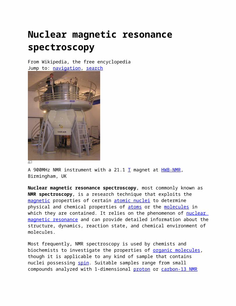
A 900MHz NMR instrument with a 21.1 T magnet at HWB-NMR, Birmingham, UK
Nuclear magnetic resonance spectroscopy, most commonly known as NMR spectroscopy, is a research technique that exploits the magnetic properties of certain atomic nuclei to determine physical and chemical properties of atoms or the molecules in which they are contained. It relies on the phenomenon of nuclear magnetic resonance and can provide detailed information about the structure, dynamics, reaction state, and chemical environment of molecules.
Most frequently, NMR spectroscopy is used by chemists and biochemists to investigate the properties of organic molecules, though it is applicable to any kind of sample that contains nuclei possessing spin. Suitable samples range from small compounds analyzed with 1-dimensional proton or carbon-13 NMR spectroscopy to large proteins or nucleic acids using 3 or 4-dimensional techniques. The impact of NMR spectroscopy on the sciences has been substantial because of the range of information and the diversity of samples, including solutions and solids.
Contents
1 Basic NMR techniques o 1.1 Chemical shift o 1.2 J-coupling
1.2.1 Second-order (or strong) coupling 1.2.2 Magnetic inequivalence
3 Solid-state nuclear magnetic resonance 4 Biomolecular NMR spectroscopy
o 4.1 Proteins o 4.2 Nucleic acids o 4.3 Carbohydrates
5 See also 6 References 7 External links
Basic NMR techniques
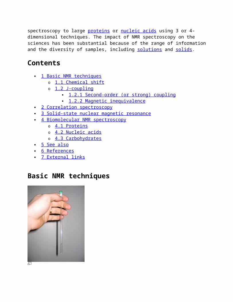
The NMR sample is prepared in a thin-walled glass tube - an NMR tube.
When placed in a magnetic field, NMR active nuclei (such as 1H or 13C) absorb electromagnetic radiation at a frequency characteristic of the isotope. The resonant frequency, energy of the absorption, and the intensity of the signal are proportional to the strength of the magnetic field. For example, in a 21 Tesla magnetic field, protons resonate at 900 MHz. It is common to refer to a 21 T magnet as a 900 MHz magnet, although different nuclei resonate at a different frequency at this field strength in proportion to their nuclear magnetic moments.
Chemical shift
Main article: Chemical shift
A spinning charge generates a magnetic field that results in a magnetic moment proportional to the spin. In the presence of an external magnetic field, two spin states exist (for a spin 1/2 nucleus): one spin up and one spin down, where one aligns with the magnetic field and the other opposes it. The difference in energy (ΔE) between the two spin states increases as the strength of the field increases, but this difference is usually very small, leading to the requirement for strong
NMR magnets (1-20 T for modern NMR instruments). Irradiation of the sample with energy corresponding to the exact spin state separation of a specific set of nuclei will cause excitation of those set of nuclei in the lower energy state to the higher energy state.
For spin 1/2 nuclei, the energy difference between the two spin states at a given magnetic field strength are proportional to their magnetic moments. However, even if all protons have the same magnetic moments, they do not give resonant signals at the same field/frequency values. This is because this is dependent on the electrons surrounding the proton in covalent compounds. Upon application of an external magnetic field, these electrons move in response to the field and generate local magnetic fields that oppose the much stronger applied field. This local field thus "shields" the proton from the applied magnetic field, which must therefore be increased in order to achieve resonance (absorption of rf energy). Such increments are very small, usually in parts per million (ppm). The difference between 2.3487T and 2.3488T is therefore about 42ppm. However a frequency scale is commonly used to designate the NMR signals, even though the spectrometer may operate by sweeping the magnetic field, and thus the 42 ppm is 4200 Hz for a 100 MHz reference frequency (rf).
However given that the location of different NMR signals is dependent on the external magnetic field strength and the rf frequency, the signals are usually reported relative to a reference signal, usually that of TMS (tetramethylsilane). Additionally, since the distribution of NMR signals is field dependent, these frequencies are divided by the spectrometer frequency. However since we are dividing Hz by MHz, the resulting number would be too small, and thus it is multiplied by a million. This operation therefore gives a locator number called the "chemical shift" with units of parts per million.[1] To detect such small frequency differences the applied magnetic field must be constant throughout the sample volume. High resolution NMR spectrometers use shims to adjust the homogeneity of the magnetic field to parts per billion (ppb) in a volume of a few cubic centimeters. In general, chemical shifts for protons are highly predictable since the shifts are primarily determined by simpler shielding effects (electron density), but the chemical shifts for many heavier nuclei are more strongly influenced by other factors including excited states ("paramagnetic" contribution to shielding tensor).
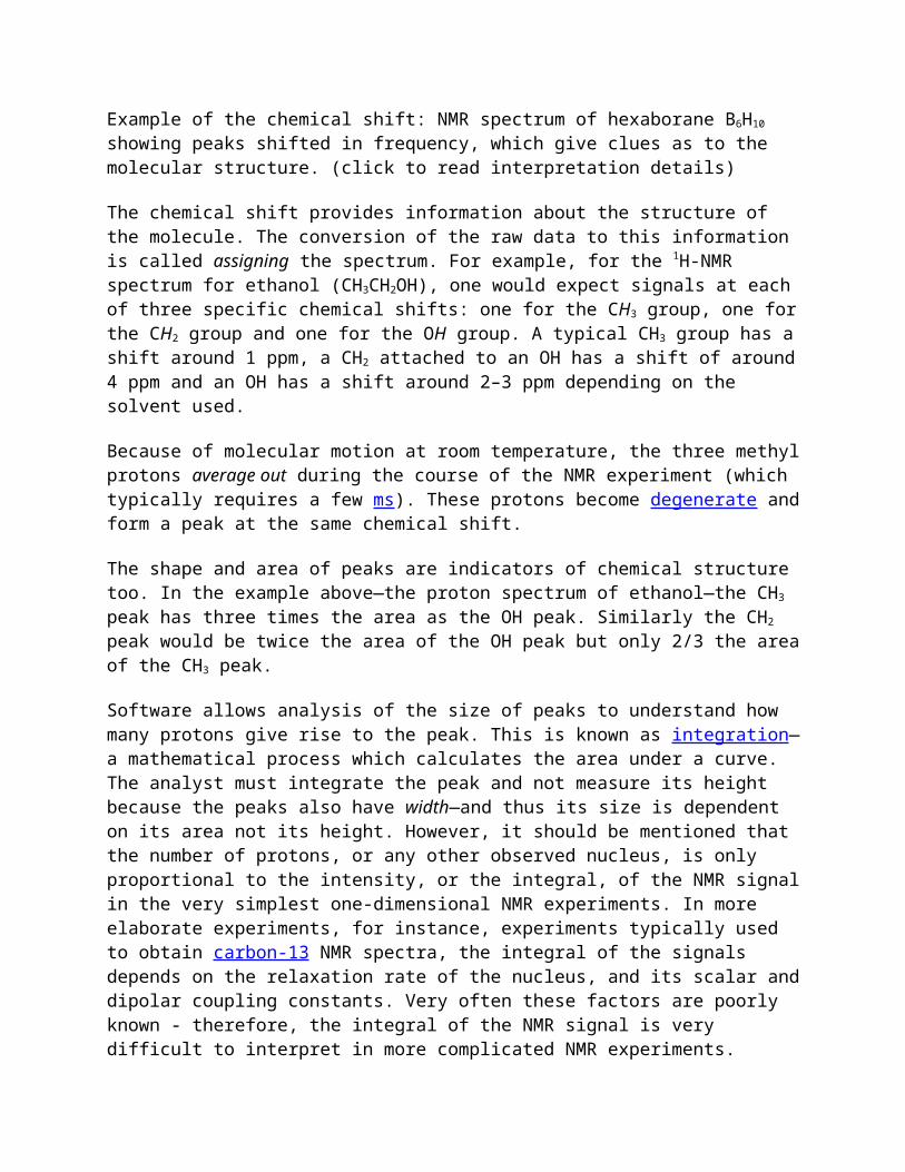
Example of the chemical shift: NMR spectrum of hexaborane B6H10 showing peaks shifted in frequency, which give clues as to the molecular structure. (click to read interpretation details)
The chemical shift provides information about the structure of the molecule. The conversion of the raw data to this information is called assigning the spectrum. For example, for the 1H-NMR
spectrum for ethanol (CH3CH2OH), one would expect signals at each of three specific chemical shifts: one for the CH3 group, one for the CH2 group and one for the OH group. A typical CH3 group has a shift around 1 ppm, a CH2 attached to an OH has a shift of around 4 ppm and an OH has a shift around 2–3 ppm depending on the solvent used.
Because of molecular motion at room temperature, the three methyl protons average out during the course of the NMR experiment (which typically requires a few ms). These protons become degenerate and form a peak at the same chemical shift.
The shape and area of peaks are indicators of chemical structure too. In the example above—the proton spectrum of ethanol—the CH3 peak has three times the area as the OH peak. Similarly the CH2 peak would be twice the area of the OH peak but only 2/3 the area of the CH3 peak.
Software allows analysis of the size of peaks to understand how many protons give rise to the peak. This is known as integration—a mathematical process which calculates the area under a curve. The analyst must integrate the peak and not measure its height because the peaks also have width—and thus its size is dependent on its area not its height. However, it should be mentioned that the number of protons, or any other observed nucleus, is only proportional to the intensity, or the integral, of the NMR signal in the very simplest one-dimensional NMR experiments. In more elaborate experiments, for instance, experiments typically used to obtain carbon-13 NMR spectra, the integral of the signals depends on the relaxation rate of the nucleus, and its scalar and dipolar coupling constants. Very often these factors are poorly known - therefore, the integral of the NMR signal is very difficult to interpret in more complicated NMR experiments.
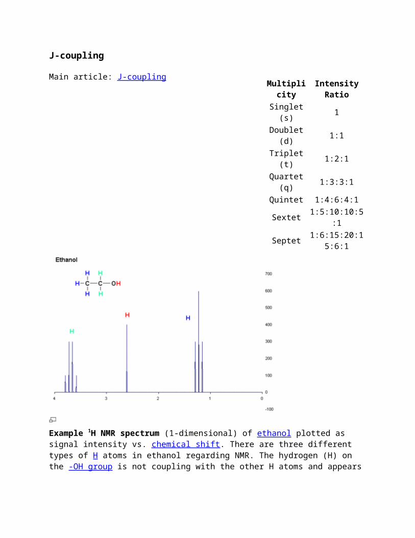
Example 1H NMR spectrum (1-dimensional) of ethanol plotted as signal intensity vs. chemical shift. There are three different types of H atoms in ethanol regarding NMR. The hydrogen (H) on the -OH group is not coupling with the other H atoms and appears as a singlet, but the CH3- and the -CH2- hydrogens are coupling with each other, resulting in a triplet and quartet respectively.
Some of the most useful information for structure determination in a one-dimensional NMR spectrum comes from J-coupling or scalar coupling (a special case of spin-spin coupling) between NMR active nuclei. This coupling arises from the interaction of different spin states through the chemical bonds of a molecule and results in the splitting of NMR signals. These splitting patterns can be complex or simple and, likewise, can be straightforwardly interpretable or deceptive. This coupling provides detailed insight into the connectivity of atoms in a molecule.
Coupling to n equivalent (spin ½) nuclei splits the signal into a n+1 multiplet with intensity ratios following Pascal's triangle as described on the right. Coupling to additional spins will lead to further splittings of each component of the multiplet e.g. coupling to two different spin ½ nuclei with significantly different coupling constants will lead to a doublet of doublets (abbreviation: dd). Note that coupling between nuclei that are chemically equivalent (that is, have the same chemical shift) has no effect of the NMR spectra and couplings between nuclei that are distant (usually more than 3 bonds apart for protons in flexible molecules) are usually too small to cause observable splittings. Long-range couplings over more than three bonds can often be observed in cyclic and aromatic compounds, leading to more complex splitting patterns.
For example, in the proton spectrum for ethanol described above, the CH3 group is split into a triplet with an intensity ratio of 1:2:1 by the two neighboring CH2 protons. Similarly, the CH2 is split into a quartet with an intensity ratio of 1:3:3:1 by the three neighboring CH3 protons. In principle, the two CH2 protons would also be split again into a doublet to form a doublet of
quartets by the hydroxyl proton, but intermolecular exchange of the acidic hydroxyl proton often results in a loss of coupling information.
Coupling to any spin ½ nuclei such as phosphorus-31 or fluorine-19 works in this fashion (although the magnitudes of the coupling constants may be very different). But the splitting patterns differ from those described above for nuclei with spin greater than ½ because the spin quantum number has more than two possible values. For instance, coupling to deuterium (a spin 1 nucleus) splits the signal into a 1:1:1 triplet because the spin 1 has three spin states. Similarly, a spin 3/2 nucleus splits a signal into a 1:1:1:1 quartet and so on.
Coupling combined with the chemical shift (and the integration for protons) tells us not only about the chemical environment of the nuclei, but also the number of neighboring NMR active nuclei within the molecule. In more complex spectra with multiple peaks at similar chemical shifts or in spectra of nuclei other than hydrogen, coupling is often the only way to distinguish different nuclei.
Second-order (or strong) coupling
The above description assumes that the coupling constant is small in comparison with the difference in NMR frequencies between the inequivalent spins. If the shift separation decreases (or the coupling strength increases), the multiplet intensity patterns are first distorted, and then become more complex and less easily analyzed (especially if more than two spins are involved). Intensification of some peaks in a multiplet is achieved at the expense of the remainder, which sometimes almost disappear in the background noise, although the integrated area under the peaks remains constant. In most high-field NMR, however, the distortions are usually modest and the characteristic distortions (roofing) can in fact help to identify related peaks.
Second-order effects decrease as the frequency difference between multiplets increases, so that high-field (i.e. high-frequency) NMR spectra display less distortion than lower frequency spectra. Early spectra at 60 MHz were more prone to distortion than spectra from later machines typically operating at frequencies at 200 MHz or above.
Magnetic inequivalence
More subtle effects can occur if chemically equivalent spins (i.e., nuclei related by symmetry and so having the same NMR frequency) have different coupling relationships to external spins. Spins that are chemically equivalent but are not indistinguishable (based on their coupling relationships) are termed magnetically inequivalent. For example, the 4 H sites of 1,2-dichlorobenzene divide into two chemically equivalent pairs by symmetry, but an individual member of one of the pairs has different couplings to the spins making up the other pair. Magnetic inequivalence can lead to highly complex spectra which can only be analyzed by computational modeling. Such effects are more common in NMR spectra of aromatic and other non-flexible systems, while conformational averaging about C-C bonds in flexible molecules tends to equalize the couplings between protons on adjacent carbons, reducing problems with magnetic inequivalence.
Correlation spectroscopy
For more details on this topic, see 2D-NMR.
Correlation spectroscopy is one of several types of two-dimensional nuclear magnetic resonance (NMR) spectroscopy or 2D-NMR. This type of NMR experiment is best known by its acronym, COSY. Other types of two-dimensional NMR include J-spectroscopy, exchange spectroscopy (EXSY), Nuclear Overhauser effect spectroscopy (NOESY), total correlation spectroscopy (TOCSY) and heteronuclear correlation experiments, such as HSQC, HMQC, and HMBC. Two-dimensional NMR spectra provide more information about a molecule than one-dimensional NMR spectra and are especially useful in determining the structure of a molecule, particularly for molecules that are too complicated to work with using one-dimensional NMR. The first two-dimensional experiment, COSY, was proposed by Jean Jeener, a professor at Université Libre de Bruxelles, in 1971[citation needed]. This experiment was later implemented by Walter P. Aue, Enrico Bartholdi and Richard R. Ernst, who published their work in 1976.[2]
Solid-state nuclear magnetic resonance
For more details on this topic, see Solid-state NMR.
A variety of physical circumstances does not allow molecules to be studied in solution, and at the same time not by other spectroscopic techniques to an atomic level, either. In solid-phase media, such as crystals, microcrystalline powders, gels, anisotropic solutions, etc., it is in particular the dipolar coupling and chemical shift anisotropy that become dominant to the behaviour of the nuclear spin systems. In conventional solution-state NMR spectroscopy, these additional interactions would lead to a significant broadening of spectral lines. A variety of techniques allows to establish high-resolution conditions, that can, at least for 13C spectra, be comparable to solution-state NMR spectra.
Two important concepts for high-resolution solid-state NMR spectroscopy are the limitation of possible molecular orientation by sample orientation, and the reduction of anisotropic nuclear magnetic interactions by sample spinning. Of the latter approach, fast spinning around the magic angle is a very prominent method, when the system comprises spin 1/2 nuclei. A number of intermediate techniques, with samples of partial alignment or reduced mobility, is currently being used in NMR spectroscopy.
Applications in which solid-state NMR effects occur are often related to structure investigations on membrane proteins, protein fibrils or all kinds of polymers, and chemical analysis in inorganic chemistry, but also include "exotic" applications like the plant leaves and fuel cells.
Biomolecular NMR spectroscopy
Proteins
Main article: Nuclear magnetic resonance spectroscopy of proteins
Much of the innovation within NMR spectroscopy has been within the field of protein NMR spectroscopy, an important technique in structural biology. A common goal of these investigations is to obtain high resolution 3-dimensional structures of the protein, similar to what can be achieved by X-ray crystallography. In contrast to X-ray crystallography, NMR spectroscopy is usually limited to proteins smaller than 35 kDa, although larger structures have been solved. NMR spectroscopy is often the only way to obtain high resolution information on partially or wholly intrinsically unstructured proteins. It is now a common tool for the determination of Conformation Activity Relationships where the structure before and after interaction with, for example, a drug candidate is compared to its known biochemical activity. Proteins are orders of magnitude larger than the small organic molecules discussed earlier in this article, but the basic NMR techniques and some of the NMR theory also applies. Because of the much higher number of atoms present in a protein molecule in comparison with a small organic compound, the basic 1D spectra become crowded with overlapping signals to an extent where direct spectra analysis becomes untenable. Therefore, multidimensional (2, 3 or 4D) experiments have been devised to deal with this problem. To facilitate these experiments, it is desirable to isotopically label the protein with 13C and 15N because the predominant naturally occurring isotope 12C is not NMR-active, whereas the nuclear quadrupole moment of the predominant naturally occurring 14N isotope prevents high resolution information to be obtained from this nitrogen isotope. The most important method used for structure determination of proteins utilizes NOE experiments to measure distances between pairs of atoms within the molecule. Subsequently, the obtained distances are used to generate a 3D structure of the molecule by solving a distance geometry problem.
Nucleic acids
Main article: Nuclear magnetic resonance spectroscopy of nucleic acids
"Nucleic acid NMR" is the use of NMR spectroscopy to obtain information about the structure and dynamics of polynucleic acids, such as DNA or RNA. As of 2003, nearly half of all known RNA structures had been determined by NMR spectroscopy.[3]
Nucleic acid and protein NMR spectroscopy are similar but differences exist. Nucleic acids have a smaller percentage of hydrogen atoms, which are the atoms usually observed in NMR spectroscopy, and because nucleic acid double helices are stiff and roughly linear, they do not fold back on themselves to give "long-range" correlations.[4] The types of NMR usually done with nucleic acids are 1 H or proton NMR , 13 C NMR , 15 N NMR , and 31 P NMR . Two-dimensional NMR methods are almost always used, such as correlation spectroscopy (COSY) and total coherence transfer spectroscopy (TOCSY) to detect through-bond nuclear couplings, and nuclear Overhauser effect spectroscopy (NOESY) to detect couplings between nuclei that are close to each other in space.[5]
Parameters taken from the spectrum, mainly NOESY cross-peaks and coupling constants, can be used to determine local structural features such as glycosidic bond angles, dihedral angles (using the Karplus equation), and sugar pucker conformations. For large-scale structure, these local parameters must be supplemented with other structural assumptions or models, because errors add up as the double helix is traversed, and unlike with proteins, the double helix does not have a
compact interior and does not fold back upon itself. NMR is also useful for investigating nonstandard geometries such as bent helices, non-Watson–Crick basepairing, and coaxial stacking. It has been especially useful in probing the structure of natural RNA oligonucleotides, which tend to adopt complex conformations such as stem-loops and pseudoknots. NMR is also useful for probing the binding of nucleic acid molecules to other molecules, such as proteins or drugs, by seeing which resonances are shifted upon binding of the other molecule.[5]
Carbohydrates
Main article: Nuclear magnetic resonance spectroscopy of carbohydrates
Carbohydrate NMR spectroscopy addresses questions on the structure and conformation of carbohydrates.
Spektroskopi NMRDitulis oleh Yoshito Takeuchi pada 03-01-2009
a. Prinsip
Banyak inti (atau lebih tepat, inti dengan paling tidak jumlah proton atau neutronnya ganjil) dapat dianggap sebagai magnet kecil. Inti seperti proton (1H atau H-1) dan inti karbon-13 (13C atau C-13; kelimpahan alaminya sekitar 1%). Karbon -12 (12C), yang dijadikan standar penentuan massa, tidak bersifat magnet.
Bila sampel yang mengandung 1H atau 13C (bahkan semua senyawa organik) ditempatkan dalam medan magnet, akan timbul interaksi antara medan magnet luar tadi dengan magnet kecil (inti). Karena ada interaksi ini, magnet kecil akan terbagi atas dua tingkat energi (tingkat yang sedikit agak lebih stabil (+) dan keadaan yang kurang stabel (-)) yang energinya berbeda. Karena dunia inti adalah dunia mikroskopik, energi yang berkaitan dengan inti ini terkuantisasi, artinya tidak kontinyu. Perbedaan energi antara dua keadaan diberikan oleh persamaan.
E = γhH/2π(13.4)
H kuat medan magnet luar (yakni magnet spektrometer), h tetapan Planck, γ tetapn khas bagi jenis inti tertentu, disebut dengan rasio giromagnetik dan untuk proton nilainya 2,6752 x 10⁸ kg-1 s A (A= amper)??
Bila sampel disinari dengan gelombang elektromagnetik ν yang berkaitan dengan perbedaan energi E, yakni,
inti dalam keadaan (+) mengabsorbsi energi ini dan tereksitasi ke tingkat energi (-). Proses mengeksitasi inti dalam medan magnetik akan mengabsorbsi energi (resonansi) disebut nuclear magnetic resonance (NMR)??
Frekuensi gelombang elektromagnetik yang diabsorbsi diungkapkan sebagai fungsi H.
ν = γH/2π(13.6)
Bila kekuatan medan magnet luar, yakni magnet spektrometer, adalah 2,3490 T(tesla; 1 T = 23490 Gauss), ν yang diamati sekitar 1 x 10⁸ Hz = 100 MHz??ilai frekuensi ini di daerah gelombang mikro.
Seacara prinsip, frekuensi gelombang elektromagnetik yang diserap ditentukan oleh kekuatan magnet dan jenis inti yang diamati. Namun, perubahan kecil dalam frekuensi diinduksi oleh perbedaan lingkungan kimia tempat inti tersebut berada. Perubahan ini disebut pergeseran kimia.
Dalam spektroskopi 1H NMR, pergeseran kimia diungkapkan sebagai nilai relatif terhadap frekuensi absorpsi (0 Hz) tetrametilsilan standar (TMS) (CH3)4Si??ergeseran kimia tiga jenis proton dalam etanol CH3CH2OH adalah sekitar 105??25 dan 490 Hz bila direkam dengan spektrometer dengan magnet 2 1140 T (90 MHz) (Gambar 13.6(a))??arena frekuensi absorpsi proton adalah 0,9 x 10⁸Hz (90 MHz), pergeseran kimia yang terlibat hanya bervariasi sangat kecil.
Gambar 13.6 1H spektra NMR etanol CH3CH2OH (a) spektrum resolusi rendah,(b) resolusi tinggi. Garis bertangga adalah integral intensitas absorpsi.
Frekuensi resonansi (frekuensi absorpsi) proton (atau inti lain) sebanding dengan kekuatan magnet spektrometer. Perbandingan data spektrum akan sukar bila spektrum yang didapat
dengan magnet berbeda kekuatannya. Untuk mencegah kesukaran ini, skala δ, yang tidak bergantung pada kekuatan medan magnet, dikenalkan. Nilai δ didefinisikan sebagai berikut.
δ = ( ν/ν) x 106 (ppm) (13.7)
ν perbedaan frekuensi resonansi (dalam Hz) inti yang diselidiki dari frekuensi standar TMS (dalam banyak kasus) dan ν frek uensi (dalam Hz) proton ditentukan oleh spektrometer yang sama. Anda harus sadar bahwa Hz yang muncul di pembilang dan penyebut persamaan di atas dan oleh karena itu saling meniadakan. Karena nilai ν/ν sedemikian kecil, nilainya dikalikan dengan 106. Jadi nilai δ diungkapkan dalam satuan ppm.
Untuk sebagian besar senyawa, nilai δ proton dalam rentang 0-10 ppm. Nilai δ tiga puncak etanol di Gambar 13. 6 adalah 1,15; 3,6 dan 5,4??
Penemuan pergeseran kimia memberikan berbagai kemajuan dalam kimia. Sejak itu spektroskopi NMR telah menjadi alat yang paling efektif untuk menentukan struktur semua jenis senyawa. Pergeseran kimia dapat dianggap sebagai ciri bagian tertentu struktur. Misalnya, pergeseran kimia proton dalam gugus metil sekitar 1 ppm apappun struktur bagian lainnya. Lebih lanjut, seperti yang ditunjukkan di Gambar 13.6, dalam hal spektra 1H NMR, intensitas sinyal terintegrasi sebanding dengan jumlah inti yang relevan dengan sinyalnya. Hal ini akan sangat membantu dalam penentuan struktur senyawa organik.
Selingan- Penemuan pergeseran kimia
Tahun 1964 adalah tahun yang tidak terlupakan sejarah kimia organik Jepang. Spektroskopi NMR awalnya diteliti oleh fisikawan yang tertarik pada sifat magnetik inti. Pengamatan pertama sinyal NMR dilakukan secara independen dan hampir simultan oleh dua fisikawan Amerika Felix Bloch (1905-1983) dan Edward Mills Purcell (1912-1987). Keduanya mendapatkan hadiah Nobel tahun 1952.
Menurut teori ini, frekuensi resonansi proton air dan parafin (hidrokarbon) identik sepanjang inti, proton yang sama yang diukur. Namun, beberapa perbedaan kecil mungkin diamati antara nilai satu frekuensi resonansi dua sampel. Pertanyaan yang timbul adalah apakah perbedaan ini adalah sifat khas alami, atau karena ketidakpastian percobaan.
Tak sengaja masalah ini diketahui oleh kimiawan yang kemudian menyarankan agar mereka mengukur spektrum etanol, dengan mengatakan bahwa etanol memiliki dua jenis proton, satu seperti air dan satunya seperti parafi. Saran ini diterima dan hasilnya sungguh menakjubkan. Jadi, pergeseran kimia ditemukan akibat kerjasama fisika dan kimia.
Contoh soal 13.3 spektrum 1H NMR
Sketsakan bentuk kira-kira spektrum 1H NMR 1-propanol CH3CH2CH2OH, dan identifikasi asal tiap sinyal. Prosedur ini disebut dengan penandaan (assignment).
Jawab
Pola spektrumnya dekat dengan pola spektrum etanol kecuali satu sinyal tambahan dari -CH2. Sinyal ini diharapkan muncul antara δ 1 dan δ 5 di Gambar 13.5. Anda harus memperhatikan bahwa proton dekat atom oksigen akan beresonansi pada medan rendah (yakni spektrum sisi kiri).
b. Kopiling spin-spin
Bahkan bila pergeseran kimia adalah satu-satunya informasi yang dihasilkan oleh spektroskopi NMR, nilai informasi dalam penentuan struktural senyawa organik sangat besar maknanya. Selain itu, spektroskopi NMR dapat memberikan informasi tambahan, yakni informasi yang terkait dengan kopling spin-spin.
Sebagaimana sudah Anda pelajari, tingkat energi inti (yakni, proton) terbelah menjadi keadaan berenergi tinggi dan rendah. Selain itu, tingkat-tingkat energi ini membelah lebih lanjut karena interaksi dengan inti tetangganya (inti-inti adalah magnet-magnet sangat kecil juga). Pembelahan ini sangat kecil tetapi akan memiliki akibat yang penting, yakni, pembelahannya tidak
dipengaruhi oleh kekuatan medan magnet spektrometer. Pembelahannya hanya bergantung pada interaksi inti-inti.
Bila spektrum 1H NMR etanol diukur dengan kondi si lebih baik (uakni resolusi lebih baik), sinyal CH3- dan CH2- tebelah menjadi multiplet (Gambar 13.6(b)). Pembelahan ini karena adanya kopling spin-spin antar proton. Spektra yang menunjukkan pembelahan kopling spin-spin ini disebut spektra resolusi tinggi. Sedang spektra yang tidak menunjukkan pembelahan ini disebut spektra resolusi rendah.
Latihan
Pertanyaan 13.1 Prediksi spektrum 1H NMR
Gambarkan sketsa spektra 1H NMR resolusi rendah dengan grafik batang.
suatu molekul yang dianalisis. Pada dasarnya spektrometri NMR merupakan bentuk lain dari spektroskopi absorbsi sama halnya dengan UV-VIS dan IR. Perbedaan dengan IR dan UV-VIS adalah
1. Sistem absorbsi dibawah pengaruh medan magnet dan hal ini tidak ada pada UV-VIS dan IR.
2. Pada NMR energi radiasi elektromagnetik pada daerah frekuensi radio.
Spekktroskopi NMR sangat penting artinya dalam analisis kualitatif, khususnya dalam penentuan struktur molekul zat organik. Lebih tepatnya letak suatu atom dalam molekulnya.
Seperti yang diketahui semua inti atom bermuatan karena mengandung proton dan juga mempunyai spin inti. Sifat inti atom dan karakter spinnya menyebabkan beberapa inti bersifat magnet.
Perputaran elektron pada porosnya (spin) menyebabkan dihasilkan momen dipol magnet. Perilaku dipol magnetik ini dicirikan oleh bilangan kuantum spin inti megnet yang dinyatakan atau diberi simbol I.
Apabila inti diletakan pada suatu medan magnet (medan magnet eksternal) maka akan terjadi interaksi inti dengan magnet ekternal tersebut. Interaksinya tergantung pada jenis inti yang berinteraksi. Berikut merupakan kriteria penggunaaan medan magnet pada spektroskopi NMR:
1. Medan magnet harus kuat. Karena kepekaan spektroskopi NMR makin tinggi seiring meningkatnya kekuatan medan magnet.
2. Medan magnet harus cukup homogen terhadap semua sampel yang dianalisis. Apabila tidak terjadi kemogenan medan magnet akan menghasilkan pita-pita yang melebar dan terjadi distorsi sinyal.
3. Medan magnet harus sangat stabil. Dengan kestabilan yang tinggi menjadikan analisis secara akurat dari detik ke detik bahkan hingga orde jam.
Seperti yang telah disinggung bahwa berhubungan dengan karakter inti dari suatu atom dalam suatu molekul, oleh sebab itu spektroskopi NMR digunakan untuk mendeteksi berbagai jenis inti sesuai dengan sifat khas inti, misalnya 1H, 13C, 19F dan 31P.
Karakter jenis inti yang dapat dideteksi menggunakan spkektroskopi NMR yaitu jenis kategori inti yang dalam kaitannya dengan bilangan kuantum spin inti, yakni:
Kategori I, yakni inti dengan I = 0. Inti dalam kategori ini tidak berinteraksi dengan medan magnet yang diterapkan pada NMR (medan magnet eksternal) sehingga disebut tidak ada kromofor NMR atau tidak aktif NMR. Inti dengan I = 0 adalah atom-atom dengan jumlah proton genap dan jumlah netron yang genap pula. Inti dengan I = 0 misalnya 12C, 16O dan 32S. Walaupun tidak dapat dicermati namun ketiga atom tersebut terdapat isotop yang dapat di deteksi.
Kategori 2 yakni inti dengan I = ½. Inti ini memiliki nomor massa ganjil sehingga mempunyai momen magnet tidak sama dengan nol. Hal inilah yang meneyebabkan inti dapat berinteraksi dengan medan magnet eksternal, oleh sebab itu disebut ada kromofor NMR. Inti dengan kategori ini misalnya 1H. 13C, 19F.
Kategori 3 yakni inti dengan proton dan netron ganjil. Inti ini memiliki I = 1, 2 atau lebih tinggi. Yang tergolong kategori ini adalah 2H, 14N, 10B. Isotop-isotop ini lebih sukar diamati dan pola spektranya melebar.
Geseran Kimia Dalam Spektroskopi NMR
Dalam spektroskopi NMR setiap jenis inti yang memiliki sifat yang khas dinyatakan dengan istilah geseran kimia (chemical shift) dan kopling spin-spin (Spin-spin coupling). Kedua besaran atau fenomena ini merefleksikan lingkungan kimia spin inti yang diamati dalam eksperimen NMR dan ini dapat dipandang sebagai efek kimia dalam spektroskopi NMR.
Frekuensi resonansi yang dialami inti bergantung pada besarnya kuat medan magnet yang diterapkan. Jadi frekuensi resonansi sebanding dengan medan magnet yang dialami oleh inti yang diamati. Makin besar spektrometer NMR, maka perpisahan antar puncak resonansi pada spektrum NMR makin besar dan kondisi demikian dikenal dengan NMR resolusi tinggi.
Geseran kimia inti yang terbaca dalam spektrometer NMR sebagai ppm (part per million) dan dilambangkan δ. Perlu diperhatikan bahwa ppm disini tidak sama dengan ppm konsentrasi. Nilai ppm tergantung pada frekuensi alat yang di gunakan yang ditulis denga persamaan berikut.
Δv = frekuensi sampel – 0 (frekuensi senyawa pembanding biasanya nol)
v = frekuensi yang dipasang atau digunakan
Senyawa Pembanding dalam NMR
Dalam mempelajari NMR digunakan suatu senyawa sebagai pembanding. Suatu senyawa pembanding yang biasa di gunakan adalah tetrametilsilana, (CH3)4Si atau yang disingkat TMS. Struktur TMS diberikan pada Gambar.
TMS biasanya langsung ditambahkan ke dalam larutan sampel yang akan diuji. TMS digunakan sebagai pembanding karena memiliki beberapa keunggulan antara lain:
1. Bersifat inert.2. Tingkat simetri yang tinggi, dalam hal ini semua atom H dan C berada pada
lingkungan kimia yang sama sehingga memberikan puncak absorbsi tunggal karena semua atom H dan C ekivalen.
3. Volatil, memiliki titik didih 27°C.4. Nonpolar sehingga mudah larut dalam pelarut organik.5. Geseran kimia TMS tidak dipengaruhi oleh kekompleksan pelarut atau tidak
dipengaruhi pelarut karena tidak mengandung gugus-gugus polar.
Selain TMS terdapat pula beberapa senyawa pembanding lain yaitu Na-2,2-dimetil-2-silapentana-5-sulfonat (DSS) dan Na-2,2,3,3-tetradeuterio-4-4-dimetil-4silapentanoat (TSP-d4). Struktur kedua senyawa tersebut sebagai berikut.
Spektrometer dan penanganan Sampel
Spektrometer NMR adalah alat atau instrumen untuk mengukur resosnansi magnetik inti. Intrumen ini menghasilkan medan magnet pada tingkat energi gelombang radio dan digunakan untuk mendeteksi radiasi yang dipancarkan pleh suatu inti. Kualitas spektrometer NMR tergantung pada dua hal yakni:
1. Kekuatan dan kehomogenan medan magnet yang digunakan.
2. Kestabilan kekuatan medan magnet selama digunakan.
Sampel atau cuplikan yang akan dianalisa dipreparasi dalam bentuk larutan. Larutan yang akan dianalisa menggunakan NMR memiliki beberapa kriteri sebagai berikut:
1. Spektrometer NMR 60 MHz. Masa sampel ±5-10 mg dalam ±0,4 mL pada tabung gelas dengan diameter 5 mm dan kedalaman tabung 35 mm. Sedangkan untuk spektrometer NMR 500 MHz diperlukan jumlah cuplikan < 1 mg (mikrogram) dalam tabung mikro pula.
2. Kualitas hasil sprktrum yang dihasilkan tergantung pada.
3. Tabung untuk cuplikan di buat dari gelas sangat tipis, mudah pecah dan sangat rapus terutama pada saat dibuka tutupnya.
4. Jika tabung yang digunakan tidak dipecahkan (mungkin disebabkan jumlah sampel yang sedikit dan harganya relatif mahal) maka segera dicuci dengan aseton atau dikloroetana bila telah selesai digunakan, dikeringkan dengan blower dalam udara bersih atau nitrogen dengan menggunakan pelat tipis dari logam selanjutnya dijaga dan disimpan pada tempat yang aman. Pengeringan tabung menggunakan oven atau dengan cara pemanasan sangat tidak dianjurkan.
Pelarut yang digunakan untuk mempreparasi sampel memiliki beberapa kriteria, yakni:
1. Tidak mengandung inti yang akan dideteksi atau diamati. Misalnya untuk 1H-NMR pelarutnya tidak boleh mengandung hidrogen-1 sedangkan untuk 13C-NMR pelarutnya tidak boleh mengandung 13-C.
2. Bersifat iner, 3. Nopolar 4. Titik didih rendah. 5. Tidak mahal.
Dari semua sifat di atas, CCl4 merupakan pelarut yang ideal yang hampir memenuhi semua persyaratan, tetapi pelarut ini sangat nonpolar sehingga mempunyai kapsitas pelarutan yang relatif rendah. Misalnya tidak dapat melarutkan senyawa-senyawa yang bersifat polar. Karena hal-hal tersebut maka terdapat beberapa pelarut yang sering digunakan pada spektrometer NMR yakni pelarut yang telah terdeuterasi, misalnya
Geseran kimia yang menunjukan terjadinya resonansi spin inti dalam lingkungan kimia yang berbeda pada suatu molekul digambarkan atau ditunjukan dalam bentuk grafik. Grafik NMR menggambarkan nilai δ (geseran kimia) dari setiap inti tertentu dalam lingkungan kimia yang tertentu pula.
Berdasarkan perjanjian atau yang telah ditetapkan pada ujung kanan memiliki geseran kimia sama dengan nol (0) merupakan inti yang memiliki atau memerlukan frekuensi kuat medan magnet besar (biasanya disebut juga kuat medan atas), sedangkan pada ujung kiri merupakan inti yang memiliki atau memerlukan frekuensi kuat medan magnet yang kecil (biasanya disebut juga kuat medan bawah). Secara ringkas dapat digambarkan sebagai berikut.
Inti Terlindungi Dan Kurang Terlindungi
setiap inti dilindungi atau dilingkupi oleh elektron-elektron yang megelilininya. Akibatnya setiap inti akan mengalami atau menerima pengaruh medan magnet eksternal atau medan magnet alat yang berbeda pula dan hal ini bergantung pada beberapa efek keterlindungan ini. Karena hal inilah inti-inti yang berbeda keterlindungannya akan mempunyai geseran kimia yang berbeda pada spektrum NMR-nya.
Hal ini memberikan magna bahwa, jumlah sinyal dalam spektrum NMR menunjukan banyaknya inti dengan lingkungan kimia yang berbeda dari molekul yang dianalisis. Inti yang efek keterlindungan tinggi (inti makin
terlindung) maka inti akan beresonansi pada kuat medan magnet yang tinggi sehingga mempunyai geseran kimia (δ) yang rendah dibanding senyawa standar (TMS). Sebaliknya inti yang memiliki efek keterlindungan rendah (inti semakin
tidak terlindung) maka inti akan beresonansi pada kuat medan magnet yang rendah sehingga mempunyai geseran kimia (δ) yang tinggi dibanding senyawa
pembanding (TMS).
Dari penjelasan ini dapat digambarkan sebagai berikut.
Secara umum inti-inti yang mengalami geseran diamagnetik dan paramagnetik dijelaskan sebagai berikut.
1. Distribusi awan elektron disekita inti. Distribusi awan elektron disekita inti sangat menentukan derajat keterlindungan inti. Makin besar kerapan distribusi awan elektron disekita inti makin besar dan makin efektif derajat keterlindungan dan menyebabkan inti harus beresonansi pada kuat medan magnet tinggi (medan magnet atas) dan mempunyai geseran kimia yang kecil atau semakin mendekati TMS = 0. Hal ini tentu berlaku juga untuk kondisi yang sebaliknya.
2. Gugus atau substituen penarik elektron. Gugus-gugus atau substituen penarik elektron seperti –OH, -OR, -OCOOH, -OCOR, -NO2, -halogen, yang terikat pada rantai alifatik menyebabkan derajat keterlindungan inti dan merubah geseran kimia ke arah medan rendah.
3. Karakter aniostropik magnetik. Contoh sirkulasi elektron dalam cincin bensena. Pengaruh anisotropik terhadap keterlindungan inti ini bekerja pada senyawa-senyawa aromatik, karbonil dan alkuna. Pengaruh karakter ini menyebabbkan inti semakin terlindung dan menggeser nilai geseran kimia pada kuat medan bawah atau kuat medan rendah. Nilai geseran kimia dalam ppm semakin besar dibanding TMS.
4. Karakter hibridisasi atom karbon dalam molekul. Perbedaan jenis atom karbon, yakni sp3, sp2, atau sp mempengaruhi derajat keterlindungan inti dalam spektroskopi NMR. Distribusi awan elektron pada atom karbon sp3 lebih rendah daripada sp2, dan lebih rendah dibanding sp akibatnya nilai geseran kimia sp3<sp2<sp dibanding senyawa pembanding (TMS).