HOW CAN NEUROIMAGING HELP UNDERSTAND, DIAGNOSE, AND DEVELOP TREATMENTS FOR ALZHEIMER'S DISEASE? Part C – AD brain scans - anatomical. NUCLEAR MEDICINE GRAND ROUNDS Stanford University J. Wesson Ashford, M.D., Ph.D. - PowerPoint PPT Presentation
11
HOW CAN NEUROIMAGING HELP UNDERSTAND, DIAGNOSE, AND DEVELOP TREATMENTS FOR ALZHEIMER'S DISEASE? Part C – AD brain scans - anatomical NUCLEAR MEDICINE GRAND ROUNDS Stanford University J. Wesson Ashford, M.D., Ph.D. Clinical Professor (affiliated), Department of Psychiatry and Behavioral Sciences Senior Research Scientist, Stanford / VA Aging Clinical Research Stanford University and VA Palo Alto Health Care System January 5, 2010 Slides at: www.medafile.com (Dr. Ashford’s lectures)
Transcript
HOW CAN NEUROIMAGING HELP UNDERSTAND, DIAGNOSE, AND
DEVELOP TREATMENTS FOR ALZHEIMER'S DISEASE?
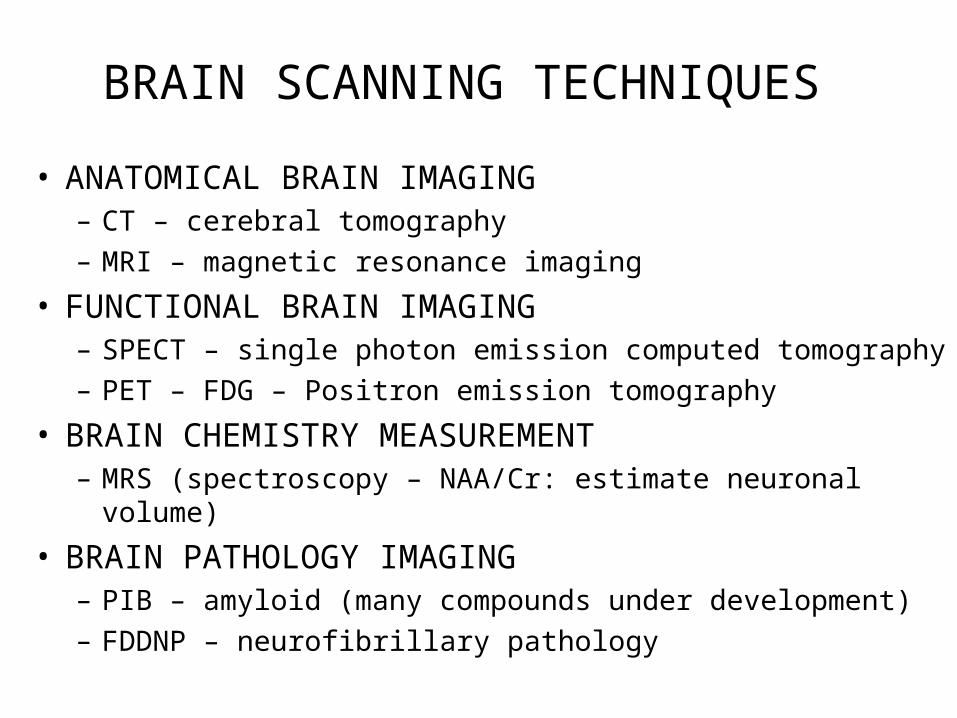
Part C – AD brain scans - anatomical NUCLEAR MEDICINE GRAND ROUNDS
Stanford University
J. Wesson Ashford, M.D., Ph.D.
Clinical Professor (affiliated), Department of Psychiatry and Behavioral SciencesSenior Research Scientist, Stanford / VA Aging Clinical Research
Stanford University and VA Palo Alto Health Care System