MQP-BIO-DSA-2973 Nucleoporin 85 is Crucial for Nuclear Import of HIV-1 A Major Qualifying Project Report Submitted to the Faculty of the WORCESTER POLYTECHNIC INSTITUTE in partial fulfillment of the requirements for the Degree of Bachelor of Science in Biology and Biotechnology by ______________________ James Gaudette January 12, 2007 APPROVED: ____________________ ____________________ Mario Stevenson, Ph.D. David Adams, Ph.D. Program in Molecular Medicine Prof, Biology and Biotech Umass Medical Center WPI Project Advisor Major Advisor
Transcript
�
�
MQP-BIO-DSA-2973
Nucleoporin 85 is Crucial for Nuclear Import of HIV-1
A Major Qualifying Project Report
Submitted to the Faculty of the
WORCESTER POLYTECHNIC INSTITUTE
in partial fulfillment of the requirements for the
Degree of Bachelor of Science
in
Biology and Biotechnology
by
______________________ James Gaudette
January 12, 2007
APPROVED:
____________________ ____________________ Mario Stevenson, Ph.D. David Adams, Ph.D. Program in Molecular Medicine Prof, Biology and Biotech Umass Medical Center WPI Project Advisor Major Advisor �
�
� ��
�
�
ABSTRACT
�
Because HIV and other lentiviruses can uniquely stably replicate within non-
dividing cells, we hypothesized that lentiviruses must utilize a special mechanism
allowing them to interact with host cell nuclear pore complexes, thus granting the HIV
pre-integration complex (PIC) access to the nucleoplasm by a common nuclear import
process. To prove this hypothesis, we performed RNAi-mediated knockdown screening
of several nuclear pore complex proteins. This led to the identification of nucleoporin-85
(nup85) as indispensable for HIV replication in macrophages. To eliminate siRNA non-
specific events as contributory to the observed inhibition, an siRNA resistant mutant
� The people and experiences that this project has allowed me to encounter have
forever changed the way I view science and research. As a result of the internship
granted to me in order to complete this project, I now leave undergraduate school, not in
fear of a lack of direction, but in utter confidence of the future course of my life. First
and foremost, I would like to thank Dr. Mario Stevenson for taking me on as a student in
his research lab, which has allowed me to experience first hand the frustrations and
rewards of daily work in the lab. Secondly, I want to thank Rajnish Kaushik for
personally training me in the techniques required to accomplish this project. These skills
will surely carry over into my next laboratory endeavor. Furthermore, I’m in deep
gratitude of his allowance of me in contributing to his own research in the lab, and for his
patience in dealing with the rigors of teaching a student with little entry knowledge of
formal laboratory practice. Also, I would like to extend a vast gesture of appreciation to
the entire team working in Dr. Stevenson’s lab. They’ve treated me with the utmost
kindness and respect during my stay in the lab, and were, every day, a source of positive
influence. I can only hope to find another lab replete with as many warm and hospitable
individuals. Lastly, I wish to thank Prof. Dave Adams, not only for advising my MQP,
but also for becoming my academic advisor in the final months of my matriculation at
WPI, when I had suddenly become absent of an advisor. Dave Adams was my initial
source of inspiration for heading a course towards the exciting field of virology, a
direction that I fully intend on committing my future to.
� �
�
� ��
BACKGROUND
HIV
Overview �
Research of human immunodificiency virus (HIV) is of dire importance to the
world today. Around 39.5 million people currently live with the virus, and about 4.3
million were newly infected in 2006 (UNAIDS, 2006). Figure 1 depicts the distribution
of infected individuals across the world.
�
Figure 1: AIDS Distribution Map. This map shows the distribution of HIV-infected adults over the world. The darker the color, the more prevalent the virus is in that region (UNAIDS, 2006). �
HIV is classified as a subclass of retroviruses termed lentiviruses, which are
characterized by a distinct morphology; including, a cylindrical or cone-shaped nucleoid
in the mature virion, and the presence of several regulatory genes (e.g., tat and rev). The
virus was first discovered in 1983, and is universally accepted as the cause of AIDS. The
pathological state exhibited by AIDS patients is distinguished by a dramatic loss of CD4+
lymphocytes, which gives rise to a variety of immune disorders and increased
�
� ��
susceptibility to opportunistic infections. Additionally, neurological disorders and
cancers can also arise from HIV infection (Levy, 1998). Virus titers high enough to be
infectious are present in both blood and genital fluids, and thus HIV can be transferred
either intravenously or sexually (Jaffe et al, 1983).
There are two distinct subtypes of HIV, HIV-1 is the predominant form and is
prevalent in all parts of the world, whereas HIV-2 is mainly present in only West African
countries (Levy, 1998). The strong genetic similarities between HIV and the monkey
version, simian immunodeficiency virus (SIV), support the theory that the virus became
present in humans by means of cross-species transmission, from monkeys to humans
(Coffin et al, 1996).
HIV Early Phase �
� Two distinct phases have been designated for the retroviral life cycle (Figure 2).
The early phase encompasses the events spanning from the attachment of virus to the cell
until integration of the viral cDNA into the cell genome, whereas the late phase involves
the expression of viral genes, and continues until the budding of mature virions from the
plasma membrane (Fig. 2; Nisole and Saïb, 2004).
�
� ��
�
�
Figure 2: The HIV Lifecycle. A schematic view of early and late stages of the retroviral replication cycle is represented. Examples of cellular factors interfering with early steps are indicated: Lv1/Ref1, CEM15, Fv1, Fv2. (Figure taken from Nisole and Saïb, 2004)
� In the early phase, retroviruses must pass two barriers, the plasma and nuclear
membranes.� The first barrier is overcome by attachment of the virion to the target cell
and then subsequent unzipping of the plasma membrane at the attachment site. The
process begins when the HIV envelope glycoprotein gp120 recognizes and attaches to
CD4 receptor on target cells. Conformational changes then take place in both of the
proteins, and co-receptors belonging to the chemokine receptor family, mainly CXCR4
and CCR5 are recruited (Berger et al, 1999). One of these receptors and gp120 then go
through a new conformational change, allowing viral gp41 fusion peptide to be inserted
into the host target membrane, thus fusing both viral and cellular membranes, allowing
entry of the viral core into the cytoplasm (Kwong et al, 1998). Refer to figure 3 for a
visual representation of viral entry. Alternatively, HIV can also gain entry to the cell by
�
� ��
simple endocytosis after binding of CD4 and available coreceptor, this latter method is
actually favored by most viral particles (Marechal et al, 1998).
�
Figure 3: HIV-1 Fusion Model. This figure shows the role of the HIV-1 surface glycoproteins during fusion. During this stage of infection the viral glycoproteins undergo conformational changes to facilitate the fusion of the virion, and eventual entrance of the viral core into the cell. (Figure taken from Knipe and Howley, 2001).
Before the second barrier to HIV is reached, the viral core must first be uncoated
and viral RNA reverse transcribed. Directly following the entry of the viral core into the
cytoplasm, the complex goes through a progressive disassembly. The resulting particles
make up what is called the reverse-transcription complex (RTC) where viral protein
reverse transcriptase creates a DNA copy of the double stranded viral RNA (Zhang et al,
2000). The new cDNA, plus remaining particles from the RTC, form the pre-integration
complex (PIC), which is distinct in the fact that it is competent for nuclear import,
although since the uncoating event is a progressive process, the exact differences between
the RTC and PIC are loosely defined (McDonald et al, 2002).�
�
� �
� At this point, the viral cDNA must now find a way into the nucleus in order to
integrate itself into the host genome (Sherman and Greene, 2002). For most retroviruses,
they must wait in the PIC stage until the nuclear envelope is broken down during the
replication cycle, however some cell types that HIV infects do not divide, thus HIV PICs
use an alternative mode of entry (Roe et al., 1993). By a method that is not well
understood, HIV is able to gain entry through small pores in the nuclear membrane
created by complexes of proteins called nucleoporins. Once the reverse transcribed viral
RNA has made its way into the nucleus, viral protein integrase can insert the viral cDNA
into the host chromosome, thus insuring the stable replication and transcription of
proviral DNA.
Nuclear Import via the Nuclear Pore Complex
Overview � The nuclear pore complex (NPC) is the only way in or out of the nucleus. The NPC
is large supramolecular protein structure that is embedded in the nuclear membrane and
spans its entire width, from cytoplasm to nucleoplasm. The method by which large
macromolecules, up to 39nm in diameter, enter the nucleus is called signal-mediated
nuclear import. The process involves the interaction of nuclear localization signals
(NLS) on the proteins being imported with nuclear membrane proteins of the karyopherin
family, also known as importins. NLSs are usually short stretches of amino acids that are
basic amino acid-rich and interact with the receptor importin (Fried and Kutay, 2003).
Interactions such as these, namely an NLS sequence with importin 7, are thought to play
a key role in the nuclear import of HIV-1 PICs in primary macrophages (Fassati et al,
2003).
�
� ��
NPC Structure �
Electron tomography of the NPC in yeast and in higher eukaryotes has elucidated
the 3D structure of the complex (Hinshaw et al., 1992). The 3D generated models
suggest that the NPC consists of an eight-fold symmetry central framework, called the
‘spoke complex’, which represents the transmembrane portion of the NPC (Figure 4a).
The spoke complex is located between a cytoplasmic and a nuclear ring moiety. Eight
short, kinked, cytoplasmic filaments are attached to the cytoplasmic ring moiety, whereas
the nuclear ring moiety is capped by a basket type structure (Stoffler et al., 2003). The
NPC central framework surrounds a pore of 90nm at its opening and 45-50nm at its
narrowest point, which serves as the passage way for nucleocytoplasmic transport.
�
�
�
Figure 4: Nuclear Pore Complex and Linear Pore Dimensions. This figure shows, (a) consensus model of the 3D architecture of the NPC and (b) linear dimensions of the central pore of the NPC. Blue boxes represent the cytoplasmic ring moiety of the NPC, orange boxes represent the nuclear ring moiety of the NPC. (Fahrenkrog et al., 2004) �
�
� �
Nucleoporins �
� The�NPC is comprised of a number of different proteins termed nucleoporins. It
is thought that the NPC consists of around 30 unique nucleoporins. Adhering to the 8-
symmetry observed in the 3D structure of the NPC, a single nucleoporin is present in 8
copies, or a multiple of 8. Within the complex, unique nucleoporin copies are mainly
located symmetrically with the two faces of the NPC, cytoplasmic and nucleoplasmic, as
opposed to asymmetric at one face (Cronshaw et al., 2002). Many nucleoporins have a
highly conserved region of phenylalanine-glycine (FG) repeats, which mediate
interactions between the nucleoporin and transport receptors (Rout et al., 2003). The fact
that the NPC is composed of so many different proteins with different structures and
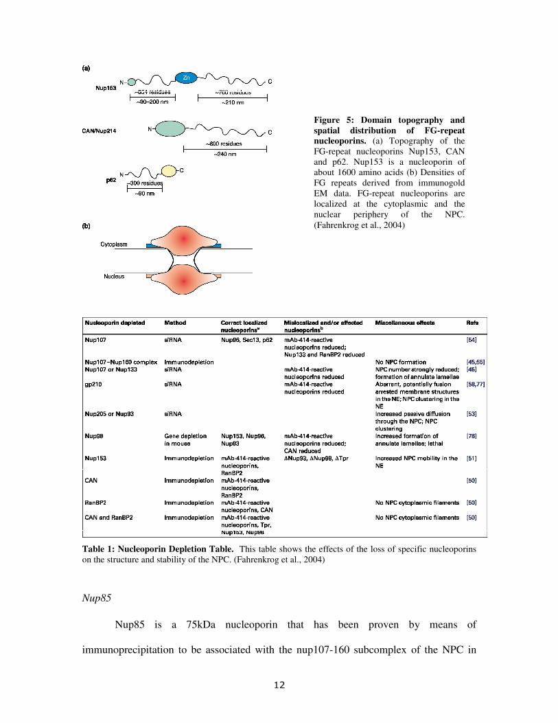
functions allows for the complex to take on multiple roles. Figure 5 shows an illustration
of different nucleoporin structures, as well as the distribution of FG containing
nucleoporins within the complex, and table 6 is a phenotypic screening of nucleoporin
knockdowns, both support the conclusion that individual nucleoporins offer unique
functional contributions to the NPC.
�
� ��
�
�
�
Table 1: Nucleoporin Depletion Table. This table shows the effects of the loss of specific nucleoporins on the structure and stability of the NPC. (Fahrenkrog et al., 2004)
Nup85 �
� Nup85 is a 75kDa nucleoporin that has been proven by means of
immunoprecipitation to be associated with the nup107-160 subcomplex of the NPC in
Figure 5: Domain topography and spatial distribution of FG-repeat nucleoporins. (a) Topography of the FG-repeat nucleoporins Nup153, CAN and p62. Nup153 is a nucleoporin of about 1600 amino acids (b) Densities of FG repeats derived from immunogold EM data. FG-repeat nucleoporins are localized at the cytoplasmic and the nuclear periphery of the NPC. (Fahrenkrog et al., 2004)
�
� ��
vertebrates (Loïodice et al., 2004). This particular subcomplex is of special importance
because it has been recently identified as essential for stable NPC assembly (Walther et
al., 2003). In nup85 partially depleted cells, a loss of localization of several nucleoporins
was observed. Furthermore, an overall decrease in NPC number in the nuclear
membrane was recorded (Walther et al., 2003). �
RNAi
Overview �
Since the RNA silencing has been used to knockdown the expression of specific
proteins of interest in this MQP project, a brief discussion of this process is included
here. RNA interference (RNAi) is initiated by double stranded RNA (dsRNA) and leads
to the ultimate degradation of homologous messenger RNA (mRNA). The first evidence
that the expression of individual genes could be silenced was in transgenic plants, where
the newly introduced gene was seen to have a suppressive effect on an endogenous gene
in trans (Napoli et al., 1990). Originally, scientists used anti-sense strand RNAs to
suppress gene function, however a group doing this noted that the sense strand also
silenced genes (Guo and Kemphues, 1995). The mystery was finally solved when Fire
and colleagues discovered that dsRNA had a more powerful silencing affect, and could
function in much smaller doses. The finding led to the assumption that RNAi must
involve a catalytic or amplification process for silencing RNA (Fire et al., 1998). �
Mechanism �
� Once it was determined that RNAi was activated with a derivative of dsRNA, a
great effort was invested in elucidating the complete mechanism. A premier contribution
�
� ��
came with the observation that 25 nucleotide RNAs complementary to silenced genes in
transgenic plants were found (Hamilton and Baulcombe, 1999). It was next found that
dsRNA was being processed into 21-23 nucleotide-duplex short interfering RNAs
(siRNAs), and that a dsRNA-specific endoribonuclease, DICER, was doing the
processing (Tuschl et al., 1999; Hammond et al., 2000). These siRNAs are then
incorporated into an RNA-induced silencing complex (RISC), which disassociates the
antisense strand from the sense strand, and subsequently is directed by the antisense half
to homologous mRNAs. Finally, the target mRNA is cleaved by an endonucleolytic
process (Elbashir et al., 2001). This entire mechanism has been illustrated below in
figure 6.
�
� ��
�
Using RNAi to Study HIV Activity �
� The ability to silence one specific target gene allows investigators to determine
the function of a particular protein by observing the resulting phenotype of the deficient
cell. This same approach can be applied to the study of viruses as well. Researchers can
obtain new knowledge regarding which cellular proteins are required for HIV function.
For example, the first experiment done with this method involved the suppression of
tumor-suppressor gene 101 (Tsg101), which led to the conclusion that Tsg101 has a
crucial role in the budding of HIV virions (Garrus et al., 2001). Those interested in
investigating potential therapeutic targets utilizing RNAi must be careful that the cellular
Figure 6: RNAi Mechanism. This figure is a cartoon representation of the mechanism by which the RNAi response processes dsRNA into siRNA whose antisense strand is then isolated and designated towards target mRNA for nucleolytic cleavage. (Stevenson, 2003)
�
� ��
co-factor being suppressed does not have devastating effects on the cell, especially since
many proteins that interact with HIV have crucial cellular functions as well. �
RNAi Induced Interferon Response The RNAi research approach does have a notable drawback. Global upregulation
of interferon inducible genes has been witnessed after siRNA treatment of cells (Sledz,
2003). Due to the fact that interferon (IFN) acts as a cell’s primary defense against viral
infections and can result in the inhibition of RNA transcription and degradation of
cellular RNAs, this finding is tremendously significant because it becomes hard to
distinguish the negative impact on HIV replication of a specific mRNA knockdown
versus HIV inhibition from IFN-inducible genes. IFN involves the initiation of a
signaling cascade which is mediated by a variety of proteins, including the Janus family
tyrosine kinases, Jak1 and Tyk2, the signal transducers and activators of transcription,
STAT1 and 2, and the IRF9 transcription factor, ultimately resulting in the induction of
interferon stimulated genes (ISGs) within the nucleus (Haque, 1998). siRNAs may also
activate this anti-viral reponse by engaging the toll-like receptor-3 (TLR-3) (Figure 7).
�
� ��
�
Figure 7: Primary and secondary signaling by siRNA. This diagram shows how siRNA can activate TLR3 (Toll-like receptor 3) and/or PKR to induce both primary and secondary transcriptional events, namely interferon � and p56, protein-synthesis dependent ISGs. (Sledz and Williams, 2004)
�
� ��
PROJECT PURPOSE �
�
�
The purpose of this research project is to study the role that nucleoporins play in
the nuclear import of HIV. RNAi-screening of several nuclear pore complex proteins led
to the identification of nucleoporin-85 (nup85) protein as indispensable for HIV
replication in macrophages. To eliminate siRNA non-specific events as contributory to
the observed inhibition, replication in human HeLa and 293T cells was rescued by an
siRNA-resistant mutant form of nup85. Furthermore, it was proven that the introduction
of dsRNA into macrophages does not elicit an interferon response potent enough to make
cells refractory to HIV, indicating the siRNA knockdown of nup85 itself affects HIV
replication.
�
� �
METHODS
Cell Culture �
� Three cell types were used for our methods, HeLa, Macrophage, and 293T.
Macrophages were obtained in elutriated form, and seeded at a concentration of 1x10⁶/ml
in Dulbecco’s modified Eagles’s medium (DMEM), with 10% Human AB male serum,
L-glutamine, Gentamicin, and 10 ng/ml MCSF. Macrophages were incubated at 37°C for
3 days, and then the same conditioned media lacking MCSF was added in order to dilute
MCSF. Every 3 days, half the media was removed and fresh media, without MCSF, was
added back.
HeLa cells were maintained in 2% FBS containing DMEM, and 293T cells were
maintained in 10% FBS containing DMEM, and both media’s were supplemented with
L-glutamine, streptomycin, and penicillin. Both cell types were subcultured at a 1-10
ratio every 3-4 days.
Isolation and Reverse Transcription of RNA �
Macrophages, seeded and maintained as previously discussed, were harvested in
lysis buffer containing proteinase K, and RNA was isolated from the lysate using an
RNeasy mini kit (Qiagen). Macrophage RNA was reverse transcribed with random
hexamers using Applied Biosystem’s, TaqMan® RT Kit.
PCR � To obtain wild-type (wt) nup85 DNA, the cDNA reverse-transcribed from
macrophages RNA was included in a one-step PCR reaction using SIGMA JumpStarttm
�
� ���
RedAccuTaqtm LA DNA polymerase, and then purified from an agarose gel with a
Promega gel extraction kit. Forward and reverse primers were designed based on
sequence data gathered from the GenBank database, to attach to both ends of the nup85
DNA coding region. The upstream (forward) primer included at the 5 prime end a
sequence which coded for an HA (Hemagglutinin) peptide.
PCR Mutagenesis of Nup85�
A mutant version of nup85 (nup85m) was created by site-directed mutagenesis to
be resistant to RNAi knockdown, while still encoding the same nup85 protein. Point
mutations were introduced into three consecutive amino acid coding triplets. All three
nucleotide changes resulted in silent (amino acids were the same) mutations. The
forward and reverse mutant primers possessed the mutated sequences. In the first PCR
reaction, both the mutant and end primers were utilized. PCR fragments were purified,
and used as template in another PCR reaction with only the end primers. The same
conditions were employed in all PCR reactions. Reaction conditions were:
Initial Denature => 02:00 min @ 94°C 35 Cycles of: Denature => 00:20 min @ 94°C Anneal => 00:20 min @ 65°C Extend => 01:30 min @ 72°C The middle primers were (Point mutations colored red): Forward => 5’– CCC CTG GAT AAT ATT TTG TTG GC –3’ Reverse => 5’– GCC AAC AAA ATA TTA TCC AGG GG –3’ The end primers were (HA-tag colored orange): Forward => 5’- ATG GCT TAC CCA TAC GAT GTT CCA GAT TAC GCT AGC TTG GGT GGT CAT ATG GAG GAG CTC GAT GGC GA -3’ Reverse => 5’- TCA GGA ACC TTC CAG TGA GC -3’
�
� ��
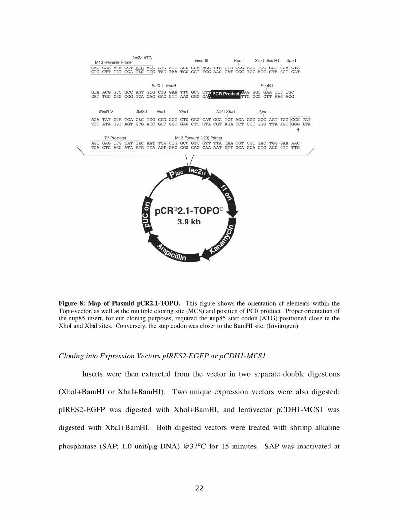
Cloning of PCR Amplicons into Plasmid TOPO-pCR2.1 � �
Nup85 and Nup85m PCR fragments were treated with taq-polymerase in order to
create 3’ oligo dA ends on the amplified DNA. The taq treated fragments were
separately ligated into an intermediary 3’ dT-tailed vector, TOPO-pCR2.1 (Figure-8;
Invitrogen), designed for easy direct ligation of taq-amplified PCR products. Ligation
and transformation procedures were performed with direct adherence to the TOPO-
cloning manual, and employed TOP10 chemically competent cells, as well as
LB+Ampicillin plates for transformation. Plasmid DNA was isolated from cultures of
individual ampr colonies using a Qiagen mini-prep kit, and were screened by restriction
digestion analysis for proper orientation of the nup85 gene within the vector’s cloning
site.
�
� ���
�
Figure 8: Map of Plasmid pCR2.1-TOPO. This figure shows the orientation of elements within the Topo-vector, as well as the multiple cloning site (MCS) and position of PCR product. Proper orientation of the nup85 insert, for our cloning purposes, required the nup85 start codon (ATG) positioned close to the XhoI and XbaI sites. Conversely, the stop codon was closer to the BamHI site. (Invitrogen)
Cloning into Expression Vectors pIRES2-EGFP or pCDH1-MCS1
Inserts were then extracted from the vector in two separate double digestions
(XhoI+BamHI or XbaI+BamHI). �Two unique expression vectors were also digested;
pIRES2-EGFP was digested with XhoI+BamHI, and lentivector pCDH1-MCS1 was
digested with XbaI+BamHI. Both digested vectors were treated with shrimp alkaline
phosphatase (SAP; 1.0 unit/µg DNA) @37°C for 15 minutes. SAP was inactivated at
�
� ���
65°C for 15 minutes. Next, XhoI+BamHI digested nup85 and nup85m DNAs were
ligated into digested pIRES2-EGFP, likewise XbaI+BamHI digested inserts were ligated
into correspondingly digested pCDH1 lentivector. Ligation reactions were carried out at
a 3:1 (insert:vector) ratio, using a Promega rapid DNA ligation kit. Recombinants were
transformed into Stbl-2 cells, and plated on LB plus antibody plates. Plates and
incubation temperatures were as follows: LB+ampicillin for pCDH1 recombinants
(grown at 32°C overnight) or kanamycin for pIRES2-EGFP plasmids (grown at 37°C
overnight). Individual colonies were screened for nup85 or nup85m inserts, and then
cultured in 250 mL of LB+ampicillin or LB+kanamycin, for pCDH1 or pIRES2-EGFP
respectively for larger scale (maxi-prep) plasmid isolations. Plasmids were purified with
Qiagen maxi-prep kit, no major changes were made in the provided maxi-prep protocol.
Figures 9 and 10 show the features and MCS contents of each expression vector.
�
� ���
�
Figure 9:�Map of the pIRES2-EGFP Expression Vector. Figure shows the properties of pIRES2-EGFP and contents of its MCS. The gene of interest is controlled by a CMV promoter with a ribosomal entry site located between the MCS and EGFP, allowing for GFP and gene sequence to be translated into one bicistronic mRNA. (Applied Biosystems)
�
� ���
�
Figure 10: Map of the Lentivector pCDH1. Figure shows the elements present in the pCDH1 lentivector, along with the contents of its MCS. This vector is designed ready for packaging into pseudotyped viral particles, but will also express a gene of interest when cells are transfected directly with the vector. The GFP element in this vector relies on a separate promoter (EF1) than the gene of interest (GFP). (System Biosciences)
DNA Sequencing �
Sequencing reactions of pCDH1 and pIRES plasmid constructs were prepared
with BigDye V3.1 sequencing reagent (Applied Biosystems). Reactions were cleaned on
DyeEx spin columns. Primers targeting sites on opposing sides of both vector MCS
regions were used so that the complete nup85 gene would be detected.
� �
Transfections �
Transfections of HeLa, 293T cells, and macrophages were performed by using
TransIt (Invitrogen), Lipofectamine2000 (Invitrogen), or siPort NeoFX (Ambion)
�
� ���
reagents. The siRNA for knockdown experiments was purchased from Qiagen custom
siRNA service (5’-AAC CCC TGG ACA ACA TCT TGTT-3’).
SDS-Page Gel Electrophoresis and Western Blot � �
� Transfected cells were harvested in lysis buffer containing SDS, run in a 10%
SDS-Page gel, and then transferred to a Hybond-C protein membrane. The membranes
were blocked in 0.2% I-Block. Primary probing was done with an anti-HA antibody, and
then the membrane was reprobed with a secondary antibody (anti-mouse).
Infection �
� My advising postdoc performed all infections, including the creation of VSVG-
pseudotyped virus. Briefly, VSV G-pseudotyped HIV-1 was made by co-transfecting
293T cells with a pNL-GFP proviral clone & pMD-G, expressing VSV-G. The
supernatant from the transfected cells was used as source of virus. Macrophages were
infected with 100 µl of virus supernatant and replaced with fresh macrophages medium
after 4 hours. Cells were harvested at indicated time points for DNA isolation.
RNA/DNA Quantification
Total DNA and RNA from transfected or infected macrophages were collected
and isolated using RNeasy and DNeasy Kits (Qiagen). RNA was reverse transcribed as
previously described. Synthesized cDNA was probed with interferon gene specific
primers and SYBR Green reagent in a real-time PCR reaction. PCR involving collected
�
� ���
DNA utilized specific probes designed to detect CCR5, HIV late product (LP), and
closed circular 2 long terminal repeats (2LTRs). CCR5 numbers were utilized in
normalizing the actual numbers obtained from both, LP and 2LTR’s.
�
� ���
RESULTS �
HIV and other lentiviruses in general harbor a unique characteristic that allows
them to efficiently establish stable replication within non-dividing cells. We
hypothesized that lentiviruses must utilize a special mechanism allowing them to interact
with host cell nuclear pore complexes, thus granting the HIV pre-integration complex
(PIC) access to the nucleoplasm by a common nuclear import process. To prove this
hypothesis, we constructed a series of experiments that demonstrate the importance of
nucleoporin-85 for the successful transportation of the HIV PIC from the cytoplasm to
the nucleoplasm. Prior to this MQP, initial research depleting specific nuclear pore
proteins (Fahrenkrog et al., 2004) led to the identification of several nucleoporins as
likely targets for inhibition of nuclear translocation. Fahrenkrog and colleagues noted that
cells deficient in the NPC subcomplex nup107-160 resulted in little to no NPC formation.
Based on the finding that nup85 associates with the nup107-160 subcomplex (Loïodice et
al., 2004), we decided to test whether nup85 depletion would disrupt HIV replication.
Knockdown of nup85 mRNA in Macrophages �
In order to determine the role of nup85 in HIV replication, macrophages were
transfected with siRNA complexes targeted to the human nup85 gene transcript (85i). As
a control, macrophages were also transfected with a random siRNA oligo-duplex (scr),
not intended to target any specific gene transcripts. After 24 hrs, macrophages were
infected with VSV G-pseudotyped GFP HIV. Total cell RNA and DNA was harvested
after 24hrs, post-infection. To visualize the potency of the gene knockdown, nup85
�
� ��
RNAs from scr and 85i transfected samples were quantitated by reverse-transcription and
PCR, then plotted on a bar graph (Figure 11). Quantitative RT-PCR showed more than 7-
fold depletion of nup85 mRNA in 85i-transfected cells as compared to scr-transfected
macrophages.�
�
���
�����
����
����
�����
�����
�����
�����
�����
�����
�����
����
�� ���
�����
���
�
Figure 11: siRNA Knockdown of nup85 mRNA in Macrophages. nup85 mRNA quantitative RT-PCR. Bar graph was created from a single quantitative RT-PCR, where macrophages were transfected with 60 pmol of scr (scrambled sequence) or 85i siRNA (against nup85), indicated on x-axis, and cells were harvested 24hrs later. The y-axis shows the relative number of mRNA transcripts (multiplied by a factor of 10^2). RNA levels in different samples were normalized relative to GAPDH.
�
�
Assay of HIV Replication in nup85 Knockdown Macrophages �
�
� To confirm the effect of nup85 depletion on HIV infected macrophages,
quantitative PCR DNA analysis was done to estimate the number of viral cDNAs as well
as 2LTR circles, shown in figures 12 and 13, respectively. Reverse-transcribed HIV late
products (LP) and 2LTR circles were produced at much lower levels, within 85i-
transfected macrophages, compared to the negative control scr-transfected cells.
�
� ���
�
�
�
�
�
�
�
�
�
�
�
�
Figure 12: Reduction of Late Product HIV DNA in nup85 Knockdown Macrophages. HIV late product DNA was assayed using quantitative PCR. Bar graph created from a single quantitative PCR where macrophages were transfected with 60 pmol of scr (scrambled) or 85i siRNA (against nup85), indicated on x-axis, and then subsequently infected 24hrs later with pseudotyped VSV-G/GFP HIV. Cells were harvested 24hrs post-infection. The y-axis shows the relative number of late HIV products (multiplied by a factor of 10^2).
������
��������
����
���������
���������
���������
���������
���������
���������
���������
���������
���������
����������
�� ���
�����
�����������
�
� ��
��������
�����
����
������
�������
�������
��������
��������
��������
��������
��������
��������
��������
�� ���
�����
�����
�
Figure 13: Reduction of HIV 2LTR DNA in nup85 Knockdown Macrophages. HIV 2LTR DNA was assayed by qPCR. Bar graph created from a single quantitative PCR where macrophages were transfected with 60 pmol of scr or 85i siRNA, indicated on x-axis, and then subsequently infected 24hrs later with pseudotyped VSV-G/GFP HIV. Cells were harvested 24hrs post-infection. The y-axis shows the relative number of 2LTRs (multiplied by a factor of 10^2).
�
Interferon Gene MxA Is Induced by RNA Duplexes
� To determine whether the lack of HIV replication in nup85 knockdown
macrophages resulted from a non-specific induction of an interferon antiviral reponse by
the 85i duplex DNA, the expression of a commonly induced interferon gene MxA was
assayed for by qRT-PCR (Figure-14). Mxa levels in 85i transfections were higher by
about five fold in comparison to scr. In the literature, it has been suggested that a very
high fold-induction in IFN-responsive gene expression is needed to elicit an anti-viral
response. Other IFN-induced genes are currently being analyzed from the same samples
to determine if the IFN response is widespread.
�
� ���
������
���������
����
���������
���������
���������
����������
����������
�� ���
�����
��
�
Figure�14: Assay of Interferon-Induced Gene MxA mRNA by qRT-PCR. Bar graph created from a single quantitative RT-PCR. Macrophages were transfected with 60 pmol of scr or 85i siRNA, indicated on the x-axis, and cells were harvested 24hrs later for RNA isolation. The y-axis shows the relative number of mRNA transcripts, normalized with respect to GAPDH (multiplied by a factor of 10^2).
�
Design of a Nup85 Mutant (nup85m) Resistant to RNAi Knockdown �
� In order to show a nup85-specific effect on HIV-1 replication, a siRNA-resistant
nup85 clone was generated which has three mismatches with the 85i siRNA. Previous
studies have indicated that 1-2 mismatches is enough to negate the effect of a specific
siRNA. The mutant clone (designated nup85m) was generated by site-directed
mutagenesis. After sequencing the mutated clone, the data was aligned with its wild-type
progenitor and analyzed for mismatched base pairs (Figure 15). As expected, in three
consecutive amino acids, three base pairs did not align with the wild-type sequence.
Mutations all involved the switching of a thymidine to a cytosine, resulting in silent
mutations, just as earlier proposed in the methodology.
�
� ���
�
Figure 15: Sequence Analysis of the Mutagenized nup85m DNA. This figure shows the alignment of both the mutated nup85 gene (upper sequence) (within our expression vector) and the wildtype nup85 gene sequence (lower sequence) (obtained from GenBank: NM_024844). Colored blocks represent mismatched basepairs. The green bar indicates homology between the two sequences above; a break in the green bar represents an inconsistency between the two strands of DNA. (Geneious v2.5; Drummond et al., 2006)
�
� To test whether the created mutations in nup85 resulted in a clone resistant to
knockdown by the 85i siRNA, 293T cells were co-transfected with siRNA and the cloned
nup85 or nup85m genes within a GFP expression vector. The pIRES2-EGFP vector
utilizes a second ribosomal entry site just before the GFP promoter, resulting in a single
bicistronic mRNA transcript. The rationale in this mechanism was that the siRNA
targeted against nup85 gene product will also knockout GFP expression, since both genes
were coded for on the same stand of mRNA. Figure 16 shows the results of this
experiment using scrambled scr siRNA negative controls and wild-type nup85 positive
control vectors. In 293T cells transfected with nup85m containing plasmid and 85i
siRNA (Figure 16, C and D), the number of GFP cells was equivalent to those cells
transfected with similar plasmid, but scrambled scr siRNA (Figure 16, A and B).
Conversely, cells co-transfected with 85i and wild-type nup85 gene showed significantly
less fluorescence compared to the scr control group (figure 16, G and H). Thus these data
demonstrate expression of the nup85m mutant is not knocked down by the 85i siRNA
treatment in 293T cells.
�
� ���
�
�
Figure�16: Lack of Mutant nup85m Knockdown by 85i siRNA in 293T Cells. Inverted Fluorescence Microscopy. Photos show the relative densities of plated cells (left column) (no UV or fluorescence filter applied), and the amount of GFP fluorescence (right column) in those same regions (UV light and Green FITC filter applied). 2x10^5 Cells were co-transfected with 500 ng of plasmid DNA and 60 pmol siRNA, and then observed 24hrs post-transfection. A,B) scr siRNA against pIRES2-EGFP/nup85m plasmid. C,D) 85i siRNA against pIRES2-EGFP/nup85m plasmid. E,F) scr siRNA against pIRES2-EGFP/nup85. G,H) 85i siRNA against pIRES2-EGFP/nup85.
The lentivirus-encoding vector pCDH1 does not utilize a single bicistronic mRNA
containing GFP, hence the effect of 85i on wt/mutant nup85 expression was observed
directly by a nup85 western blot. Fortunately, the vector is also designed to enable
expression of inserted genes while in its closed circular form, so we were able to confirm
nup85 protein expression in transfected Hela cells (Figure 17), rather than the more
complicated infection method in macrophages. Total cellular proteins from Hela cells
were separated by SDS-page gel electrophoresis, transferred to a protein membrane, and
�
� ���
then stained with HA and mouse antibodies to indirectly detect nup85 knockdown.
Figure 17 analyzed proteins from cells transfected with only plasmid. Empty pCDH1 and
pIRES vectors were used as negative controls. The lanes representing plasmids
containing nup85 and nup85m inserts showed nup85 bands properly migrated to the same
lengths, between 50kDa and 75kDa.
�
�
Figure 17: Expression of Nup85 and Mutant Nup85m Proteins in Hela Cells. Cell lysates from GFP-pIRES or GFP-pCDH1 expressing Hela cells were run on a 12% SDS-page gel and then transferred to hyperbond-C protein membranes. Membranes were probed with primary, anti-HA (1:1000), and secondary, anti-mouse (1:10000), antibodies to detect HA-tagged nup85. Membranes were treated with western blot lighting reagent and exposed for 1hr on X-ray film. The lane labels are the particular expression vector and gene being over-expressed. The right label indicates the proper position of nup85 protein.
�
To prove that knockdown of nup85 protein has occured in wt nup85-encoding
plasmids, but not in nup85m-encoding plasmids, the protein levels were compared after
siRNA co-transfections, figure 18. Scrambled (scr) siRNA groups served as the controls,
and in those lanes (labeled above the lane), a solid band resolved for both pCDH1 and
pIRES. As expected, expression of wt nup85 was downregulated when cotransfected
with 85i siRNA.
�
�
�
� ���
�
�
Figure 18: Nup85 and Mutant Nup85m Protein Levels Following Knockdown in 293T Cells. 293T cell lysates, co-transfected with GFP-pIRES or GFP-pCDH1 vectors and random (scr) or specific nup85 (85i) siRNAs, were run on a 12% SDS-page gel and then transferred to hyperbond-C protein membranes. Membranes were probed with primary, anti-HA (1:1000) (to detect HA-tagged nup85), and secondary, anti-mouse (1:10000), antibodies. Membranes were treated with western blot lighting reagent and exposed for 1hr on Polaroid film. The lane labels denote the particular expression vector, gene, and siRNA used for co-transfections. The right label indicates the proper position of nup85 protein.
�
�
�
�
� ���
DISCUSSION
�
� The data from this MQP indicate that nucleoprotein-85 (nup85) has an important
role in the nuclear transportation of the HIV pre-integration complex (PIC). In primary
macrophage cells, nup85 mRNA was successfully knocked down using 85i siRNA,
which also decreased HIV replication. In 293T cells, the 85i-induced knockdown of
nup85 appeared to be sequence specific since a mutant nup85m (whose sequence would
not strongly hybridize to 85i) produced no knockdown. The siRNA-resistant mutant
form of the nup85 protein has also been successfully cloned into a lentiviral gene delivery
vector for future experiments. Initial research, regarding the depletion of certain nuclear
pore proteins (Fahrenkrog et al., 2004), led to the identification of several nucleoporins as
likely targets for inhibition of nuclear translocation. Fahrenkrog and colleagues noted that
cells deficient in the NPC subcomplex nup107-160 resulted in little to no NPC formation.
Taking in account that nup85 associates with the nup107-160 subcomplex (Loïodice et
al., 2004), we decided to test whether or not nup85 depletion would disrupt HIV
replication.
The initial data from this MQP suggests that lack of nup85 expression has in fact
limited HIV infection to the reverse transcription stage without nuclear import. The
levels of HIV late products and 2LTRs, figures 12 and 13 respectively, have been
significantly reduced in cells exhibiting reduced levels of nup85 mRNA (figure 11).
These observations support a link between nup85 and HIV replication. Furthermore,
since LTRs are created after the PIC has translocated to the nucleoplasm and has
attempted to integrate viral cDNA into the host genome, we can conclude that the
�
� ���
inhibition occurs at the reverse transcription step, where the mature PIC is denied entry
into the nucleus. Interestingly HIV late product (LP), which supposedly forms before
nuclear import, decreased with loss of nup85 (Figure 13). This dilemma gives light to the
HIV reverse transcription mechanism. It is possible that reverse transcription only
initiates after the PIC has bound its signal translocation receptor in the NPC. Since
nuclear pore complex (NPC) formation is limited when nup85 is depleted, reverse
transcription may never reach its maximum efficiency. Further, these findings may point
at the true location of reverse transcription. Late product may in fact, mainly occur in the
nucleoplasm rather than the cytoplasm.
When RNA interference was performed in this project, it was imperative to show
that non-specific effects were not responsible for the observed phenotype. The
assumption that interferon genes would be upregulated in response to RNAi was
supported by the quantitation of MxA mRNA transcripts in siRNA transfected cells
(Figure 14). However, whether or not the approximate 5-fold interferon response that
was induced was strong enough to inhibit HIV remains inconclusive. MxA mRNA was
upregulated in cells after siRNA transfection, yet the overall MxA mRNA level was
relatively low compared to those seen in cells significantly affected by the interferon
response. In light of this, we proceeded in establishing an siRNA-resistant nup85
macrophage cell line, in order to rescue HIV infection. Fluorescence microscopy of
nup85 and nup85m knockdown experiments (Figure 16) proves that the mutated nup85 is
resistant to the 85i siRNA. Furthermore, the western blot analysis, confirms production
of nup85m in the pCDH1 lentivector (Figure 17). The nup85 western band where siRNA
85i is used against pCDH1-nup85m resolves in a similar intensity to that of scrambled scr
�
� ��
siRNA against pCDH1-nup85, so this is clear indication that the lentivector does harbor a
resistant nup85 gene.
The future holds many prospects for further investigation. Lentiviral system will
be used to express wt/mutant nup85 in macrophages, and experiments involving 85i-
transfections will be performed to determine the stability of siRNA-resistant nup85. The
proposed theory is that resistant cell lines will allow for HIV infection to persist, whereas
in nup85-depleted cells the virus will not be permissive. If this event is observed, we can
rule out that non-specific effects of siRNA have caused cells to behave refractory towards
HIV, and that nup85 alone is responsible for loss of viral replication in macrophages.
Additional research will involve elucidating the mechanism by which the HIV
PIC interacts with nup85 and is granted access through the NPC. It is possible that nup85
does not have a unique role in the mechanism, and that the phenotype observed through
our experiments are due to the inability of the NPC to assemble, or the lack of other key
nucleoporins from localizing to proper positions in the nucleus. The main experiments
for these purposes will include pull-down assays, in order to identify the proteins that the
PIC interacts with at the nuclear membrane.
�
� ���
BIBLIOGRAPHY Berger, EA et al. (1999) Chemokine receptors as HIV- 1 coreceptors: roles in viral entry,
tropism, and disease. Annu Rev Immunol 1999, 17, 657-700. Coffin, JM et al. (1996) Retroviruses. Cold Spring Harbor Laboratory Press. Cronshaw, J.M. et al. (2002) Proteomic analysis of the mammalian nuclear pore complex.
J. Cell Biol. 158, 915–927. Drummond AJ, Kearse M, Heled J, Moir R, Thierer T, Ashton B, Wilson A, Stones-
Havas S (2006) Geneious v2.5, Available from http://www.geneious.com/ Elbashir, S. et al. (2001) Duplexes of 21-nucleotide RNAs mediate RNA interference in
cultured mammalian cells. Nature 411, 494–498. Fahrenkrog B., Koser J., and Aebi U. (2004) The nuclear pore complex: a jack of all
trades? Trends BioChem. Sci. 29(4), 175-182. Fassati, A et al. (2003) Nuclear import of HIV-1 intracellular reverse transcription
complexes is mediated by importin 7. Embo J, 22, 3675-3685. Fire, A. et al. (1998) Potent and specific genetic interference by double-stranded RNA in
Caenorhabditis elegans. Nature 391, 806–811. Fried, H and Kutay, U (2003) Nucleocytoplasmic transport: taking aninventory. Cell Mol
Life Sci, 60, 1659-1688. Garrus, J. E. et al. (2001) Tsg101 and the vacuolar protein sorting pathway are essential
for HIV-1 budding. Cell 107, 55–65. Guo, S. and Kemphues, K. (1995) par-1, a gene required forestablishing polarity in C.
elegans embryos, encodes a putative Ser/Thr kinase that is asymmetrically distributed. Cell 81, 611–620.
Haque, S. J. and Williams, B. R. G. (1998) Signal transduction in the interferon system.
Semin. Oncol. 25, 14–22. Hamilton, A. and Baulcombe, D. (1999) A species of small antisense RNA in
posttranscriptional gene silencing in plants. Science 286, 950–952. Hammond, S., Bernstein, E., Beach, D. and Hannon, G. (2000) An RNA-directed
Hinshaw, J.E. et al. (1992) Architecture and design of the nuclear pore complex. Cell 69, 1133 – 1141.
Jaffe, HW et al. (1983) Acquired Immunodeficiency Syndrome in the United States the
first 1,000 cases. Journal of Infectious Diseases, 148, 339-345. Knipe D.M. and Howley P.M. (2001) Fields Virology, Volume 2, 4th edition. Lippincott
Williams & Wilkins, Philadelphia, PA. Kwong, PD et al. (1998) Structure of an HIV gp120 envelope glycoprotein in complex
with the CD4 receptor and a neutralizing human antibody. Nature, 393, 648-659. Levy, JA (1998) HIV and the pathogenesis of AIDS. Second edition. ASM Press,
Washington, D.C. Loiodice et al. (2004) The entire nup107-160 complex, including three new members, is
targeted as one entity to kinetochores in mitosis. Mol. Biol. Cell 15, 3333–3344. Marechal, V et al. (1998) Cytosolic Gag p24 as an index of productive entry of human
immunodeficiency virus type 1. J Virol, 72, 2208-2212. McDonald, D et al. (2002) Visualization of the intracellular behavior of HIV in living
cells. J Cell Biol, 159, 441-452. Napoli, C., et al. (1990) Introduction of a chimeric chalcone synthse gene into Petunia
results in reversible co-suppression of homologous genes in trans. Plant Cell. 2, 279–289.
Nisole, S. and Saïb, A. (2004) Early steps of retrovirus replicative cycle. Retrovirology,
1(9), 1-20. Roe T, et al. (1993) Integration of murine leukemia virus DNA depends on mitosis. Embo
J. 12, 2099-2108. Rout, M.P. et al. (2003) Virtual gating and nuclear transport: the whole picture. Trends
Cell Biol. 13, 622 – 628. Sherman, MP and Greene WC (2002) Slipping through the door: HIV entry into the
nucleus. Microbes Infect, 4, 67-73. Sledz, C. A., Holko, M., De Veer, M. J., Silverman, R. H. and Williams, B. R. (2003)
Activation of the interferon system by short interfering RNAs. Nature Cell Biol. 5, 834–839.
Sledz, C.A. and Williams, B. R. G. (2004) RNA interference and double-stranded-
Stevenson, M. (2003) Dissecting HIV-1 through RNA interference. Nature Rev. Immu. 3,
951-958. Stoffler, D. et al. (2003) Cryo-electron tomography provides novel insights into nuclear
pore architecture: implications for nucleocytoplasmic transport. J. Mol. Biol. 328, 119–130.
Tuschl, T., Zamore, P. D., Lehmann, R., Bartel, D. and Sharp, P. Targeted (1999) mRNA
degradation by double-stranded RNA in vitro. Genes Dev. 15, 3191–3197. UNAIDS (2006) AIDS Epidemic Update. Joint United Nations Programme on
HIV/AIDS (UNAIDS) and World Health Organization (WHO). Walther, T.C. et al. (2003) The conserved Nup107–160 complex is critical for nuclear
pore complex assembly. Cell 113, 195–206. Zhang, H et al. (2000) Morphologic changes in human immunodeficiency virus type 1
virions secondary to intravirion reverse transcription: evidence indicating that reverse transcription may not take place within the intact viral core. J Hum Virol. 3, 165-172.


![LONO1 Encoding a Nucleoporin Is Required for Embryogenesis … · LONO1 Encoding a Nucleoporin Is Required for Embryogenesis and Seed Viability in Arabidopsis1[C][W][OA] Christopher](https://static.documents.pub/doc/80x56/5f33c74a6e74b45879570c2c/lono1-encoding-a-nucleoporin-is-required-for-embryogenesis-lono1-encoding-a-nucleoporin.jpg)


![PySession4 - it.uu.se filePySession4 February 5, 2019 In [1]: import pandas as pd import numpy as np import matplotlib.pyplot as plt import sklearn.preprocessing as skl_pre import](https://static.documents.pub/doc/80x56/5cb2863a88c993f5708be449/pysession4-ituuse-february-5-2019-in-1-import-pandas-as-pd-import-numpy.jpg)


