+ All Categories
Home > Documents > Nucleotide sequence of gene VII and of a hypothetical gene (IX) in ...

Nucleotide sequence of gene VII and of a hypothetical gene (IX) in ...

Date post: 03-Feb-2017
Category:
Upload: lamtu
View: 219 times
Download: 0 times
Share this document with a friend
22
Volume 5 Number 12 December 1978 Nucleic Acids Research Nucleotide sequence of gene VII and of a hypothetical gene (IX) in bacteriophage Ml3 T.Hulsebos and J.G.G.Schoenmakers Laboratory of Molecular Biology, University of Nijmegen, Nijmegen, Netherlands Received 5 September 1978 ABSTRACT A DNA fragment containing gene VII of bacteriophage M13 has been transcribed and the nucleotide sequence of this 169-nucleotides long tran- script was determined by RNA sequencing methods. Additionally, the nucleo- tide sequence of this gene and parts of its neighbouring genes V and VIII has been determined by the dimethylsulphate-hydrazine technique. The reading frame of gene VII has been established by determining the nucleotide changes occurring in the transcripts of two amber mutants of this gene. From these combined data it is apparent that gene VII is only 99 nucleotides long and is immediately followed by the termination codon UGA. Its initiation codon AUG is separated from gene V by only a single nucleotide. It was noted that between the UGA termination codon of gene VII and the initiation codon of the next gene (gene VIII) there is space for another, hitherto unknown gene. This gene (IX) most probably codes for the small polypeptide ("C-protein") present in mature M13 phage particles. INTRODUCTION Bacteriophage Ml 3 is a small filamentous coliphage, closely related to the phages fd, fl and ZJ2. The genome of these phages consists of a circular single-stranded DNA which comprises only 6400 bases. In the last few years a rapid progress is being made toward an under- standing of the molecular biology of these phages (for a review see 1,2). In particular, genetic mapping, the process of viral DNA replication and the mechanism of transcription and translation has been studied in detail. The Ml3 genome is known to code for at least nine gene products, some of which have been well characterized regarding their biological function. In particular, the proteins encoded by gene II (nickase) and gene V (DNA- binding protein) are functional elements in the process of viral DNA repli- cation whereas the proteins encoded by genes III and VIII are constituents of the mature phage particle. The biological function of the genes I, IV, VI and VII are still unknown although there is evidence that the products © Information Retrieval Limited 1 Falconberg Court London W 1 V 5FG England 4677 Downloaded from https://academic.oup.com/nar/article-abstract/5/12/4677/2380725 by guest on 04 April 2018
Transcript

Volume 5 Number 12 December 1978 Nucle ic Ac ids Research

Nucleotide sequence of gene VII and of a hypothetical gene (IX) in bacteriophage Ml3

T.Hulsebos and J.G.G.Schoenmakers

Laboratory of Molecular Biology, University of Nijmegen, Nijmegen, Netherlands

Received 5 September 1978

ABSTRACT

A DNA fragment containing gene VII of bacteriophage M13 has beentranscribed and the nucleotide sequence of this 169-nucleotides long tran-script was determined by RNA sequencing methods. Additionally, the nucleo-tide sequence of this gene and parts of its neighbouring genes V and VIIIhas been determined by the dimethylsulphate-hydrazine technique.

The reading frame of gene VII has been established by determining thenucleotide changes occurring in the transcripts of two amber mutants ofthis gene. From these combined data it is apparent that gene VII is only99 nucleotides long and is immediately followed by the termination codonUGA. Its initiation codon AUG is separated from gene V by only a singlenucleotide. It was noted that between the UGA termination codon of geneVII and the initiation codon of the next gene (gene VIII) there is spacefor another, hitherto unknown gene. This gene (IX) most probably codesfor the small polypeptide ("C-protein") present in mature M13 phageparticles.

INTRODUCTION

Bacteriophage Ml 3 is a small filamentous coliphage, closely related to

the phages fd, fl and ZJ2. The genome of these phages consists of a circular

single-stranded DNA which comprises only 6400 bases.

In the last few years a rapid progress is being made toward an under-

standing of the molecular biology of these phages (for a review see 1,2).

In particular, genetic mapping, the process of viral DNA replication and

the mechanism of transcription and translation has been studied in detail.

The Ml3 genome is known to code for at least nine gene products, some of

which have been well characterized regarding their biological function.

In particular, the proteins encoded by gene II (nickase) and gene V (DNA-

binding protein) are functional elements in the process of viral DNA repli-

cation whereas the proteins encoded by genes III and VIII are constituents

of the mature phage particle. The biological function of the genes I, IV,

VI and VII are still unknown although there is evidence that the products

© Information Retrieval Limited 1 Falconberg Court London W 1 V 5FG England 4677

Downloaded from https://academic.oup.com/nar/article-abstract/5/12/4677/2380725by gueston 04 April 2018

Nucleic Acids Research

of these genes are involved in the process of phage maturation.

By coupled transcription-translation of Ml3 replicative form (RF) DNA

(3,4) or restriction fragments (5,6) each M13 gene product has now been

identified and characterized regarding their molecular weight. The ex-

ceptions are genes VI and VII the products of which have neither been ob-

served among the products in the infected cell (7) nor among the synthetized

products in the cell-free systems applied (3-6,8). Also in minicells har-

bouring Ml3 RF, the synthesis of these two proteins could not yet be demon-

strated (9) .

The low level of gene Vll-protein synthesis is rather intruiging.

Previously we have shown that the transcription of Ml3 RF is initiated at

nine different promotor sites and terminates at a single unique site

(6,8,10,11). This central termination site has been localized immediately

distal to gene VIII (11). Since gene VII is positioned on the genetic map

between gene V and gene VIII (Fig. 1) and transcription proceeds in only one

direction along the genetic map, gene VII is located in a region where the

highest transcriptional activity of the DNA genome has been demonstrated (8).

Moreover, an RNA transcript has been isolated which encompasses the coding

information of genes V, VII and VIII, but upon translation of this poly-

cistronic RNA in an in vitro protein synthesizing system only the abundant

synthesis of the proteins of genes V and VIII is apparent (8). This strong-

ly suggests that the expression of gene VII is controlled at the level of

translation.

As a step towards the elucidation of the structural features of this

gene we have undertaken to sequence the region encompassing gene VII and

parts of its neighbouring genes. In the meantime, the sequence was deter-

mined of RNA transcripts derived from Ml3 DNA restriction fragments carrying

several amber mutations in gene VII. From the results of these studies the

reading frame could be determined, ultimately leading to a detailed know-

ledge of the primary structure of gene VII.

MATERIALS AND METHODS

Materials

Replicative form DNA, either derived from wild-type or amber mutant M13

phages, was prepared from E.coli C89 (su ) or E.aoli K37 (sul )-infected

cells by the procedure described previously (12). The restriction enzymes

R.ftzp II and R.Hae III were prepared as described in a previous report (13)

4678

Downloaded from https://academic.oup.com/nar/article-abstract/5/12/4677/2380725by gueston 04 April 2018

Nucleic Acids Research

EndoR.Hha I was purchased from New England Biolabs and EndoR.Taqi was from

the Microbiol.Res.Establishment, Porton. E.coli RNA polymerase holoenzyme

was a generous gift of Dr. R. Schilperoort, Leiden. The Ml3 nonsense mutants

aml-U2 r,nd amT-H3 were provided by Dr. D. Pratt, Davis. The primer dinucleo-

tides GpC, CpG, ApA and ApG were purchased from Boehringer, Germany.

Restriction fragments

M13 RF (200 pg), enriched with uniformly labelled (32P)-M13 RF, was

digested with the appropriate endonuclease in excess using the conditions

previously described (14). The digest was layered on a 3% discontinuous

polyacrylamide slab gel (40 cm x 20 cm x 0.2 cm) formed in 40 mM Tris,

20 mM sodium acetate, 2 mM EDTA, pH 7.8. After electrophoresis for 16 h

at 40 mA, the gels were covered with Saran wrap and autoradiographed. Gel32

segments corresponding to the P-radioactive bands were excised and the

DNA fragments were extracted from the gel as described by Van den Hondel

et al. (14).

Preparation of transcripts.

RNA synthesis was carried out in a reaction mixture (0.15 ml) contain-

ing 40 mM Tris-HCl, pH 7.6, 150 mM KC1, 0.1 mM dithiothreitol, 4 mM EDTA,

15 mM MgCl , 0.1% Tween-80, 20 pM nucleoside (a- P)-triphosphate, 200 pM

of each of the other ribonucleoside triphosphates, 2 pmol of "300-fragment"

and about 20 pmol of E.coli RNA polymerase holoenzyme.

The transcription was started by the addition of MgCl . After 30 min

at 37 the reaction was terminated by the addition of 150 pi of 10 mM Tris-

HCl, 1 mM EDTA, pH 7.6 (buffer A) containing 150 pg of carrier tRNA per ml

and 0.1% SDS. The mixture was extracted with an equal volume of freshly

distilled phenol, then 0.1 vol. of 3 M sodium acetate, pH 5.6, was added to

the aqueous phase and the RNA was precipitated twice with 2.5 vol. of

ethanol for 1 h at -80 . The RNA was dried in Vaauo and dissolved in 20 pi

of 40 mM Tris, 20 mM sodium acetate, 2 mM EDTA, pH 7.8 buffer containing

7 M urea, 20% glycerol, 0.1% SDS and 0.1% bromophenol blue. After dissolu-

tion the RNA was heated for one min at 90 , rapidly chilled and subjected

to electrophoresis on polyacrylamide gels.

The conditions for primer-dependent RNA synthesis were identical to

the standard conditions except that a primer was added to a final concentra-

tion of 400 pM and the concentration of all ribonucleoside triphosphates,32

one of which was labelled with P in the a-position, were 10 pM.

4679

Downloaded from https://academic.oup.com/nar/article-abstract/5/12/4677/2380725by gueston 04 April 2018

Nucleic Acids Research

Gel electrophoresis and recovery of RNA from the gel

RNA products were fractionated by electrophoresis on 4% polyacrylamide

slab gels (20 cm x 20 cm x 0.2 cm) which were prepared in 40 mM Tris, 20 mM

sodium acetate, 2 mM EDTA, pH 7.8, containing 7 M urea and 0.1% SDS. After

electrophoresis for 5 h at 30 mA (about 75 V) and autoradiography, the

portions of the gel containing the RNA were cut out, crushed by piercing

through a hypodermic syringe and extracted twice for 4 h with 2 ml of

buffer A containing 0.1% SDS and 10 yg of carrier tRNA. The extracts were

combined and the RNA was precipitated with ethanol. The precipitate was

spun down, dissolved in 0.3 ml of buffer A and reprecipitated with ethanol.

The RNA precipitate was dried in vaouo and finally dissolved in about 15)jl32

of buffer A. The recovery of P-labelled RNA after this isolation procedure

was 70-80%.

RNA sequencing methods

Standard RNA sequencing methods were used according to Brownlee and

Sanger (15) and Barrell ( 16). Digestion of RNA with RNase Tl (Sankyo Co.)

was carried out in 10 ul of 10 mM Tris-HCl, 1 mM EDTA, pH 7.4 for 30 min

at 37 using a ratio of enzyme/carrier RNA of 1:20. The resulting Tl-oligo-

nucleotides were fractionated by electrophoresis at pH 3.5 on cellulose

acetate (Schleicher-Schull) in the first dimension followed by homochromato-

graphy on DEAE-cellulose thin layer plates (Machery-Nagel, CEL 300/HR) in

the second dimension. As developing medium homomixture "C" was used (15).

Tl-oligonucleotides eluted from fingerprints were digested with pancreatic

RNase (Worthington) for 60 min at 37° with an enzyme/RNA ratio of 1:10.

The pancreatic RNase products were characterized by electrophoresis on

DEAE-paper (Whatman DE 81) at pH 3.5. Most of the secondary digestion

products were further analysed by complete digestion with 0.5 N NaOH for

16 h at 37 . The resulting mononucleotides were fractionated by electro-

phoresis on Whatman 540-paper at pH 3.5 and the distribution of P in

mononucleotides was determined.

To determine the sequences of oligonucleotides for which unique

sequences were not deduced by nearest neighbour analysis, partial digestion

was carried out with spleen phosphodiesterase. The Tl-oligonucleiotides,

labelled with (a- P)-GTP and containing approximately 150 ug of carrier RN.

were dissolved in 60 yl of 25 mM ammonium acetate, pH 5.7. A sample of this

mixture (20 ul) was heated for 3 min at 90°. After cooling to 37 , 5 pi of

spleen phosphodiesterase solution (3 mg/ml) was added and 5 ul-aliquots

4680

Downloaded from https://academic.oup.com/nar/article-abstract/5/12/4677/2380725by gueston 04 April 2018

Nucleic Acids Research

were removed at 15 min intervals. The aliquots were rapidly chilled, pooled

and dried in vacuo and subsequently dissolved in 5 yl water. The partial

digestion products were fractionated by two-dimensional homochromatography

using homomix C. The sequences, indicated in Table 1 by underlining, were

deduced from the mobility shift pattern.

32

Labelling of fragments with P at a single 5'-OH terminus

The 5'-ends of restriction fragments were dephosphorylated with

bacterial alkaline phosphatase essentially as described by Maxam and

Gilbert (17). Labelling of the 5'-0H ends of fragments was performed with

(y- P)-ATP and polynucleotide kinase (17). The dephosphorylated fragments

(3-4 pmol) were dissolved in 45 yl of 10 mM glycine-NaOH, pH 9.5, 1 mMspermidine. 0.1 mM EDTA. The fragments were denatured by heating at 100

for 3 min, then quickly chilled and transferred to an Eppendorf tube con-32

taining 100 pmol of dried (y- P)-ATP (spec.act. >2000 Ci/mmol). After addi-

tion of 5 yl of 0.5 M glycine-NaOH, pH 9.5, 0.1 M MgCl2, 50 mM dithio-

threitol, the phosphorylation was started by adding 2-3 units of polynucleo-

tide kinase (P.L.Biochemicals). After 30 min at 37 the reaction was ter-

minated with phenol. Carrier tRNA (10 yg) was added and after two extrac-

tions with phenol the labelled fragments were precipitated with ethanol.

The precipitate was dissolved in 70 yl of buffer A, the solution was heated

at 100 for 3 min and the DNA fragments were renatured by incubation at 67

for 2 h. Thereafter the appropriate restriction enzyme and buffer was added

and the volume adjusted to 100 yl with buffer A. After a digestion at 37

for 2 h the 5'-labelled fragments were separated on 5% polyacrylamide gels

essentially as described by Maxam and Gilbert (17).

DNA sequencing methods

Partial digestion of DNA with snake venom phosphodiesterase was carried

out as described by Manlatis et at. (19). To 5 yl of fragment labelled at

one 5'-terminal end (about 0.5 pmol) was added 5 yl of 10 mM Tris-HCl,

pH 7.4, 10 mM MgCl_, 10 mM mercaptoethanol, 6 mM KC1 buffer and 1 yg of

sonicated calf-thymus DNA, 10 yg of carrier tRNA, 7.5 ng of DNase I

(Boehringer) and 2.5 ng of snake venom phosphodiesterase (Worthington).

Aliquots of 2 yl, taken at 10 min intervals, were rapidly chilled, pooled,

dried and finally dissolved in 5 yl ice-cold water. The partial digestion

products were fractionated by two-dimensional homochromatography using

homomix V (18) in the second dimension. The smaller products were eluted

4681

Downloaded from https://academic.oup.com/nar/article-abstract/5/12/4677/2380725by gueston 04 April 2018

Nucleic Acids Research

from fingerprints and their sequences were determined by comparing their

electrophoretic mobility on Whatman 3 MM-paper at pH 3.5 with the mobility

of markers of known composition.

For DNA sequencing by chemical degradation the protocol of Maxam and

Gilbert (17) was followed. Purine residues were partially methylated by

dimethyl sulphate. Cleavage at Guanine was obtained by heating at neutral

pH and subsequent treatment with 0.1 N NaOH at 90 . Preferential cleavage

at Adenine was achieved by treatment with 0.1 N HC1 followed by treatment

with 0.1 N alkali at 90 . Cleavage at Cytosine and Thymine was obtained by

partial hydrazinolysis followed by treatment with 0.5 M piperidine. Hydra-

zinolysis at Thymine was suppressed by the presence of 2 M NaCl.

Reaction mixtures were fractionated on 15% and 20% polyacrylamide slab

gels (40 cm x 30 cm x 0.1 cm) which were prepared using an acrylamide/bis-

acrylamide ratio of 30:1 in 50 mM Tris-borate, pH 8.3, 1 mM EDTA and 7 M

urea.

RESULTS

Localization of gene VII

Previously we demonstrated that the restriction fragments HapIX-B^ and

/7aelII-B (Fig. 1) contain genetic markers of gene VII. A more accurate

position of these markers has recently been deduced from marker rescue

experiments which showed that the Ml 3 mutant aml-'Sl, which is an amber

mutation in gene VII, is rescued not only with fragment Taql-C but also

with the very small Hhal-l, fragment which is only 90 base pairs long (12)

(data not shown). A second amber mutant, <am7-H3, was rescued by both the

wild-type fragments Taql-H and Hhal-L. From this we infer that the latter

fragments form parts of gene VII and that this gene is most probably located

on the left-hand side of the "300-fragment" which constitutes the overlap

between fragment HapII-B^ and flaelll-B (Fig. 1).

To substantiate this assumption, the "300-fragment" was terminally32

labelled with polynucleotide kinase and (y- P)-ATP, and after subsequent

cleavage with restriction enzyme Hha-T the fragments labelled at a single

5'-end were separated by gel electrophoresis. Each fragment was partially

digested with pancreatic DNase and snake-venom phosphodiesterase as de-

scribed by Maniatis et at. (19). The degradation products were fractionated

by electrophoresis on cellulose acetate at pH 3.5 followed by homochromato-

graphy on DEAE-cellulose thin layer plates. Autoradiographs of the products

generated from each 5"-terminally labelled fragment are shown in Fig. 2a

4682

Downloaded from https://academic.oup.com/nar/article-abstract/5/12/4677/2380725by gueston 04 April 2018

Nucleic Acids Research

Genes

Hap I

Hae l

Hha 1

TaqI

T

, I ' ,

A , M ,

C

; "H :

I ,

: m i

B,

s

H ,

N

II

C

0

F

Fig. 1. Schematic dia-gram of a segment of thegenetic map and of therestriction enzyme clea-vage maps of bacteriophageM13 DNA. The Roman numeralsrefer to the genes. Thecapital letters refer tothe DNA fragments whichare obtained after diges-tion of this part of theMl 3 genome with the va-rious restriction endo-nucleases.

and 2b. The derived sequences are summarized in Fig. 7. Interestingly, the

nucleotide sequence at the left-hand terminus of the "300-fragment" corre-

sponds to the sequence expected for the C-terminal amino acid residues

-Pro-Ala-Lys-OH of gene V-protein (21), which is followed by the termination

codon UAA whereas the nucleotide sequence at the right-hand terminus of this

fragment corresponds exactly with the 5th to 9th amino acid residues

-Asp-Pvo-Ala-Lys-Ala at the N-terminal end of the major capsid protein en-

coded by gene VIII (22). Since the order of genes is V-VII-VIII (14) gene

VII is therefore most probably confined to the "300-fragment" only.

I*

Fig. 2. Autoradiographs of two-dimensional fingerprints of oligonucleo-tides derived after partial digestion with snake venom phosphodiesteraseof the left-hand-(A) and right-hand-(B) boundary of the "300-fragment"icf. Fig. 3).

4683

Downloaded from https://academic.oup.com/nar/article-abstract/5/12/4677/2380725by gueston 04 April 2018

Nucleic Acids Research

Transcription of "300-fragment"

Previously we have demonstrated that a strong promoter, designated

G , is located on the "300-fragment" (10,11,20). Upon transcription of

this fragment, the major product formed is an RNA species which is initiated

at this promoter and which is terminated at the terminal end of the DNA

fragment (11). This RNA, marked G'-RNA, is approximately 210 nucleotides

long (Fig. 3 and 4). In addition, two minor RNA species are formed, readily

separated from the major product on the polyacrylamide gel and which have

been denoted G"-RNA and (-)RNA.

Analysis and comparison of the Tl- and pancreatic oligonucleotide

products obtained from the transcripts G'-RNA and G"-RNA have shown that

the latter product is a prematurely terminated product consisting of the

first 45 nucleotides of G'-RNA (data not shown). The (-)RNA, which is

approximately 170 nucleotides long, gives rise to a completely different

set of oligonucleotide products which originate from the non-codogenic

viral strand (Fig. 3). If it is assumed that termination of transcription

has occurred at the end of the template viral strand of the "300-fragment",

the- (-)RNA should cover extensive parts of fragment Hhal-l. and Taql-H

(of. Fig. 1) and, hence, it should be considered as a "reversed transcript"

of (a large part of) gene VII. For this reason we have deduced the nucleo-

tide sequence of (-)RNA transcribed from wild-type "300-fragment" and of

(-)RNA transcribed from 300-fragments bearing various amber-7 mutations.

These data enabled us to localize exactly the position of the amber muta-

tions and allowed deduction of the reading frame of gene VII.

Under standard conditions of transcription the yield of (-)RNA is too

low for nucleotide sequence analysis. To improve the yield several di-

nucleotide primers were tested for their capacity to stimulate the syn-

thesis of (-)RNA. It appeared that the addition of GpC to the reaction

mixture suppressed G'-RNA synthesis but di,d enhance the synthesis of (-)RNA

several folds (Fig. 4b). Also high concentrations of rCTP had a stimulatory

effect on (-)RNA synthesis (Fig. 4c) whereas no significant effects were

observed with CpC (Fig. 4d) , ApA, ApG and the other ribonucleoside triphos-

phates. Therefore, all further transcription experiments were performed

with primer GpC in the reaction mixtures (final concentration 400 yM) .

Analysis of Tl-oligonucleotides of wild-type (-)RNA

Synthesis of (-)RNA on wild-type "300-fragment" was performed under

primer-dependent transcription conditions with each of the four (a- P)

4684

Downloaded from https://academic.oup.com/nar/article-abstract/5/12/4677/2380725by gueston 04 April 2018

Nucleic Acids Research

Hapl-Bj

- ' 3 0 0 fragment"

G-RNA

G'-RNA

C"-RNA

H a e l

-)RNA

"500 fragment"-

Fig. 3. Transcriptionmap of fragment Hapll-B^and the "300-fragment".P and T refer to thepromoter GQ ._ and thecentral terminator TQ 2 5 <

respectively.

ribonucleoside triphosphates. The transcription products were fractionated

on 5% polyacrylamide slab gels and after isolation and subsequent purifi-

cation, the (-)RNA was completely digested with RNase Tl.

A typical fingerprint of wild-type (-)RNA, labelled with (a- P)-GTP,

is shown in Fig. 5A. The distribution of P in each spot was determined to

estimate relative molar yields. All Tl-oligonucleotides obtained were

further characterized by digestion with pancreatic RNase and fractionation

of the products by electrophoresis on DEAE-paper (16). Oligonucleotide

products were further subjected to alkaline hydrolysis for nearest neigh-

bour analysis and determination of 'the base composition. The results ob-

tained are summarized in Table 1, in which nucleotide numbers correspond

to the spot numbers given in Fig. 5. These analysis established the

sequence of most RNase Tl-oligonucleotides. The nucleotide sequences of

T13, T14, T17, T22 and T23 for which unique sequences were not deduced by

4G--RNA

Fig. 4. Electrophoretic analysis on 4%polyacrylamide gels of the RNA productsformed upon transcription of the "300-fragment" in the absence (A) and presenceof the dinucleotide GpC (B) and CpC (D)or in the presence of high concentrationsof CTP (C) (e/. Fig. 3).

4685

Downloaded from https://academic.oup.com/nar/article-abstract/5/12/4677/2380725by gueston 04 April 2018

Nucleic Acids Research

- ' 1-t INt Ol

A

# •

i

,J ~ - ^ ~ ,JFig. 5. Autoradiographs of the T -fingerprints of (-)RNA. Fingerprintsof wild-type (-)RNA (A,B) , aml~n2 (-)RNA (Cl and OW7-H3 (-)RNA W.The RNA species have been labelled with (a- P)-GTP (A) or with (a- P)-UTP(B, C and D). Oligonucleotides la and lb (Table 1) are not labelled underthese labelling conditions.

4686

Downloaded from https://academic.oup.com/nar/article-abstract/5/12/4677/2380725by gueston 04 April 2018

Nucleic Acids Research

Table 1 . RNase A digestion products of Tl RNase resistant oligonucleotides.

Oligonucleotide3'

T1a,b

T2

T 3 . . b

I 4 a , b

T5a

b

T6

T7

T8

T9

110

T i l

T12

T13

TH

115

T16

T17

T18

T19

T20

T21

T22

T23

T24

X

y

<mT22

pppG

-

U

C

AG.JG

c

c

n,c

Jf,u

A*G

u

u

AAJG

AU

hc

G.AA!

c

u

kAAXG

AAG

AAAAJ

u

AAH?,

AU

AAG

u

(-)PNA labe l l ed t

PPPA

G

-

G

AG

G

-

r

G

JAG

--

MAG

c

u

u,c

U.AJ.JAC

AAAU

G,U,C

AU.MAU

c

MAG.UAJ

U.C.AU.JAG

G,U,2C,AAC

MXAC

G.U.C.AZ.SAB,

SOW

U.C.AJ.MAG

u,c

U.C.AB.JAG

U.JUU.WAU

n vitro with (a-

pppU

-

G

-

-

-

-

-

-

C

-

G.U.M

c

-

G.C.Xu

2U,AJ

AU

AAJU

G.IU.AAJU

-

AAAS

SS.Ju

c

2U,C,!U.2A!,

Ajtu

A!

G.C.Ju.A?

«J.JSu

G,C,AIU,AAIU

3 2P)NTPb

pppC

G

G

U

U,G

C

-

c

-

-

u.c

AS.XC

G.2AU

Jc.AiSc

G.C.AAAU

C,2Sc

-

3C.AG.IS

AAJC

« . A A 8

X£,AJJ,AAA!C

U,AU,3Sc,

AAASC

C,2iSc

u.c.H.Jc

K.AAG

c

Sequencec

G(A) ;S(C)

UG(U)

CG{A);CG{C)

AG(G);AG(A{

UCG(A)

UCG(C)

CCG(G)

CAG(G)

CCUG(A)

AAG(G)

AUUUG(U)

ACCUG(C)

AAAG(A)

CUCCAUG(U)

UUACUUAG(C)

UAUCAUCG(C)

UACAACG(G)

AAAUCCG(C)

CCACUACG(A)

AUAAAUJG(U)

ACCCCCAG(C)

AAACAAAG(U)

AUUAUACCAAG(C)

Q ACC.AACC1 UAAAACG(A)

CCACUACAAAG(G)

ACCUACUCCAUG(U)

AUUAUACCAAG(C)+

CCUAAUAAAUUG(U)

Relative

molar yields

1;2

1

4;1

l ; l

1

<0.5

<0.5

<0.5

1

1

1

1

1

1

1

1

1

1

1

1

1

1

1

1

1

1

1

1

1

a. The numbers refer to the fingerprints shown in Figure 5. X, Y and omT22 refer to oligonucleotides which are not present

anong the digestion products of (-)RNA transcribed from wild-type DNA, but which are present in the digestion products

of (-)RNA transcribed from a "300-fragmenf having an amber mutation in gene VI I (CT»7-H2, X and Y; <OT7-H3, <OTT22).

b. pppG.pppA, etc. refer to the (o-3 ZP)- label led ribonudeoside triphosphate precursor used to label (-)RNA.

c. The proposed nearest-neighbour bases are indicated in parenthesis. The underlined sequences were derived by part ial spleen

phosphodiesterase dirjestion analysis.

!lat

(a-32P)NTP's.

4687

Downloaded from https://academic.oup.com/nar/article-abstract/5/12/4677/2380725by gueston 04 April 2018

Nucleic Acids Research

nearest neighbour analysis were determined in conjunction with information

obtained by partial spleen phosphodiesterase digestion of these oligonucleo-

tides. No attempts were made to resolve completely the sequence of oligo-

nucleotide T24.

All Tl-oligonucleotides produced from (-)RNA occurred in one or more

mole-equivalents, except for nucleotides UCG(C), CCG(G), and CAG(G) which

were present- in much lower amounts (0.2 - 0.4 moles). Since no unique Tl-

nucleotide containing the 3'OH-end was identified in the digest of (-)RNA

and emphasizing that the 5'-end of the DNA template contains the endoR.

Hapll recognition sequence (C.CGG) it is assumed that these extra nucleo-

tides are incorporated to the 3'-end of the resulting transcript (-)RNA

after transcription has reached to the end of the template. Evidence for

such an aberrant termination of transcription at the template end is

provided in the next section.

Ordering of Tl-oligonucleotides of wild-type (-)RNA

Since G'-RNA is transcribed from position 90 -»• 300 on the "300-

fragment" and (-)RNA most probably is transcribed from position 170 •+ 1

(in the opposite direction) a region of about 80 nucleotides in both trans-

cripts is complementary to each other (of. Fig. 3). This region represents

the 5'-terminal end of the messenger RNA which codes for the precursor of

the major capsid protein of phage M13, i.e. the product of gene VIII. The

complete nucleotide sequence of this mRNA has recently been solved (23;

Hulsebos and Schoenmakers, unpublished results). Initially, the sequence

of this RNA was used to help specify the relative order of certain of the

oligonucleotides of (-)RNA. About 50% of the Tl-products, representing the

5'-terminal part of (-)RNA could be ordered in a unique sequence.

To order all RNase Tl products of wild-type (-)RNA we have deduced

the DNA sequence of the region from which it is transcribed, using the

chemical procedures introduced by Maxam and Gilbert (17). For this purpose,

the fragments Taql-H, HhaX-H, Taql-C, HapII-1. and the "300-fragment" were32

labelled at their 5'-hydroxy termini with (y- P)-ATP and polynucleotide

kinase. Each fragment was then cleaved with the appropriate restriction

enzyme to produce fragments with a single 5'-labelled end. After electro-

phoretic separation, each labelled fragment was subjected to the dimethyl-

sulphate hydrazine degradation procedure and the partial products were

analysed on the sequencing gels. Representative autoradiographs of the

sequencing gels are shown in Fig. 6. The DNA sequences derived from the

4688

Downloaded from https://academic.oup.com/nar/article-abstract/5/12/4677/2380725by gueston 04 April 2018

Nucleic Acids Research

AG>» C C-T

B C

Fig. 6. Autoradiographs of DNA sequencing gels obtained after chemicaldegradation of: (A) the right-hand 5'-end of fragment Taql-C; (B) the left-hand 5'-end of the "300-fragment"; (c) the left-hand 5'-end of the fragmentHhal-H and (D) the left-hand 5'-end of fragment Taql-H. After labellingof the 5'-ends the first two fragments were digested with endoR. Hhal,while the latter two were digested with endoR. Haelll.

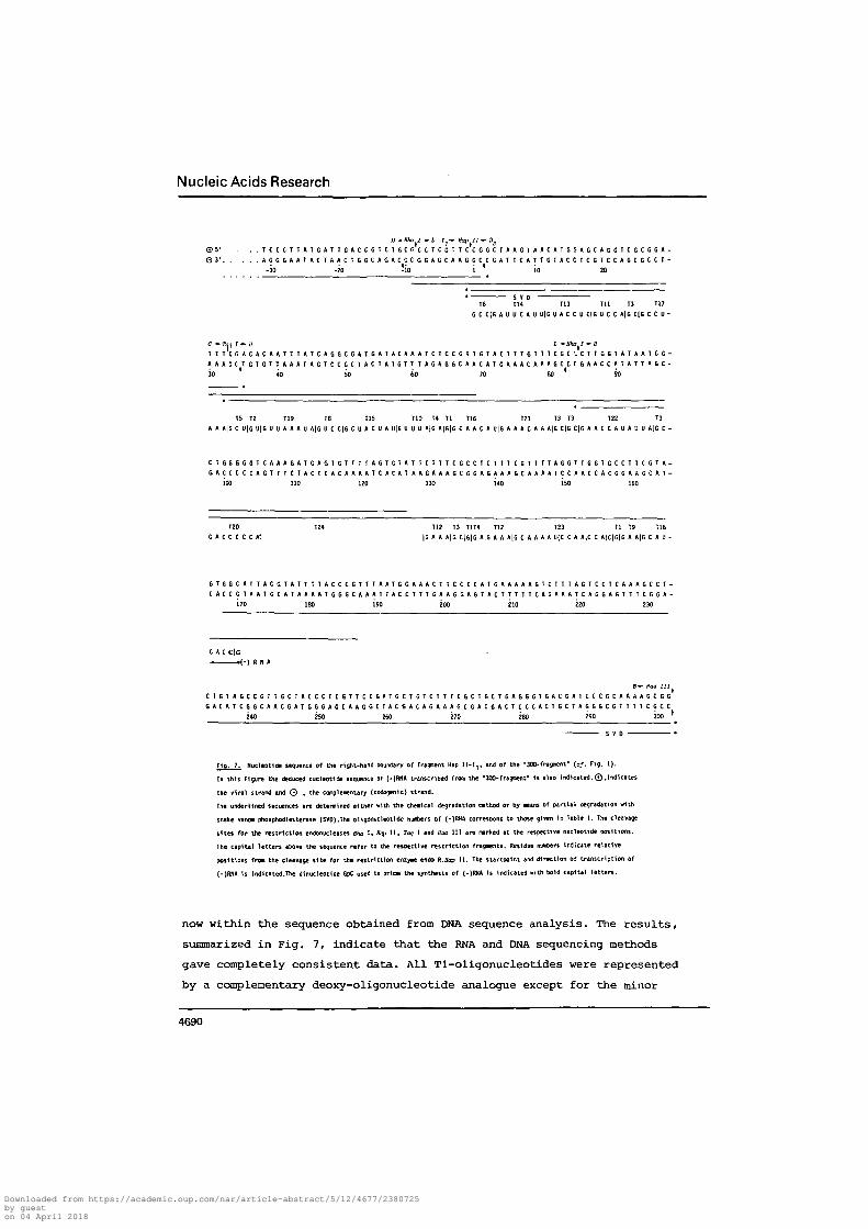
autoradiographs are presented in Fig. 7. As expected, the sequences reveal-

ed a considerable overlap with each other resulting in a complete unique

sequence of about 330 base pairs. They also confirmed the sequence pre-

dicted from the known specificity of the restriction enzyme cleavage sites

and the sequences at the 5'-end of the 300-fragment as deduced from partial

hydrolysis with pancreatic DNase and snake venom phosphodiesterase.

Given the catalogue of all Tl-RNase products obtained from the RNA

sequence analysis of (-)RNA, as presented in Table 1, they can be ordered

4689

Downloaded from https://academic.oup.com/nar/article-abstract/5/12/4677/2380725by gueston 04 April 2018

Nucleic Acids Research

© 5 ' . . . T C C C T T A T G A T T G A C C G T C i G C G C C T C G T T C C G G C T A A G T A A C A T G G A G C A G G T C G C G G A -

© 3 ' A G G G A A T A C T A A C T G G C A G A C C C G G A G C A A G G C C G A T T C A T T G T A C C T C G T C C A G C G C C T -

•30 -20 -io i io 20

S V DT6 T14 T13 T U T3 T17

G C C | G A U U C A U U | G U A C C U C | G U C C A | G C l G C C U -

C *Tta I *• tt L —Hha I * H

T T T C G A C A C A A T T T A T C A G G C G A T G A T A C A A A T C T C C G T T G T A C T T T G T T T C G C ' - C T T G G T A T A A T C G -

A A A G C T G T G T T A A A T A G T C C G C T A C T A T G T T T A G A G G C A A C A T G A A A C A A A G C C C G A A C C A T A T T A G C -

30 40 50 60 70 60 90

T5 T2 T19 T8 T15 T10 T4 Tl T16 T21 T3 T3 T2Z T3

A A A|G C U|G U(G U U A A A U A|G U C C|G C U A C U A U|G U U U A|G A|G|G C A A C A U|G A A A C A A A|G C|G C|G A A C C A U A U U A|G C -

C T G G G G G T C A A A G A T G A G T G T T T T A G T G T A T T C T T T C G C C T C T T T C G T T T T A 6 G T T G G T G C C T T C G T A -

G A C C C C C A G T T T C T A C T C A C A A A A T C A C A T A A G A A A G C G G A G A A A G C A A A A T C C A A C C A C G G A A G C A T -

100 HO 120 130 i*0 150 160

T20 TZ4 T12 T3 TIT* T12 T23 Tl T9 T18

G A C C C C C K\ |G A A A|G C|G|G A|G A A A|G C A A A A U(C C A A,C C A)C|G[G A A|G C A U -

G T G G C A T T A C G T A T T T T A C C C G T T T A A T G G A A A C T T C C T C A T G A A A A A G T C T T T A G T C C T C A A A G C C T -

C A C C G T A A T G C A T A A A A T G G G C A A A T T A C C T T T G A A G G A G T A C T T T T T C A G A A A T C A G G A G T T T C G G A -

170 ISO 190 ZOO 210 220 230

C A C C|G- .<-) R N A

B * Haa III

C T G T A G C C G T T G C T A C C C T C G T T C C G A T G C T G T C T T T C G C T G C T G A G G G T G A C G A T C C C G C A A A A G C G G

G A C A T C G G C A A C G A T G G G A G C A A G G C T A C G A C A G A A A G C G A C G A C T C C C A C T G C T A G G G C G T T T T C G C Cflin 9tn flCft "nn ^on ?Qfi i rv l *

Fig. 7. Nucleotide sequence of the right-hand boundary of freojwnt Hap 11-Ij, and of the -300-frig^enf {of. Fig. 1).

In this Figure the deduced nucleotide sequence of {-)RNA transcribed from the "300-fragment" Is ilso Indicated.©.indicates

the viral strand and 0 . the complementary (codogenic) strand.

The underlined sequences are determined either with the chemical degradation method or by means of partial degradation with

snake venom phosphodiesterase (SVD).The oligonucleotide nuobers of (-)RNA correspond to those given In Table 1. The cleavage

sites for the restriction endonucleases BHa I , «ap I I , Taq I and Hoc I I I are narked at the respective nucleotide positions.

The capital letters above the sequence refer to the respective restriction fragment*. Residue iu *en indicate relative

positions from the cleavage site for the restriction enzyme endo K.Bap I I . The startpoint and direction of transcription of

{-)RNA is indicated.The dinucleotide GpC used to prime the synthesis of (-)RNA is indicated with bold capital letters.

now within the sequence obtained from DNA sequence analysis. The results,

summarized in Fig. 1, indicate that the RNA and DNA sequencing methods

gave completely consistent data. All Tl-oligonucleotides were represented

by a complementary deoxy-oligonucleotide analogue except for the minor

4690

Downloaded from https://academic.oup.com/nar/article-abstract/5/12/4677/2380725by gueston 04 April 2018

Nucleic Acids Research

products CCG(G), CAG(G) and UCG(C). Since the product CCG can only be

derived from the ffapll-end of the template and we also noted that the

other nucleotides CAG(G) and UCG(C) are just complementary to the sequence

at position 19 •* 26 of the complementary DNA strand, we assume that RNA

polymerase, when reached to the end of the template, is able to switch

transcription from one strand to the other. Our findings that these nucleo-

tides are present in less than one molar yield, strongly suggest that

reading back as well as termination of transcription after short complemen-

tary RNA chain growth does not occur at-a fixed nucleotide position but more

or less randomly. Although the mechanism involved in this reaction is quite

unknown, its occurrence has already been noted by others (23). Also the

pancreatic RNase digestion data of T24, the sequence of which was ambiguous,

were completely consistent with those predicted from the DNA sequence

deduced. We therefore feel confident to conclude that the sequence of 169

nucleotide residues representing (-)RNA is correct.

Localization of nucleotide sequence changes in amber-7 mutants

RNA sequencing techniques were used to determine exactly the positions

of the single-point mutations within the nucleotide sequence of several

amber mutants of gene VII. For this purpose, 300-fragments were isolated

from M13 RF bearing hydroxylamine-induced amber-7 mutations and the indi-

vidual 300-fragments were transcribed in the presence of the primer GpC and

each of the four (a- P)-ribonucleoside triphosphates. The resulting (-)RNA

species were isolated as described under Methods and subsequently digested

with RNase Tl. A Tl-RNase fingerprint of (a~ p)UTP-labelled (-)RNA tran-

scribed from a 300-fragment bearing the am7-H2 mutation is shown in Fig. 5c.

32For comparison, a Tl-RNase fingerprint of (a- p)UTP-labelled wild-type

(-)RNA is also included in this figure (Fig. 5b). It is easily recognized

that aw7-H2 is a double mutant. The Tl-fingerprint of om7-H2 (-)RNA namely

contains two oligonucleotides, denoted (X) and (Y), which are not present

in the fingerprint of wild-type (-)RNA. In turn, the wild-type Tl-oligo-

nucleotides Til, T13 and T18 are missing in the amber mutant (-)RNA. All

Tl-oligonucleotides of am7-H2 (-)RNA were further analysed by pancreatic

RNase digestion and alkaline hydrolysis of the secondary digestion products.

It turned out that all Tl-oligonucleotides from the amber mutant (-)RNA

were completely identical to those derived from wild-type (-)RNA (data not

shown), except the oligonucleotides (X) and (Y). The nucleotide sequences

of both Tl-products, as deduced from the standard RNA sequencing analysis

4691

Downloaded from https://academic.oup.com/nar/article-abstract/5/12/4677/2380725by gueston 04 April 2018

Nucleic Acids Research

are given in Table 1.

Given these nucleotide sequences and emphasizing that T13 is adjacent

to Til in the wild-type (-)RNA [of. Fig. 7) one has to conclude that due

to G •* A transition in Til a fusion product ACCUTlCUCCAUG is generated.

A transition of G + ft in (-)RNA, as a consequence of a C + T transition

in the viral DNA strand is in accordance with the specificity of hydroxyl-

amine-induced mutagenesis.

In analogy, the deduced sequence of oligonucleotide (X) suggests this

sequence to be generated by a fusion of T9 and T18. The fusion is caused

then as the result of a G •+ A transition in T18. Oligonucleotide T9, being32

AAG(G), will not be labelled by transcription with (a- P)-UTP and its

disappearance, therefore, cannot be detected in the fingerprint shown in

Fig. 5b.

Using the same approach, nucleotide sequence analyses have also32

been carried out with (a- P)UTP-labelled (-)RNA derived from a second

amber-7 mutant, namely am7-H3. The Tl-RNase fingerprints of this am7-H3

(-)RNA is shown in Fig. 5d. It is clear that in this case the wild-type

oligonucleotides T8 and T19 are missing in the fingerprint of the am7-H3

mutant (-)RNA, whereas the relative intensity of T22 in the fingerprint of

the mutant (-)RNA has markedly increased. Further analysis by pancreatic

RNase digestion and alkaline hydrolysis have shown that the actual differ-

ences in both types of RNA are restricted to these oligonucleotides only.

The pancreatic RNase digestion products of the T22-analogue present in the

COT7-H3 (-)RNA are shown in Table 1. This spot is actually composed of two

oligonucleotides: the original oligonucleotide T22 and a new product

CCUAAUAAAUUG(U). If it is emphasized that the sequence of T8 is CCUG(A) and

of T19 is AUAAAUUG(U) and that both Tl-products are contiguous , the new

product then must be the fusion product of wild-type T8 and T19. This

fusion is generated by a G -* A transition in T8 which is the result of a

C -*• T transition in the phage am7-H3 viral DNA strand.

Nucleotide sequence of gene VII

Since phage Ml3 messenger RNAs are solely transcribed from the non-

viral strand of the DNA template, the DNA region analysed has been written

for convenience in its codogenic RNA sequence. This is presented in Fig. 8.

Also the nucleotide changes which are found in the amber mutants <2m7-H2

and aw7-H3 are included in this figure. Both single-point mutations have

changed a glutamine codon (CAG) into a nonsense codon (UAG). Both mutated

4692

Downloaded from https://academic.oup.com/nar/article-abstract/5/12/4677/2380725by gueston 04 April 2018

Nucleic Acids Research

u. , i

5 ' . . U C C C U U A U G A U U G A C C G U C U G C G C C U C G U U C C G G C U A A G U A A C A U G G A G C A G G U C G C G G A U -

- 3 0 - 2 0 - 1 0 ! 10 2 0 3 0

Ctr Lci< 'tzz Zlc Aep Arg Lc^ Arj Leu Val Pro Ala Lya Fret Glu Gin Val Ala Acp

U U C G A C A C A A U U U A U C A G G C G A U G A U A C A A A U C U C C G U U G U A C U U U G U U U C G C G C U U G G U A U A A U C -

40 50 60 70 80 90

The Asp Thr lie ?yr Gin Ala He: He Gin lie Ser Val Val Leu Cyg Phe Ala Leu Gly lie He

— CCnc VII protein

• G -RNA

G C U G G G G G U C A A A G A l l G * G U G U U U U A G U G U A U U C U U U C G C C U C U U U C G U U U U A G G U U G G U G C C U U -

100 110 120 130 140 150 160

Ala Cly CIj, Cln Arg

Ser Val Lou Val Tyr Ser Phe Ala Set- Phe Val Leu Gly 7Vp Cys Leu

gene IX protein .

tC G U A G U G G C A U U A C G U A U U U U A C C C G U U U A A U G G A A A C U U C C U C A U G A A A A A G U C U U U A G U C C U C -

170 i80 190 200 210 220

Fmet Lya Lya Ser Leu Vat Leu

Arg Sar Gly lie Thr Tyr Pha Thr Arg Leu Met Glu Tkr Ser Ser

A A A G C C U C U G U A G C C G U U G C U A C C C U C G U U C C G A U G C U G U C U U U C G C U G C U G A G G G U G A C G A U C C C -

230 2flO 250 260 270 . 280 290

Lye Ala Ser Val Ala Val Ala Thr Leu Val Pro f!et Leu Ser Phe Ala Ala Glu Gly Asp Asp Pro

—— gene VIII protein —

G C A A A A G C G G

300

Ala lya Ala

f ' g . 8. HucieotliJe sequence of ;he RHA complementary to the established nucleotide sequence of the codogenic strand

p "eseritco in r i ^ /» in tnis rin*ure the anino acid sec uences of (dJ ^ene VII proteint (DJ the C™tenoina• part of 9ene V protein

the hypothetical gene IX protein, and of (d) the H-terninal part of the pre-coat protein encoded by gene VIII are indicated

The vertical bar refers to the position where this pre-coat protein is cleaved during processing into the nature coat protein.

Init iat ion codons for translation are overlined and termination codons are underlined. The transitions of the am7-H2 and OTI7-H3

mutations are marked above the positions 19,46 and 162 respectively (cf. Table 1).

The S'-end of the RNA that codes for the pre-coat protein (G-RNA) is as Indicated {cf. Pig. 3}.

The nucleotides present within the ribqsome binding site of gene VII and which are conplementary to the J'-terninus of 16S rRNA (29)

are indicated with an asterisk.

codons are in phase to each other. Given these data, the coding region of

(-)RNA and, hence of gene VII can be deduced now. As shown in Fig. 8, the

initiation triplet AUG is at the 2nd triplet-frame upstream the mutated CAG

codon in am7~H2, whereas a termination codon (UGA) is located 22 triplets

downstream the mutated codon of am7-H3 and a second termination codon (UAG)

4693

Downloaded from https://academic.oup.com/nar/article-abstract/5/12/4677/2380725by gueston 04 April 2018

Nucleic Acids Research

3 triplets more downstream the first termination codon. It is worth

mentioning that in the am7-H2 mutant in fact two point mutations have been

introduced. One C -*• U substitution in the CAG codon of gene VII and a

second C •+• u substitution far more downstream and outside gene VII. Its

position was found at 48 nucleotides downstream the UGA termination codon

of this gene and within a region which has a hypothetical coding function

(see discussion). Our inference that the AUG triplet mentioned is the

initiation codon is based upon the observation that an in phase nonsense

triplet UGA is present at 36 nucleotides upstream the initiation codon and

that no other AUG or GUG codons are contained in this particular part of

the sequence. However, a translational start of gene VII might be well

further upstream if one considers the UGA codon at position -24 as a leaky

terminator (of. 29). Which of these two possibilities have to be favoured

must await ribosomal binding studies and/or isolation and sequence deter-

mination of the gene Vll-protein (see discussion).

From these sequence data it is further concluded that gene VII is

separated from its next gene, i.e. gene VIII, by a region of 94 nucleotides.

This region most probably has a coding function since it contains a purine-

rich sequence with features of a ribosome binding site (24) and the

appropriate codons for the initiation and termination of protein synthesis.

The initiation codon (AUG) of this hypothetical gene forms an integral part

of the termination signal of gene VII whereas its termination codon (UGA)

overlaps with the initiation codon of gene VIII.

DISCUSSION

As a part of our studies on the regulation of transcription and trans-

lation of the small circular Ml3 genome we have determined the nucleotide

sequence of gene VII. This has been achieved by a combination of RNA and

DNA sequencing techniques. Although each approach generated certain ambi-

guities, in conjunction these methods gave completely consistent data and

allowed deduction of an unambiguous sequence of about 330 nucleotides

encompassing the complete gene VII, a still hypothetical gene, designated

gene IX, and parts of the neighbouring genes V and VIII. It is shown that

gene VII is only 99 nucleotides long and that this gene is immediately

followed by gene IX which, in turn, partially overlaps with gene VII on one

side and with gene VIII on the other.

The complete nucleotide sequence of phage fd has recently been

established (25). A comparison of this sequence with the one reported in

4694

Downloaded from https://academic.oup.com/nar/article-abstract/5/12/4677/2380725by gueston 04 April 2018

Nucleic Acids Research

this study for Ml 3 shows that the sequences of the corresponding genes VII

and IX are completely identical. This is quite different for several other

genes of phage Ml3 and fd, the tentative sequences of which revealed several

second-base substitutions and approximately 2-3% base-changes in the third

position of the triplets. Hence, the gene VII and gene IX sequences in both

filamentous phages are rather conserved.

As already pointed out in the Introduction, no clear explanation can

be given yet for the undetectable level of gene Vll-protein synthesis

in vivo and in vitro. The low synthesis of this protein is not caused by a

very low frequency of transcription of this gene. It has clearly been shown

that transcription of the Ml3 genome occurs in a cascade-like fashion (8,20),

which means that the amounts of the individual gene transcripts increase

proportionally in the direction of (gene) II-V-VII-VIII with a maximal level

of transcription at gene VIII. This suggests that the expression of gene

VII is mainly controlled at the level of translation. A possible reason for

the low synthesis of its product might be that gene VII has a low capacity

of ribosome recognition. All potential ribosome binding sites of the genes

of fl, fd and M13 have now been sequenced (25,26; van Wezenbeek et al. ,

unpublished results). They all show the potential to form Watson-Crick base

pairs with the 3'-terminal end of 16S ribosomal RNA. From the sequence

reported here it is clear that only the first, third, tenth and eleventh

base upstream the initiation codon of gene VII are complementary to the

3'-end of 16s RNA (of. Fig. 8). This means that gene VII has the lowest

potential of Shine-Dalgarno base pairing among all filamentous phage genes.

If there exists a correlation between the number of potential Shine -Dalgarno

base pairs and the efficiency of ribosome binding (24,27), the low level of

gene Vll-protein synthesis could be explained by its low potential of base

pairing with 16S RNA. That complementarity to 16S RNA, however, is not sole-

ly determining for ribosome binding has recently been evidenced by Taniguchi

and weissmann (28) . They showed that also the interaction of the ribosome

binding site with fMet-tRNA plays an essential role in the formation of the

70S initiation complex. Ribosome binding was substantially enhanced in case

the first base following the AUG initiation triplet was mutated from a G

into an A residue. From our sequence studies we know that all ribosome

binding sites in Ml3 DNA have an A residue following their initiation

triplet. Interestingly, the exceptions are gene VII (AUGG), gene VI (AUGC)

and gene" I (AUGG) of which it is known that protein synthesis under the

direction of these genes is extremely low (3-5).

4695

Downloaded from https://academic.oup.com/nar/article-abstract/5/12/4677/2380725by gueston 04 April 2018

Nucleic Acids Research

As already pointed out in the Results section, the predicted transla-

tional start of gene VII might be well further upstream if the UGA codon at

position -24 is considered as a leaky terminator. In the nucleotide sequence

of gene V of phage M13 and fd (25, Hulsebos and Schoenmakers, unpublished

data) an AUG codon is present, which is preceded by a potential Shine-

Dalgarno sequence, and which is in phase with the predicted initiation

codon but not in phase with the gene V reading frame. If translation starts

at this position (position -87), an overlap of the N-terminal region of

gene VII distal to the leaky UGA terminator with the C-terminal end of gene

V being an essential part for its function cannot be excluded. Furthermore,

it seems likely that translation of gene v, the product of which is made in

large amounts, would obstruct binding of ribosomes at the gene VII initia-

tion site. This, together with a read-through at the UGA signal, might not

only provide new insights in the polarity among genes V and VII but might

also be a convenient control mechanism if gene VII protein is only required

in small amounts.

From their nucleotide sequence studies Schaller and Takanami (25) were

first to postulate that between genes VII and VIII a hitherto unidentified

gene, i.e. gene IX, might be located. If true, this gene should code for a

polypeptide which is 32 amino acids lonJ and which does not contain the

amino acid residues His, Fro, Lys and Asp. This property has enabled us

to find out whether such a polypeptide exists among the products present

in the M13-infected cells. Recently we have discovered that within the M13

virion a third capsid protein is present with a molecular weight of about

3300 (G. Simons, R. Konings and J. Schoenmakers, in preparation). The

presence of this polypeptide, designated "C-protein", could not be detected

in phage particles which were labelled with either His, Pro and Lys.

Interestingly, a polypeptide with exactly identical properties has also

been observed in minicells harbouring M13 RF (9; Simons et al. unpublished

data). The latter polypeptide was absent, however, in minicells harbouring

RF which carry a certain amber mutant in gene VII. Studies are in progress

to substantiate our findings that C-protein originates from this hypo-

thetical gene and that its synthesis is regulated by its proximal genes.

ACKNOWLEDGEMENTS

We thank Mrs. Joke de Witte for performing the SVD-digests analyses.

Dr. Rob Schilperoort for providing us with RNA polymerase and Drs. Ruud

Konings and Peter van Wezenbeek for critical comments and support. We also

4696

Downloaded from https://academic.oup.com/nar/article-abstract/5/12/4677/2380725by gueston 04 April 2018

Nucleic Acids Research

thank Dr. Heinz Schaller for communicating the sequence of phage fd DNA.

This research was supported by a grant from the Netherlands Foundation for

Chemical Research (SON) with financial aid from The Netherlands Organization

of Pure Research (ZWO).

REFERENCES

1. Denhardt, D.T. (1975) In: CRC Critical Reviews in Microbiology, TheChemical Rubber Company, Cleveland, Ohio, 161-223.

2. Ray, D.S. (1977) In: Comprehensive Virology, Fraenkel-Conrat, H. andWagner, R.R., Eds. Plenum Publishing Corporation, New York, 105-178.

3. Konings, R.N.H., Hulsebos, T. and van den Hondel, C.A. (1975) VirologyIS, 570-584.

4. Model, P. and Zinder, N.D. (1974) J. Mol. Biol. 83, 231-251.5. Hondel van den, C.A.M., Konings, R.N.H. and Schoenmakers, J.G.G. (1975)

Virology 67, 487-497.6. Edens, L., Konings, R.N.H. and Schoenmakers, J.G.G. (1978) J. Virol,

(in press).7. Henry, T.J. and Pratt, D. (1969) Proc. Nat. Acad. Sci. U.S.A. S2>

800-807.8. Edens, L., Konings, R.N.H. and Schoenmakers, J.G.G. (1978) Virology

86, 354-368.9. Smits, M.A., Simons, G., Konings, R.N.H. and Schoenmakers, J.G.G.

(1978) Biochim. Biophys. Acta, in press.10. Edens, L., van Wezenbeek, p., Konings, R.N.H. and Schoenmakers, J.G.G.

(1975) Eur. J. Biochem. 70, 577-587.11. Edens, L., Konings, R.N.H. and Schoenmakers, J.G.G. (1975) Nucleic

Acids Res. 2, 1811-1820.12. Hondel van den, C.A. and Schoenmakers, J.G.G. (1976) J. Virol. 13,

1024-1039.13. Hondel, van den, C.A. and Schoenmakers, J.G.G. (1976)

Eur. J. Biochera. 68, 55-70.14. Hondel van den, C.A., Weijers, A., Konings, R.N.H. and Schoenmakers,

J.G.G. (1975) Eur. J. Biochem. 53, 559-567.15. Brownlee, G.G. and Sanger, F. (1969) Eur. J. Biochem. H, 395-399.16. Barrell, B.G. (1971) In: Procedures in Nucleic Acid Research, Cantoni,

C.L. and Davies, D.R., Eds. Harper and Row, New York, Vol. 2, 751-795.17. Maxara, A.M. and Gilbert, W. (1977) Proc. Nat. Acad. Sci. U.S.A. 74,

560-564.18. Jay, E., Bambara, R., Padmanabhan, R. and Wu, R. (1974) Nucleic Acids

Res. 1_, 331-35319. Maniatis, T., Jeffrey, A. and Kleid, D.G. (1975) Proc. Nat. Acad. Sci.

U.S.A. 72, 1184-1188.20. Konings, R.N.H. and Schoenmakers, J.G.G. (1978) In: Single-Stranded

DNA Phages, Denhardt, D.T., Ray, D.T. and Dressier, D., Eds. Cold21. Spring Harbor Laboratory, in press.

Cuypers, T., van der Ouderaa, F.J. and de Jong, W. (1974) Biochem.Biophys. Res. Commun. 59, 557-563.

22. Asbeck, F., Beyreuther, K., Kohler, H., von Wettstein, G. and Braunitzer,G. (1969) Hoppe Seyler's Z. Physiol. Chem. 350, 1047-1066.

23. Sugimoto, K., Sugisaki, H., Okamoto, T. and Takanami, M. (1977) J. Mol.Biol. Ill, 487-507.

24. Shine, J. and Dalgarno, L. (1974) Proc. Nat. Acad. Sci. U.S.A. 71,1342-1346.

4697

Downloaded from https://academic.oup.com/nar/article-abstract/5/12/4677/2380725by gueston 04 April 2018

Nucleic Acids Research

25. Schaller, H. and Takanami, M. (1978) In: Single-Stranded DNA Phages,Denhardt, D.T., Ray, D.T. and Dressier, D., Eds. Cold Spring HarborLaboratory, in press.

26. Pieczenik, G., Model, P. and Robertson, H.D. (1974) J. Mol. 90,191-214.

27. Steitz, J.A. and Jakes, K. (1975) Proc. Nat. Acad. Sci. U.S.A. 72,4734-4738.

28. Taniguchi, T. and Weissmann, C. (1978) J. Mol. Biol. 118, 533-565.29. Yates, J.L., Gette, W.R., Furth, M.E. and Nomura, M. (1977) Proc. Nat.

Acad. Sci. U.S.A. 74, 689-693.

4698

Downloaded from https://academic.oup.com/nar/article-abstract/5/12/4677/2380725by gueston 04 April 2018


Recommended