NucliSens HIV-1 QT Summary of Safety and Effectiveness 1 I. General Information Device Generic Name: In vitro nucleic acid amplification test for the quantitation of Human Immunodeficiency Virus Type 1 (HIV -1) RNA in human plasma Device Tradename: NucliSens HIV-1 QT Applicant’s Name and Address : bioMérieux, Inc. 100 Akzo Avenue Durham, North Carolina 27712 Premarket Approval Application (PMA) Number: BP010001 II. Intended use The NucliSens HIV-1 QT is an in vitro nucleic acid amplification test for the quantitation of Human Immunodeficiency Virus Type 1 (HIV-1) RNA in human plasma. The test can quantitate HIV-1 RNA over the range of 176 to 3.47X10 6 copies/mL. The test is intended for use in conjunction with clinical presentation and other laboratory markers of disease progression for prognostic assessment of HIV-1 infected patients, and for monitoring the effects of anti-retroviral therapy by serial measurements of plasma HIV-1 RNA for pediatric and adult patients with baseline viral loads greater than 93,000 and 28,000 copies of HIV-1 viral RNA/mL respectively. The NucliSens HIV-1 QT assay is not intended to be used as a screening test for HIV-1 nor is it to be used as a diagnostic test to confirm the presence of HIV-1 infection. III. Device Description (a) Test Principles The NucliSens HIV-1 QT assay comprises five separate stages as described below. Nucleic acid release The sample is added to NucliSens Lysis Buffer containing guanidine thiocyanate and Triton X- 100, which causes the lysis of viral particles and cells in the sample and the inactivation of RNases and DNases. Nucleic acid is thus released. Nucleic Acid Isolation Three synthetic RNAs (Qa, Qb, Qc) of known high, medium and low concentration, respectively, are added to the Lysis Buffer containing the released nucleic acid. These RNAs serve as internal calibrators, each differing from the HIV-1 wild-type (WT) RNA by only a short length of sequence. Under high salt conditions, all nucleic acid in the buffer, including the calibrators, binds to silicon dioxide particles. (3) These particles, acting as the solid phase, are washed several times. Finally, the nucleic acid is eluted from the solid phase.
Transcript
NucliSens HIV-1 QT Summary of Safety and Effectiveness
1
I. General Information
Device Generic Name: In vitro nucleic acid amplification test for the quantitation of Human Immunodeficiency Virus Type 1 (HIV -1) RNA in human plasma
Device Tradename: NucliSens HIV-1 QT Applicant’s Name and Address: bioMérieux, Inc.
The NucliSens HIV-1 QT is an in vitro nucleic acid amplification test for the quantitation of Human Immunodeficiency Virus Type 1 (HIV-1) RNA in human plasma. The test can quantitate HIV-1 RNA over the range of 176 to 3.47X10⁶ copies/mL. The test is intended for use in conjunction with clinical presentation and other laboratory markers of disease progression for prognostic assessment of HIV-1 infected patients, and for monitoring the effects of anti-retroviral therapy by serial measurements of plasma HIV-1 RNA for pediatric and adult patients with baseline viral loads greater than 93,000 and 28,000 copies of HIV-1 viral RNA/mL respectively. The NucliSens HIV-1 QT assay is not intended to be used as a screening test for HIV-1 nor is it to be used as a diagnostic test to confirm the presence of HIV -1 infection.
III. Device Description (a) Test Principles The NucliSens HIV-1 QT assay comprises five separate stages as described below.
Nucleic acid release The sample is added to NucliSens Lysis Buffer containing guanidine thiocyanate and Triton X-100, which causes the lysis of viral particles and cells in the sample and the inactivation of RNases and DNases. Nucleic acid is thus released. Nucleic Acid Isolation Three synthetic RNAs (Qa, Qb, Qc) of known high, medium and low concentration, respectively, are added to the Lysis Buffer containing the released nucleic acid. These RNAs serve as internal calibrators, each differing from the HIV -1 wild-type (WT) RNA by only a short length of sequence. Under high salt conditions, all nucleic acid in the buffer, including the calibrators, binds to silicon dioxide particles.(3) These particles, acting as the solid phase, are washed several times. Finally, the nucleic acid is eluted from the solid phase.
NucliSens HIV-1 QT Summary of Safety and Effectiveness
2
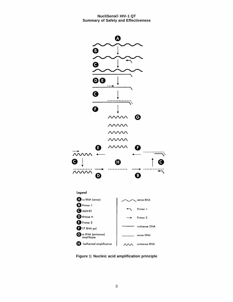
Nucleic Acid Amplification (see Figure 1 for illustration) WT HIV-1 RNA present in the eluted nucleic acid is co-amplified along with the three internal calibrators. As illustrated in figure 1, amplification is based on repeated cycles of transcription reactions. Multiple copies of each WT and calibrator RNA target sequence are synthesized by T7-RNA polymerase by means of an intermediate DNA molecule that contains a double-stranded T7-RNA polymerase promoter. Each transcribed RNA molecule enters a new amplification cycle. The DNA intermediate is generated through a process that involves the binding of a primer to the RNA template, the extension of primer by AMV-RT (Avian Myeloblastosis Virus Reverse Transcriptase) to form an RNA -DNA duplex, the degradation of the RNA strand of the duplex by RNase H, the binding of a second primer to the remaining DNA strand and, finally, the extension of the second primer to form the double-stranded T7-RNA polymerase promoter needed for transcription. Once transcription is initiated, the RNA transcripts which are ‘negatives’ of the original RNA present in the sample will be subject to the same process, only in this case extension is not restricted to the second primer, since the extension product of the first primer will also be extended. The primers (one of which contains the sequence of the T7-RNA polymerase promoter) are complementary to two different parts of the HIV-1 RNA. This pair of primers defines, and allows the amplification of, a sequence within the HIV -1 gag region. Since the Nucleic Acid Sequence-Based Amplification (NASBA) process requires no strand separation, amplification is isothermal and continuous.
NucliSens HIV-1 QT Summary of Safety and Effectiveness
3
Figure 1: Nucleic acid amplification principle
NucliSens HIV-1 QT Summary of Safety and Effectiveness
4
Nucleic Acid Detection The quantitation of HIV-1 RNA in a sample is based on the measurement of electrochemiluminescence (ECL) with the NucliSens Reader. To differentiate among the amplicons (WT, Qa, Qb and Qc), aliquots of the amplified sample are added to four hybridization solutions, each specific for one type of amplicon. Here, the respective amplicons are hybridized with a bead-oligo (i.e. an oligo bound to streptavidin coated paramagnetic beads acting as the solid phase) and a ruthenium-labeled probe. The paramagnetic beads carrying the hybridized amplicon/probe complex are captured on the surface of an electrode by means of a magnet. Voltage applied to this electrode triggers the (ECL) reaction. The light emitted by the hybridized ruthenium-labeled probes is proportional to the amount of amplicons, which in turn are proportional to the HIV -RNA in the input samples. Calculation based on the relative amounts of the four amplicons gives an estimation of the amounts of HIV-1 RNA in the sample. Quantitation of HIV-1 RNA The NucliSens HIV-1 QT system uses three internal calibrators for quantitation. These internal calibrator RNAs are produced in vitro from plasmid constructs encoding the wild-type gag region sequence. These three calibrator RNAs are identical to the wild-type sequence except for a 20 base sequence recognized specifically by the detector probe. In the case of calibrator RNAs (designated Qa, Qb, and Qc), the detector probe site was mutagenized to generate three distinct sequences, but similar in A, C, G, and T content to the wild type sequence. Thus, the calibrator RNAs are amplified with the same kinetics as the wild-type target, each reaction product is captured onto the magnetic bead with the same efficiency, and each calibrator reaction product can be distinguished from the other and from the wild-type product by the detector probes with distinct sequences. Because the A, C, G, and T content in each detector site is maintained, the melting temperature of all four probes is similar. The internal calibrators for quantitation are introduced into the specimen immediately after the initial lysis step. Importantly, the three calibrators are included at known copy numbers (Qa ~ 105, Qb ~ 104.3, Qc ~ 103.6). These calibrators are co-extracted and co-amplified with the wild-type nucleic acid in a single tube. The reaction product is then divided into four independent detection assays, each with one of the calibrator detector probes or with the wild-type detector probe. At the completion of the hybridization reactions, the four products are loaded into the NucliSens Reader. The ECL signal for each of the hybridization reactions is determined and the input copy number for wild-type HIV-1 RNA, relative to the input quantities of calibrator RNAs is determined by calculating the ratio of ECL signals for WT to Qa, Qb, and Qc. The actual computation of the input quantity of wild-type RNA is achieved through the application of a specific curve fitting program.(12) Importantly, the inclusion of the calibrators in the analysis of the independent samples permits individual determinations of the acceptance of each wild-type calculation through a validated algorithm designed to evaluate the relationship of the calibrator signals to each other. The three calibrator signals (Qa, Qb, Qc) are corrected for background noise and checked against a number of parameters: upper detection limit, fixed minimum values, variance, and correlation. If the Qc calibrator is discarded as a result of these checks, the remaining two calibrators (Qa and Qb) are used to calculate the wild-type RNA concentration.
NucliSens HIV-1 QT Summary of Safety and Effectiveness
5
(b) HIV-1 Target Description
The region of HIV-1 RNA targeted for amplification by the NucliSens HIV-1 QT is a highly conserved region of the gag gene. The oligonucleotide primers were designed to amplify a region spanning nucleotides 1358 – 1499 of HXB2, AC K03455. Primer 1 is comprised of a T7 promoter sequence and a sequence complementary to nucleotides 1471 - 1499 of HIV-1. Primer 2 is comprised of a sense sequence corresponding to nucleotides 1358 - 1386 of HIV-1.
(c) HIV-1 RNA Quantitation
The relationship between the calibrator signals and the calibration input amounts is used for the determination of the unknown amount of Wild Type (WT) RNA in a sample. Within the linear range of the assay, the ECL signal for each target or standard is proportional to the amount of HIV-1 RNA, Qa RNA, Qb RNA, or Qc RNA present in the sample tube and therefore, proportional to the total amount of individual RNA present in the original sample. This relationship is given by the following equation:
ECL signal = constant x RNA input.
Since the three calibrators are added to the sample prior to isolation, amplification, and detection the proportionality between each RNA and its corresponding signal is the same. So for each sample there are four equations:
ECL signal Qa = constant x Qa RNA input (1)
ECL signal Qb = constant x Qb RNA input (2)
ECL signal Qc = constant x Qc RNA input (3)
ECL signal WT = constant x WT RNA input (4)
As the ECL signal and the Q input amount are known for each level of the calibrators, three estimates of the constant can be determined. That is:
Constant Qa = ECL signal Qa/Qa RNA input
Constant Qb = ECL signal Qb/Qb RNA input
Constant Qc = ECL signal Qc/Qc RNA input
The average constant then is:
Constant Qa + Constant Qb + Constant Qc . 3
This average constant can then be used to estimate the WT RNA input amount by the formula:
WT RNA input = ECL signal WT/average constant.
As mentioned above, each of the calibrator signals are measured against a number of parameters. This is to ensure that a calibrator that gives an unreasonable estimate of the constant, which can skew the calculation of the initial WT RNA input amount, is not included in the calculations. If two calibrators fail to meet the parameters above, the test is considered invalid and marked as such.
(d) Primers and Probes
NASBA amplification uses two primers to amplify the viral target as well as the three internal calibrators. The P1 primer has a length of 54 nucleotides, 29 nucleotides on the 3’-end code are complementary to the HIV sequence while the other nucleotides facilitate transcription by T7 RNA polymerase. Essential is a 20 nucleotide consensus sequence (5’-TAATACGAC-
NucliSens HIV-1 QT Summary of Safety and Effectiveness
6
TCACTATAGGG) for the T7 RNA polymerase promoter. In addition, an extra sequence of 5 nucleotides (5’-AATTC) flanks the consensus sequence. This extra sequence serves as a “buffer” for the core sequence that is required for T7 RNA polymerase activity. Because of this design choice, partial length oligos n-1 through n-5 are expected to show equivalent functionality compared to the full length oligo since neither (1) the core consensus sequence for the T7 RNA polymerase nor (2) the 3’-end with the HIV specific sequence where chain elongation takes place is affected. The P2 primer has a length of 29 nucleotides, which is completely HIV specific. A typical minimal oligo length needed for sufficient hybridization efficiency and to ensure sequence specificity is 20 nucleotides. Partial length oligos lack nucleotides on the 5’-end since DNA synthesis takes place in the 3’ to 5’ direction. This means that hybridization characteristics on the 3’-end of the oligo are not affected which is important since chain elongation during the amplification reaction takes place in the 5’ to 3’ direction. Therefore, it is expected that partial length oligos missing up to approximately 5 nucleotides will show equivalent performance as the full-length oligo. The generic capture probe is an oligonucleotide comprised of a sense sequence that corresponds to nucleotides 1388 – 1411 of the HIV -1 RNA gag sequence. Four separate detection probes are used to detect the amount of amplified WT RNA and each of the calibrator RNAs present in the hybridized mixture. The detection probe for the WT is an oligonucleotide comprised of a sense sequence that corresponds to nucleotides 1419 – 1442 of the HIV -1 sequence. In order to have a unique detection probe for each of the calibrators a unique replacement is made in this position of the sequence for each calibrator RNA. Each replacement consists of a randomized sequence of the same length as present in the WT RNA sequence (24 nucleotides). Each sequence contains one less ‘G’ and one more ‘A’ than the WT sequence. The number of ‘T’ and ‘C’ bases is the same in all detection probes. Therefore, the efficiency of amplification for each of the calibrators is the same as that of the WT target.
(e) Quality Control
The integrity of each individual result can be monitored by reference to the performance of the three internal calibrators. However, it is recommended that a High Positive Control, Low Positive Cont rol, and a Negative Control be included with the first run of each kit lot to verify product performance. Furthermore, the inclusion of a Low Positive and a Negative Control with each subsequent run of the same kit lot is also recommended. Refer to the control manufacturer’s package insert for instructions. As with any new laboratory procedure, new operators should consider the use of additional controls to establish a high degree of confidence in performing the assay. If using the NucliSens HIV-1 RNA Controls, the expected range for the Positive Controls is stated in the package insert for the controls. The copy number per ml for each Positive Control should fall within the range indicated in the package insert. The Negative Control should give a less than lower detection limit result. If using commercially available controls or internally prepared controls, a typical acceptance range is approximately ± 3 standard deviations from the established copy level. If controls are not as expected, the run should be re-evaluated and a determination made as to whether the sample results are acceptable or should be repeated.
NucliSens HIV-1 QT Summary of Safety and Effectiveness
each tube contained in a foil pack with desiccant. Color code: yellow capped tube
Enzymes 5 tubes 6.5 mg Lyophilized sphere containing AMV-RT, RNase H, T7-RNA polymerase and BSA, each tube contained in a foil pack with desiccant. Color code: red capped tube
Enzyme Diluent
5 tubes 0.5 mL TRIS/HCl. Color code: red capped tube
Primers 5 tubes 10 mg Lyophilized sphere with synthetic primers, nucleotides, and stabilizers; each tube contained in a foil pack with silica gel desiccant. Color code: blue capped tube
Primer Diluent
5 vials 0.5 mL TRIS/HCl, 30% DMSO. Color code: blue capped tube
NucliSens HIV-1 QT Summary of Safety and Effectiveness
8
Nucleic Acid Detection
Components in each Detection Reagent Module
Component Size Volume Composition Bead-Oligo 2 tubes 1.68 mL DNA oligo bound to streptavidin-coated
paramagnetic beads with preservative. Color code: pink capped tube
WT probe 1 vial 0.84 mL Ruthenium-labeled DNA oligo with preservative. Color code: white capped tube
Qa probe 1 vial 0.84 mL Ruthenium-labeled DNA oligo with preservative. Color code: red capped tube
Qb probe 1 vial 0.84 mL Ruthenium-labeled DNA oligo with preservative. Color code: yellow capped tube
Qc probe 1 vial 0.84 mL Ruthenium-labeled DNA oligo with preservative. Color code: blue capped tube
Detection Diluent
2 vials 15 mL TRIS/HCl with preservative.
Instrument Reference Solution
1 vial 1.7 mL Streptavidin-coated paramagnetic beads.
IV. Warnings and Limitations
The NucliSens HIV-1 QT is an in vitro nucleic acid amplification test for the quantitation of
The NucliSens HIV-1 QT is an in vitro nucleic acid amplification test for the quantitation of Human Immunodeficiency Virus Type 1 (HIV-1) RNA in human plasma. The test can quantitate HIV-1 RNA over the range of 176 to 3.47X10⁶ copies/mL. The NucliSens HIV-1 QT assay can accurately detect a 0.3 log 10 (2-fold) or greater change in HIV -1 RNA for patients whose viral load is between 3500 copies/mL and 3,500,000 copies/mL. The NucliSens HIV-1 QT can accurately detect a 0.9 log 10 (9-fold) change in HIV-1 RNA for patients whose viral load is between 180 and 850 copies/ml. Use of this product should be limited to personnel who have been trained in all aspects and techniques of the NucliSens HIV-1 QT assay. Attention should be paid to adequate specimen collection, transporting, storage and processing procedures. Good laboratory techniques should be used in all procedures to ensure proper performance of this assay. Because of the high analytical sensitivity of this assay and the need to avoid all possible sources of contamination,
• Monitoring the effects of anti-retroviral therapy by serial measurement of plasma HIV -1 RNA has only been validated for patients with baseline viral loads = 93,000 copies/mL for pediatric patients and = 28,000 copies/mL for adult patients.
• The performance of the NucliSens HIV-1 QT assay has only been adequately validated
with HIV-1 subtype B specimens.
• NucliSens HIV-1 QT assay is generally insensitive for detection of HIV -2 RNA. However, samples from individuals infected with HIV-2 may exhibit cross-reactivity in this assay.
• When testing specimens with viral load < 300 copies/mL, the user should consider the use
of well-characterized reference materials titered from 150 copies/mL to 300 copies/mL, inclusive.
NucliSens HIV-1 QT Summary of Safety and Effectiveness
9
extreme care should be taken to maintain the purity of the reagents and mixtures. All reagents should be monitored for purity. Discard any reagents that may be suspect or have exceeded their recommended shelf life. As with any diagnostic test, results from the NucliSens HIV-1 QT assay should be interpreted with consideration of all clinical and laboratory findings. A negative test result does not exclude the possibility of exposure to, or infection with, HIV-1. The assay must be performed in strict accordance with the instructions in this package insert (especially those regarding contamination risks) and the NucliSens Reader Operator Manual to obtain accurate, reproducible results. Plasma should be used for determination of HIV-1 RNA levels. EDTA, citrate, or heparin may be used as anticoagulants. In addition to quantitative HIV -1 RNA load, other virological or immunopathological factors may contribute to variable rates of CD4+ T-cell counts and clinical outcome of the disease.(8)
V. Alternative Practices and Procedures
There are various types of direct and indirect methods for detection and quantitation of human immunodeficiency virus in clinical samples. These methods allow the measurement of disease progression as a result of HIV infection and monitoring of patients’ response to anti-retroviral therapy. Examples of these methods are listed below.
a. HIV-1 Antigen assays including p24 core Antigen and Immune Complex Dissociation p24
Antigen tests b. Nucleic acid probe technologies for direct detection and quantitation of circulating viral
particles c. Surrogate markers including CD4 and CD8 cell surface receptors d. Quantitative Cell Culture.
VI. Potential Adverse Effects of the Device on Health
Incorrect test results are possible in all in vitro diagnostic products. An erroneous low test result may lead to inappropriate treatment, or instill a false sense of security in a patient, which could lead to deterioration of the patient’s condition. The possibility of incorrect results can happen with assignable causes such as a technician’s error in following the procedures in the package insert or a device malfunction. An erroneous high test result on the other hand may contribute to unnecessary treatment or create anxiety or trauma to the patient. However, if the appropriate direction is followed as stated in the package insert, the likelihood of erroneous results are minimal from the use of this device.
The performance of the product in the clinical studies indicates that the benefit to the patient far outweighs any potential risk of adverse effect to the patient as a result of its use.
VII. Marketing History
The first generation NASBA HIV-1 RNA QT assay for quantitative determination of HIV-1 RNA “viral load” was introduced at the Biotechnology Against AIDS meeting in Florence, Italy in April of 1994. In 1997, the second generation NucliSens HIV-1 QT assay was introduced. Organon Teknika markets this assay in more than 30 countries throughout North America, South America, Europe, Africa, Asia and Australia.
Since these initial placements in 1994, the NASBA/NucliSens has been adopted by many users throughout the world. The device has also been introduced to investigators studying a wide
NucliSens HIV-1 QT Summary of Safety and Effectiveness
10
variety of parameters related to AIDS. HIV -1 RNA “viral load” is now widely accepted as an important surrogate marker in research on the pathogenesis of HIV infection and AIDS. Research studies utilizing the NASBA assay include antiviral effects on progression of disease, mother to infant transmission, early detection of transmission, relationship of plasma viral load to HIV-1 RNA quantitation in other body compartments (e.g., CSF, seminal plasma, tissues), natural history cohorts, and others.
The NucliSens HIV-1 QT assay has proven to be a valuable tool for studying the AIDS epidemic worldwide. Since introduction of the first NASBA assay and the subsequent NucliSens HIV-1 QT, the product has not been withdrawn from marketing in any country for reasons related to safety or effectiveness or for any other reasons. VIII. Summary of Studies
Potentially Interfering Substances Elevated levels of lipids, bilirubin, and hemoglobin in specimens do not interfere with the quantitation of HIV-1 RNA by the NucliSens HIV-1 QT assay. The presence of antinuclear antibodies in specimens had no detectable detrimental effect on the performance of the assay. Specimens from individuals known to be positive for rheumatoid factor, multiparous women and pregnant women showed no detectable detrimental effect on the quantitation of HIV-1 RNA. In addition, the presence of platelets in plasma did not appear to interfere with the performance of the NucliSens HIV-1 QT assay.(30)
The following compounds were found not to interfere with the quantitation of viral load by this assay: AZT, ddI, d4T, ddC, 3TC, indinavir, ritonavir, saquinavir, ganciclovir, acyclovir, zithromax-azithromycin, biaxin-clarithromycin, clofazamine, ethionamide, pentamidine, bactrin-trimethoprim sulfamethoxazole, dapsone, and diflucan. Three anticoagulants, (EDTA, citrate, and heparin) have been evaluated for use with the assay. None of them were found to exhibit any significant interference on the quantitation of HIV-1 RNA load by the NucliSens HIV-1 QT assay. Linearity and Limit of Detection/Quantitation The linear range (linearity) and limits of detection/quantitation of NucliSens HIV-1 QT was assessed with specimens derived from a well-characterized HIV -1 RNA stock. Linearity Testing of diluted specimens derived from the HIV-1 RNA stock indicated a direct proportional relationship between the dilutions tested and the number of HIV-1 RNA copies reported. The linear range of the assay was determined using the data combined from three kit lots and linear regression analysis. Based on this study, the quantitation of HIV-1 RNA using this assay was determined to be linear over a range of 51 to 5,390,000 HIV -1 RNA copies.
NucliSens HIV-1 QT Summary of Safety and Effectiveness
11
Figure 2: NucliSens HIV-1 QT Linearity
NucliSens HIV-1 QT / VQA Dilutional Panel
Clin. Batches 1, 2, and 3
Log
Ob
serv
ed
1_m
l B
as
is
1
2
3
4
5
6
7
Log Expected 1_ml Basis1 2 3 4 5 6 7
y= -0.286+1.042x and R=0.993
Limits of Detection and Quantitation. • The limit of detection (LOD) is defined as the lowest HIV-1 RNA input level where at least
95% of the tests produce a result indicative of reactivity of the input sample for HIV -1.
• The limit of quantitation (LOQ) is defined as the lowest HIV-1 RNA input level where at least 95% of the tests produce quantifiable (within linear range) results with reasonable accuracy and precision.
The limits of detection and quantitation for NucliSens HIV-1 QT were determined by testing at three sites of specimens containing HIV-1 RNA concentrations ranging from 659 to 41 copies/ml. Five specimens representing a range of HIV-1 RNA concentrations were selected and tested 72 times each using three lots of reagents. Logistic regression was used to determine the relationship between the proportion detected and the log nominal input. The limit of detection at a 95% rate (LOD) was calculated to be 176 HIV-1 RNA copies/ml. This HIV-1 RNA input level for the Limit of Detection was verified by the results of panel member testing (Table 1). Panel member 16 (nominal input = 164 HIV-1 RNA copies) gave a positivity rate of 95.8% while lower viral concentrations resulted in positivity rates of less than 95% (Table 1).
NucliSens HIV-1 QT Summary of Safety and Effectiveness
12
Table 1: Analysis of NucliSens HIV-1 QT Detection Rates
The 95% detection rate was determined by logistic regression analysis to be 176 copies/ml. The coefficients of variation (CV) for panel members 15 and 16 with input concentrations of 329 and 164 copies/ml, respectively, were 53% and 94% (Table 9-1). Based on these results the Limit of Quantitation was determined to be equal to the Limit of Detection, i.e. 176 copies/ml. Terminal Dilution of Clinical Specimens To evaluate the relative sensitivity of NucliSens HIV-1 QT, dilutions of six clinical specimens from HIV -1 infected individuals were made with human plasma negative for HIV-1. In each case, the response in copy number was a function of the dilution factor, with a diminishing HIV-1 RNA copy number as dilution factor increased. The detection of HIV-1 RNA was similar for each of the three NucliSens HIV-1 QT lots evaluated. Comparison to FDA Approved Device (Roche AMPLICOR HIV-1 MONITOR ) Dilution panel members used for the linearity study, clinical specimens, and panels provided by CBER (FDA) and Boston Biomedica (BBI panel 709)) were tested with NucliSens HIV-1 QT and the Roche AMPLICOR HIV-1 MONITOR Test, standard procedure. If available, samples with results reported as <400 copies/ml with the Standard MONITOR procedure were re-tested with the AMPLICOR UltraSensitive procedure. The results are shown in Table 2 below.
Table 2: Comparison of NucliSens HIV-1 QT to Roche AMPLICOR HIV-1 MONITOR (Standard)
Number of Specimens Reported with Copy Number Group Number NucliSens HIV-1 QT Roche AMPLICOR HIV-1
MONITOR (Standard)
VQA Dilutions
14 14 9
Clinical Specimens
76 76 63
BBI & CBER Panels
13 12 8
Totals (%) 103 102 (99%) 80 (78%)* *Three of these not reported were above the Monitor upper limit of >750,000 copies/ml
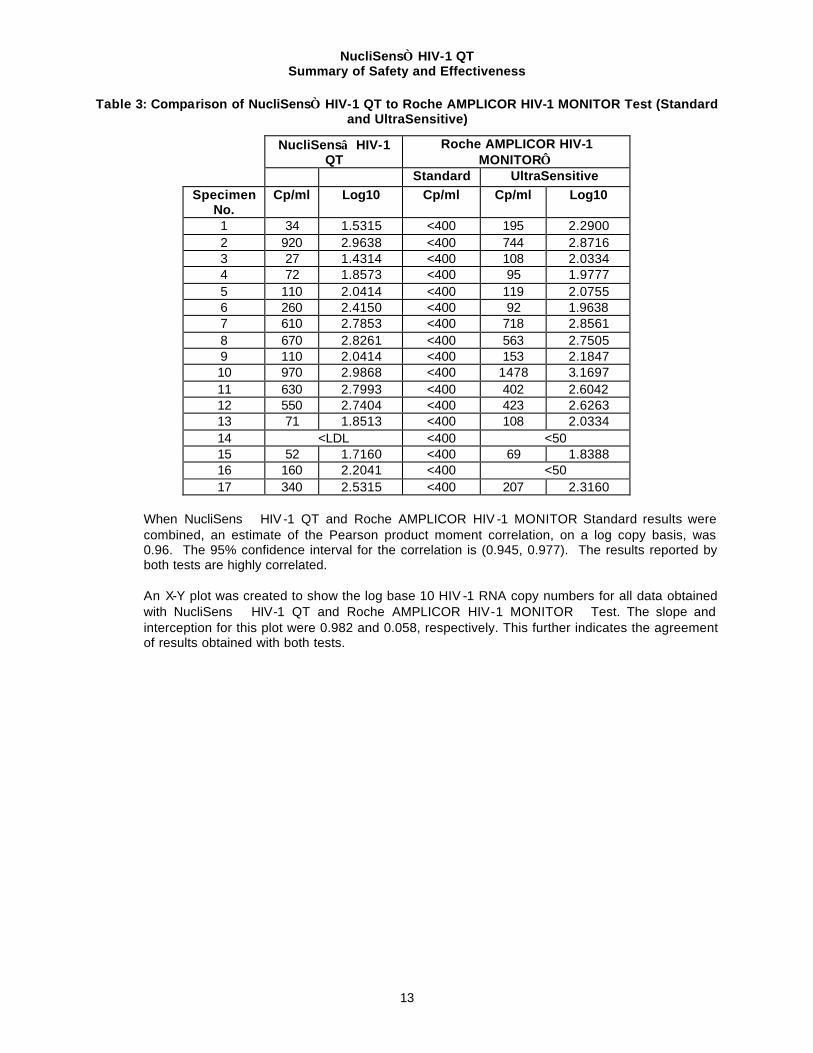
Seventeen (17) specimens that were <400 in the Standard Roche AMPLICOR HIV-1 MONITOR Test were re-tested with the UltraSensitive Roche AMPLICOR HIV -1 MONITOR procedure. Of the 17 specimens, NucliSens HIV-1 QT detected 16 (94%) and the UltraSensitive Roche AMPLICOR HIV-1 MONITOR procedure detected 15 (88%). One specimen was not detected by either test (CBER-B2). Results are presented in Table 3 below
NucliSens HIV-1 QT Summary of Safety and Effectiveness
13
Table 3: Comparison of NucliSens HIV-1 QT to Roche AMPLICOR HIV-1 MONITOR Test (Standard and UltraSensitive)
When NucliSens HIV -1 QT and Roche AMPLICOR HIV -1 MONITOR Standard results were combined, an estimate of the Pearson product moment correlation, on a log copy basis, was 0.96. The 95% confidence interval for the correlation is (0.945, 0.977). The results reported by both tests are highly correlated. An X-Y plot was created to show the log base 10 HIV -1 RNA copy numbers for all data obtained with NucliSens HIV-1 QT and Roche AMPLICOR HIV-1 MONITOR Test. The slope and interception for this plot were 0.982 and 0.058, respectively. This further indicates the agreement of results obtained with both tests.
NucliSens HIV-1 QT Summary of Safety and Effectiveness
14
Figure 3: The Log Results for the NucliSens HIV-1 QT and Roche AMPLICOR HIV-1 MONITOR
Test are Plotted Compared to the Line of Perfect Agreement
NucliSens HIV-1 QT versus Roche AMPLICOR HIV-1 MONITOR TestVQA Dilutions, Clinical Specimens, BBI and CBER panels
GROUP BBI/CBER panels UltrasensitiveVQA dilutions clinical spec
Reproducibility Reproducibility was determined by testing a panel (5 specimens) with varying levels of HIV-1 RNA with three lots at three sites by multiple technicians over four days. The variance components associated with run and replicate within run were used to estimate inter-assay variability and intra-assay variability, respectively. Inter-assay variability includes variability due to site, lot, and day. Intra-assay variability is replicate testing variability. The following table shows total inter-assay and intra-assay precision by specimen on an HIV-1 RNA copy basis.
Table 4: Per Specimen Precision Estimates - Copy Units
TOTAL INTER-ASSAY INTRA-ASSAY SID N MEAN SD CV SD CV SD CV
NucliSens HIV-1 QT Summary of Safety and Effectiveness
15
As shown in Table 4, the NucliSens HIV-1 QT was found to have relatively low variability. However, higher variability was observed for specimens with lower HIV -1 RNA concentrations. Analytical Specificity Analytical specificity was evaluated by testing a panel of specimens from individuals exhibiting the following infections or diseases, but serologically negative for HIV-1 infection. The majority of these specimens were tested with or without spiked HIV-1 RNA to assess false positive or false negative reactivity. Of the specimens tested, there was no evidence of false reactivity as shown in Table 5. The specificity was 100% across all sixteen-specimen categories listed below with an overall 95% confidence interval of 97.9% - 100%. Overall recovery of spiked HIV-1 RNA ranged from 97% to 101%.
Table 5: Possible Interfering Specimens Tested
Specimen Category
Number of Specimens
Tested
Unspiked Results1
Mean Spiked RNA Copies Recovered
Mean Recovery Relative to Expected2
HIV-2 positive 10 Not detected 24,500 97% HTLV antibody positive 10 Not detected 33,500 100% Herpes Simplex antibody positive 10 Not detected 28,100 100% Icteric 10 Not detected 25,600 98% Hemolyzed 10 Not detected 28,200 98% Antinuclear Antibody 10 Not detected 24,900 99% Lupus 10 Not detected 26,100 99% Rheumatoid factor Positive 10 Not detected 35,000 100% Specimens from Multiparous Woman 10 Not detected 26,500 98% Specimens from Pregnant Woman 10 Not detected 24,900 97% Syphilis (RPR) antibody positive 10 Not detected 51,500 Not Determined Mycobacterium tuberculosis 10 Not detected 30,600 101% Bacterially Contaminated 10 Not detected 69,300 Not Determined Antibiotic/ Anti-Viral therapy 10 Not detected 60,410 Not Determined Platelet 10 Not detected 26,400 101% Lipemic 10 Not detected 30,600 99% Summary Means 160 Not detected 34,132 99% 1 All specimens with no spiked HIV-1 virus were tested nonreactive (less than lower limit of detection). 2 Mean percent recovery was calculated by dividing the test result in log copy number with
expected log copy number of the spiked specimens.
To evaluate the performance of NucliSens HIV-1 QT assay on non-clade B HIV -1 subtypes, a panel of specimens with non-clade B subtypes obtained from BBI was tested using the NucliSens HIV -1 QT assay and Roche AMPLICOR HIV-1 MONITOR Test (Standard). The results are presented in Table 6. Except for clade C, the NucliSens HIV-1 QT assay showed improved detection efficiency as compared to the Roche AMPLICOR HIV-1 MONITOR Test. However, the difference of detection efficiency may not be statistically significant since only one specimen was tested for each subtype.
NucliSens HIV-1 QT Summary of Safety and Effectiveness
16
Table 6: NucliSens HIV-1 QT and Roche AMPLICOR HIV-1 MONITOR Test Results with BBI Panel PRD201
NucliSens Results Roche AMPLICOR HIV-1 MONITOR
Results Specimen Observed copy/ml
(log) Observed copy/ml
(log) PRD201-01 Clade A 2,400 (3.3802) Not detected PRD201-02 Clade B 140,000 (5.1461) 149,360 (5.1742) PRD201-03 Clade C 9,400 (3.9731) 23,861 (4.3776) PRD201-04 Clade D 210,000 (5.3222) 394,638 (5.5961) PRD201-05 Clade E 55,000 (4.7403) 12,387 (4.0929) PRD201-06 Clade F 130,000 (5.1139) 50,423 (4.7026) PRD201-07 Clade G 110,000 (5.0413) 8,401 (3.9243) PRD201-08 Clade H 77,000 (4.8864) 1,508 (3.1784) PRD201-09 (Diluent) <LDL Not Detected
<LDL = Less than Detectable Limit. Clinical specificity The clinical specificity of NucliSens HIV-1 QT was assessed by testing a total of 502 plasma specimens collected from a low risk whole blood donor population. These specimens were non-reactive for antibodies to HIV-1 and HIV-2 as determined with an FDA licensed assay. The specificity of NucliSens HIV-1 QT in this population was 100% (95% confidence limits of 99.27% to 100%). Clinical Performance – Patient Prognosis and Drug Monitoring Study Descriptions The use of the assay was evaluated for patient prognosis and drug monitoring in conjunction with two clinical studies: 1) ACTG (AIDS Clinical Trial Group) Study 0152(31) and 2) the Delta Trial.(32) ACTG 0152 evaluated the efficacy, safety, and long-term tolerance of zidovudine (AZT, also abbreviated as ZDV) or didanosine (ddI) as monotherapy or AZT and ddI in combination in a pediatric population. Children who were three months to eighteen years of age and who had received no or less than six weeks of previous anti-retroviral therapy and who had laboratory evidence of HIV infection were eligible for enrollment. Multiple specimens from 295 patients were tested with the assay. Two hundred eighty-seven (287) patients had reportable subsequent measurements for the calculation of proportion of progression to endpoint (Table 7). However, all 295 patients had reportable baseline HIV-1 RNA measurements prior to randomization into intent-to-treat groups (Figures 4 and 5). The Delta Trial was a multi-center study evaluating the efficacy and safety of combinations of the anti-retroviral drug AZT plus ddI or AZT plus zalcitabine (ddC) compared to AZT alone in HIV infected adults. The Delta Trial began in March, 1992 and continued through 1995. Participants in the Delta Trial included one hundred seventy-five (175) centers from Europe, Australia and New Zealand. All testing was performed at two sites on repository specimens from this study. In order to be eligible for the extended virology study, samples must have been stored frozen at –70°C or colder and must have been taken from individuals with a baseline sample and at least one later sample. Multiple samples from 1280 HIV infected participants in the Delta Trial were evaluated. Twelve hundred fifty-six (1256) had reportable subsequent measurement for the calculation of proportion of progression to endpoint (Table 8), of which eleven hundred thirty-three
NucliSens HIV-1 QT Summary of Safety and Effectiveness
17
(1133) also had reportable baseline HIV -1 RNA measurement prior to randomization into intent-to-treat groups (Figures 6 and 7). Study Objectives In each virology sub-study tied to these two investigations, the clinical utility was evaluated by 1) examining HIV-1 RNA measurement as a prognostic indicator of disease progression and 2) assessing the association, if any, of decreased HIV-1 RNA level with a change in patient therapy. Patient Prognosis To assess the association of RNA as measured by the assay with HIV-1 disease progression, a logistic function was used to model the probability of progressing to endpoint as a function of logarithm baseline HIV -1 RNA level. This model fits well, as measured by the Pearson Chi-Square Goodness-of-Fit Test for both the ACTG 152 study (p=0.4304) and Delta study (p=0.7863). For both studies, an overall association across the entire range of HIV -1 RNA concentrations was demonstrated with the test of slope coefficient (p<0.001). The estimate of slope was 0.976 for the ACTG 152 study and 0.999 for the Delta study. The estimates of slope for both studies are very similar and close to one. Using the logistic model, the odds ratio was defined and used to calculate how much difference the assay can distinguish in terms of risk of clinical endpoints. For an individual with a viral load of 10,000 copies/ml, the risk of disease progression is 1.35 times as high as for an individual with a viral load of 5,000 copies/ml. These data suggest that a higher viral load is associated with an increased risk in both studies. For each study, the distributions of actual baseline HIV -1 RNA levels for patients progressing and not progressing to endpoint were plotted. Figures 4 and 5 show the distribution of HIV-1 RNA levels for patients progressing and not progressing to endpoint, respectively, for the ACTG 152 study. Similarly, Figures 6 and 7 show the distribution of HIV-1 RNA levels for patients progressing and not progressing to endpoint, respectively, for the Delta Study.
NucliSens HIV-1 QT Summary of Safety and Effectiveness
18
Figure 4: Distribution of Baseline HIV-1 RNA Levels for Patients (N=90) Progressing to Endpoint – ACTG 152 Study
ACTG 152 Study
Distribution of Baseline HIV-1 RNA Levelsfor Patients Progressing to Endpoint
FREQ.CUM.
FREQ. PCT.CUM.PCT.
57 57 63.33 63.33
14 71 15.56 78.89
9 80 10.00 88.89
4 84 4.44 93.33
3 87 3.33 96.67
0 87 0.00 96.67
1 88 1.11 97.78
0 88 0.00 97.78
0 88 0.00 97.78
0 88 0.00 97.78
0 88 0.00 97.78
0 88 0.00 97.78
0 88 0.00 97.78
0 88 0.00 97.78
0 88 0.00 97.78
2 90 2.22 100.00
Bas
el
ine
HIV
-1 R
NA
Lev
el
>= 3.00E7
2.80E7 - 2.99E7
2.60E7 - 2.79E7
2.40E7 - 2.59E7
2.20E7 - 2.39E7
2.00E7 - 2.19E7
1.80E7 - 1.99E7
1.60E7 - 1.79E7
1.40E7 - 1.59E7
1.20E7 - 1.39E7
1.00E7 - 1.19E7
8.0E6 - 9.9E6
6.0E6 - 7.9E6
4.0E6 - 5.9E6
2.0E6 - 3.9E6
< 2.0E6
FREQUENCY0 10 20 30 40 50 60
Figure 5: Distribution of Baseline HIV-1 RNA Levels for Patients (N=205) Not Progressing to Endpoint – ACTG 152 Study
ACTG 152 Study
Distribution of Baseline HIV-1 RNA Levelsfor Patients Not Progressing to Endpoint
FREQ.CUM.
FREQ. PCT.CUM.PCT.
176 176 85.85 85.85
18 194 8.78 94.63
3 197 1.46 96.10
3 200 1.46 97.56
1 201 0.49 98.05
0 201 0.00 98.05
0 201 0.00 98.05
0 201 0.00 98.05
1 202 0.49 98.54
1 203 0.49 99.02
0 203 0.00 99.02
0 203 0.00 99.02
0 203 0.00 99.02
1 204 0.49 99.51
0 204 0.00 99.51
1 205 0.49 100.00
Ba
seli
ne
HIV
-1 R
NA
Le
vel
>= 3.00E7
2.80E7 - 2.99E7
2.60E7 - 2.79E7
2.40E7 - 2.59E7
2.20E7 - 2.39E7
2.00E7 - 2.19E7
1.80E7 - 1.99E7
1.60E7 - 1.79E7
1.40E7 - 1.59E7
1.20E7 - 1.39E7
1.00E7 - 1.19E7
8.0E6 - 9.9E6
6.0E6 - 7.9E6
4.0E6 - 5.9E6
2.0E6 - 3.9E6
< 2.0E6
FREQUENCY0 50 100 150 200
NucliSens HIV-1 QT Summary of Safety and Effectiveness
19
Figure 6: Distribution of Baseline HIV-1 RNA Levels for Patients (N=144) Progressing to Endpoint – Delta Study
Delta Study
Distribution of Baseline HIV-1 RNA Levelsfor Patients Progressing to Endpoint
FREQ.CUM.
FREQ. PCT.CUM.PCT.
61 61 42.36 42.36
35 96 24.31 66.67
16 112 11.11 77.78
14 126 9.72 87.50
6 132 4.17 91.67
2 134 1.39 93.06
3 137 2.08 95.14
2 139 1.39 96.53
1 140 0.69 97.22
1 141 0.69 97.92
0 141 0.00 97.92
0 141 0.00 97.92
0 141 0.00 97.92
0 141 0.00 97.92
1 142 0.69 98.61
2 144 1.39 100.00
Ba
sel
ine
HIV
-1 R
NA
Le
vel
>= 3.00E6
2.80E6 - 2.99E6
2.60E6 - 2.79E6
2.40E6 - 2.59E6
2.20E6 - 2.39E6
2.00E6 - 2.19E6
1.80E6 - 1.99E6
1.60E6 - 1.79E6
1.40E6 - 1.59E6
1.20E6 - 1.39E6
1.00E6 - 1.19E6
8.0E5 - 9.9E5
6.0E5 - 7.9E5
4.0E5 - 5.9E5
2.0E5 - 3.9E5
< 2.0E5
FREQUENCY0 10 20 30 40 50 60 70
Figure 7: Distribution of Baseline HIV-1 RNA Levels for Patients (N=989) Not Progressing to Endpoint – Delta Study
Delta Study
Distribution of Baseline HIV-1 RNA Levelsfor Patients Not Progressing to Endpoint
FREQ.CUM.
FREQ. PCT.CUM.PCT.
690 690 69.77 69.77
141 831 14.26 84.02
61 892 6.17 90.19
36 928 3.64 93.83
15 943 1.52 95.35
11 954 1.11 96.46
6 960 0.61 97.07
9 969 0.91 97.98
5 974 0.51 98.48
3 977 0.30 98.79
2 979 0.20 98.99
1 980 0.10 99.09
2 982 0.20 99.29
1 983 0.10 99.39
1 984 0.10 99.49
5 989 0.51 100.00
Base
lin
e H
IV-1
RN
A L
eve
l
>= 3.00E6
2.80E6 - 2.99E6
2.60E6 - 2.79E6
2.40E6 - 2.59E6
2.20E6 - 2.39E6
2.00E6 - 2.19E6
1.80E6 - 1.99E6
1.60E6 - 1.79E6
1.40E6 - 1.59E6
1.20E6 - 1.39E6
1.00E6 - 1.19E6
8.0E5 - 9.9E5
6.0E5 - 7.9E5
4.0E5 - 5.9E5
2.0E5 - 3.9E5
< 2.0E5
FREQUENCY0 200 400 600 800
NucliSens HIV-1 QT Summary of Safety and Effectiveness
20
The relationship between baseline RNA levels grouped by decile and the percent of subjects in each group progressing to endpoint in each decile was evaluated. Analyses were performed to look at differences in the probability of disease progression by sequentially testing combinations of data from different groups of deciles. With this approach, the proportion of subjects responding in decile 1 was contrasted with the proportion responding in the group formed by combining deciles 2-10. Next, the group formed by combining deciles 1 and 2 was contrasted against combined deciles 3-10, and so on. Tables 7 and 8 show decile, HIV -1 RNA copy number representation, and the numbers of individuals from the ACTG 152 and Delta Studies, respectively, progressing to endpoint and total number in each decile across all three treatment groups. The tables also show the contrast statement results of the test for differences between the proportions in sequential groupings of deciles. P-values associated with the contrasts for each study are not adjusted for multiple testing. These results show that the biggest increase in significance between successive decile groupings occurs between deciles two and three in both studies. Thus, in the ACTG 152 study, this jump occurs around 93,000 copies/mL; in the Delta around 28,000 copies/mL.
Table 7: Proportion to Endpoint by RNA Decile – ACTG 152 Study
Decile Midpoint Range Progressing N Proportion Cumulative
NucliSens HIV-1 QT Summary of Safety and Effectiveness
21
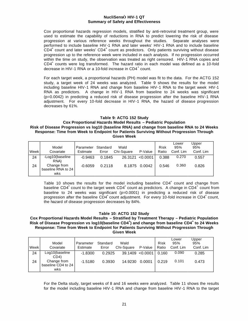
Cox proportional hazards regression models, stratified by anti-retroviral treatment group, were used to estimate the capability of reductions in RNA to predict lowering the risk of disease progression at various reference weeks throughout the studies. Separate analyses were performed to include baseline HIV -1 RNA and later weeks’ HIV-1 RNA and to include baseline CD4+ count and later weeks’ CD4+ count as predictors. Only patients surviving without disease progression up to the reference week were included in each analysis. If no progression occurred within the time on study, the observation was treated as right censored. HIV-1 RNA copies and CD4+ counts were log transformed. The hazard ratio in each model was defined as a 10-fold decrease in HIV-1 RNA or a 10-fold increase in CD4+ count. For each target week, a proportional hazards (PH) model was fit to the data. For the ACTG 152 study, a target week of 24 weeks was analyzed. Table 9 shows the results for the model including baseline HIV-1 RNA and change from baseline HIV-1 RNA to the target week HIV-1 RNA as predictors. A change in HIV -1 RNA from baseline to 24 weeks was significant (p=0.0042) in predicting a reduced risk of disease progression after the baseline HIV-1 RNA adjustment. For every 10-fold decrease in HIV-1 RNA, the hazard of disease progression decreases by 61%.
Table 9: ACTG 152 Study Cox Proportional Hazards Model Results – Pediatric Population
Risk of Disease Progression vs log10 (baseline RNA) and change from baseline RNA to 24 Weeks Response: Time from Week to Endpoint for Patients Surviving Without Progression Through
Table 10 shows the results for the model including baseline CD4+ count and change from baseline CD4+ count to the target week CD4+ count as predictors. A change in CD4+ count from baseline to 24 weeks was significant (p=0.0001) in predicting a reduced risk of disease progression after the baseline CD4+ count adjustment. For every 10-fold increase in CD4+ count, the hazard of disease progression decreases by 84%.
Table 10: ACTG 152 Study Cox Proportional Hazards Model Results – Stratified by Treatment Therapy – Pediatric Population Risk of Disease Progression vs log10(baseline CD4+) and change from baseline CD4+ to 24 Weeks
Response: Time from Week to Endpoint for Patients Surviving Without Progression Through Given Week
For the Delta study, target weeks of 8 and 16 weeks were analyzed. Table 11 shows the results for the model including baseline HIV -1 RNA and change from baseline HIV -1 RNA to the target
NucliSens HIV-1 QT Summary of Safety and Effectiveness
22
week HIV-1 RNA as predictors. A change in HIV -1 RNA from baseline to 8 or 16 weeks was significant (p<0.0001) in predicting a reduced risk of disease progression after the baseline HIV-1 RNA adjustment. For every 10-fold decrease in baseline HIV-1 RNA, the hazard of disease progression decreases by 75%.
Table 11: Delta Study Cox Proportional Hazards Model Results – Stratified by Treatment Therapy – Adult Population Risk of Disease Progression vs log10 (baseline RNA) and change from baseline RNA to Given
Weeks Response: Time from Week to Endpoint for Patients Surviving Without Progression Through
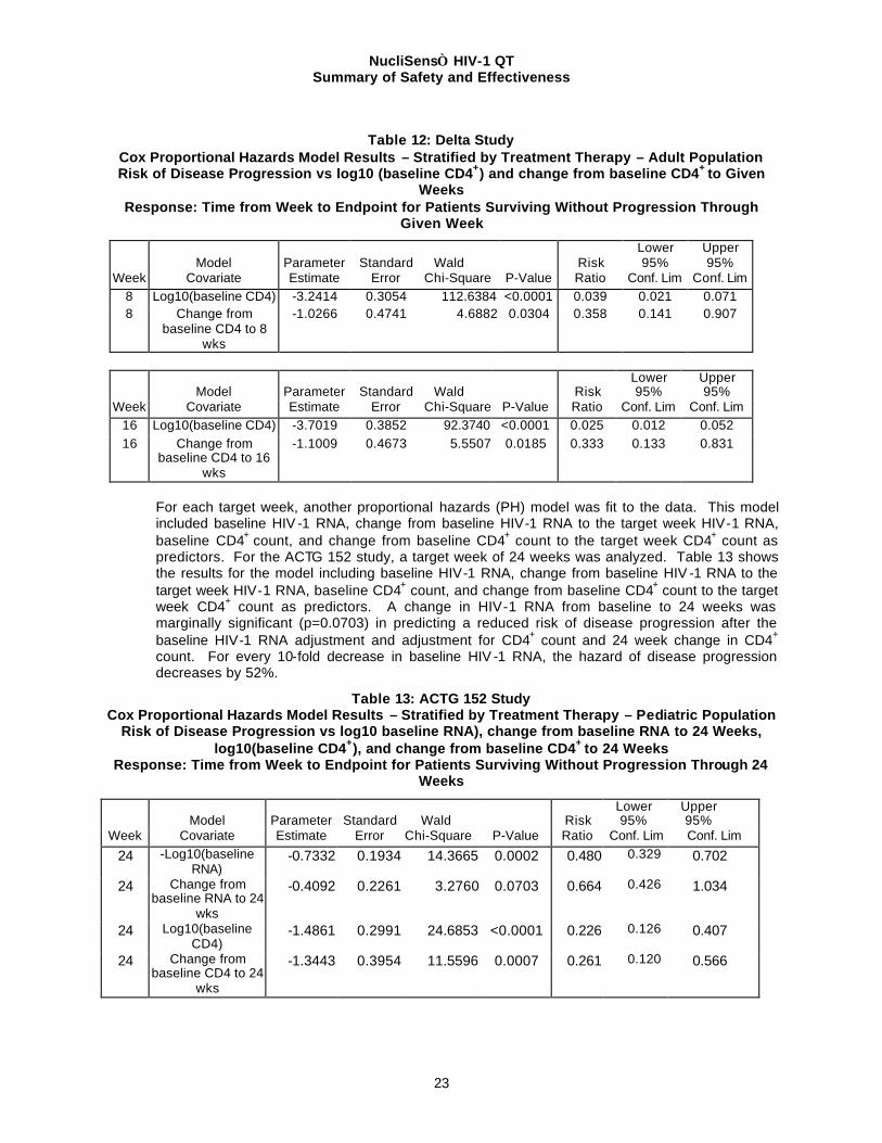
Table 12 shows the results for the model including baseline CD4+ count and change from baseline CD4+ count to the target week CD4+ count as predictors. A change in CD4+ count from baseline to 8 or 16 weeks was significant (p=0.0304 and p=0.0185, respectively) in predicting a reduced risk of disease progression after the baseline CD4+ count adjustment. For every 10-fold increase in CD4+ count, the hazard of disease progression decreases by 96-97%.
NucliSens HIV-1 QT Summary of Safety and Effectiveness
23
Table 12: Delta Study
Cox Proportional Hazards Model Results – Stratified by Treatment Therapy – Adult Population Risk of Disease Progression vs log10 (baseline CD4+) and change from baseline CD4+ to Given
Weeks Response: Time from Week to Endpoint for Patients Surviving Without Progression Through
For each target week, another proportional hazards (PH) model was fit to the data. This model included baseline HIV -1 RNA, change from baseline HIV-1 RNA to the target week HIV-1 RNA, baseline CD4+ count, and change from baseline CD4+ count to the target week CD4+ count as predictors. For the ACTG 152 study, a target week of 24 weeks was analyzed. Table 13 shows the results for the model including baseline HIV-1 RNA, change from baseline HIV -1 RNA to the target week HIV-1 RNA, baseline CD4+ count, and change from baseline CD4+ count to the target week CD4+ count as predictors. A change in HIV-1 RNA from baseline to 24 weeks was marginally significant (p=0.0703) in predicting a reduced risk of disease progression after the baseline HIV-1 RNA adjustment and adjustment for CD4+ count and 24 week change in CD4+ count. For every 10-fold decrease in baseline HIV -1 RNA, the hazard of disease progression decreases by 52%.
Table 13: ACTG 152 Study Cox Proportional Hazards Model Results – Stratified by Treatment Therapy – Pediatric Population
Risk of Disease Progression vs log10 baseline RNA), change from baseline RNA to 24 Weeks, log10(baseline CD4+), and change from baseline CD4+ to 24 Weeks
Response: Time from Week to Endpoint for Patients Surviving Without Progression Through 24 Weeks
NucliSens HIV-1 QT Summary of Safety and Effectiveness
24
For the Delta study, target weeks of 8 and 16 weeks were analyzed. Tables 14 and 15 show the results for the model including baseline HIV -1 RNA, change from baseline HIV -1 RNA to the target week HIV-1 RNA, baseline CD4+ count, and change from baseline CD4+ count to the target week CD4+ count as predictors. A change in HIV -1 RNA from baseline to 8 or 16 weeks was significant (p=0.0001) in predicting a reduced risk of disease progression after the baseline HIV-1 RNA adjustment and adjustment for CD4+ count and 8 or 16 week change in CD4+ count. For every 10-fold decrease in baseline HIV-1 RNA, the hazard of disease progression decreases by 64-69%.
Table 14: Delta Study Cox Proportional Hazards Model Results – Stratified by Treatment Therapy – Adult Population Risk of Disease Progression vs log10 (baseline RNA), change from baseline RNA to 8 Weeks,
log10(baseline CD4+), and change from baseline CD4+ to 8 Weeks Response: Time from Week to Endpoint for Patients Surviving Without Progression Through 8
Table 15: Delta Study Cox Proportional Hazards Model Results – Adult Population
Risk of Disease Progression vs log10 (baseline RNA), change from baseline RNA to 16 Weeks, log10(baseline CD4+), and change from baseline CD4+ to 16 Weeks
Response: Time from Week to Endpoint for Patients Surviving Without Progression Through 16 Weeks
NucliSens HIV-1 QT Summary of Safety and Effectiveness
25
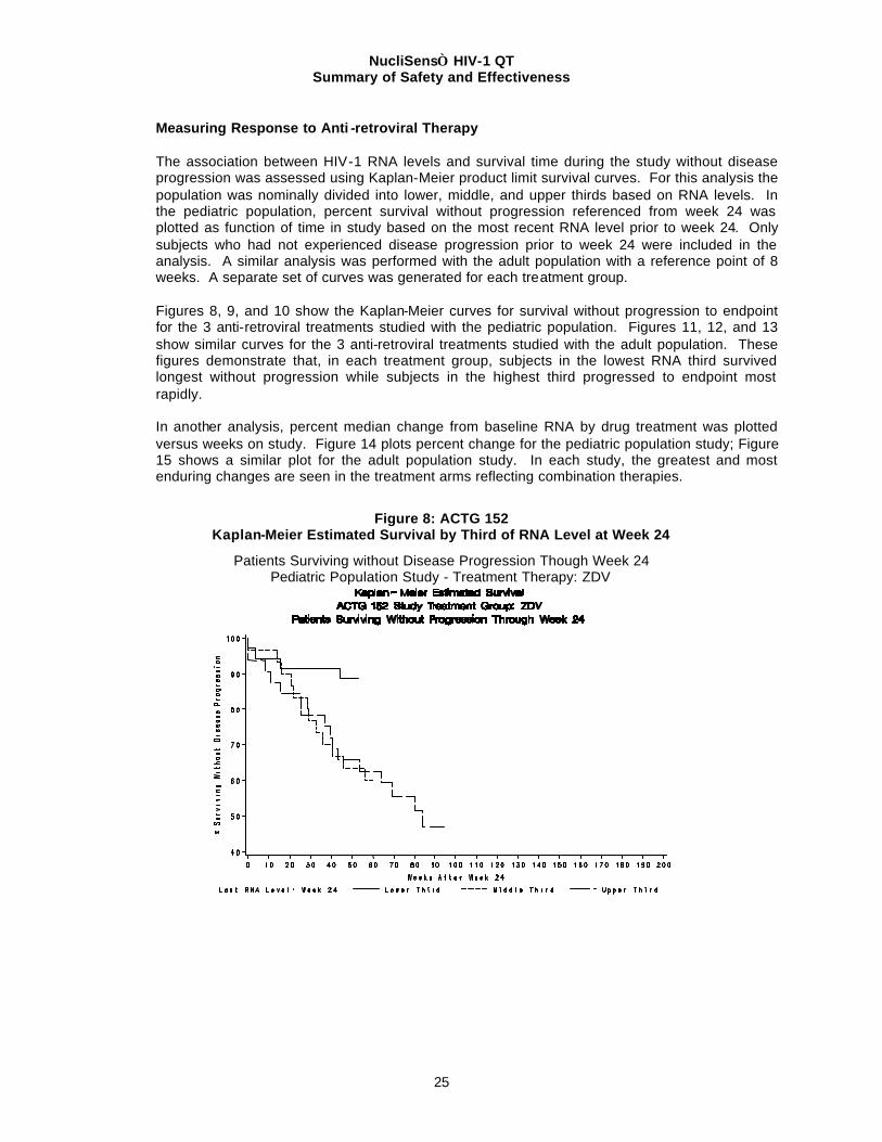
Measuring Response to Anti -retroviral Therapy The association between HIV-1 RNA levels and survival time during the study without disease progression was assessed using Kaplan-Meier product limit survival curves. For this analysis the population was nominally divided into lower, middle, and upper thirds based on RNA levels. In the pediatric population, percent survival without progression referenced from week 24 was plotted as function of time in study based on the most recent RNA level prior to week 24. Only subjects who had not experienced disease progression prior to week 24 were included in the analysis. A similar analysis was performed with the adult population with a reference point of 8 weeks. A separate set of curves was generated for each treatment group. Figures 8, 9, and 10 show the Kaplan-Meier curves for survival without progression to endpoint for the 3 anti-retroviral treatments studied with the pediatric population. Figures 11, 12, and 13 show similar curves for the 3 anti-retroviral treatments studied with the adult population. These figures demonstrate that, in each treatment group, subjects in the lowest RNA third survived longest without progression while subjects in the highest third progressed to endpoint most rapidly. In another analysis, percent median change from baseline RNA by drug treatment was plotted versus weeks on study. Figure 14 plots percent change for the pediatric population study; Figure 15 shows a similar plot for the adult population study. In each study, the greatest and most enduring changes are seen in the treatment arms reflecting combination therapies.
Figure 8: ACTG 152 Kaplan-Meier Estimated Survival by Third of RNA Level at Week 24
Patients Surviving without Disease Progression Though Week 24 Pediatric Population Study - Treatment Therapy: ZDV
NucliSens HIV-1 QT Summary of Safety and Effectiveness
26
Figure 9: ACTG 152 Kaplan-Meier Estimated Survival by Third of RNA Level at Week 24
Patients Surviving without Disease Progression Though Week 24 Pediatric Population Study - Treatment Therapy: ddI
Figure 10: ACTG 152 Kaplan-Meier Estimated Survival by Third of RNA Level at Week 24
Patients Surviving without Disease Progression Though Week 24 Pediatric Population Study - Treatment Therapy: ZDV+ ddI
NucliSens HIV-1 QT Summary of Safety and Effectiveness
27
Figure 11: Delta Trial Kaplan-Meier Estimated Survival by Third of RNA Level at Week 8
Patients Surviving without Disease Progression Though Week 8 Adult Population Study - Treatment Therapy: AZT
Figure 12: Delta Trial Kaplan-Meier Estimated Survival by Third of RNA Level at Week 8
Patients Surviving without Disease Progression Though Week 8 Adult Population Study - Treatment Therapy: AZT+ddI
NucliSens HIV-1 QT Summary of Safety and Effectiveness
28
Figure 13: Delta Trial Kaplan-Meier Estimated Survival by Third of RNA Level at Week 8
Patients Surviving without Disease Progression Though Week 8 Adult Population Study - Treatment Therapy: AZT+ddC
Figure 14: ACTG 152 Percent Change in Median RNA Level from Baseline RNA Level as A Function of Weeks on Study
by Treatment Therapy
NucliSens HIV-1 QT Summary of Safety and Effectiveness
29
Figure 15: Delta Trial Percent Change in Median RNA Level from Baseline RNA Level as A Function of Weeks on Study
by Treatment Therapy
Validation of Blood Collection and Processing
• Plasma should be used as the sample input. The standard sample volume is 1.0 ml (in the 9.0 ml Lysis Buffer tube).
• Blood should be collected in sterile tubes using EDTA, citrate or heparin as an anticoagulant. Clinical results indicate that there is no significant difference in the quantitation of HIV-1 RNA with the NucliSens HIV-1 QT system for these three anticoagulants.
• Whole blood specimens collected using EDTA as an anticoagulant can be stored at room temperature (15-30°C) for up to 24 hours before processing without detectable loss of HIV-1 RNA. EDTA plasma can be stored at 2-8º C for up to 14 days.
• Plasma collected using EDTA, citrate or heparin is stable for > 1 year at –70oC and can be frozen and thawed up to three times with no significant loss of HIV-1 RNA reported by the NucliSens HIV-1 RNA QT system. Specimens repeatedly frozen and thawed or those containing particulate matter may give erroneous results.
• In Lysis Buffer, EDTA plasma specimens can be stored: -up to one year at –70°C or -for a maximum of 14 days at 2-8°C or -for a maximum of 24 hours at room temperature (15-30°C). Note: Do not store specimens in Lysis Buffer at –20°C.
• Purified RNA eluate (post Isolation) can be stored for 12 months at –20 or –70oC or for 14 days at 2-8oC.
• Amplified material may be stored for 12 months at –20oC.
NucliSens HIV-1 QT Summary of Safety and Effectiveness
30
Specimen transport If the shipment can be accomplished within 24 hours of collection, the specimen may be shipped at room temperature (15-30oC). Otherwise, the specimen should be shipped on dry ice. Specimens may be transported to the laboratory via courier, air freight, or regular mail in accordance with applicable Federal, state and local regulations that apply to the transportation of diagnostic specimens.
Note: Handle tubes stored at –70°C (or on dry ice) with care to avoid breakage associated with low temperature storage. Note: Specimens in Lysis Buffer should be shipped at a temperature at or below –30°C (i.e, on dry ice).
IX Conclusions Drawn from the Studies The studies demonstrate that the NucliSens HIV-1 QT assay is safe and effective for use in
measuring HIV -1 RNA within a range between 176 to 3.47 X 10⁶ copies/mL. The data from the clinical studies contained in this application support the prognostic value of
HIV-1 RNA measurements and demonstrate its clinical utility as a tool for monitoring the effects of antiretroviral therapy. For example, for an individual with a viral load of 10,000 copies/ml, the risk of disease progression is 1.35 times as high as for an individual with a viral load of 5,000 copies/ml. These data suggest that a higher HIV-1 RNA viral load is associated with an increased risk of disease progression in both the ACTG 152 and Delta studies. In both the ACTG and Delta studies, the greatest and most enduring changes for better clinical outcome were seen in the treatment arms with patients under combination therapies, which suggest that anti-retroviral therapies may reduce the measurable HIV-1 RNA and the progression of disease.
X. Benefit Analysis The NucliSens HIV-1 QT is an in vitro nucleic acid amplification test for the quantitation of HIV-1
RNA in human plasma. The test is intended for use in conjunction with clinical presentation and other laboratory markers as an indicator of disease prognosis by measuring baseline HIV-1 RNA levels, and for monitoring the effects of anti-retroviral therapy by serial measurements of plasma HIV-1 RNA. The NucliSens HIV-1 QT is not intended to be used as a screening test for the HIV-1 virus nor is it to be used as a test to confirm the presence of an HIV -1 infection.
Quantitative measurements of HIV-1 viremia in the peripheral blood showed that higher levels of viral load correlated with a higher risk of clinical progression toward AIDS or death. Therefore, determination of viral load is a valuable marker for the prediction of disease progression. In addition, monitoring the efficacy of anti-viral therapy through serial measurement of viral loads may improve the clinical outcome of the infected patients under therapy (19,20,21). The performance of the product in the clinical studies indicates that, when used properly, this assay could be used for the measurement of baseline HIV-1 RNA levels for disease prognosis and for the monitoring of effectiveness of anti-viral drug therapy, both of which would significantly benefit the patient while inflicting little, if any, adverse effect to the patient as a result of its use.
XI. FDA Decision
The FDA concluded that the results of the clinical studies demonstrated that the NucliSens HIV-1 QT is a safe and effective test for the intended use. An approval letter was thus issued to bioMérieux Inc. for the intended use of this device.
NucliSens HIV-1 QT Summary of Safety and Effectiveness
31
XII. References 1 Kievits T, van Gemen B, Lens P, et al: NASBATM isothermal enzymatic in vitro nucleic acid
amplification optimized for the diagnosis of HIV -1 infection, J Vir Meth 1991; 35:273-286. 2 van Gemen B, Kievits T, Lens P, et al: Qualitative and quantitative detection of HIV -1 RNA by
nucleic acid sequence-based amplification, AIDS 1993, 7 (suppl 2):S107-S110. 3 Boom R, Sol CJA, van der Noordaa J, et al: Rapid and simple method for purification of nucleic
acids, J Clin Microbiol 1990; 28:495-503. 4 Bruisten S, van Gemen B, Huisman H, et al: Detection of HIV -1 Distribution in Different Blood
Fractions of HIV-1 Seropositive Persons by Two Nucleic Acid Amplification Assays, AIDS Research and Human Retroviruses 1993; (9):259-265.
5 Bagnarelli P, Menzo S, Clementi M, et al: Detection of Human Immunodeficiency Virus Type 1 Genomic RNA in Plasma Samples by Reverse-Transcription Polymerase Chain Reaction, J Med Vir 1991;34:89-95.
6 Ottmann M, Innocenti P, Seigneurin J-M, et al: The polymerase chain reaction for the detection of HIV-1 genomic RNA in plasma from infected individuals, J Vir Meth 1991; 31:273-284.
7 van Gemen B, Kievits T, Lens P, et al: Quantification of HIV -1 RNA in plasma using NASBATM during HIV-1 primary infection, J Vir Meth 1993; 43:177-188.
8 Piatak M, Saag MS, Lifson JD, et al: High Levels of HIV-1 in Plasma During All Stages of Infection Determined by Competitive PCR, Science 1993; 259:1749-1754.
9 Kessler HA, Blaauw B, Spear J, et al: Diagnosis of human immunodeficiency virus infection in seronegative homosexuals presenting with an acute viral syndrome, JAMA 1987; 258:1196-1199.
10 Lu W, Andrieu JM: Early Identification of Human Immunodeficiency Virus -Infected Asymptomatic Subjects Susceptible to Zidovudine by Quantitative Viral Coculture and Reverse Transcription-Linked Polymerase Chain Reaction, J Inf Dis 1993; 167:1014-1020.
11 Semple M, Loveday C, Weller I, Tedder R: Direct Measurement of Viraemia in Patients Infected With HIV-1 and Its Relationship to Disease Progression and Zidovudine Therapy, J Med Vir 1991; 35:38-45.
12 van Gemen B, van Beuningen R, Kievits T, et al: A one-tube quantitative HIV -1 RNA NASBA nucleic acid amplification assay using electrochemiluminescent (ECL) labelled probes, J Vir Meth, 1994; 49:157-168.
13 Nedelman J, Heagerty P, Lawrence C: Quantitative PCR with internal controls, CABIOS 1992; 8:65-70.
14 Nedelman J, Heagerty P, Lawrence C: Quantitative PCR: procedures and precisions, Bull Math Biol 1992; 54:477-502.
15 Blackburn GF, Shah HP, Kenten JH, Leland J, Kamin RA, Link J, Peterman J, Powell MJ, Shah A, Talley DB et al: Electrochemiluminescence detection for development of immunoassays and DNA probe assays for clinical diagnostics, Clin Chem 1991; 37:1534-1539.
16 Kenten JH, Gudibande S, Link J, Willey JJ, Curfman B, Major EO, Massey RJ: Improved electrochemiluminescent label for DNA probe assays: rapid quantitative assays of HIV-1 polymerase chain reaction products, Clin Chem 1992; 38:873-879.
17 Ho DD, Neumann AU, Markowitz M, et al: Rapid turnover of plasma virions and CD4 lymphocytes in HIV-1 infection. Nature 1995; 373:123-126.
18 Perelson AS, Neumann AU, Ho DD, et al: HIV-1 dynamics in vivo: virion clearance rate, infected cell life-span and viral generation time. Science 1996; 271:1582-1586.
19 Mellors JW, Kingsley LA, Kokka RP et al: Quantitation of HIV-1 RNA in plasma predicts outcome after seroconversion. Ann. Intern. Med. 1995; 122:573-579.
20 Jurriaans S, van Gemen B, Goudsmit J, et al: The natural history of HIV-1 infection: viral load and virus phenotype independent determinants of clinical course? Virology 1994; 204:223-233.
21 Mellors JW, Rinaldo CR, Kingsley LA, et al: Prognosis in HIV-1 infection predicted by the quantity of virus in plasma. Science 1996; 272:1167-1170.
22 de Wolf F, Spijkerman I, Schellekens PT, et al: Reciprocal predictive value of the quantity of HIV-1 RNA versus the number and function of CD4+ T-cells in time since seroconversion, Published yet?
23 O’Brien WA, Hartigan PM, Martin D, Esinhart D; Changes in plasma HIV-1 RNA and CD4+ lymphocyte count relative to treatment and progression to AIDS. N. Engl. J. Med. 1996; 334:426-431.
NucliSens HIV-1 QT Summary of Safety and Effectiveness
32
24 Carpenter CJ, Fischl MA, Volberding PA, et al: Antiretroviral therapy for HIV infection in 1996. JAMA 1996; 146-154.
25 Hammer SM for the ACTG 175 Virology Substudy Team. Virological markers and outcome in ACTG 175. In: Third conference on retroviruses and opportunistic infections, Washington DC. 1996, abstract S24.
26 Gazzard B for the International Coordinating Committee. Further results from European/Australian Delta Trial. In: Third conference on retroviruses and opportunistic infections, Washington DC, 1996, abstract LB5A.
27 Gulick R, Mellors J, Havlir D: Potent and sustained antiretroviral activity of indinavir in combination with zidovudine and lamivudine. In: Third conference on retroviruses and opportunistic infections. Washington DC, 1996, abstract LB7.
28 Bruisten SM, Oudshoorn P, Cuypers HTM, et al: Stability of HIV-1 RNA in whole blood, plasma and serum during specimen handling and storage prior to amplification by NASBA-QT. published yet?
29 Holodniy M, Mole L, Jackson JB, et al: Comparative stabilities of human immunodeficiency virus RNA in plasma from samples collected in VACUTAINER CPT, VACUTAINER PPT, and standard VACUTAINER tubes. J. Clin. microbiol. 1995;33:1562-1566.
30 Witt, DJ, Kemper, M: Techniques for the evaluation of nucleic acid amplification technology performance with specimens containing interfering substances: efficacy of Boom methodology for extraction of HIV-1 RNA, J Virological Methods 1999, 79:97-111.
31 Palumbo, PE, Raskino, C, Fiscus, S, Pawha, S, Fowler, MG, Spector, SA, Englund, JA, Baker, CJ: Predictive Value of Quantitative Plasma HIV RNA and CD4+ Lymphocyte Count in HIV -Infected Infants and Children, JAMA 1998, 279:756-761.
32 Delta Coordinating Committee and Delta Virology Committee: HIV-1 RNA response to antiretroviral treatment in 1280 participants in the Delta Trial: an extended virology study, AIDS 1999, 13:57-65.
NucliSens HIV-1 QT Summary of Safety and Effectiveness
33
PMA REVIEW COMMITTEE Signature Date ____________________________________ <First name> <Last Name>, Chairperson ____________________________________ <First name> <Last Name>, Member ____________________________________ <First name> <Last Name>, Member ____________________________________ <First name> <Last Name>, Member ____________________________________ <First name> <Last Name>, Member ____________________________________ <First name> <Last Name>, Member ____________________________________ <First name> <Last Name>, Member