NEW ZEALAND OBSTETRIC DOPPLER GUIDELINE This is a New Zealand guideline for the performance of obstetric Doppler examinations. The guideline incorporates detailed notes on correct Doppler technique, interpretation and reference charts for each of commonly used obstetric Doppler examinations. All health practitioners involved in the performance and interpretation of obstetric ultrasound examinations are encouraged to use this document and incorporate the principles and reference values into their clinical practice. Unaltered copies of this guideline may be freely reproduced and distributed. Contributors: Martin Necas, Dr Emma Parry, Professor Lesley McCowan
Transcript
New Zealand Obstetric Doppler Guideline, NZMFMN , revised September 2014, Page 1
NEW ZEALAND OBSTETRIC DOPPLER GUIDELINE
This is a New Zealand guideline for the performance of obstetric Doppler examinations. The guideline incorporates detailed notes on correct Doppler technique, interpretation and reference charts for each of commonly used obstetric Doppler examinations. All health practitioners involved in the performance and interpretation of obstetric ultrasound examinations are encouraged to use this document and incorporate the principles and reference values into their clinical practice.
Unaltered copies of this guideline may be freely reproduced and distributed.
Contributors: Martin Necas, Dr Emma Parry, Professor Lesley McCowan
New Zealand Obstetric Doppler Guideline, NZMFMN , revised September 2014, Page 2
Which Doppler Test When?
SGA or IUGR Umbilical artery PI and Mean uterine artery PI
Umbilical artery PI raised Specialist obstetric review
within a week
Absent or reversed end-‐diastolic flow
Specialist obstetric review now
Normal >34 weeks Add MCA PI
At risk of early onset maternal hypertensive disorder or SGA
Mean uterine artery PI at 20 or 24 weeks
MCA PSV
MCDA Twins with TTTS Both twins:
Umbilical artery PI MCA PSV DV PI
Suspected fetal anemia
New Zealand Obstetric Doppler Guideline, NZMFMN , revised September 2014, Page 3
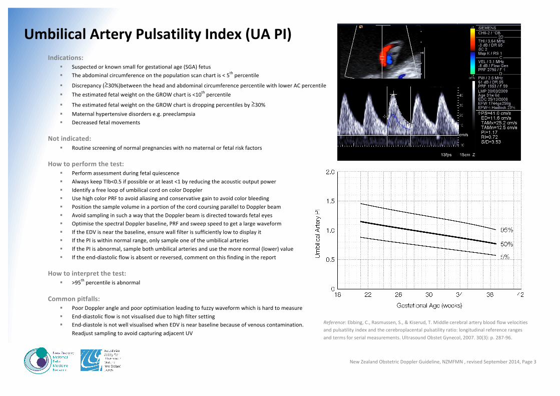
Umbilical Artery Pulsatility Index (UA PI) Indications:
§ Suspected or known small for gestational age (SGA) fetus § The abdominal circumference on the population scan chart is < 5th percentile
§ Discrepancy (≥30%)between the head and abdominal circumference percentile with lower AC percentile § The estimated fetal weight on the GROW chart is <10th percentile
§ The estimated fetal weight on the GROW chart is dropping percentiles by ≥30% § Maternal hypertensive disorders e.g. preeclampsia § Decreased fetal movements
Not indicated: § Routine screening of normal pregnancies with no maternal or fetal risk factors
How to perform the test: § Perform assessment during fetal quiescence § Always keep TIb<0.5 if possible or at least <1 by reducing the acoustic output power § Identify a free loop of umbilical cord on color Doppler § Use high color PRF to avoid aliasing and conservative gain to avoid color bleeding § Position the sample volume in a portion of the cord coursing parallel to Doppler beam § Avoid sampling in such a way that the Doppler beam is directed towards fetal eyes § Optimise the spectral Doppler baseline, PRF and sweep speed to get a large waveform § If the EDV is near the baseline, ensure wall filter is sufficiently low to display it § If the PI is within normal range, only sample one of the umbilical arteries § If the PI is abnormal, sample both umbilical arteries and use the more normal (lower) value § If the end-‐diastolic flow is absent or reversed, comment on this finding in the report
How to interpret the test: § >95th percentile is abnormal
Common pitfalls: § Poor Doppler angle and poor optimisation leading to fuzzy waveform which is hard to measure § End-‐diastolic flow is not visualised due to high filter setting § End-‐diastole is not well visualised when EDV is near baseline because of venous contamination.
Readjust sampling to avoid capturing adjacent UV
Reference: Ebbing, C., Rasmussen, S., & Kiserud, T. Middle cerebral artery blood flow velocities and pulsatility index and the cerebroplacental pulsatility ratio: longitudinal reference ranges and terms for serial measurements. Ultrasound Obstet Gynecol, 2007. 30(3): p. 287-‐96.
New Zealand Obstetric Doppler Guideline, NZMFMN , revised September 2014, Page 4
Middle Cerebral Artery Pulsatility Index (MCA PI) Indications:
§ SGA but normal UA PI in a fetus after 34 weeks § MCDA twin gestation with TTTS
How to perform the test:
§ Perform assessment during fetal quiescence § Always keep TIb<0.5 if possible or at least <1 by reducing the acoustic output power § Start with the BPD view § Move caudally to visualise the butterfly shape of suprasellar cisterns and the sphenoid § Use the coronal suture/sphenoidal fontanelle as an acoustic window § Use high definition (write) zoom § Activate color Doppler to visualise the MCA § Assess the MCA which is closer to the transducer § Move anteriorly and angle back to align the MCA flow direction with the Doppler beam § Position a small (0.5-‐1mm) sample volume 2mm beyond from the MCA origin § Optimise the spectral Doppler baseline and PRF to get a large waveform
How to interpret the test:
§ <5th percentile is abnormal
Common pitfalls:
§ Poor Doppler angle and poor optimisation leading to fuzzy waveform which is hard to measure § Gate too close to MCA origin where multidirectional contamination from ACA and PCoA occurs § Sample positioned too peripherally in the MCA where velocities fall § PCA misidentified as MCA § Poor visualisation due to inadequate zoom
Reference: Ebbing, C., Rasmussen, S., & Kiserud, T. Middle cerebral artery blood flow velocities and pulsatility index and the cerebroplacental pulsatility ratio: longitudinal reference ranges and terms for serial measurements. Ultrasound Obstet Gynecol, 2007. 30(3): p. 287-‐96.
New Zealand Obstetric Doppler Guideline, NZMFMN , revised September 2014, Page 5
Cerebroplacental Ratio (CPR) Indications:
§ If MCA PI assessment was performed, the CPR should be calculated and recorded
How to calculate CPR:
§ The CPR is defined the ratio of MCA PI and UA PI
How to interpret the test:
§ <5th percentile is abnormal § It may be helpful to plot the MCA and CPR results on serial assessments to
determine the trend
Reference: Ebbing, C., Rasmussen, S., & Kiserud, T. Middle cerebral artery blood flow velocities and pulsatility index and the cerebroplacental pulsatility ratio: longitudinal reference ranges and terms for serial measurements. Ultrasound Obstet Gynecol, 2007. 30(3): p. 287-‐96.
New Zealand Obstetric Doppler Guideline, NZMFMN , revised September 2014, Page 6
Ductus Venosus Pulsatility Index (DV PI) Indications:
§ Raised UA PI (>95th ) and reduced MCA PI in preterm SGA § MCDA twin gestation with TTTS or selective IUGR
How to perform the test:
§ Perform assessment during fetal quiescence § Always keep TIb<0.5 if possible or at least <1 by reducing the acoustic output power § Sagittal and transverse approaches are acceptable as long as Doppler angle is 0-‐60 degrees § Activate color Doppler to identify DV at the end of UV § Enlarge the image § 0.5-‐1mm gate placed in the inlet of DV § Set wall filter low, sweep speed high § Optimise the spectral Doppler baseline and PRF to get a large waveform § If PI>95th percentile, assess umbilical vein for pulsatility
How to interpret the test:
§ >95th percentile is abnormal
Common Pitfalls
§ Color PRF too low and gain too high leading to difficulty in DV identification amongst other vessels § Sample size too large, leading to contamination from other vessels § Sample not placed at the inlet of the DV § Adjacent hepatic vein or celiac axis misidentified as DV § Poor Doppler angle and poor optimisation leading to fuzzy waveform which is hard to measure § Fetal breathing activity may result in false impression of absent A wave
Reference: Kessler, J., Rasmussen, S., Hnson, M., & Kiserud, T. Longitudinal reference ranges for ductus venosus flow velocities and waveform indices. Ultrasound Obstet Gynecol, 2006. 28: 890-‐898.
New Zealand Obstetric Doppler Guideline, NZMFMN , revised September 2014, Page 7
MCA Peak Systolic Velocity (MCA PSV) Indications:
§ Maternal-‐fetal isoimmunisation § Any suspicion of fetal anaemia § Unexplained hydrops § MCDA twin gestation with suspicion of TTTS or TAPS
How to do perform the test:
§ Perform assessment during fetal quiescence § Always keep TIb<0.5 if possible or at least <1 by reducing the acoustic output power when required § Start with the BPD view § Move caudally to visualise the butterfly shape of suprasellar cisterns and the sphenoid § Use the coronal suture/sphenoidal fontanelle as an acoustic window § Use high definition (write) zoom § Activate color Doppler to visualise the MCA § Assess the MCA which is closer to the transducer § Move anteriorly and angle back to align the MCA flow direction with the Doppler beam § Ideal interrogation angle is 0 degrees but 30 degrees or less is acceptable § Position a small (0.5-‐1mm) sample volume 2 mm beyond from the MCA origin § Optimise the spectral Doppler baseline and PRF to get a large waveform § If the Doppler angle is not zero, angle correction must be used § If PSV>1.5 multiples of the median (MoM), obtain 3 high quality samples and use the highest value
How to interpret the test:
§ >1.5MoM is abnormal
Common Pitfalls:
§ Poor Doppler angle and poor optimisation leading to fuzzy waveform which is hard to measure § Gate too close to MCA origin where multidirectional contamination from ACA and PCoA occurs § Sample positioned too peripherally in the MCA where velocities fall § PCA misidentified as MCA § Failure to angle-‐correct at angles other than 0 leading to underestimation of PSV § Poor visualisation due to inadequate zoom References:
Mari, G. Noninvasive Diagnosis by Doppler ultrasonography of fetal anemia due to maternal red-‐cell alloimmunization. N Engl J Med 2000; 342:9-‐14, p 9-‐14. Mari, G. Middle cerebral artery peak systolic velocity for the diagnosis of fetal anemia: the untold story. Ultrasound Obstet Gynecol 2005 Apr;25(4):323-‐30.
New Zealand Obstetric Doppler Guideline, NZMFMN , revised September 2014, Page 8
Mean uterine Artery Pulsatility Index Indications:
§ Screen patients at high risk of early preeclampsia or early SGA at 20 or 24 weeks § If abnormal at 20 weeks, repeat at 24 weeks § Early onset IUGR § Current hypertensive disorder in pregnancy § Full assessment of suspected SGA pregnancy
How to perform the test:
§ Locate the maternal anterior superior iliac spine and angle medially § Alternatively visualize the external iliac artery (EIA) § The uterine artery is typically seen crossing the EIA anteriorly § Select a portion of the uterine artery which courses at a favourable Doppler angle 0-‐60 degrees § Optimise the spectral Doppler baseline and PRF to get a large waveform § Measure the right and left PI and calculate the mean value
How to interpret the test:
§ >95th percentile is abnormal § Bilateral notching after 24 weeks is abnormal
Common Pitfalls:
§ Failure to identify the uterine artery by not scanning inferiorly enough
Reference: Gomez O., et al. Reference ranges for uterine mean pulsatility index at 11-‐41 weeks of gestation. Ultrasound Obstet Gynecol 2008; 32: 128-‐132.
New Zealand Obstetric Doppler Guideline, NZMFMN , revised September 2014, Page 9
Quick Reference Tables
Umbilical Artery PI v MCA PI v
Cerebroplacental Ratio (CPR) v
Mean Uterine Artery PI ö
CPR = MCA PI/UA PI Mean PI=(RT PI + LT PI)/2 >95th percentile is abnormal <5th percentile is abnormal <5th percentile is abnormal >95th percentile is abnormal
References: Ô Acharya G, et al. Reference ranges for serial measurements of blood velocity and pulsatility index at the intra-‐abdominal portion, and fetal and placental ends of umbilical artery. Ultrasound Obstet Gynecol 2005; 26:162-‐169. v Ebbing, C., Rasmussen, S., & Kiserud, T. Middle cerebral artery blood flow velocities and pulsatility index and the cerebroplacental pulsatility ratio: longitudinal reference ranges and terms for serial measurements. Ultrasound Obstet Gynecol, 2007. 30(3): p. 287-‐96. ø Baschat AA, Gembruch U. The cerebroplacental Doppler ratio revisited. Ultrasound Obstet Gynecol 2003; 21:124-‐127. ö Gomez O, et al. Reference ranges for uterine mean pulsatility index at 11-‐41 weeks of gestation. Ultrasound Obstet Gynecol 2008; 32: 128-‐132.
New Zealand Obstetric Doppler Guideline, NZMFMN , revised September 2014, Page 10
Obstetric Doppler reference charts
New Zealand Obstetric Doppler Guideline, NZMFMN , revised September 2014, Page 11
Reference: Ebbing, C., Rasmussen, S., & Kiserud, T. Middle cerebral artery blood flow velocities and pulsatility index and the cerebroplacental pulsatility ratio: longitudinal reference ranges and terms for serial measurements. Ultrasound Obstet Gynecol, 2007. 30(3): p. 287-‐96.
New Zealand Obstetric Doppler Guideline, NZMFMN , revised September 2014, Page 12
Reference: Ebbing, C., Rasmussen, S., & Kiserud, T. Middle cerebral artery blood flow velocities and pulsatility index and the cerebroplacental pulsatility: longitudinal reference ranges and terms for serial measurements. Ultrasound Obstet Gynecol, 2007. 30(3): p. 287-‐96.
New Zealand Obstetric Doppler Guideline, NZMFMN , revised September 2014, Page 13
Reference: Ebbing, C., Rasmussen, S., & Kiserud, T. Middle cerebral artery blood flow velocities and pulsatility index and the cerebroplacental pulsatiliy ratio: longitudinal reference ranges and terms for serial measurements. Ultrasound Obstet Gynecol, 2007. 30(3): p. 287-‐96.
New Zealand Obstetric Doppler Guideline, NZMFMN , revised September 2014, Page 14
Reference: Gomez O., et al. Reference ranges for uterine mean pulsatility index at 11-‐41 weeks of gestation. Ultrasound Obstet Gynecol 2008; 32: 128-‐132.
New Zealand Obstetric Doppler Guideline, NZMFMN , revised September 2014, Page 15
Reference: Kessler, J., Rasmussen, S., Hnson, M., & Kiserud, T. Longitudinal reference ranges for ductus venosus flow velocities and waveform indices. Ultrasound Obstet Gynecol, 2006. 28: 890-‐898.
New Zealand Obstetric Doppler Guideline, NZMFMN , revised September 2014, Page 16
References: Mari, G. Noninvasive Diagnosis by Doppler ultrasonography of fetal anemia due to maternal red-‐cell alloimmunization. N Engl J Med 2000; 342:9-‐14, p 9-‐14.
Mari, G. Middle cerebral artery peak systolic velocity for the diagnosis of fetal anemia: the untold story. Ultrasound Obstet Gynecol 2005 Apr;25(4):323-‐30.


![Index [assets.cambridge.org]assets.cambridge.org/97805211/96611/index/9780521196611_index.pdf · Index More information ... inpregnancy,55,56 alemtuzumab,477,563,564,572, 623 sideeffects,573](https://static.documents.pub/doc/80x56/5b0803d37f8b9a3d018b96c2/index-more-information-inpregnancy5556-alemtuzumab477563564572-623.jpg)

