Inorg. Chem. 1989, 28, 1357-1361 1357 Con c I u s i o n s The prominent 8983-eV feature present on the absorption edge of Cu(1) complexes is sensitive not only to overall coordination number (as documented previously by Kau and co-workers26) but also to the details of the symmetry of the complex. Thus, in all the complexes studied a correlation was found between the in- tensity of this feature and the degree pf distortion of the complex from two- or three-coordinate planar geometry toward tetrahedral. These correlations have been applied to predict the geometry of the Cu(1) sites of SOD, DBH, and deoxy-Hc. Assignment of Cu( 1)-SOD as a planar (or closely planar) three-coordinate center is possible. In the latter two cases the presence of more than one structural type of Cu( I) complicates the analysis, but comparison of the edge profile of deoxy-Hc with those of the model systems suggests distorted three- or four-coordination and hence the possible presence of an endogenous hydroxyl bridge. The technique is particularly valuable in the study of ligand binding to Cu(1) centers in proteins. Here, comparative changes in absorption edge features allow ligand binding to be detected and any consequent changes in coordination geometry to be predicted. Using the latter approach, we have demonstrated the existence of a CO complex of DBH. In the case of B. canali- culatum deoxy-Hc, changes in the absorption edge upon CO binding to a single Cu atom43are consistent with trigonal-py- ramidal or pseudotetrahedral geometry at each copper center of the dinuclear unit. The data indicate that each Cu center in HcTCO is three- or four-coordinate and argue against previous proposals of two- plus four-coordination in Hc-CO. Acknowledgment. We thank the Daresbury Laboratory for provision of beam time, computational facilities, and other nec- essary equipment for this study. Financial support is gratefully acknowledged from the SERC to N.J.B., NIH to K.D.K., and ZWO to J.R. We also thank the NATO Scientific Affairs Bureau for the award of a travel grant (RG.82/0139) to N.J.B. and K.D.K. Supplementary Material Available: Listings of bond lengths, bond angles, anisotropic temperature factors, and hydrogen coordinates and temperature factors for complex 3-(PF6),.CH2CI2 (Tables 4-7) (10 pages); listings of observed and calculated structure factors for 3- (PF6)2.CH2C12 (Table 8) (38 pages). Contribution from the Department of Chemistry, University of Georgia, Athens, Georgia 30602, and Department of Physics, Emory University, Atlanta, Georgia 30322 Observation of S = 2 EPR Signals from Ferrous Iron-Thiolate Complexes. Relevance to Rubredoxin-Type Sites in Proteins Mark T. Werth,? Donald M. Kurtz, Jr.,*,? Barry D. Howes,* and Boi Hanh Huynh* Received September 15, 1988 In frozen solution the pseudotetrahedral ferrous iron-thiolate complexes with 2-mercaptoethanol, dithiothreitol, and glutathione (y-glutamylcysteinylglycine) exhibit a low-field (g - 10) EPR signal below 30 K. This signal is ascribed to a transition within the I f 2 ) doublet of the S = 2 spin state, and these complexes appear to be the first examples of pseudotetrahedral ferrous complexes that give rise to such a signal. A detailed study was undertaken to ascertain the origin of this signal in the case of the complex with 2-mercaptoethanol. Ba[Fe(SCHZCH20H),] was prepared and shown to contain an FeS, site of approximate tetrahedral symmetry by X-ray crystallography. For [Fe(SCH2CHZ0H)4]2' in water, the 1-/2- midpoint reduction potential (-0.35 V vs SCE) and IH NMR (6(Fe-SCH2CHZ0H) 203 ppm at 25 "C) and 57Fe Mossbauer (AEQ = 3.48 f 0.03 mm/s, 6 = 0.73 f 0.02 mm/s at 4.2 K) spectra show that this complex, and essentially only this complex, survives in solution, when the mole ratio of thiolate to iron is 4 or greater. Mossbauer spectra obtained in applied fields up to 7 T reveal at least two species in frozen aqueous solutions of [Fe(SCH2CH20H),J2-. Spectral simulations show that the major (75%) and minor (25%) species have spin Ham- iltonian parameters strongly resembling those of reduced rubredoxin and reduced desulforedoxin,respectively. The minor species has values of zero-field splitting parameters (D = -5.0 f 2.0 cm-I, E/D = 0.17 f 0.05) that are consistent with observation of an S = 2 EPR signal at low field. The slightly larger values of these parameters for reduced desulforedoxin preclude the observation of an S = 2 EPR signal from this protein. The two species of [Fe(SCH2CH20H),I2- in solution are ascribed to different sets of S-Fe-S angles. Since variations in these angles are expected to have relatively low energy barriers, observation of an S = 2 EPR signal from biological Fe(Cys-S), sites is likely to be strongly dependent on the protein matrix. As part of a search for new spectroscopic probes of high-spin ferrous centers in proteins, we have examined the simplest such prototype for iron-sulfur proteins, namely, the pseudotetrahedral Fe(Cys-S),, site in reduced rubredoxin (Cys-S = cysteine thiolate). We have recently added 'H NMR to the list of spectroscopies that can elicit characteristic signals from the ferrous Fe(Cys-S),, site in rubredoxin.' Judging from a number of recent EPR signals arising from integer spin states represent an in- creasingly important electronic probe of iron sites in proteins. We now report that the S = 2 state of ferrous Fe(SR)4complexes can exhibit a characteristic EPR signal. We also delineate some structural and spectroscopic parameters that favor observation of such a signal. Experimental Section All manipulations were carried out at room temperature under an Ar atmosphere in either Schlenk-type glassware or septum-capped vials attached to a vacuum manifold. Solutions were added and transferred +University of Georgia. *Emory University. via steel tubing or gastight syringes. The thiols used for this study, 2-mercaptoethanol, dithiothreitol, glutathione, and lipoic acid, were ob- tained commercially and used without further purification. D,L-Di- hydrolipoate was prepared from the oxidized compound as previously described? The following general procedure was used to prepare ferrous Fe(SR), complexes in situ. The thiol was dissolved in the desired solvent (either water or DMSO) with sufficient LiOH to deprotonate thiol groups plus any carboxylate groups. Iron was then added as solid FeCI2 or Fe(S04)-7H20. A thiolate/iron mol ratio 1 4 was maintained in all solutions. This procedure was previously shown to generate stable solu- tions of the ferrous Fe(SR)4 complexes in water.' Specific reagent concentrations are given in the figure legends. Solutions for electro- chemistry were -4 mM in iron and -100 mM in total thiol. These (1) Werth, M. T.; Kurtz, D. M., Jr.; Moura, I.; LeGall, J. J. Am. Chem. Soc. 1987, 109, 213-275. (2) Hendrich, M. P.; Debrunner, P. G. J. Magn. Reson. 1988.78, 133-141. (3) Reem, R. C.; Solomon, E. I. J. Am. Chem. Soc. 1987, 109, 1216-1226. (4) Hagen, W. R.; Dunham, W. R.; Johnson, M. K.; Fee, J. A. Eiochim. Biophys. Acta 1985, 828, 369-374. (5) Hagen, W. R. Biochim. Biophys. Acta 1982, 708, 82-98. (6) Bonomi, F.; Werth, M. T.; Kurtz, D. M., Jr. Inorg. Chem. 1985, 24, 433 1-4335. 0020-1669/89/1328-1357$01.50/0 0 1989 American Chemical Society
Transcript
Inorg. Chem. 1989, 28, 1357-1361 1357
Con c I u s i o n s The prominent 8983-eV feature present on the absorption edge
of Cu(1) complexes is sensitive not only to overall coordination number (as documented previously by Kau and co-workers26) but also to the details of the symmetry of the complex. Thus, in all the complexes studied a correlation was found between the in- tensity of this feature and the degree pf distortion of the complex from two- or three-coordinate planar geometry toward tetrahedral.
These correlations have been applied to predict the geometry of the Cu(1) sites of SOD, DBH, and deoxy-Hc. Assignment of Cu( 1)-SOD as a planar (or closely planar) three-coordinate center is possible. In the latter two cases the presence of more than one structural type of Cu( I ) complicates the analysis, but comparison of the edge profile of deoxy-Hc with those of the model systems suggests distorted three- or four-coordination and hence the possible presence of an endogenous hydroxyl bridge.
The technique is particularly valuable in the study of ligand binding to Cu(1) centers in proteins. Here, comparative changes in absorption edge features allow ligand binding to be detected and any consequent changes in coordination geometry to be
predicted. Using the latter approach, we have demonstrated the existence of a CO complex of DBH. In the case of B. canali- culatum deoxy-Hc, changes in the absorption edge upon CO binding to a single Cu atom43 are consistent with trigonal-py- ramidal or pseudotetrahedral geometry at each copper center of the dinuclear unit. The data indicate that each Cu center in HcTCO is three- or four-coordinate and argue against previous proposals of two- plus four-coordination in Hc-CO.
Acknowledgment. We thank the Daresbury Laboratory for provision of beam time, computational facilities, and other nec- essary equipment for this study. Financial support is gratefully acknowledged from the SERC to N.J.B., NIH to K.D.K., and ZWO to J.R. We also thank the NATO Scientific Affairs Bureau for the award of a travel grant (RG.82/0139) to N.J.B. and K.D.K.
Supplementary Material Available: Listings of bond lengths, bond angles, anisotropic temperature factors, and hydrogen coordinates and temperature factors for complex 3-(PF6),.CH2CI2 (Tables 4-7) (10 pages); listings of observed and calculated structure factors for 3- (PF6)2.CH2C12 (Table 8) (38 pages).
Contribution from the Department of Chemistry, University of Georgia, Athens, Georgia 30602, and Department of Physics, Emory University, Atlanta, Georgia 30322
Observation of S = 2 EPR Signals from Ferrous Iron-Thiolate Complexes. Relevance to Rubredoxin-Type Sites in Proteins
Mark T. Werth,? Donald M. Kurtz, Jr.,*,? Barry D. Howes,* and Boi Hanh Huynh* Received September 15, 1988
In frozen solution the pseudotetrahedral ferrous iron-thiolate complexes with 2-mercaptoethanol, dithiothreitol, and glutathione (y-glutamylcysteinylglycine) exhibit a low-field (g - 10) EPR signal below 30 K. This signal is ascribed to a transition within the If2) doublet of the S = 2 spin state, and these complexes appear to be the first examples of pseudotetrahedral ferrous complexes that give rise to such a signal. A detailed study was undertaken to ascertain the origin of this signal in the case of the complex with 2-mercaptoethanol. Ba[Fe(SCHZCH20H),] was prepared and shown to contain an FeS, site of approximate tetrahedral symmetry by X-ray crystallography. For [Fe(SCH2CHZ0H)4]2' in water, the 1-/2- midpoint reduction potential (-0.35 V vs SCE) and IH NMR (6(Fe-SCH2CHZ0H) 203 ppm at 25 "C) and 57Fe Mossbauer (AEQ = 3.48 f 0.03 mm/s, 6 = 0.73 f 0.02 mm/s at 4.2 K) spectra show that this complex, and essentially only this complex, survives in solution, when the mole ratio of thiolate to iron is 4 or greater. Mossbauer spectra obtained in applied fields up to 7 T reveal at least two species in frozen aqueous solutions of [Fe(SCH2CH20H),J2-. Spectral simulations show that the major (75%) and minor (25%) species have spin Ham- iltonian parameters strongly resembling those of reduced rubredoxin and reduced desulforedoxin, respectively. The minor species has values of zero-field splitting parameters (D = -5.0 f 2.0 cm-I, E / D = 0.17 f 0.05) that are consistent with observation of an S = 2 EPR signal at low field. The slightly larger values of these parameters for reduced desulforedoxin preclude the observation of an S = 2 EPR signal from this protein. The two species of [Fe(SCH2CH20H),I2- in solution are ascribed to different sets of S-Fe-S angles. Since variations in these angles are expected to have relatively low energy barriers, observation of an S = 2 EPR signal from biological Fe(Cys-S), sites is likely to be strongly dependent on the protein matrix.
As part of a search for new spectroscopic probes of high-spin ferrous centers in proteins, we have examined the simplest such prototype for iron-sulfur proteins, namely, the pseudotetrahedral Fe(Cys-S),, site in reduced rubredoxin (Cys-S = cysteine thiolate). We have recently added 'H N M R to the list of spectroscopies that can elicit characteristic signals from the ferrous Fe(Cys-S),, site in rubredoxin.' Judging from a number of recent EPR signals arising from integer spin states represent an in- creasingly important electronic probe of iron sites in proteins. We now report that the S = 2 state of ferrous Fe(SR)4 complexes can exhibit a characteristic EPR signal. We also delineate some structural and spectroscopic parameters that favor observation of such a signal. Experimental Section
All manipulations were carried out at room temperature under an Ar atmosphere in either Schlenk-type glassware or septum-capped vials attached to a vacuum manifold. Solutions were added and transferred
+University of Georgia. *Emory University.
via steel tubing or gastight syringes. The thiols used for this study, 2-mercaptoethanol, dithiothreitol, glutathione, and lipoic acid, were ob- tained commercially and used without further purification. D,L-Di- hydrolipoate was prepared from the oxidized compound as previously described? The following general procedure was used to prepare ferrous Fe(SR), complexes in situ. The thiol was dissolved in the desired solvent (either water or DMSO) with sufficient LiOH to deprotonate thiol groups plus any carboxylate groups. Iron was then added as solid FeCI2 or Fe(S04)-7H20. A thiolate/iron mol ratio 1 4 was maintained in all solutions. This procedure was previously shown to generate stable solu- tions of the ferrous Fe(SR)4 complexes in water.' Specific reagent concentrations are given in the figure legends. Solutions for electro- chemistry were -4 mM in iron and -100 mM in total thiol. These
( 1 ) Werth, M. T.; Kurtz, D. M., Jr.; Moura, I . ; LeGall, J. J . Am. Chem. Soc. 1987, 109, 213-275.
(2) Hendrich, M. P.; Debrunner, P. G. J . Magn. Reson. 1988.78, 133-141. ( 3 ) Reem, R. C.; Solomon, E. I . J . Am. Chem. Soc. 1987, 109, 1216-1226. (4) Hagen, W. R.; Dunham, W. R.; Johnson, M. K.; Fee, J. A. Eiochim.
Biophys. Acta 1985, 828, 369-374. (5) Hagen, W. R. Biochim. Biophys. Acta 1982, 708, 82-98. (6) Bonomi, F.; Werth, M. T.; Kurtz, D. M., Jr. Inorg. Chem. 1985, 24,
433 1-4335.
0020-1669/89/1328-1357$01.50/0 0 1989 American Chemical Society
1358 Inorganic Chemistry, Vol. 28, No. 7 , 1989
latter solutions typically contained 100 mM Et4NBF4 as supporting electrolyte.
Ba[Fe(SCH2CH20H)4]. To 50 mL of water were sequentially added 6.42 g (20.4 mmol) of Ba(OH),.8H20, 7.1 mL (100 mmol) of 2- mercaptoethanol, and 2.79 g of (10.0 mmol) Fe(S04).7H20. The het- erogeneous reaction mixture was stirred for 75 min, after which a white solid (presumably BaS04) settled from a dark yellow solution. The top 20-30 mL of solution was transferred to another flask. Solvent was then removed from this solution by evacuation until a thick oil was obtained. Slow addition of roughly 8 volumes of acetone with stirring produced the sudden precipitation of off-white crystals. Separation of the crystals from solvent, washing with methanol, and thorough drying under dynamic vacuum resulted in a 54% yield of analytically pure Ba[Fe(SCH2CH20- H)4]. Anal. Calcd for C8H2,,O4BaFeS4: C, 19.15; H , 4.02; Ba, 27.38; Fe,11.13;S,25.56. Found: C,19.28;H,4.12;Ba,27.60;Fe,11.11;S, 25.94. IH N M R (DMSO-& 6 in ppm): 211, -12, 6.2. Absorption spectrum (DMSO; A,,,, nm (cM)): 316 (5800), 340 sh (3200). A single-crystal X-ray diffraction study of this compound was carried out by Elizabeth Holt a t Oklahoma State U n i ~ e r s i t y . ~
Physical Measurements. ' H N M R spectra were obtained at -23 OC on a Bruker Model AM-250 spectrometer. Shifts downfield of the Me4Si reference are designated as positive. Cyclic voltammetry was performed on Ar-purged solutions with a Bioanalytical Systems Model 100 elec- trochemical analyzer equipped with a glassy-carbon working electrode and an Ag/AgCI reference electrode. Scan rates in the range 10-250 mV/s were used. Experimentally obtained values of E, , , were corrected to the SCE reference electrode by subtraction of 0.01 V. First-derivative EPR spectra were recorded on an IBM Model ER-220D spectrometer equipped with an Oxford Instruments Model ESR-9 cryostat. Samples were placed in 4-mm-0.d. quartz tubes under Ar and frozen in liquid N,.
Solutions of [Fe(SCH2CH20H)4]2- for Mossbauer spectroscopy were prepared according to the general procedure in aqueous solution using the natural isotopic abundance of S7Fe (2.2%). Approximately 0.3 mL of solution was transferred to a nylon cylindrical Mossbauer sample cup that had been chilled to liquid N, temperature. Samples were thereafter stored frozen at liquid N2 temperature. S7Fe Mossbauer spectra were recorded on two spectrometers, one for weak-field and one for strong-field measurements. Both spectrometers used a constant acceleration and standard transmission arrangement. The weak-field spectrometer was equipped with a Ranger VT-700 velocity transducer and a top-loading Janis 8DT SuperVaritemp cryostat. The strong-field spectrometer was equipped with a toploading Janis 12 CNDT/SC SuperVaritemp cryostat complete with an American Magnetics 8-T superconducting magnet and a home-built Doppler velocity transducer. The y-ray source was pur- chased from New England Nuclear and was made by electrodeposition of 50-mCi 57C0 onto a 0.006 mm thick rhodium foil. Absorber tem- peratures were controlled by a Lake Shore 520 cryogenic temperature controller. The velocity scale was calibrated by using the room-tem- perature Mossbauer spectrum of a metallic iron foil absorber. Zero velocity was taken to be the centroid of the iron-foil spectrum.
Results A quasi-reversible wave (AE, = 75 mV, ia/ic = 1.04) was
observed for Ba[Fe(SCH2CH20H),] in DMSO by cyclic vol- tammetry yielding a midpoint reduction potential, E l / 2 = -0.81 V vs SCE. This wave, which requires excess thiol for quasi-re- versibility, is reasonably assigned to the [ Fe(SCH2CH20H),] couple. The value of E I j 2 is more positive than the -1.08 V measured for [Fe(SCH2CH3),] '-I2- (in CH3CN);9 the more positive value can be rationalized as due predominantly to the electron-withdrawing hydroxyl substituent.loJ' A single-crystal
( 7 ) General procedures for collection and reduction of the X-ray data and solution and refinement of the structure are given elsewhere.8 Crystal data: Ba[Fe(SCH,CH,OH),], mol wt = 501.7, tetragonal Pa, Z = 1, dcald = 1.945 g / ~ m ' ~ , a = b = 6.900 (4) A, c = 8.995 ( 5 ) A, a = = y = 90.00°, F(000) = 246, = 36.02 cm-l for Mo Ka radiation ( A = 0.7 10 69 A, graphite monochromated), number of measured reflections = 305, number of observed reflections = 299, R = xllFol,l - lF,ll/ZlF,,l = 9.7%. While the diffraction data were of sufficient quality to establish the positions of the Ba, Fe, and S atoms, disorder of the hydroxyethyl groups is clearly evident in the bond distances derived for these atoms from the positions of the final refinement and in the magnitudes of the anisotropic thermal parameters for these atoms (deposited as supple- mentary material). The FeS4 site is centered on the crystallographic 4 symmetry element and, therefore, has Du point group symmetry. The Fe-S bond length is 2.30 (2) A. The S-FeS bond angles are 108.3 (6) and 1 1 1.9 (6)".
(8) King, R. B.; Wu, F.-J.; Holt, E. M. J . Am. Chem. SOC. 1987, 109, 7764-7775.
(9) Hagen, K . S.; Holm, R. H. J . Am. Chem. SOC. 1982, 104, 5496-5497.
Werth et al.
I , , I , I n
10 ' J \
~ \ dllhlothreitol 'r
I 1 1 I
0.0 0.1 0.2 0.3 H (Tesla)
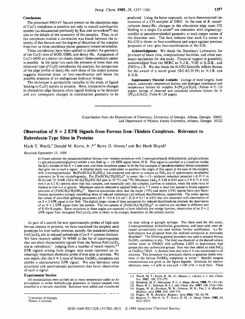
Figure 1. EPR spectra of ferrous Fe(SR)4 complexes of, from top to bottom, 2-mercaptoethanol, dithiothreitol, glutathione, and D,L-di- hydrolipoate. Solutions were prepared in water as described in the Ex- perimental Section and in ref 1. The solutions were 40 mM in thiol (20 mM in dithiol) and LiOH and 10 mM in FeC1,. The lowest spectrum is of an - 11 mM aqueous solution of FeS04. EPR conditions: T, 5-7 K; frequency, 9.41 GHz; power, 0.2 mW; modulation, 16 G at 100 kHz; time constant, 0.16 s. Positions of g = 10 and 4.3 are indicated on the top spectrum. The g = 4.3 feature is due to a small amount of rhombic high-spin Fe3+ impurity.
X-ray diffraction study confirmed the pseudotetrahedral FeS4 structure in Ba[ Fe(SCH2CH20H),].7
Due to the high-absorption cross section of barium for y-rays, Ba[ Fe(SCH2CH20H),] could not be examined by Mossbauer spectroscopy. The powder EPR spectrum of this solid shows only a broad feature extending from g,,, - 9 to gmi, - 3, which we attribute to an integer spin state. This EPR spectrum is pre- sumably complicated by magnetic dipolar interactions among neighboring iron atoms, which are C7 8, apart in the crystal. In order to circumvent these difficulties, concentrated aqueous so- lutions of [Fe(SCH2CH20H),12- were prepared as described in the Experimental Section for detailed examinations by Mossbauer and EPR spectroscopies. These solutions exhibited absorption (A,,, 309 nm) and 'H N M R (G(Fe-SCH2CH20H) 203 ppm at 25 "C in D20) spectra' very similar to those of Ba[Fe(SCH,C- H20H),] in DMSO. The IH N M R spectra revealed no reso- nances attributable to ferrous iron-thiolate clusters, such as [Fe2(SR),I2- or [Fe4(SR)',J2-, that have pseudotetrahedral co- ordination g e ~ m e t r i e s . ' ~ J ~ Furthermore, the reduced paramag- netism of these clusters relative to that of the mononuclear com- plexes is indicative of antiferromagnetic coupling within the clusters. No such coupling was evident in the magnetic Mossbauer spectra obtained in the present work (vide infra). Aqueous so- lutions of [Fe(SCH2CH2OH),I2- reproducibly exhibited quasi- reversible waves (e.g., AE, = 103 mV, ia/ic = 1.04) yielding E l j 2 = -0.35 V vs SCE. The increase of -0.46 V in the value of the midpoint reduction potential on changing the solvent from DMSO to water is similar in magnitude to that observed for the cubane
(10) Millar, M.; Lee, J. F.; Koch, S. A,; Fikar, R. F. Inorg. Chem. 1982, 21 , 4 105-41 06.
(1 1) Ueyama, N.; Nakata, M.; Fuji, M.; Terakawa, T.; Nakamura, A. Inorg. Chem. 1985, 24, 2190-2196.
(12) Hagen, K. S . ; Holm, R. H. Inorg. Chem. 1984, 23, 418-427. ( I 3) [Fe4(SCH2CH20H),,J2- was evident in 'H NMR spectra (6(Fe-SCH2) - 140 (b) and - 110 ppm (t)) only when the mole ratio of thiolatei
ferrous iron was <4.14 (14) Werth, M. T. Ph.D. Thesis, Iowa State University. 1989
S = 2 EPR Signals from Ferrous Fe(SR), Complexes Inorganic Chemistry, Vol. 28, No. 7, 1989 1359
0.10
A '
I I I I I L D.0 0.1 0.2
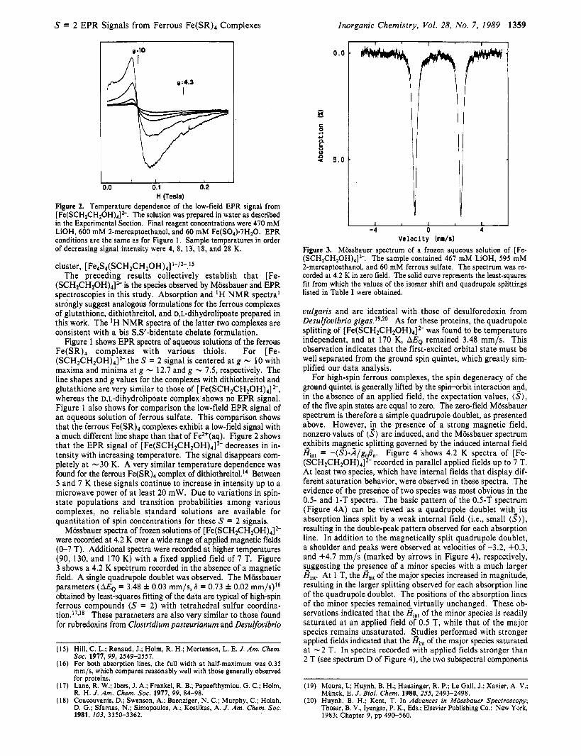
H (Tesla) Figure 2. Temperature dependence of the low-field EPR signal from [Fe(SCH2CH20H)4]2-. The solution was prepared in water as described in the Experimental Section. Final reagent concentrations were 470 mM LiOH, 600 mM 2-mercaptoethanol, and 60 mM Fe(S04).7H20. EPR conditions are the same as for Figure 1 . Sample temperatures in order of decreasing signal intensity were 4, 8, 13, 18, and 28 K.
cluster, [ Fe4S4(SCH2CH20H)4] '-/2-.15
The preceding results collectively establish that [Fe- (SCH2CH20H)4]2- is the species observed by Mossbauer and EPR spectroscopies in this study. Absorption and 'H N M R spectra' strongly suggest analogous formulations for the ferrous complexes of glutathione, dithiothreitol, and D,L-dihydrolipoate prepared in this work. The 'H NMR spectra of the latter two complexes are consistent with a bis S,S'-bidentate chelate formulation.
Figure 1 shows EPR spectra of aqueous solutions of the ferrous Fe(SR), complexes with various thiols. For [Fe- (SCH2CH20H)4]2- the S = 2 signal is centered at g - 10 with maxima and minima at g - 12.7 and g - 7.5, respectively. The line shapes and g values for the complexes with dithiothreitol and glutathione are very similar to those of [Fe(SCH2CH20H)4]2-, whereas the D,L-dihydrolipoate complex shows no EPR signal. Figure 1 also shows for comparison the low-field EPR signal of an aqueous solution of ferrous sulfate. This comparison shows that the ferrous Fe(SR), complexes exhibit a low-field signal with a much different line shape than that of Fe2+(aq). Figure 2 shows that the EPR signal of [Fe(SCH2CH20H)4]2- decreases in in- tensity with increasing temperature. The signal disappears com- pletely at -30 K. A very similar temperature dependence was found for the ferrous Fe(SR), complex of dithiothreit01.l~ Between 5 and 7 K these signals continue to increase in intensity up to a microwave power of at least 20 mW. Due to variations in spin- state populations and transition probabilities among various complexes, no reliable standard solutions are available for quantitation of spin concentrations for these S = 2 signals.
Mijssbauer spectra of frozen solutions of [ Fe(SCH2CH20H),] 2- were recorded at 4.2 K over a wide range of applied magnetic fields (0-7 T). Additional spectra were recorded at higher temperatures (90, 130, and 170 K) with a fixed applied field of 7 T. Figure 3 shows a 4.2 K spectrum recorded in the absence of a magnetic field. A single quadrupole doublet was observed. The Mijssbauer parameters (MQ = 3.48 f 0.03 mm/s, 6 = 0.73 f 0.02 mm/s)I6 obtained by least-squares fitting of the data are typical of high-spin ferrous compounds (S = 2) with tetrahedral sulfur coordina- t i ~ n . ' ' , ' ~ These parameters are also very similar to those found for rubredoxins from Clostridium pasteurianum and Desulfovibrio
(15) Hill, C. L.; Renaud, J.; Holm, R. H.; Mortenson, L. E. J . Am. Chem. SOC. 1917. 99. 2549-2551. , , ~ . ~ ~ . .
(16) For both absorption lines, the full width at half-maximum was 0.35 mm/s, which compares reasonably well with those generally observed for proteins.
(17) Lane, R. W.; Ibers, J. A,; Frankel, R. B.; Papaefthymiou, G . C.; Holm, R . H . J . Am. Chem. SOC. 1911, 99, 84-98.
(18) Coucouvanis, D.; Swenson, A,; Baenziger, N . C.; Murphy, C.; Holah, D. G.; Sfarnas, N.; Simopoulos, A,; Kostikas, A. J . Am. Chem. Soc. 1981, 103, 3350-3362.
0.0
- - x
c 0 .r(
Y
0 u)
a
n a 5 . 0
-4 0 4 Velocity (mn/s)
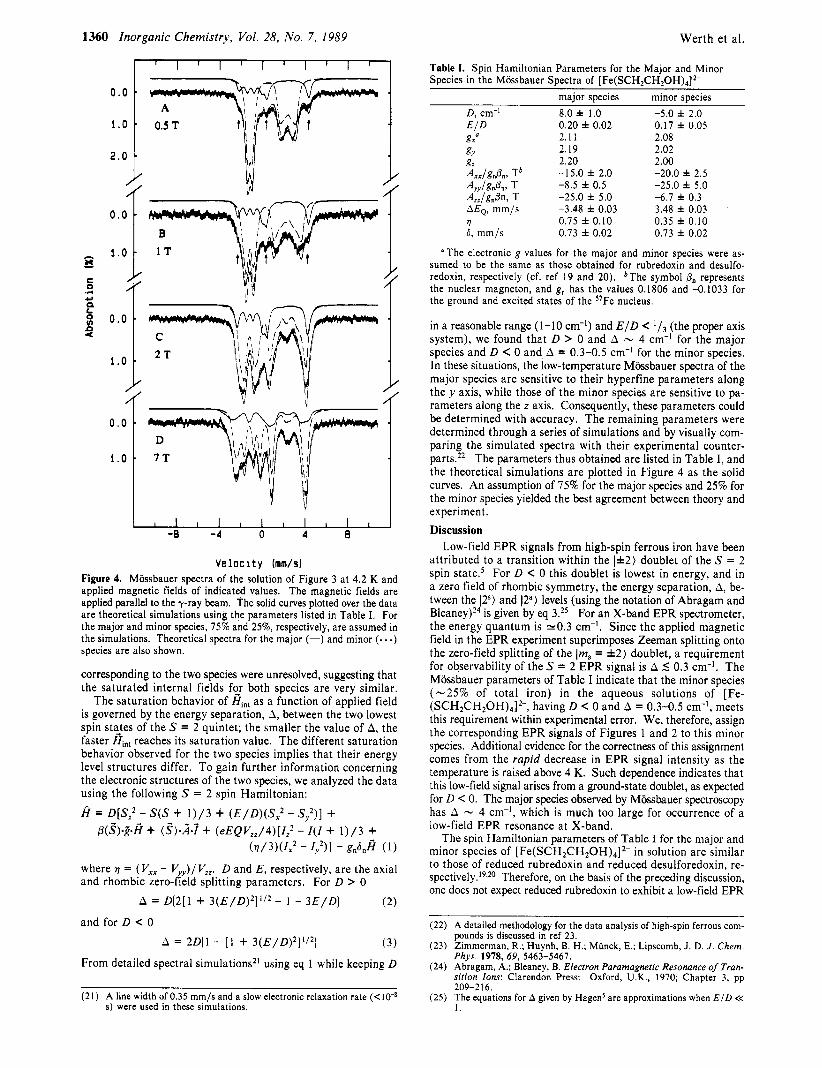
Figure 3. Mossbauer spectrum of a frozen aqueous solution of [Fe- (SCH2CH20H)4]2-. The sample contained 467 mM LiOH, 595 mM 2-mercaptoethanol, and 60 mM ferrous sulfate. The spectrum was re- corded at 4.2 K in zero field. The solid curve represents the least-squares fit from which the values of the isomer shift and quadrupole splittings listed in Table I were obtained.
vulgaris and are identical with those of desulforedoxin from Desuvovibrio g i g a ~ . I ~ , ~ O As for these proteins, the quadrupole splitting of [ Fe(SCH2CH20H)4]2- was found to be temperature independent, and at 170 K, AEQ remained 3.48 mm/s. This observation indicates that the first-excited orbital state must be well separated from the ground spin quintet, which greatly sim- plified our data analysis.
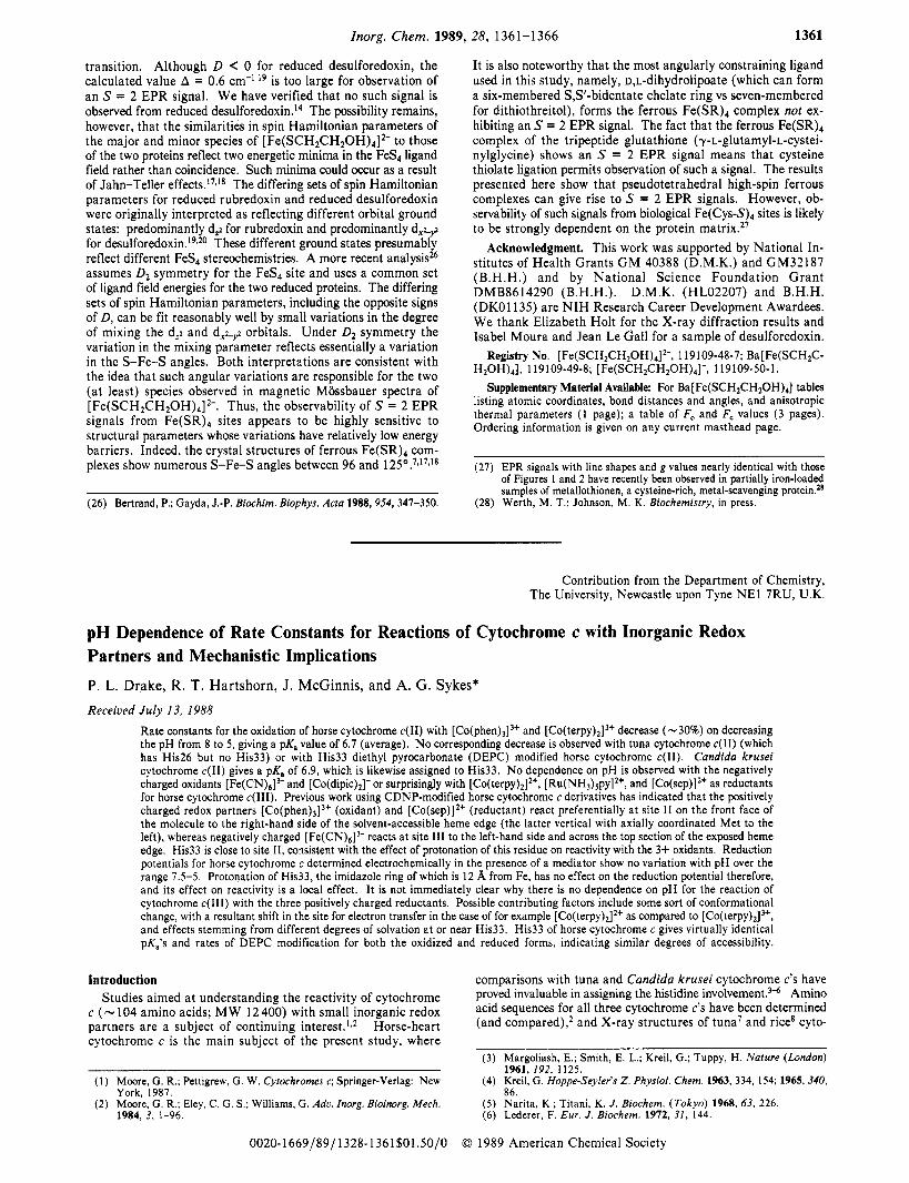
For high-spin ferrous complexes, the spin degeneracy of the ground quintet is generally lifted by the spin-orbit interaction and, in the absence of an applied field, the expectation values, (S), of the five spin states are equal to zero. The zero-field Miissbauer spectrum is therefore a simple quadrupole doublet, as presented above. However, i,n the presence of a strong magnetic field, nonzero values of (S) are induced, and the Mossbauer spectrum e_xhibits magnetic splitting governed by the induced internal field H,,, = - (S) .A/g,@,. Figure 4 shows 4.2 K spectra of [Fe- (SCH2CH20H)4]2- recorded in parallel applied fields up to 7 T. At least two species, which have internal fields that display dif- ferent saturation behavior, were observed in these spectra. The evidence of the presence of two species was most obvious in the 0.5- and 1-T spectra. The basic pattern of the 0.5-T spectrum (Figure 4A) can be viewed as a quadrupole doublet with-its absorption lines split by a weak internal field (Le., small (S)), resulting in the double-peak pattern observed for each absorption line. In addition to the magnetically split quadrupole doublet, a shoulder and peaks were observed at velocities of -3.2, +0.3, and +4.7 mm/s (marked by arrows in Figure 4), respectively, s_uggesting the prgsence of a minor species with a much larger H,,,. At 1 T, the Hh, of the major species increased in magnitude, resulting in the larger splitting observed for each absorption line of the quadrupole doublet. The positions of the absorption lines of the minor species remained_ virtually unchanged. These ob- servations indicated that the Hi,, of the minor species is readily saturated at an applied field of 0.5 T, while that of the major species remains unsaturated. SJudies performed with stronger applied fields indicated that the H,, of the major species saturated at -2 T. In spectra recorded with applied fields stronger than 2 T (see spectrum D of Figure 4), the two subspectral components
(19) Moura, I.; Huynh, B. H.; Hausinger, R. P.; Le Gall, J.; Xavier, A. V.; Miinck, E. J . Biol. Chem. 1980, 255, 2493-2498.
(20) Huynh. B. H.; Kent, T. In Advances in Mossbauer Spectroscopy; Thosar, B. V., Iyengar, P. K., Eds.; Elsevier Publishing Co.: New York, 1983; Chapter 9, pp 490-560.
Velocity (mm/s) Figure 4. Mossbauer spectra of the solution of Figure 3 at 4.2 K and applied magnetic fields of indicated values. The magnetic fields are applied parallel to the y-ray beam. The solid curves plotted over the data are theoretical simulations using the parameters listed in Table I. For the major and minor species, 75% and 2570, respectively, are assumed in the simulations. Theoretical spectra for the major (-) and minor (- - -) species are also shown.
corresponding to the two species were unresolved, suggesting that the saturated internal fields f y both species are very similar.
The saturation behavior of Hi,, as a function of applied field is governed by the energy separation, A, between the two lowest spin sta_tes of the S = 2 quintet; the smaller the value of A, the faster Hi,, reaches its saturation value. The different saturation behavior observed for the two species implies that their energy level structures differ. To gain further information concerning the electronic structures of the two species, we analyzed the data using the following S = 2 spin Hamiltonian:
A = D[S: - S(S + 1 ) / 3 + ( E / D ) ( S ? - S;)] + p(!?)-&fi + (!?)-k-? + ( eEQVz , /4 ) [ I : - 1(1 + 1 ) / 3 +
(7 /3 ) (1x2 - I,Z)I - gn6nfi ( 1 )
where 17 = (V,, - V ) / Vzz. D and E , respectively, are the axial and rhombic zero-Kld splitting parameters. For D > 0
( 2 )
( 3 )
From detailed spectral simulations2' using eq 1 while keeping D
A = D(2[1 + 3 ( E / D ) 2 ] ' / 2 - 1 - 3 E / D )
A = 2D(1 - [ l + 3 ( E / D ) 2 ] ' / 2 ]
and for D < 0
(21) A line width of 0.35 mm/s and a slow electronic relaxation rate (<lo-* s) were used in these simulations.
Table I. Spin Hamiltonian Parameters for the Major and Minor Species in the Mossbauer Spectra of [Fe(SCH2CH20H),]*-
major species minor species D , cm-' 8.0 f 1.0 E I D g,"
gz 2.20 AxxIgnPm Tb -15.0 f 2.0 AyyIgnPm T -8.5 f 0.5 A,,lg"@n, T -25.0 f 5.0 AEQ, mm/s -3.48 f 0.03
0.20 f 0.02 2.1 1
gu 2.19
?l 0.75 f 0.10 6, mm/s 0.73 f 0.02
-5.0 f 2.0 0.17 f 0.05 2.08 2.02 2.00 -20.0 f 2.5 -25.0 f 5.0 -6.7 f 0.3 3.48 f 0.03 0.35 f 0.10 0.73 f 0.02
"The electronic g values for the major and minor species were as- sumed to be the same as those obtained for rubredoxin and desulfo- redoxin, respectively (cf. ref 19 and 20). bThe symbol 6, represents the nuclear magneton, and g, has the values 0.1806 and -0.1033 for the ground and excited states of the "Fe nucleus.
in a reasonable range (1-10 cm-I) and E / D < (the proper axis system), we found that D > 0 and A - 4 cm-' for the major species and D < 0 and A = 0.3-0.5 cm-' for the minor species. In these situations, the low-temperature Mossbauer spectra of the major species are sensitive to their hyperfine parameters along the y axis, while those of the minor species are sensitive to pa- rameters along the z axis. Consequently, these parameters could be determined with accuracy. The remaining parameters were determined through a series of simulations and by visually com- paring the simulated spectra with their experimental counter- parts.22 The parameters thus obtained are listed in Table I, and the theoretical simulations are plotted in Figure 4 as the solid curves. An assumption of 75% for the major species and 25% for the minor species yielded the best agreement between theory and experiment. Discussion
Low-field EPR signals from high-spin ferrous iron have been attributed to a transition within the 1*2) doublet of the S = 2 spin state.' For D 0 this doublet is lowest in energy, and in a zero field of rhombic symmetry, the energy separation, A, be- tween the 12s) and 12a) levels (using the notation of Abragam and B l e a n e ~ ) ~ ~ is given by eq 3.25 For an X-band EPR spectrometer, the energy quantum is -0.3 cm-'. Since the applied magnetic field in the EPR experiment superimposes Zeeman splitting onto the zero-field splitting of the 1112, = f 2 ) doublet, a requirement for observability of the S = 2 EPR signal is A 5 0.3 cm-I. The Mossbauer parameters of Table I indicate that the minor species ( -25% of total iron) in the aqueous solutions of [Fe- (SCH2CH20H),12-, having D C 0 and A = 0.3-0.5 cm-', meets this requirement within experimental error. We, therefore, assign the corresponding EPR signals of Figures 1 and 2 to this minor species. Additional evidence for the correctness of this assignment comes from the rapid decrease in EPR signal intensity as the temperature is raised above 4 K. Such dependence indicates that this low-field signal arises from a ground-state doublet, as expected for D < 0. The major species observed by Mossbauer spectroscopy has A - 4 cm-I, which is much too large for occurrence of a low-field EPR resonance at X-band.
The spin Hamiltonian parameters of Table I for the major and minor species of [Fe(SCH2CH20H),12- in solution are similar to those of reduced rubredoxin and reduced desulforedoxin, re- spectively. 19,20 Therefore, on the basis of the preceding discussion, one does not expect reduced rubredoxin to exhibit a low-field EPR
(22) A detailed methodology for the data analysis of high-spin ferrous com- pounds is discussed in ref 23.
(23) Zimmerman, R.; Huynh, B. H.; Miinck, E.; Lipscomb, J . D. J . Chem. Phys. 1978, 69, 5463-5467.
(24) Abragam, A,; Bleaney, B. Electron Paramognetic Resonance of Tran- sition Ions; Clarendon Press: Oxford, U.K., 1970 Chapter 3, pp 209-2 16.
(25) The equations for A given by Hagens are approximations when E / D << 1 .
Znorg. Chem. 1989, 28, 1361-1366 1361
transition. Although D C 0 for reduced desulforedoxin, the calculated value A = 0.6 cm-' I9 is too large for observation of an S = 2 EPR signal. We have verified that no such signal is observed from reduced desulf~redoxin. '~ The possibility remains, however, that the similarities in spin Hamiltonian parameters of the major and minor species of [Fe(SCH2CH20H)4]2- to those of the two proteins reflect two energetic minima in the FeS4 ligand field rather than coincidence. Such minima could occur as a result of Jahn-Teller effects.l7-I8 The differing sets of spin Hamiltonian parameters for reduced rubredoxin and reduced desulforedoxin were originally interpreted as reflecting different orbital ground states: predominantly dg for rubredoxin and predominantly d+,,2 for d e s u l f o r e d o ~ i n . ' ~ ~ ~ ~ These different ground states presumably reflect different FeS4 stereochemistries. A more recent analysis26 assumes D2 symmetry for the FeS4 site and uses a common set of ligand field energies for the two reduced proteins. The differing sets of spin Hamiltonian parameters, including the opposite signs of D, can be fit reasonably well by small variations in the degree of mixing the dz2 and dX2+ orbitals. Under D2 symmetry the variation in the mixing parameter reflects essentially a variation in the S-Fe-S angles. Both interpretations are consistent with the idea that such angular variations are responsible for the two (at least) species observed in magnetic Mossbauer spectra of [Fe(SCH2CH20H)4]2-. Thus, the observability of S = 2 EPR signals from Fe(SR)4 sites appears to be highly sensitive to structural parameters whose variations have relatively low energy barriers. Indeed, the crystal structures of ferrous Fe(SR), com- plexes show numerous S-Fe-S angles between 96 and 125°.7J7J8
It is also noteworthy that the most angularly constraining ligand used in this study, namely, D,L-dihydrolipoate (which can form a six-membered S,S'-bidentate chelate ring vs seven-membered for dithiothreitol), forms the ferrous Fe(SR)4 complex not ex- hibiting an S = 2 EPR signal. The fact that the ferrous Fe(SR)4 complex of the tripeptide glutathione (y-L-glutamyl-L-cystei- nylglycine) shows an S = 2 EPR signal means that cysteine thiolate ligation permits observation of such a signal. The results presented here show that pseudotetrahedral high-spin ferrous complexes can give rise to S = 2 EPR signals. However, ob- servability of such signals from biological F e ( c y ~ - S ) ~ sites is likely to be strongly dependent on the protein matrix.27
Acknowledgment. This work was supported by National In- stitutes of Health Grants GM 40388 (D.M.K.) and GM32187 (B.H.H.) and by National Science Foundation Grant DMB8614290 (B.H.H.). D.M.K. (HL02207) and B.H.H. (DKO1135) are N I H Research Career Development Awardees. We thank Elizabeth Holt for the X-ray diffraction results and Isabel Moura and Jean Le Gall for a sample of desulforedoxin.
Supplementary Material Available: For Ba[ Fe(SCH2CH20H)4] tables listing atomic coordinates, bond distances and angles, and anisotropic thermal parameters (1 page); a table of F, and F, values (3 pages). Ordering information is given on any current masthead page.
(27) EPR signals with line shapes and g values nearly identical with those of Figures 1 and 2 have recently been observed in partially iron-loaded samples of metallothionen, a cysteine-rich, metal-scavenging protein.28
(28) Werth, M . T.: Johnson, M. K. Biochemistry, in press.
Contribution from the Department of Chemistry, The University, Newcastle upon Tyne NE1 7RU, U.K.
pH Dependence of Rate Constants for Reactions of Cytochrome c with Inorganic Redox Partners and Mechanistic Implications P. L. Drake, R. T. Hartshorn, J. McGinnis, and A. G. Sykes* Received J u l y 13, 1988
Rate constants for the oxidation of horse cytochrome c(I1) with [Co(phen)J" and [C~(terpy)~]~+ decrease (-30%) on decreasing the pH from 8 to 5 , giving a pK, value of 6.7 (average). No corresponding decrease is observed with tuna cytochrome c(I1) (which has His26 but no His33) or with His33 diethyl pyrocarbonate (DEPC) modified horse cytochrome c(I1). Candida krusei cytochrome c(I1) gives a pK, of 6.9, which is likewise assigned to His33. No dependence on pH is observed with the negatively charged oxidants [Fe(CN)$ and [C~(dipic)~]- or surprisingly with [Co(terpy),l2+, [ R U ( N H ~ ) ~ ~ ~ ] * ' , and [Co(sep)12' as reductants for horse cytochrome c(II1). Previous work using CDNP-modified horse cytochrome c derivatives has indicated that the positively charged redox partners [C~(phen)~]~ ' (oxidant) and [Co(sep)12+ (reductant) react preferentially at site I1 on the front face of the molecule to the right-hand side of the solvent-accessible heme edge (the latter vertical with axially coordinated Met to the left), whereas negatively charged [Fe(CN)6]3- reacts at site 111 to the left-hand side and across the top section of the exposed heme edge. His33 is close to site 11, consistent with the effect of protonation of this residue on reactivity with the 3+ oxidants. Reduction potentials for horse cytochrome c determined electrochemically in the presence of a mediator show no variation with pH over the range 7.5-5. Protonation of His33, the imidazole ring of which is 12 %, from Fe, has no effect on the reduction potential therefore, and its effect on reactivity is a local effect. It is not immediately clear why there is no dependence on pH for the reaction of cytochrome c(II1) with the three positively charged reductants. Possible contributing factors include some sort of conformational change, with a resultant shift in the site for electron transfer in the case of for example [C~(terpy)~]~+ as compared to [C~(terpy)~]", and effects stemming from different degrees of solvation at or near His33. His33 of horse cytochrome c gives virtually identical pK,'s and rates of DEPC modification for both the oxidized and reduced forms, indicating similar degrees of accessibility.
Introduction Studies aimed at understanding the reactivity of cytochrome
c ( - 104 amino acids; M W 12 400) with small inorganic redox partners are a subject of continuing interest.ls2 Horse-heart cytochrome c is the main subject of the present study, where
( I ) Moore, G. R.; Pettigrew, G. W. Cytochromes c; Springer-Verlag: New York, 1987.
(2) Moore, G. R.; Eley, C. G. S.; Williams, G. Adc. Inorg. Bioinorg. Mech. 1984, 3, 1-96.
0020-1669/89/1328-1361$01.50/0
comparisons with tuna and Candida krusei cytochrome c's have proved invaluable in assigning the histidine in~olvement.~" Amino acid sequences for all three cytochrome c's have been determined (and compared),2 and X-ray structures of tuna7 and rice" cyto-
(3) Margoliash, E.; Smith, E. L.; Kreil, G.; Tuppy, H. Nature (London) 1961. 192. 1125.
(4) Kreil: G. Hoppe-Seyler's Z . Physiol. Chem. 1963, 334, 154; 1965, 340, 86.