18
| Date post: | 26-Mar-2016 |
| Category: |
Documents |
| Upload: | elliot-schatmeier |
| View: | 229 times |
| Download: | 3 times |


You are reading Obstetrics and Gynecology: The Essentials of Clinical Care.
Visit www.thieme.com to purchase this title.

91
10 Intrapartum Care and Fetal SurveillanceCarolina Ghia, Luciana Prozzillo, and Gustavo F. Leguizamón
In the last 50 years different techniques have been devel-oped to assess fetal health during labor. From intermit-tent auscultation to the most recent invasive techniques, the objective of these methods is to detect fetal stress early, so that complications, such as intrapartum fetal hy-poxia leading to cognitive impairment, cerebral palsy, or even fetal death, can be prevented.
Persistent intrapartum hypoxia, which complicates about 1 % of all labors, can lead to severe acidemia, which in turn may compromise those vital tissues requiring strict oxygen levels, such as in the renal, cardiovascular, and central nervous systems. The latter is the most vul-nerable to oxygen deprivation and is, therefore, frequent-ly involved in long-term sequelae.
Thus, an ideal method of intrapartum fetal surveil-lance should be able to differentiate between transient hypoxia without metabolic acidosis and pathologic hy-poxia leading to acidosis and tissue damage. This is of ut-most importance, since it allows accurate intervention and prevention of long-term sequelae without increas-ing unnecessary cesarean sections. In other words, it re-quires a method with a high degree of sensitivity and a low false-positive rate.
Since alterations in maternal blood pressure, heart rate, and uterine contractions have direct effects on fe-tal oxygenation, maternal vital signs should be monitored during labor, especially in the event of a non-reassuring fetal pattern.
Fetal heart rate and its variations is a good parameter of fetal response to labor events. In particular, knowledge of normal and abnormal patterns will allow both detection of fetal distress and accurate intervention. Other meth-ods of measuring metabolic status, such as fetal blood sampling, pulse oximetry, and lactate measurement, have been developed in order to complement fetal assessment. These methods are discussed in detail in this chapter.
Definitions
Acceleration: This describes a short-term rise in fetal heart rate of greater than 15 beats per minute (bpm) that lasts for more than 15 seconds.
Acidemia: This condition arises from increased hydrogen content in the blood.
Acidosis: This term describes a state of increased hydro-gen content in the tissues.
Respiratory acidosis: The accumulation of CO2 leads to respiratory acidosis. When the umbilical cord is com-pressed, CO2 rapidly accumulates in the fetal blood.
Metabolic acidosis: During the peak of uterine contrac-tion, intramyometrial pressure exceeds uterine arterial pressure and therefore, the blood flow decreases, leav-ing the fetus in a transient state of hypoperfusion. In the event of basal inadequate oxygen delivery to the fetus, this transitory lack of perfusion leads to fetal hypoxia, which can result in metabolic acidosis.
Asphyxia: This is a state of hypoxia with metabolic aci-dosis.
Deceleration: This describes a fall in the fetal heart rate of greater than 15 bpm that lasts for more than 15 sec-onds (but less than 10 minutes).
Hypoxemia: This condition occurs following decreased oxygen concentration in the blood.
Hypoxia: This term describes decreased oxygen concen-tration in the tissues.

Section I Normal Obstetrics92
Pa
rt II
O
bste
tric
s Fetal Heart Rate: Normal and Pathologic Patterns
The normal fetal heart rate at term varies between 110 and 160 bpm. This is measured by a cardiotachometer during intrapartum fetal monitoring and is defined as the baseline heart rate, which is much higher in the second trimester and declines thereafter with increasing gesta-tional age. This decline in baseline heart rate is a good in-dicator of development of the vagal tone.
A heart rate above 160 bpm that persists for more than 10 minutes (a shorter period could represent a transient acceleration) is referred to as tachycardia. Among the most frequent causes of tachycardia are maternal fever (as seen in chorioamnionitis), the use of drugs that el-evate heart rate (e. g., ritodrine), fetal anemia, and fetal arrhythmias. However, if tachycardia is persistent, it can also indicate fetal hypoxia.
A heart rate of less than 110 bpm that persists for more than 10 minutes (as distinct from transient deceleration) is known as bradycardia. Some fetuses have normal base-lines of 100–105 bpm. However, in most cases, it indicates some metabolic alteration (e. g., maternal hypothermia or hypoglycemia), use of drugs that diminish heart rate (e. g., magnesium sulfate), or cardiac abnormalities (e. g., heart block, umbilical cord occlusion).
Variability
The difference in heart rate from beat to beat, which is registered by a device known as a cardiotachometer as a trace moving over and under the baseline, is known as variability (Fig. 10.1). Normal variability ranges from 5 bpm to 25 bpm, and is an indicator of a well-functioning fetal brain. However, heart rate variability increases with gestational age, reaching a stable pattern at approximate-ly 28 weeks of gestation. Variations between fetuses are also observed.
Decreased variability is defined as less than 5 bpm for longer than 80 minutes (Fig. 10.2). It may reflect differ-ent physiological conditions, including fetal sleep and prematurity, or may be secondary to drugs such as nar-cotics, barbiturates, tranquilizers, phenotiazines, and general anesthetics. Among the pathologic causes of de-creased heart rate variability are maternal hypoglycemia, reduced fetal oxygenation and acidosis, major anomalies of the fetal central nervous system, chorioamnionitis, and fetal heart block.
Transient Accelerations
Heart rate accelerations are usually a response to fetal movement or external stimulations, such as uterine con-tractions (Fig. 10.3). Their spontaneous presence indi-cates absence of hypoxia, especially in the context of nor-mal baseline and variability. The presence of provoked ac-celerations, even in a context of non-reassuring fetal heart rate, rules out a pH lower than 7.20 on scalp sampling.
The absence of accelerations should be interpreted in the context of other variables. In general, if the fetal heart rate pattern is non-reassuring with no accelerations, ap-proximately half of such fetuses will be acidotic. On the other hand, the lack of accelerations in an otherwise re-assuring pattern is generally not associated with an in-creased fetal risk of hypoxia.
Transient Decelerations
Decelerations are classified in three groups, according to their location regarding uterine contractions:
• Early decelerations. These coincide with uterine con-tractions and appear as vagal reactions in response to fetal head compression during the final stages of labor and they are not associated with fetal hypoxia, however. The drop in fetal heart rate appears as a
Fig. 10.1 Fetal heart rate variability that changes with gestational age is an indicator of a well-functioning fetal brain.

10 Intrapartum Care and Fetal Surveillance 93
Fig. 10.2 Decreases in fetal heart rate variability (as indicated by the changes in peak size) may reflect aberrant physiologic conditions that may harm the fetus.
Fig. 10.3 The presence of accelerations, which can be provoked by uterine contractions, can rule out problems such as hypoxia and acidosis.

Section I Normal Obstetrics94
Pa
rt II
O
bste
tric
s V-shaped pattern coinciding with the uterine con-traction, and does not persist beyond it. They create a “mirror” image of the contraction in the monitor trace, with their nadir in coincidence with the peak of the contraction.
• Variable decelerations. These are the most common type of deceleration during labor. They are called “variable” because of the lack of a particular relation with contractions, and the absence of a consistent pattern (Fig. 10.4). These decelerations usually are not associated with fetal hypoxia. They are caused by fetal heart rate changes in response to blood pressure alterations, which are frequently due to cord com-pression. When the cord vein is compressed, CO2 ac-cumulates in fetal blood; this can produce respiratory acidosis. If compression continues, oxygen delivery becomes insufficient; producing metabolic acidosis, turning the situation into mixed acidosis.
Variable decelerations can be further classified according to their severity:
• Mild decelerations. These have duration of less than 30 seconds regardless of the depth, or heart rate not below 80 bpm regardless of duration.
• Moderate decelerations. These fall below 80 bpm.• Severe decelerations. These are decelerations that
last for more than 60 seconds and fall to less than 70 bpm.
• Late decelerations. These generally appear within 30 seconds of a contraction; their nadir is delayed with respect to the peak of the contraction, usually descend no more than 40 bpm from the baseline, and last a variable amount of time beyond the contrac-tion (Fig. 10.5). Late decelerations reflect transient periods of fetal hypoxia due to a diminished uterine– placental blood flow during a contraction. The sever-ity of hypoxia cannot be predicted from the depth of the deceleration. The persistence in time of late
Fig. 10.4 Variable decelerations are usually not associated with fetal hypoxia but are frequently caused by cord compression.

10 Intrapartum Care and Fetal Surveillance 95
Fig. 10.4 (continued)
Fig. 10.5 Late decelerations reflect transient periods of fetal hypoxia during contractions.
decelerations gives a more reliable reason to suspect hypoxia and fetal metabolic acidosis.
A fetus whose placenta suffers from any pathologic con-dition is more likely to develop late decelerations in re-sponse to decreased oxygen exchange. This condition usually includes maternal hypertension or pre-eclampsia, maternal diabetes, or collagen vascular diseases.
Sinusoidal Pattern
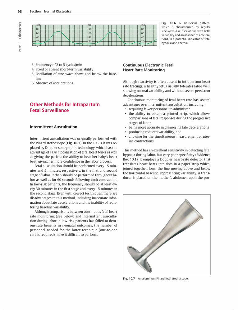
A sinusoidal pattern is strongly associated with severe fe-tal anemia and hypoxia. Its characteristics include:
1. Stable baseline fetal heart rate of 120–160 bpm with regular sine-wave–like oscillations (Fig. 10.6)
2. Amplitude of 5 to 15 bpm
Section I Normal Obstetrics96
Pa
rt II
O
bste
tric
s
3. Frequency of 2 to 5 cycles/min4. Fixed or absent short-term variability5. Oscillation of sine wave above and below the base-
line6. Absence of accelerations
Other Methods for Intrapartum Fetal Surveillance
Intermittent Auscultation
Intermittent auscultation was originally performed with the Pinard stethoscope (Fig. 10.7). In the 1950s it was re-placed by Doppler sonographic technology, which has the advantage of easier localization of fetal heart tones as well as giving the patient the ability to hear her baby’s heart beat, giving her more confidence in the labor process.
Fetal auscultation should be performed every 15 min-utes and 5 minutes, respectively, in the first and second stage of labor. It then should be performed throughout la-bor as well as for 60 seconds following each contraction. In low-risk patients, the frequency should be at least ev-ery 30 minutes in the first stage and every 15 minutes in the second stage. Even with correct techniques, there are disadvantages to this method, including inaccurate infor-mation about late decelerations and the inability of regis-tering baseline variability.
Although comparisons between continuous fetal heart rate monitoring (see below) and intermittent ausculta-tion during labor in low-risk patients has failed to dem-onstrate benefits in neonatal outcomes, the number of personnel needed for the latter technique (one-to-one care is required) make it difficult to perform.
Continuous Electronic Fetal Heart Rate Monitoring
Although reactivity is often absent in intrapartum heart rate tracings, a healthy fetus usually tolerates labor well, showing normal variability and without severe persistent decelerations.
Continuous monitoring of fetal heart rate has several advantages over intermittent auscultation, including:
• requiring fewer personnel to administer• the ability to obtain a printed strip, which allows
comparisons of fetal responses during the progressive stages of labor
• being more accurate in diagnosing late decelerations• producing reduced variability, and• allowing for the simultaneous measurement of uter-
ine contractions
This method has an excellent sensitivity in detecting fetal hypoxia during labor, but very poor specificity (Evidence Box 10.1). It employs a Doppler heart-rate detector that translates heart beats into dots in a paper strip which, joined together, form the line moving above and below the horizontal baseline, representing variability. A trans-ducer is placed on the mother’s abdomen upon the pro-
Fig. 10.6 A sinusoidal pattern, which is characterized by regular sine-wave–like oscillations with little variability and an absence of accelera-tions, is a potential indicator of fetal hypoxia and anemia.
Fig. 10.7 An aluminum Pinard fetal stethoscope.
10080604020
00
20406080 80
40
0
20
60
100 100

10 Intrapartum Care and Fetal Surveillance 97
jection of the fetal shoulder. A tocodynamometer is also placed in the mother’s abdomen, in the projection of the uterine fundus, to obtain simultaneous reading of uterine contractions.
Approximately one-half of fetuses that present with a non-reassuring pattern during labor (e. g., reduced vari-ability, persistent decelerations progressing in severity, tachycardia, or bradycardia) are born with normal Apgar scores. If the patient’s membranes are ruptured and there is difficulty in obtaining good recordings with external monitoring, an internal electrode can be applied to the fetal scalp for a more accurate tracing.
Fetal Metabolic Status Assessment
In order to improve the specificity of continuous moni-toring for fetal hypoxia/acidemia and to prevent unnec-essary operative interventions, invasive procedures have been developed to further evaluate non-reassuring pat-terns in the electronic heart-rate monitoring.
One such method is fetal pulse oximetry, whereby a specially adapted device is placed in contact with the fe-tus’ cheek to provide information about fetal oxygenation during labor. Another method involves sampling fetal blood in order to measure pH, base deficit, and lactate as direct parameters of the fetal metabolic status. This in-volves obtaining samples by puncturing the fetal scalp. However, this method can only be used if the membranes are ruptured and the woman’s a cervix is dilated at least 4–5 cm).
Fetal Scalp pH
This is the most accurate technique to diagnose fetal aci-dosis. It consists of taking a blood sample from the fetal scalp using a lancet and a capillary collection tube. Dur-ing labor, normal pH is above 7.24. A pH value between 7.20 and 7.24 indicates pre-acidemia, and a pH lower than 7.20 indicates acidosis. Complementing electronic fetal monitoring with fetal scalp pH measurement should reduce cesarean rates for non-reassuring fetal status. However, the technique is rarely used. Measurement of base excess of umbilical artery is a great tool for differen-tiating respiratory from metabolic acidosis. Base excess below −12 mmol/L has a high correlation with increased risk of neonatal neurologic injury.
Fetal Pulse Oximetry
Fetal pulse oximetry is a tool that measures fetal arterial O2 saturation using a sensor attached to the fetus’ tem-ple or cheek. Normal fetal O2 saturation during labor is 40–70 %. Values below 30 % are strongly associated with pH under 7.20. The American College of Obstetricians and Gynecologists states that further studies confirming safety and efficacy are required before recommendation of this technology. To use it, the membranes must be rup-tured and the cervix ought to be dilated 2 cm or more.
Meconium
Meconium is the normal content of the fetal gut. When the fetus is suffering from hypoxia, because of gut vaso-constriction, there can be passage of meconium to the amniotic fluid. Hypoxia also works as an important stim-ulus for fetal gasping, which can result in fetal aspiration of meconium. Thick meconium is frequently associated with oligohydramnios, which in turn is a sign of placental dysfunction. Nevertheless, with normal fetal heart rate, the presence of meconium does not always reflect fetal hypoxia.
Amnioinfusion
In variable decelerations caused by oligohydramnios, this technique can improve Apgar score and pH values and can also reduce the number of cesarean sections for non-reassuring fetal status. Amnioinfusion might reduce the cord compression that leads to hypoxia. It is also used in the presence of meconium to dilute it.
Interventions for Altered Heart Rate Patterns
Clinical Management
The following steps should be taken when an altered heart rate pattern is detected via continuous monitoring:
• Assess the mother’s vital signs and uterine tone. It is possible that fever, hypotension, hypertonic uterus, or tachysystolia may be causing fetal distress.

Section I Normal Obstetrics98
Pa
rt II
O
bste
tric
s • Place the mother in the left lateral position. This action relieves uterine compression of the vena cava, allow-ing better blood flow.
• Increase oxygen supply. Administer oxygen to the mother via a nasal cannula or a mask to improve the mother’s Po2 and therefore placental O2 exchange.
• Increase intravenous hydration. This helps to maxi-mize uterine flow and perfusion and to correct a pos-sible hypotension as a cause of the abnormal pattern.
• Discontinue oxytocin infusion (if being administered). This will improve placental perfusion as it decreases uterine contractions.
• Initiate acoustic or scalp stimulation. Fetuses who respond with increased heart rate upon stimulation have better outcomes than those who don’t. Usually, fetal response to either acoustic or scalp stimulation reflects a lack of acidosis.
Operative Management
When a fetus is considered to be “at risk” due to a per-sistent non-reassuring pattern in continuous fetal moni-toring, and back-up studies cannot offer reassurance, the physician must proceed with rapid delivery.
The choice between instrumental vaginal delivery and cesarean section will depend on the stage of labor, cervi-cal dilatation, vertex station, and the estimated time for performing each procedure. Therefore, each case must be analyzed individually in order to achieve delivery in the least amount of time and without exposing the mother and fetus to unnecessary procedures.
Key Points
• The objective of the intrapartum fetal testing is to determine whether the fetus presents hypoxia and/or metabolic acidosis.
• If hypoxia is detected, attempts to reverse it by interventions such as hydration, discontinuation of oxytocin, oxygen supply, or fetal stimulation must be performed.
• If persistent non-reassuring results are observed in spite of such interventions, expedited delivery must be initiated.
Evidence Box 10.1
Careful interpretation of specific FHR patterns can be a useful screening test for fetal asphyxia. However, supplementary tests are required to identify the large number of false-positive patterns to avoid unnecessary intervention.
Low et al. analyzed selected patterns of important fetal heart rate variables, during the last hour before delivery, for their predictive value for fetal asphyxia among a group of 71 term infants with umbilical artery base deficit >16 mmol/L, and a control group of 71 term infants with umbilical artery base deficit <8 mmol/L. The fetal heart rate variables associated with fetal asphyxia included absent and minimal
baseline variability and late and prolonged decelerations. Fetal heart rate patterns with absent baseline variability were the most specific, but identified only 17 % of the asphyxia group. The sensitivity of this test increased to 93 % with the addition of less specific patterns. The estimated positive predictive value ranged from 18.1 % to 2.6 %, and the negative predictive value ranged from 98.3 % to 99.5 %. The investigators concluded that a narrow 1-hour window of fetal heart rate patterns, including minimal baseline variability and late or prolonged decelerations, can predict fetal asphyxial exposure before decompensation and newborn morbidity. Thus, with careful interpretation, predictive fetal heart rate patterns can be a useful screening test for fetal asphyxia. However, supplementary tests are required to confirm the diagnosis and to identify the large number of false-positive patterns to avoid unnecessary intervention.
Low JA, Victory R, Derrick EJ. Predictive value of electronic fetal monitoring for intrapartum fetal asphyxia with metabolic acidosis. Obstet Gynecol 1999 Feb;93(2):285-91.
Further Reading
American College of Obstetricians and Gynecologists. Fetal Distress and Birth Asphyxia. ACOG Committee Opinion 1994, No 137, Washington, DC
American College of Obstetricians and Gynecologists. Fetal Heart Rate Monitoring, Interpretation and Management. ACOG Technical Bulletin 1995, No. 207, Washington, DC
Dildy G. Intrapartum assessment of the fetus: historical and evidence-based practice. Obstet Gynecol Clin 2005, Vol. 32, Issue 2.
National Institute of Child Health and Human Development Research Planning Workshop. Electronic fetal heart rate monitoring: research guidelines for interpretation. Am J Obstet Gynecol 1997;177(6):1385–1390
Gabbe SG, Niebyl JR, Simpson JL. Obstetrics—Normal and Problem Pregnancies. 5th ed. New York: Churchill Living-stone / Elsevier; 1997
Graham E. Intrapartum electronic fetal heart rate monitoring and the prevention of perinatal brain injury. ACOG 2006, Vol. 108, No.3, Part 1
Jibodu OA, Arulkumaran S. Intrapartum fetal surveillance. Curr Opin Obstet Gynecol 2000;12(2):123–127
Smith JF Jr, Onstad JH. Assessment of the fetus: intermit-tent auscultation, electronic fetal heart rate tracing, and fetal pulse oximetry. Obstet Gynecol Clin North Am 2005;32(2):245–254

99
11 Labor and DeliveryVanina S. Fishkel and Gustavo F. Leguizamòn
This chapter reviews the determinants of human parturi-tion, or labor. Labor is a physiologic process consisting in organized uterine contractions of adequate intensity, fre-quency, and duration to affect complete cervical dilata-tion and expulsion of the fetus, placenta, and fetal mem-branes through the birth canal.
Labor progresses from a state of uterine quiescence (latent phase) to a phase characterized by uterine con-tractions and cervical dilatation (active phase). Mater-nal structures (e.g., pelvis) and functions (e.g., uterine contractions) as well as fetal structures (e.g., presenting parts) and functions (e.g., fetal cardinal movements) are of utmost importance in the progression of labor.
Labor Physiology
The physiology of term labor initiation is not fully eluci-dated, but during the past several decades significant in-formation has been added to our understanding of this process.
Uterine activity can be classified as having four dis-tinct phases (Fig 11.1):
• Phase 0 (quiescence). This Phase is characterized by uterine quiescence and the leading hormone is pro-gesterone.
• Phase 1 (activation). In this Phase, receptors for oxy-tocin and prostaglandins are actively synthesized as well as an increased number of gap junctions, leading to a progressive increase in uterine sensitivity to dif-ferent uterotonics. Estrogens play a pivotal role in this phase.
• Phase 2 (stimulation). Once the uterus has reached its potential to respond, oxytocin and prostaglandins stimulate contractions actively.
• Phase 3 (involution). After delivery, oxytocin leads to uterine contraction and bleeding decreases signifi-cantly.
Mechanics of Labor
Adequate interaction between the fetus’ features and ma-ternal pelvis allows vaginal delivery. Thus, maternal con-tractions to propel the fetus through the birth canal and the ability of the fetus to pass through the mother’s pelvic bones are crucial to successful labor and birth.
Contractions
Uterine contractions are characterized by the following measurable parameters:
Amplitude or intensity: The ability of the external toco-dynamometer or manual palpation to determine contrac-tion intensity is limited and of no clinical value. Intensity can be measured more precisely by an intrauterine pres-sure catheter. This device is placed transcervically inside
InhibitorsProgesteroneProstacyclinRelaxinNitric oxideParathyroid hormone-related peptide
orticotropin- releasing hormone
ental lactogen
UterotropinsEstrogen
es
orticotropin- releasing hormone
UterotoninsProstaglandinsOxytocin
InvolutionOxytocin
Ute
rine
cont
ract
ility
Phase 0ence)
Phase 1(Activation)
Phase 2(S )
Phase 3(Invo )
Part n
Fig. 11.1 Uterine activity in pregnancy. Adapted from Challis JRG, Gibb W: Control of Parturition. Prenatal and Neonatal Medicine 1996;1:283.

Section I Normal Obstetrics100
Pa
rt II
O
bste
tric
s the uterine cavity after membrane rupture and above the fetal presenting part.
The Montevideo Unit attempts to measure objectively the intensity of the contraction. It is calculated by the av-erage strength of contractions in millimeters of mercury multiplied by the number of contractions in 10 minutes. During the active phase of labor, 200–250 Montevideo Units are considered normal. Since an internal catheter is not routinely used in clinical practice, the ability to mea-sure intensity of uterine contractions is limited.
Frequency: Laboring patients in the active phase usually contract 3–5 times in 10 minutes. The presence of more than 5 contractions in 10 minutes for a period of 20 min-utes is abnormal and is defined as tachysystole. When an alteration in the fetal heart rate accompanies tachysystole it is called hyperstimulation.
Duration: The sensitivity to detect the uterine contrac-tions varies with different methods. The highest sensitiv-ity is observed with internal catheters followed by palpa-tion and, finally, patient perception. External tocodyna-mometer determinations are biased by the sensitivity of the device as well as maternal body habit.
The Pelvis
Since the progression of labor and fetal descent are de-termined in part by the relationship of the fetal present-ing part and the bony pelvis, the obstetrician must be-come familiar with the evaluation of the pelvic dimen-sions, which is traditionally determined by a pelvimeter (Fig. 11.2).
Overall, four different shapes of female bony pelvis have been described. Two of them with favorable charac-teristics for vaginal delivery (gynecoid, anthropoid), and two (android, platypelloid) more frequently associated with cephalopelvic disproportion (CPD).
The bony pelvis (Fig. 11.3) is composed of the sacrum, ilium, ischium, and pubis. The pelvic brim separates the false (greater) pelvis from the true (lesser) pelvis. The true pelvis is further divided into three sections: 1) the pelvic inlet, 2) midpelvis, and 3) pelvic outlet.
Pelvic Inlet
The following are the measurable parts of the pelvic in-let.
Diagonal conjugate: This is the distance from the sacral promontory to the inferior margin of the symphysis pu-bis. This measure can be obtained by vaginal examina-tion.
Fig. 11.2 A Martin pelvimeter.
Sacroiliac joint Sacrum
Anteriorsuperioriliac spine
Sacral promontory
Ilium
Pubis
Ischium
Coxa
Symphysis pubis
Acetabulum
Obturatorforamen
Subpubic angle
Fig. 11.3 Anatomyof the bony pelvis, anterior view.

11 Labor and Delivery 101
True or obstetric conjugate: This is the distance from the sacral promontory to the superior margin of the symphy-sis pubis. It is the shortest diameter of the pelvic inlet. Values below 10 cm are frequently associated with CPD. Although this measure cannot be obtained by vaginal ex-amination, it can be calculated by subtracting 2 cm from the diagonal conjugate.
Midpelvis
The interspinous diameter: This is the distance between the ischial spines. It is the shortest diameter and mea-sures below 10 cm are associated with CPD.
Pelvic Outlet
Clinical examination allows evaluation of the pubic angle, prominence of the coccyx, and intertuberous diameter of the pelvic outlet.
The Fetus
Fetal characteristics are of utmost importance in the pro-gression of labor. Important parameters include fetal size, lie, presentation, attitude, position, and station.
Fetal size: The impact of this measure in the progression of labor is relative to the maternal pelvis. However, mac-rosomic infants (>4500 g) need to be delivered by cesar-ean section significantly more often than normal birth weight babies.
Lie: This term refers to the relation between the fetal and maternal longitudinal axis. It can be longitudinal, trans-verse, or oblique. Longitudinal lie is required to achieve a successful vaginal delivery.
Presentation: This refers to the fetal part being offered to the pelvic inlet. It can be cephalic (vertex), breech, or compound (when multiple fetal parts are offered to the pelvic inlet). The presentation is further classified accord-ing to the main bony presenting part. For example, in a cephalic presentation, different degrees of fetal cervical flexion can offer different anatomical landmarks such as occiput (vertex), the chin (mentum), and the brow.
Attitude: This consists in the position of the head with regard to the fetal spine. The optimal attitude is with the head flexed and the chin against the chest, presenting the smallest possible head diameter to the pelvic inlet, or the suboccipitobregmatic diameter.
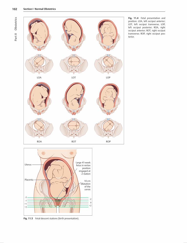
Position: In cephalic presentation the occiput is the ana-tomical reference, and its localization in relation to the maternal axis determines the fetal position. For example, if the occiput is localized anterior straight to the pubic arch, it is occiput anterior (OA), and if it is toward the mother’s right, then it is right occiput anterior (ROA). Fig-ure 11.4 depicts the possible fetal positions in vertex pre-sentation.
Station: This refers to the relation of the lowest bony fe-tal part to the ischial spines. It is a measure of descent of the fetus in labor and is classified according to the dis-tance in centimeters from the plane of the ischial spines (Fig. 11.5).
Cardinal Movements of Labor
During labor, the fetus dynamically interacts with the rig-id maternal bony pelvis to offer the best possible diame-ter to the pelvic path. Seven main movements are identi-fied in the fetus (Fig. 11.6).
Engagement: This occurs when the widest diameter of the presenting part (biparietal diameter in cephalic or bitrocantheric diameter in breech presentation) reaches a plane below the pelvic inlet. In cephalic presentation as seen at vaginal examination, the presenting part is at 0 station and the fetus is engaged.
Descent: The descent of the presenting part is through the pelvic birth canal.
Flexion: To improve the ability to pass through the birth canal, flexion of the head (the chin lies against the chest) occurs. This allows presentation of the smallest diameter of the fetal head, the suboccipitobregmatic.
Internal rotation: Most frequently, the presenting head enters the pelvis in transverse position. By internal rota-tion the head turns to OA position (towards the pubis), offering the best diameter to the pelvic canal.
Extension: Once the fetal head reaches the introitus, it extends leaving the pubis symphysis at the base of the occiput. This facilitates the delivery of the fetal head.
External rotation: Once the fetal head is delivered it ro-tates back in line with the anatomical position of the fetal body.
Section I Normal Obstetrics102
Pa
rt II
O
bste
tric
s
LOTLOA LOP
ROTROA ROP
Fig. 11.4 Fetal presentation and position. LOA, left occiput anterior; LOT, left occiput transverse, LOP, left occiput posterior; ROA, right occiput anterior; ROT, right occiput transverse; ROP, right occiput pos-terior.
Uterus
-3-2
-10+1
+2+3
Placenta 10 cmdilatation
of thecervix
Large 41-weekfetus in vertex
positionengaged at
-2 station
Fig. 11.5 Fetal descent stations (birth presentation).

11 Labor and Delivery 103
Fig. 11.6 Cardinal movements of labor.

Section I Normal Obstetrics104
Pa
rt II
O
bste
tric
s Expulsion: This describes the delivery of the rest of the fetal body. The delivery of the anterior shoulder follows the same pattern of rotation as the delivery of the head.
Progress of Labor
Stages
The three stages of labor are:• First stage. Extending from the onset of labor to full
cervical dilatation• Second stage. Extending from full cervical dilatation
until fetal delivery• Third stage. Extending from the delivery of the baby
until the placenta is delivered
Phases
There are two phases of labor: latent and active.• Latent phase. Extending from initiation of labor until
active labor is achieved. The diagnosis of labor initia-tion is subjective, and usually refers to the presence of regular contractions.
• Active phase. Labor usually is considered active when there is 80 % effacement and greater than 4 cm of cer-vical dilatation is achieved.
To objectively monitor the progress of labor and to iden-tify patients that require further evaluation, it is practical to plot the progress of labor as a labor curve (Fig. 11.7). In general, the rate of dilation during the active phase is
1.2 cm and 1.5 cm per hour for nulliparous and multipa-rous women, respectively. The use of epidural analgesia appears to prolong these time periods. Table 11.1 depicts mean and 95th percentile for duration of first and second stage of labor.
During labor, risk factors for a prolonged second stage, such as macrosomia or maternal diabetes, must be identi-fied. Once the fetal head is crowning, the physician must control the delivery of the head. It is critical to protect the perineal region with the other hand to diminish the risk for tears.
Once the head is delivered, external rotation is permit-ted or gently assisted. At this point, look for the nuchal umbilical cord and, if present, reduce it. If it is tight and reduction is not feasible, perform clamping and section at this point, before delivery of the rest of fetal body. Cur-rently, there is not robust evidence to support routine oropharynx aspiration. The anterior shoulder is then de-livered by gentle downward traction. Subsequently, the delivery of the posterior shoulder is assisted by gentle up-ward traction. Again, protection of the maternal perineal region is important at this stage.
The placenta and membranes are delivered passively. Manual intervention is only considered after 30 minutes of expectant management. After delivery, examine the placenta and membranes for integrity, and document the number of cord vessels.
0
2
4
6
8
10
20 4 6 8 10 12 14Active phaseLatent phase
Time (hours)
Accel.phase
Decel.phase
Phaseof max.slope
Sec.stage
Cerv
ical
dila
tatio
n (c
m)
Fig. 11.7 A typical labor graph.
Table 11.1 Duration of second stage of labor (mean and 95th percentile)
Parameter Mean 95th percentile
Nulliparas
Latent labor 7.3–8.6 h 17–21 h
First stage 7.7–13.3 h 16.6–19.4 h
First stage epidural
10.2 h 19 h
Second stage 53–57 min 122–147 min
Second stage epidural
79 min 185 min
Multiparas
Latent labor 4.1–5.3 h 12–14 h
First stage 5.7–7.5 h 12.5–13.7 h
First stage epidural
7.4 h 14.9 h
Second stage 17–19 min 57–61 min
Second stage epidural
45 min 131 min

11 Labor and Delivery 105
Episiotomy
Episiotomy is an incision in the perineal region performed when the fetal head crowns. The objective of this inter-vention is to reduce the risk of perineal trauma. It could be median (vertical medial midincision from the vagina toward the anal sphincter) or mediolateral (at a 45° angle from the vagina) (Fig. 11.8).
Although it was previously believed that episiotomy could favor delivery and decrease perineal trauma, cur-rent studies have shown that midline episiotomy increas-es the risk of third and fourth degree tears. Therefore, episiotomy should not be used routinely (See Evidence Box 11.1) and must be reserved for special circumstances, such us the relief of shoulder dystocia.
Operative Vaginal Delivery
In selected cases operative delivery either by forceps or vacuum extraction, instrumentation is required.
Forceps Delivery
Three general classes of forceps exist:• Classic forceps. These are designed for traction in ver-
tex presentation, but are not designed for rotation of the fetal head. The most common forceps in this group are Tucker-McLane, Simpson, and Elliot.
• Rotational forceps. This is characterized by the lack of pelvic curvature (to avoid pelvic damage on rota-tion) and sliding lock (to facilitate the application on asynclitic presentations). The most common forceps in this group is Kielland.
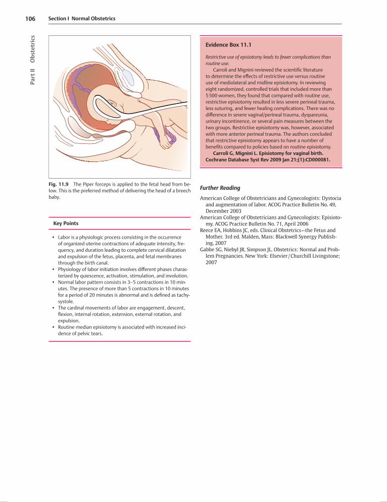
• Forceps to deliver the aftercoming head in breech pre-sentation. This forceps has a cephalic curve consisting of a reverse pelvic curve. When its application is re-quired, the trunk of the baby is held horizontally and the forceps is applied from below (Fig. 11.9). This ma-neuver is feasible because of the inverse pelvic curve. This forceps is called Piper.
Vacuum Extraction
The vacuum extraction device consists of a stainless steel or a plastic cup attached to a handle grip and a tube that connects to a vacuum source. Fundamentals for applica-tion are the same for either vacuum extraction or forceps delivery and require:
• baby to be in an engaged vertex presentation• cervix to be fully dilated• membranes to be ruptured• bladder to be drained• adequate assessment of fetal position in relation to
the maternal pelvis• availability of maternal analgesia• acquisition of informed consent• operator to be trained in forceps delivery/vacuum ex-
traction• willingness to abandon the procedure if unsuccessful
Indications to consider operative vaginal delivery are also similar for both techniques and include a prolonged sec-ond stage. For, nulliparous women, indications for vaginal delivery include the lack of progression for 2 hours when no regional anesthesia is used, and for 3 hours if analgesia was applied. For multiparous women, indicators for vagi-nal delivery include lack of progression for 1 hour when no regional anesthesia is used, and for 2 hours if analgesia was applied.
a b
Midline incision Mediolateral incision Fig. 11.8 Options for an episiotomy incision.
Section I Normal Obstetrics106
Pa
rt II
O
bste
tric
s
Key Points
• Labor is a physiologic process consisting in the occurrence of organized uterine contractions of adequate intensity, fre-quency, and duration leading to complete cervical dilatation and expulsion of the fetus, placenta, and fetal membranes through the birth canal.
• Physiology of labor initiation involves different phases charac-terized by quiescence, activation, stimulation, and involution.
• Normal labor pattern consists in 3–5 contractions in 10 min-utes. The presence of more than 5 contractions in 10 minutes for a period of 20 minutes is abnormal and is defined as tachy-systole.
• The cardinal movements of labor are engagement, descent, flexion, internal rotation, extension, external rotation, and expulsion.
• Routine median episiotomy is associated with increased inci-dence of pelvic tears.
Evidence Box 11.1
Restrictive use of episiotomy leads to fewer complications than routine use.
Carroli and Mignini reviewed the scientific literature to determine the effects of restrictive use versus routine use of mediolateral and midline episiotomy. In reviewing eight randomized, controlled trials that included more than 5 500 women, they found that compared with routine use, restrictive episiotomy resulted in less severe perineal trauma, less suturing, and fewer healing complications. There was no difference in severe vaginal/perineal trauma, dyspareunia, urinary incontinence, or several pain measures between the two groups. Restrictive episiotomy was, however, associated with more anterior perineal trauma. The authors concluded that restrictive episiotomy appears to have a number of benefits compared to policies based on routine episiotomy.
Carroli G, Mignini L. Episiotomy for vaginal birth. Cochrane Database Syst Rev 2009 Jan 21;(1):CD000081.
Further Reading
American College of Obstetricians and Gynecologists: Dystocia and augmentation of labor. ACOG Practice Bulletin No. 49, December 2003
American College of Obstetricians and Gynecologists: Episioto-my. ACOG Practice Bulletin No. 71, April 2006
Reece EA, Hobbins JC, eds. Clinical Obstetrics—the Fetus and Mother. 3rd ed. Malden, Mass: Blackwell Synergy Publish-ing, 2007
Gabbe SG, Niebyl JR, Simpson JL. Obstetrics: Normal and Prob-lem Pregnancies. New York: Elsevier / Churchill Livingstone; 2007
Fig. 11.9 The Piper forceps is applied to the fetal head from be-low. This is the preferred method of delivering the head of a breech baby.


